Preprint
Review

This version is not peer-reviewed.

Management of High Urogenital Sinus in Adults: A Scoping Review

A peer-reviewed article of this preprint also exists.

Submitted:

22 November 2024

Posted:

26 November 2024

You are already at the latest version

Abstract

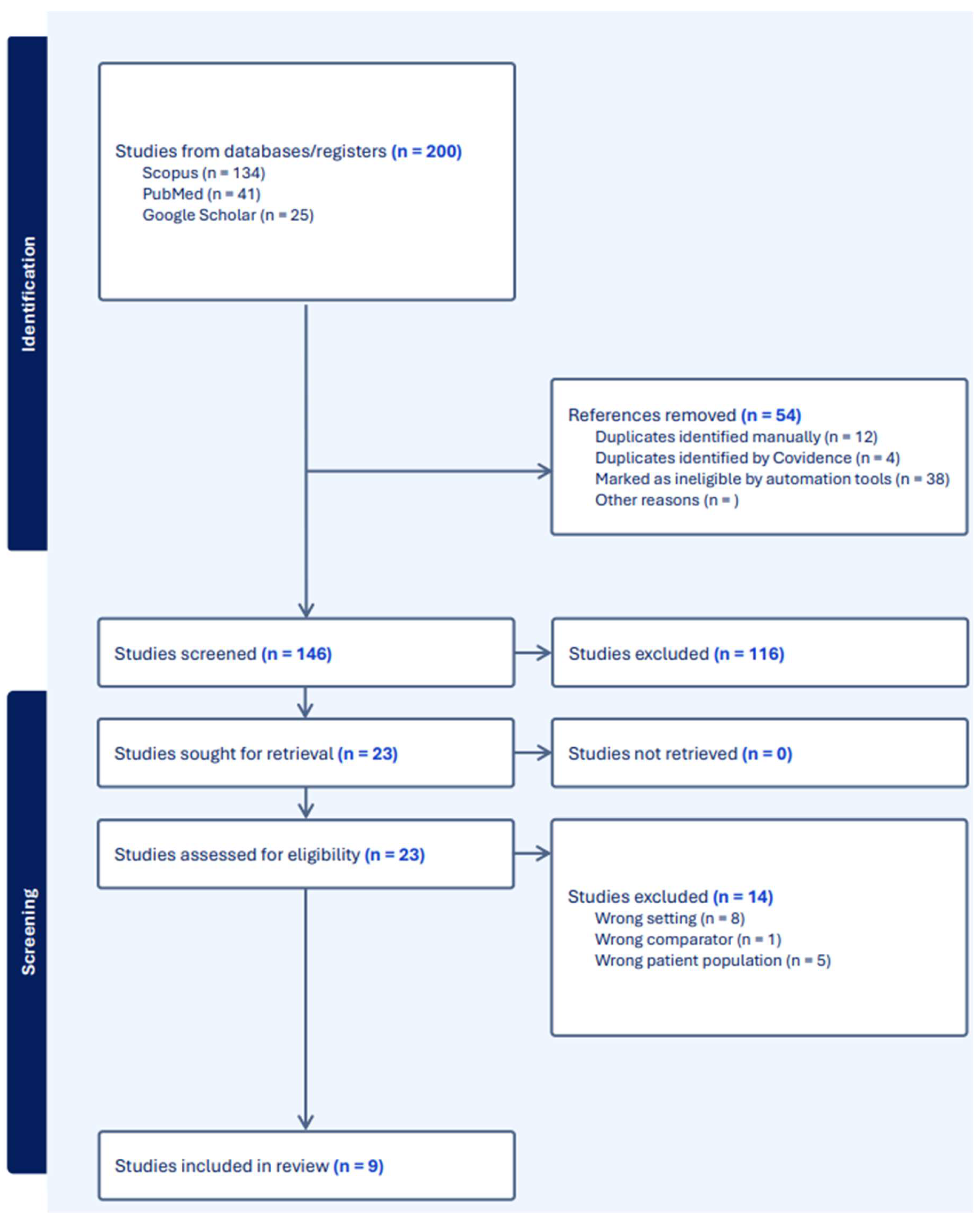
Background and Objectives: Persistent urogenital sinus (PUGS) is a rare congenital anomaly resulting from disruptions in urogenital development during embryogenesis, with an incidence of approximately 6/100,000 females. Characterized by the confluence of the vaginal canal and urethra into a single channel, it poses diagnostic and management challenges, particularly in adult patients. Surgical correction remains the primary treatment at an early age, yet there are no universally accepted guidelines regarding treatment at later stages in life. We explored current evidence on the management of high urogenital sinus (HUGS) in adults, focusing on surgical techniques, outcomes, perioperative care, and follow-up. Materials and Methods: A systematic review was conducted using PubMed, Scopus, and Google Scholar, including studies published up to August 2024. Search terms included "high urogenital sinus," "adult patients," and related conditions. Articles were included if they addressed clinical, diagnostic, or therapeutic data on HUGS in adult populations. Data extraction was performed independently by two reviewers, and a PRISMA flow diagram was used to outline the study selection process. Results: Nine studies met inclusion criteria, with data on surgical techniques, perioperative care, and long-term outcomes extracted for analysis. The included studies highlighted diverse surgical approaches such as feminizing genitoplasty, buccal mucosa vaginoplasty, posterior prone techniques, and robotic-assisted urogenital sinus mobilization. Complications like vaginal stenosis, urethrovaginal fistula, and urinary incontinence were reported. Long-term outcomes were underreported, with limited data on sexual function and patient satisfaction. Hormonal therapies, including glucocorticoids played critical roles in managing cases associated with congenital adrenal hyperplasia (CAH). Conclusions: The management of HUGS requires a multidisciplinary approach integrating advanced surgical techniques and individualized perioperative care. While short-term outcomes are generally favorable, long-term data on sexual function and quality of life remain scarce. Future research should prioritize longitudinal studies to address these gaps, aiming to optimize surgical outcomes, patient satisfaction, and psychosocial well-being.

Keywords: 
;  ;  ;  ;  

Introduction

Urogenital sinus (UGS) malformation, also known as persistent urogenital sinus (PUGS), is a rare congenital pathological disease with an incidence of approximately 6/100,000 women [1,2]. It occurs when the vaginal canal merges with the urethra into a single channel rather than opening into a distinct vulva. This confluence can occur near the bladder neck (high UGS or HUGS) or closer to the external opening (low UGS). Currently, there are no universally accepted guidelines for managing UGS, including the timing of surgery and long-term patient follow-up. This anomaly arises from disruptions in urogenital development during crucial stages of embryogenesis.

Embryology

In the development of external genitalia, the cloaca subdivides to form the urogenital sinus, and the cloacal membrane ruptures to form the urogenital plate on the surface of the perineum [3]. Between the 12th and 16th weeks of gestation, the junction between the developing lower vagina and urogenital sinus is displaced caudally until it stops at the urogenital sinus posterior wall, separating from the urethra in the vestibule to obtain a separate vaginal opening [4].
Although some studies have suggested that renal duct hypoplasia or insufficient growth of the tail urogenital wedge may cause some cases of PUGS [5,6], most are caused by the high androgen levels stimulated by CAH. The vaginal opening may be connected to the urethra instead of the vulva, with the junction of the vagina and urethra varying from the proximal confluence near the bladder neck to the distal confluence near the perineum, depending on the androgen-dependent closure of the zipper mechanism [7]. The location of the vaginal confluence depends on the descending position of the sinus ridge, which is related to androgen levels. Androgens inhibit the descending movement of the sinus ridge and can prevent the formation of the vaginal opening in the vulva [8,9].
Prenatal exposure to androgens in female mice has been found to inhibit the descent of the sinus ridge and prevent the formation of the vaginal opening in the vulva [10]. The specific time and duration of androgen exposure determines the location of the confluence of the vagina and urethra [10]. Mesenchymal cells adjacent to the urothelium are likely the primary target of androgen signaling for urogenital sinus ridge morphogenesis [10]. Recent studies also showed that the position of the sinus ridge is influenced by the amount of androgen exposure, such that higher doses of androgen result in a proximal shift of the region where the vagina and urethra meet (i.e., towards the bladder neck) [11,12,13]. It is likely that PUGS is caused by a combination of genetic, hormonal, environmental, and mechanical factors. The specific factors involved may vary depending on the individual case.
Due to the complexity of normal urogenital differentiation, adult cases of high UGS are infrequent. Surgery remains the primary treatment and is typically carried out in childhood.
However, there are still significant gaps in understanding the causes, ideal surgical techniques, and long-term outcomes of high UGS. We therefore aimed to explore the available literature for related articles on the management of HUGS.

Materials and Methods

2.1. Search Strategy and Study Selection

A systematic review of the literature was conducted to identify relevant studies on high urogenital sinus in adults. The search was performed using PubMed, Scopus, and Google Scholar covering studies published up to August 2024. The search terms included variations of "high urogenital sinus," "adult patients," and other related urological conditions. Boolean operators (AND, OR) were used to combine key terms, and the search was limited to articles in English and human subjects.
Two independent reviewers performed the literature search and screened the titles and abstracts of all identified articles. Studies were included if they met the following criteria: (1) focused on high urogenital sinus in female adult populations; (2) provided clinical, diagnostic, or therapeutic data; and (3) included original research, case series, cohort studies, reviews, or editorials. Articles were excluded if they involved pediatric populations exclusively or were non-English. Discrepancies between reviewers were resolved through discussion or consultation with a third reviewer.

2.2. Data Extraction and Quality Assessment

The two reviewers independently extracted data from the selected studies using a standardized form. The extracted data included study characteristics (authors, year, study design, sample size), patient demographics, clinical presentations, diagnostic modalities, and outcomes related to treatment and management of high urogenital sinus.

2.3 PRISMA Flow Diagram

The PRISMA (Preferred Reporting Items for Systematic Reviews and Meta-Analyses) flow diagram illustrates the study selection process (Figure 1). Initially, 200 articles were retrieved through database searches. After the removal of duplicates, 146 articles were screened by title and abstract, with 116 being excluded due to irrelevant titles or failure to meet inclusion criteria. Finally, 23 studies were assessed for eligibility, of which 9 were included in the final analysis after excluding articles with unrelated study population or wrong setting.

Results

9 studies met our inclusion and exclusion criteria (Table 1). The literature on PUGS surgical data is very limited. Most of the research data are retrospective and based on a small number of cases, focusing mainly on short-term outcomes such as the appearance of the vulva and complications after surgery. These studies do not provide strong evidence on the long-term efficacy of these procedures, particularly regarding sexual function.
In reviewing the management of urogenital sinus anomalies across various studies, a range of approaches and outcomes are presented. Braz (1999) highlighted the use of vaginal replacement with a sigmoid colon in eight patients, noting a complication of urethrovaginal fistula in one case, which was successfully corrected [14]. This was followed by a report from Mindy L. Samuelson et al. (2006) on the management of a single patient with adrenogenital syndrome, where feminizing genitoplasty and buccal mucosa vulvovaginoplasty were performed without complications [15].
Podesta et al. (2008) contributed by examining 12 cases involving urogenital sinus anomalies in DSD patients. While the surgical outcomes were generally favorable, some patients experienced glans atrophy, vaginal stricture, and urinary incontinence [16]. These findings were contrasted by Bailez et al. (2014), who described 55 cases of congenital adrenal hyperplasia treated with a urogenital sinus mobilization maneuver, reporting no adverse outcomes and no impact on voiding function [17].
Sircili et al. (2016) focused on 20 patients with congenital adrenal hyperplasia who had undergone failed previous surgeries [18]. A Y-V perineal flap technique was employed, and although one patient required reoperation, most patients had favorable outcomes, including reports of pain-free sexual activity.
In a more recent study by Wang et al. (2021), a patient with persistent urogenital sinus and associated anatomical anomalies was treated with urethral reconstruction and artificial vaginoplasty, resulting in successful urination and long-term management without complications [19] Around the same time, Oktay Ulusoy et al. (2021) explored a posterior prone approach for high urogenital sinus, confirming normal pelvic function postoperatively, supported by electromyography [20].
Ellerkamp et al. (2021) offered further insights into the treatment of secondary vaginal stenosis after prior surgeries, achieving positive outcomes through partial urogenital mobilization [21]. Jessica Yang et al. (2023) on the other hand described the use of robotic techniques in a complex case involving VACTERL syndrome, achieving successful outcomes without complications [22]. Each of these studies contributes to an evolving understanding of the best approaches for managing urogenital sinus anomalies, offering a wide array of techniques with generally positive results.
Figure 1. PRISMA flow chart.
Preprints 140562 g001

Discussion

Management Strategies and Surgical Techniques

The management of HUGS involves a variety of surgical approaches tailored to the anatomical complexities and associated conditions of each patient. Different management strategies and techniques are described in the literature.

Feminizing Genitoplasty and Buccal Mucosa Vaginoplasty

Samuelson et al. (2005) reported a case where a patient with mid-to-high urogenital sinus and adrenogenital syndrome underwent feminizing genitoplasty combined with buccal mucosa vulvovaginoplasty [15]. This approach aimed to reconstruct the external genitalia to a typical feminine appearance while addressing functional aspects. Buccal mucosa grafts were utilized due to their favorable healing properties and resistance to stenosis. The combination enhanced both functional and aesthetic outcomes.

Feminizing Genital Reconstruction with Long-term Follow-up

Podesta et al. (2008) treated 12 patients with urogenital sinus anomalies and disorders of sex development (DSD) using feminizing genital reconstruction [16]. Preoperative evaluations included history-taking, clinical examination, laboratory tests, karyotyping, ultrasound, and radiological examinations with urethroscopy. Postoperative complications such as glans atrophy, vaginal stricture, and urinary stress incontinence were noted. Despite these, a successful feminine genital appearance was achieved, emphasizing the need for long-term follow-up to monitor and manage late complications.

Perineal Prone Approach without Division of the Rectum

Rink et al. (1997) described a perineal prone approach for repairing high urogenital sinus without dividing the rectum [23]. This technique was employed in 8 cases and allowed good access to the urogenital tract while minimizing trauma to surrounding structures. Some patients experienced urethrovaginal fistulas and mild vaginal stenosis, which were managed successfully with dilation.

Partial Urogenital Mobilization and Flap Vaginoplasty

Ellerkamp et al. (2015–2018) treated 13 patients with DSD who developed vaginal stenosis after previous surgeries [21]. Management involved partial urogenital mobilization with perineal or lateral flaps and, in some cases, bowel vaginoplasty. One patient required a revision vaginoplasty, which failed due to complex anatomy. The study emphasized detailed anatomical assessment and appropriate surgical techniques to minimize complications.

Posterior Prone Approach with Pelvic Muscle Sparing

Ulusoy et al. (2021) managed 7 patients with HUGS using a posterior prone approach without dividing the rectum, focusing on preserving pelvic floor muscle function [20]. Postoperative assessments using EMG-uroflowmetry showed normal pelvic floor function, indicating that this technique effectively preserves urinary function.

UGS Mobilization Maneuver for CAH Patients

Bailez et al. (2014) reported on 55 cases of patients with CAH and high UGS, treated with the UGS mobilization maneuver [17]. This technique did not compromise voiding function or urinary continence, crucial considerations in managing high urogenital sinus, especially in CAH patients with severe anatomical malformations.

Robotic Total Urogenital Sinus Mobilization

Yang et al. (2023) described a novel approach using robotic total urogenital sinus mobilization combined with posterior sagittal anorectoplasty in a patient with VACTERL syndrome and a bicornuate uterus [22]. Robotic surgery allowed precise dissection and mobilization of urogenital structures, reducing intraoperative trauma and enhancing visibility in complex anatomical fields. Early results demonstrated promising outcomes without significant complications.

Staged Vaginal Reconstruction and Long-term Dilation

Bailez et al. (1992) conducted staged vaginal reconstruction with salt-wasting adrenal hyperplasia [24]. The initial procedure involved opening the urogenital sinus and exteriorization of the vagina through a perineal approach. A significant proportion (78.5%) required further vaginal reconstructive procedures to achieve a normal vaginal outlet. The authors highlighted the necessity of long-term follow-up and periodic dilation to prevent vaginal stenosis. Moreover, Ghoniem et al. (2024) reported a 21-year-old female misdiagnosed with transverse vaginal septum initially [25]. Her medical history included bilateral hydroureteronephrosis, leading to surgical interventions such as ureteric reimplantation and bladder flap surgery. Upon examination, her genitalia showed a HUGS, prompting further investigation. A modified ASTRA technique was used to surgically correct it, with steps to identify and close a fistula and lengthen the vaginal canal using skin flaps. Leuprolide acetate was administered to suppress menstruation and prevent complications. Postoperatively, the patient had no infections or incontinence but required serial vaginal dilations to prevent stenosis, achieving success with no further interventions after 12 months.

Peri-operative Care: Hormonal Treatment

Since most PUGS cases are secondary to congenital adrenal hyperplasia (CAH), their main treatment besides surgical female genital reconstruction is long-term glucocorticoid therapy. This therapy aims to suppress excess hormones, replace deficient hormones, and avoid potential cushing-like side effects [26]. Estrogen replacement therapy with progesterone should be initiated around physiological puberty to induce periodic bleeding (menstruation) and gradually transition to an adult regimen [27].
For patients with a normal hormonal profile, it is recommended to receive GnRH agonists for menstrual suppression perioperatively. GnRH agonists are non-contraceptive medications used for menstrual suppression by creating a hypoestrogenic state. Depot-leuprolide acetate 11.25 mg, injected intramuscularly every 12 weeks, is commonly used [28,29]. Rates of amenorrhea are as high as 96% [28,30]. Patients should be counseled to expect an initial "flare" in bleeding within 1 to 3 weeks until hormone levels are adequately suppressed [30]. Add-back therapy (e.g., norethindrone acetate 5 mg once daily) can help minimize other bothersome symptoms [28].

Follow-up

Current literature data are limited, and the results of studies differ. For example, Ellerkamp et al. reported that perineal flaps with partial urogenital mobilization provided normal anatomical results with normal sexual function in patients following female genital repair [21]. In contrast, other studies reported unsatisfactory follow-up outcomes after female genital reconstruction. A meta-analysis reported impaired clitoral sensitivity, vaginal stenosis, and pain and discomfort during intercourse [31]. Two other studies with long-term follow-up showed that postoperative outcomes in children with CAH in terms of sexual function and clitoral sensitivity were unsatisfactory [32,33].
A major etiology that complicates the outcome and success of vaginal reconstruction is patient participation with vaginal dilation. Stenosis is common after vaginal reconstruction, and long-term postoperative dilation is needed to prevent it. The ability to self-dilate the vagina or neovagina requires an understanding of one's own anatomy, the reason for the procedure, and the necessity for long-term dilation. In early teens, this is physically, emotionally, and psychologically challenging, often resulting in non-compliance and subsequent vaginal stenosis. Thorough counseling with gynecology, surgery, urology, and psychology teams is essential to assess the patient's readiness and ability to comply with dilation protocols [34].
Although patient data are heterogeneous regarding diagnosis, age, and surgical method, the success of vaginoplasty outcomes is improved when performed after puberty [35,36,37]. This improvement is believed to stem from better compliance with dilation and the estrogenization of tissue during the postoperative healing process. Many recommend undertaking these procedures when patients are ready to initiate vaginal sexual activity [35,36,38].

Psychological Well-being and Support System

Determining readiness for complex surgical reconstruction during pubertal maturation requires assessment of the patient's psychological well-being and available support. Although individuals with complex anatomies such as cloacal exstrophy and cloacal malformation have other comorbidities, they are reported to have good psychological functioning [39]. However, adolescents may have anxiety about their genital appearance and sexual activity [40]. Comprehensive psychological support and counseling are crucial components of perioperative care.

Conclusion

The management of PUGS remains complex due to its rare occurrence and the diversity of anatomical presentations. Advances in diagnostic imaging and surgical techniques have significantly improved our ability to assess and treat this congenital condition. Despite these developments, long-term outcomes, particularly concerning sexual function and patient satisfaction, remain underexplored, and further high-quality, longitudinal studies are required. While hormonal therapy and perioperative care play essential roles in the overall treatment of PUGS. Given the complexity of these cases, a multidisciplinary approach involving urology, gynecology, endocrinology, and psychology is critical to optimize outcomes. Future research should aim to improve long-term data collection, focusing on sexual function, quality of life, and the psychosocial well-being of patients undergoing these life-altering procedures.

Author Contributions

Conceptualization, MAMH, NMS, and GG; methodology, MAMH, NMS, and GG; data curation, MAMH, NMS, and GG; writing—original draft preparation, MAMH, NMS, and GG; writing—review and editing, MAMH, NMS, and GG; supervision, MAMH, NMS, and GG; project administration, MAMH, NMS, and GG; funding acquisition, MAMH, NMS, and GG. All authors have read and agreed to the published version of the manuscript.

Funding

This research received no external funding.

Data Availability Statement

The original data presented in the study are openly available.

Acknowledgments

None.

Conflicts of Interest

The authors declare no conflicts of interest.

References

  1. Valentini, A.L.; Giuliani, M.; Gui, B.; et al. Persistent Urogenital Sinus: Diagnostic Imaging for Clinical Management. What Does the Radiologist Need to Know? Am J Perinatol. 2016, 33, 425–432. [Google Scholar] [CrossRef] [PubMed]
  2. Singh, S.; Singh, P.; Singh, R.J. Persistent Urogenital Sinus. J Anat Soc India. 2010, 59, 242–244. [Google Scholar] [CrossRef]
  3. Shen, J.; Cunha, G.R.; Sinclair, A.; Cao, M.; Isaacson, D.; Baskin, L. Macroscopic whole-mounts of the developing human fetal urogenital-genital tract: Indifferent stage to male and female differentiation. Dev Hum Male Female Urogenit Tracts. 2018, 103, 5–13. [Google Scholar] [CrossRef]
  4. Schoenwolf GC, Bleyl SB, Brauer PR, Francis-West PH. Larsen’s Human Embryology. Elsevier Health Sciences; 2014.
  5. Acién, P.; Acién, M.I. The history of female genital tract malformation classifications and proposal of an updated system. Hum Reprod Update. 2011, 17, 693–705. [Google Scholar] [CrossRef]
  6. Martínez Escoriza, J.C.; Palacios Marqués, A.M.; López Fernández, J.A.; et al. Congenital vesicovaginal fistula with or without menouria: a literature review. Eur J Obstet Gynecol Reprod Biol. 2014, 175, 38–48. [Google Scholar] [CrossRef]
  7. Fernando, M.A.; Creighton, S.M.; Wood, D. The long-term management and outcomes of cloacal anomalies. Pediatr Nephrol. 2015, 30, 759–765. [Google Scholar] [CrossRef]
  8. Drews, U.; Sulak, O.; Schenck, P.A. Androgens and the development of the vagina. Biol Reprod. 2002, 67, 1353–1359. [Google Scholar] [CrossRef]
  9. Drews, U. Helper Function of the Wolffian Ducts and Role of Androgens in the Development of the Vagina. Sex Dev. 2007, 1, 100–110. [Google Scholar] [CrossRef]
  10. Larkins, C.E.; Enriquez, A.B.; Cohn, M.J. Spatiotemporal dynamics of androgen signaling underlie sexual differentiation and congenital malformations of the urethra and vagina. Proc Natl Acad Sci. 2016, 113, E7510–E7517. [Google Scholar] [CrossRef]
  11. Yucel, S.; Cavalcanti, A.G.; Wang, Z.; Baskin, L.S. The impact of prenatal androgens on vaginal and urogenital sinus development in the female mouse. J Urol. 2003, 170, 1432–1436. [Google Scholar] [CrossRef]
  12. Hotchkiss, A.K.; Furr, J.; Makynen, E.A.; Ankley, G.T.; Gray, L.E. In utero exposure to the environmental androgen trenbolone masculinizes female Sprague-Dawley rats. Toxicol Lett. 2007, 174, 31–41. [Google Scholar] [CrossRef] [PubMed]
  13. Hotchkiss, A.K.; Lambright, C.S.; Ostby, J.S.; Parks-Saldutti, L.; Vandenbergh, J.G.; Gray, L.E. Prenatal testosterone exposure permanently masculinizes anogenital distance, nipple development, and reproductive tract morphology in female Sprague-Dawley rats. Toxicol Sci Off J Soc Toxicol. 2007, 96, 335–345. [Google Scholar] [CrossRef]
  14. Braz, A. Posterior sagittal transanorectal approach in patients with ambiguous genitalia: report of eight cases. Pediatr Surg Int. 1999, 15, 108–110. [Google Scholar] [CrossRef]
  15. Samuelson, M.L.; Baker, L.A. Autologous buccal mucosa vulvovaginoplasty for high urogenital sinus. J Pediatr Urol. 2006, 2, 486–488. [Google Scholar] [CrossRef] [PubMed]
  16. Podesta, M.; Urcullo, J. Perineal mobilization of the common urogenital sinus for surgical correction of high urethrovaginal confluence in patients with intersex disorders. J Pediatr Urol. 2008, 4, 352–358. [Google Scholar] [CrossRef]
  17. Bailez, M.M.; Cuenca, E.S.; Dibenedetto, V. Urinary Continence Following Repair of Intermediate and High Urogenital Sinus (UGS) in CAH. Experience with 55 Cases. Front Pediatr. 2014, 2, 67. [Google Scholar] [CrossRef] [PubMed]
  18. Sircili, M.H.P.; Bachega, T.S.S.; Madureira, G.; Gomes, L.; Mendonca, B.B.; Dénes, F.T. Surgical Treatment after Failed Primary Correction of Urogenital Sinus in Female Patients with Virilizing Congenital Adrenal Hyperplasia: Are Good Results Possible? Front Pediatr. 2016, 4, 118. [Google Scholar] [CrossRef]
  19. Wang, Y.; Hu, S.; Wang, H. Multimodal ultrasound imaging of persistent urogenital sinus with uterus didelphys and double vagina malformation: A case report. Medicine (Baltimore) 2021, 100. https://journals.lww.com/md-journal/fulltext/2021/12300/multimodal_ultrasound_imaging_of_persistent.47.aspx. [CrossRef]
  20. Ulusoy, O.; Sabuncu, S.; Karakuş, O.Z.; et al. Urinary continence after high urogenital sinus repair conducted with posterior prone approach: electromyography-uroflowmetric assessment. Int Urol Nephrol. 2021, 53, 1813–1818. [Google Scholar] [CrossRef]
  21. Ellerkamp, V.; Rall, K.K.; Schaefer, J.; et al. Surgical Therapy After Failed Feminizing Genitoplasty in Young Adults With Disorders of Sex Development: Retrospective Analysis and Review of the Literature. J Sex Med. 2021, 18, 1797–1806. [Google Scholar] [CrossRef]
  22. Yang, J.; Syed, H.; Baker, Z.; Vasquez, E. Urogenital Sinus Diagnosed During Workup of Recurrent Urinary Tract Infections: A Case Report. Urology. 2023, 174, 165–167. [Google Scholar] [CrossRef] [PubMed]
  23. Rink, R.; Pope, J.; Kropp, B.; Smith, E.; Keating, M.; Adams, M. Reconstruction of the high urogenital sinus: early perineal prone approach without division of the rectum. J Urol. 1997, 158, 1293–1297. [Google Scholar] [CrossRef] [PubMed]
  24. Bailez, M.M.; Gearhart, J.P.; Migeon, C.; Rock, J. Vaginal reconstruction after initial construction of the external genitalia in girls with salt-wasting adrenal hyperplasia. J Urol. 1992, 148, 680-682; discussion 683-684. [Google Scholar] [CrossRef]
  25. Ghoniem, G.; Samaan, N.M.; Samir, M.; Hammad, M.A.M.; Fahmy, A.G. Successful management of high urogenital sinus in an adult female: Case report. Cont Rep. 2024, 12, 100068. [Google Scholar] [CrossRef]
  26. Auer, M.K.; Nordenström, A.; Lajic, S.; Reisch, N. Congenital adrenal hyperplasia. Lancet Lond Engl. 2023, 401, 227–244. [Google Scholar] [CrossRef]
  27. El-Maouche, D.; Arlt, W.; Merke, D.P. Congenital adrenal hyperplasia. Lancet Lond Engl. 2017, 390, 2194–2210. [Google Scholar] [CrossRef] [PubMed]
  28. Pradhan, S.; Gomez-Lobo, V. Hormonal Contraceptives, Intrauterine Devices, Gonadotropin-releasing Hormone Analogues and Testosterone: Menstrual Suppression in Special Adolescent Populations. J Pediatr Adolesc Gynecol. 2019, 32, S23–S29. [Google Scholar] [CrossRef]
  29. American College of Obstetricians and Gynecologists’ Committee on Clinical Consensus–Gynecology. General Approaches to Medical Management of Menstrual Suppression: ACOG Clinical Consensus No. 3. Obstet Gynecol. 2022, 140, 528–541. [Google Scholar] [CrossRef]
  30. Options for Prevention and Management of Menstrual Bleeding in Adolescent Patients Undergoing Cancer Treatment: ACOG Committee Opinion, Number 817. Obstet Gynecol. 2021, 137, e7–e15. [CrossRef]
  31. Almasri, J.; Zaiem, F.; Rodriguez-Gutierrez, R.; et al. Genital Reconstructive Surgery in Females With Congenital Adrenal Hyperplasia: A Systematic Review and Meta-Analysis. J Clin Endocrinol Metab. 2018, 103, 4089–4096. [Google Scholar] [CrossRef]
  32. Nordenström, A.; Frisén, L.; Falhammar, H.; et al. Sexual function and surgical outcome in women with congenital adrenal hyperplasia due to CYP21A2 deficiency: clinical perspective and the patients’ perception. J Clin Endocrinol Metab. 2010, 95, 3633–3640. [Google Scholar] [CrossRef]
  33. Rapp, M.; Duranteau, L.; van de Grift, T.C.; et al. Self- and proxy-reported outcomes after surgery in people with disorders/differences of sex development (DSD) in Europe (dsd-LIFE). J Pediatr Urol. 2021, 17, 353–365. [Google Scholar] [CrossRef] [PubMed]
  34. MILLER, R.J.; BREECH, L.L. Surgical Correction of Vaginal Anomalies. Clin Obstet Gynecol 2008, 51. https://journals.lww.com/clinicalobgyn/fulltext/2008/06000/surgical_correction_of_vaginal_anomalies.7.aspx. [CrossRef] [PubMed]
  35. Burgu, B.; Duffy, P.G.; Cuckow, P.; Ransley, P.; Wilcox, D.T. Long-term outcome of vaginal reconstruction: comparing techniques and timing. J Pediatr Urol. 2007, 3, 316–320. [Google Scholar] [CrossRef]
  36. Filipas, D.; Black, P.; Hohenfellner, R. The use of isolated caecal bowel segment in complicated vaginal reconstruction. BJU Int. 2000, 85, 715–719. [Google Scholar] [CrossRef] [PubMed]
  37. Krege, S.; Walz, K.H.; Hauffa, B.P.; Körner, I.; Rübben, H. Long-term follow-up of female patients with congenital adrenal hyperplasia from 21-hydroxylase deficiency, with special emphasis on the results of vaginoplasty. BJU Int. 2000, 86, 253-258; discussion 258-259. [Google Scholar] [CrossRef]
  38. Callens, N.; De Cuypere, G.; De Sutter, P.; et al. An update on surgical and non-surgical treatments for vaginal hypoplasia. Hum Reprod Update. 2014, 20, 775–801. [Google Scholar] [CrossRef]
  39. Mukherjee, B.; McCauley, E.; Hanford, R.B.; Aalsma, M.; Anderson, A.M. Psychopathology, psychosocial, gender and cognitive outcomes in patients with cloacal exstrophy. J Urol. 2007, 178, 630–635; discussion 634-635. [Google Scholar] [CrossRef]
  40. Ebert, A.; Scheuering, S.; Schott, G.; Roesch, W.H. Psychosocial and psychosexual development in childhood and adolescence within the exstrophy-epispadias complex. J Urol. 2005, 174, 1094–1098. [Google Scholar] [CrossRef]
Table 1. Summary of Included Studies.
No. Author(s) Year Diagnosis Method of Diagnosis Management Complications Number of Cases Remarks/Outcome
1 Braz, A 1999 Urogenital sinus, high vaginal implantation, normal rectum, sigmoidostomy Cystoscopic and radiological examinations Vaginal replacement with sigmoid colon, protective sigmoidostomy Urethrovaginal fistula (re-operated and cured) 8 One patient successfully engaged in sexual intercourse post-vaginal replacement.
2 Mindy L. Samuelsonet al 2006 Mid-to-high urogenital sinus in adrenogenital syndrome Clinical examination, endoscopic evaluation Feminizing genitoplasty, buccal mucosa vulvovaginoplasty None reported 1 -
3 Podesta, M et al 2008 Urogenital sinus anomalies in DSD Clinical history, examination, laboratory tests, karyotyping, ultrasound, radiology, urethroscopy Feminizing genital reconstruction Glans atrophy, vaginal stricture, urinary stress incontinence 12 Achieved a satisfactory feminine genital appearance; long-term follow-up needed.
4 Bailez, M.M. et al 2014 Congenital adrenal hyperplasia (CAH) with intermediate and high urogenital sinus Contrast imaging studies Urogenital sinus mobilization maneuver None reported 55 Does not compromise voiding function or urinary continence.
5 Sircili, M.H.P. et al 2016 Congenital adrenal hyperplasia (CAH) with failed previous surgery Cystoscopy Y-V perineal flap with/without partial mobilization of urogenital sinus One patient required a second reoperation 20 Eight adult patients were sexually active without experiencing dyspareunia.
6 Wang et al 2021 Persistent urogenital sinus (PUG) with uterus didelphys and double vagina Transabdominal ultrasound, transrectal ultrasound, contrast-enhanced ultrasound Urethral reconstruction, vaginal pull-through, artificial vaginoplasty, bilateral hysterosalpingectomy None reported 1 Urination normal after six months; continuous vaginal dilation used to prevent stenosis.
7 Oktay Ulusoy et al 2021 High urogenital sinus Clinical and imaging evaluation Posterior prone approach for repair without rectal division None reported 7 Electromyography and uroflowmetric tests showed normal function; pelvic EMG was also normal in all patients.
8 Ellerkamp, V et al 2021 Secondary vaginal stenosis after reconstructive surgery for urogenital sinus anomalies Genitoscopy, genitography, MRI Perineal flap with partial urogenital mobilization None reported 13 Uneventful outcomes post-surgery.
9 Jessica Yang et al 2023 VACTERL syndrome, urinary tract infection, high urogenital sinus, bicornuate uterus Ultrasound, voiding cystourethrogram (VCUG), MRI Vaginoplasty, robotic urogenital sinus mobilization, posterior sagittal anorectoplasty None reported 1 -
Disclaimer/Publisher’s Note: The statements, opinions and data contained in all publications are solely those of the individual author(s) and contributor(s) and not of MDPI and/or the editor(s). MDPI and/or the editor(s) disclaim responsibility for any injury to people or property resulting from any ideas, methods, instructions or products referred to in the content.
Copyright: This open access article is published under a Creative Commons CC BY 4.0 license, which permit the free download, distribution, and reuse, provided that the author and preprint are cited in any reuse.
Prerpints.org logo

Preprints.org is a free preprint server supported by MDPI in Basel, Switzerland.

Subscribe

Disclaimer

Terms of Use

Privacy Policy

Privacy Settings

© 2025 MDPI (Basel, Switzerland) unless otherwise stated