

Submitted:
24 November 2024
Posted:
25 November 2024
You are already at the latest version
Abstract
The interconnection between irritable bowel syndrome (IBS) and metabolic dysfunction-associated steatotic liver disease (MASLD) reveals shared molecular and physiological pathways, with microRNA-29a (miRNA-29a) emerging as a potential regulator. miRNA-29a influences inflammation, epithelial barrier integrity, mitochondrial function, and epigenetic modulation, impacting both conditions. In IBS, miRNA-29a disrupts tight junction proteins, exacerbates intestinal permeability, and heightens visceral hypersensitivity by modulating inflammatory pathways such as the NLRP3 inflammasome. In MASLD, miRNA-29a regulates lipid metabolism, mitigates mitochondrial stress, and curbs fibrotic progression through pathways involving GSK3β, SIRT1, and DNMT3b. Additionally, miRNA-29a preserves gut barrier function and combats dysbiosis, linking gut and liver pathology. The shared mechanisms underscore miRNA-29a’s role in amplifying systemic inflammation, driving IBS and MASLD progression. Understanding this interplay opens avenues for targeted therapeutic interventions to address these interconnected disorders.
Keywords:
epithelial permeability
; mitochondrial dysfunction
; inflammation pathways
; gut-liver axis
; dysbiosis
; visceral hypersensitivity
; lipid metabolism
1. Background
MicroRNA-29a (miRNA-29a) has emerged as a pivotal regulator of gene expression in gastrointestinal and hepatic disorders, garnering attention for its potential as a biomarker and therapeutic target [1]. Irritable Bowel Syndrome (IBS), a functional gastrointestinal disorder characterized by abdominal pain and altered bowel habits, affects approximately 10–15% of the global population [2,3]. The pathophysiology of IBS is complex and multifactorial, involving disruptions in gut motility, mucosal immunity, and the gut-brain axis [4,5]. Recent evidence suggests that miRNA-29a may contribute to these mechanisms, influencing intestinal barrier integrity, immune responses, and neuronal signaling pathways [6,7]. Its dysregulation has been implicated in heightened visceral hypersensitivity, a hallmark of IBS, making it a potential candidate for diagnostic and therapeutic innovations [7,8].
In the realm of liver diseases, metabolic dysfunction-associated steatotic liver disease (MASLD), formerly known as non-alcoholic fatty liver disease (NAFLD), stands as the leading cause of chronic liver conditions worldwide [9]. MASLD encompasses a spectrum ranging from simple steatosis to progressive fibrosis and cirrhosis, with metabolic syndrome as a central driver [9,10]. miRNA-29a plays a crucial role in liver fibrogenesis, acting as a regulator of extracellular matrix (ECM) remodeling and hepatic stellate cell activation [11,12]. Its dysregulation correlates with the progression of liver fibrosis, highlighting its dual function as both a biomarker for disease severity and a target for antifibrotic therapies. The overlapping metabolic and inflammatory pathways between MASLD and IBS underscore the potential of miRNA-29a as a shared molecular nexus [13,14].
Understanding the role of miRNA-29a in IBS and MASLD could unveil novel insights into their interconnected pathophysiology. As a non-invasive biomarker, miRNA-29a holds promise for early detection and monitoring of these conditions, while its regulatory capacity offers therapeutic opportunities to address their underlying molecular mechanisms [15,16,17]. This narrative review explores the emerging evidence linking IBS and MASLD via miRNA-29a, aiming to illuminate its potential in transforming diagnostic and therapeutic approaches for these prevalent disorders.
2. The connection between IBS and MASLD
The interplay between MASLD and IBS reveals a complex network of shared pathophysiological mechanisms involving inflammation, gut-liver-brain axis dysregulation, and microbial dysbiosis. Wu et al. [18] presented compelling epidemiological evidence showing that MASLD patients have a 13% higher risk of developing IBS. This association, more pronounced in females, highlights gender-specific interactions between metabolic dysfunction and gastrointestinal disturbances. The authors suggested that shared proinflammatory cytokines, such as TNF-α, IL-6, and IL-1β, disrupt intestinal barrier integrity and alter motility, thereby promoting IBS symptoms. These findings point to systemic inflammation as a potential link between the two conditions.
Ng et al. [19] found a significant association between IBS and MASLD. IBS prevalence was higher in MASLD patients (23.2% vs. 12.5%, P < 0.01), and a greater proportion of IBS patients had MASLD (65.8%–74.0%). IBS patients were three times more likely to have MASLD than non-IBS individuals (P < 0.001).
MASLD shares several points of connection with IBS, with low-grade chronic inflammation serving as a primary driver of disease progression in MASLD. Obesity activates both innate and adaptive immune pathways, while adipose tissue inflammation further exacerbates MASH [19,20]. Building on this, Scalera et al. [20] examined the gut-liver axis, emphasizing the bidirectional communication mediated through the portal vein and biliary tract. In MASLD, impaired bile acid synthesis and microbial metabolite clearance compromise intestinal homeostasis. Dysbiosis characterized by reduced beneficial bacteria and overgrowth of pathogenic strains further exacerbates intestinal barrier dysfunction, a hallmark of IBS. This interplay fosters a proinflammatory environment that perpetuates both hepatic steatosis and IBS-like symptoms. Scalera et al. [20] also noted that metabolic endotoxemia originating from the gut can fuel hepatic inflammation, creating a vicious cycle of mutual exacerbation.
Purssell et al. [21] delved into the neural pathways underpinning the liver-brain-gut axis, describing how MASLD-induced hepatic vagal afferent activation can alter gut motility and sensitivity. Disrupted neural signaling may lead to visceral hypersensitivity, a core feature of IBS. Moreover, MASLD-associated systemic low-grade inflammation impacts central nervous system function, potentially influencing the stress response and exacerbating IBS symptoms. Their work underscored the multifaceted impact of MASLD on the gut-brain axis, linking metabolic, immune, and neural dysfunctions.
These findings collectively highlight the intricate relationship between MASLD and IBS, driven by shared inflammatory pathways, microbiota alterations, and neural dysregulation. This emerging knowledge provides a foundation for targeted interventions addressing both conditions simultaneously.
3. MASLD and miRNA-29a
The interplay between miRNA-29a and MASLD unveils an intricate network influencing hepatic lipid metabolism, inflammation, and fibrosis [12,22]. Yang et al. [22] demonstrated that miRNA-29a mitigates mitochondrial stress by inhibiting the mitochondrial antiviral-signaling protein (MAVS) pathway, a key mediator in innate immune signaling. Their Western diet-fed mice with miRNA-29a overexpression exhibited lower serum levels of AST, ALT, and cholesterol, alongside reduced liver fibrosis and mitochondrial dsRNA release. This underscores miRNA-29a’s role in improving mitochondrial proteostasis and reducing oxidative stress, pivotal in MASLD pathogenesis.
Liu et al. [23] identified miRNA-29a as a critical regulator of cholesterol homeostasis in MASLD. Their studies with liver-specific Dicer1-knockout mice revealed that the absence of miRNAs, including miRNA-29a, exacerbated hepatic free cholesterol accumulation via upregulation of HMGCR, a cholesterol synthesis enzyme. miRNA-29a directly targeted HMGCR’s 3’-UTR, suppressing its expression. Their findings position miRNA-29a as a potential therapeutic target to normalize lipid metabolism and reduce cholesterol-driven hepatic injury in MASLD.
Building on miRNA-29a’s role in lipid regulation, Lin et al. [24] explored its impact on fatty acid metabolism, revealing how it influences multiple metabolic pathways. Lin et al. [24] explored miRNA-29a’s impact on fatty acid metabolism, demonstrating its suppression of CD36, a fatty acid translocase. In high-fat diet-induced MASLD models, miRNA-29a transgenic mice exhibited reduced hepatic steatosis, fibrosis, and systemic inflammation. Notably, the inhibition of CD36 by miRNA-29a attenuated lipid uptake and subsequent liver injury. The authors further linked miRNA-29a to the downregulation of pro-inflammatory and fibrotic markers, emphasizing its dual role in metabolic and immune modulation within the liver [13,24]. The appearance of CD36 on the cell surface triggers the activation of the NF-κB transcription factor. However, this pathway is suppressed when SIRT1 is expressed within the cell [25,26].
Expanding on its anti-inflammatory effects, Yang et al. [27] investigated miRNA-29a’s anti-fibrotic properties, focusing on its regulation of fibrotic signaling pathways. Yang et al. [27] expanded on miRNA-29a’s anti-fibrotic properties by illustrating its disruption of DNMT3b, a methyltransferase implicated in fibrosis progression. Their methionine-choline-deficient diet-induced MASLD models showed that miRNA-29a transgenic mice experienced reduced oxidative stress, fibrosis, and pro-inflammatory signaling. By inhibiting DNMT3b and downstream pathways like TGF-β and SMAD3, miRNA-29a emerged as a pivotal factor in curbing liver fibrosis and inflammation.
Following its anti-fibrotic actions, miRNA-29a’s role in mitigating mitochondrial dysfunction was further substantiated. The anti-inflammatory properties of miRNA-29a are further substantiated by Yang et al. [28], who linked its overexpression to reduced activation of GSK3β and SIRT1, key players in mitochondrial proteostasis and the unfolded protein response. In their MASLD models, miRNA-29a alleviated mitochondrial stress and restored liver homeostasis by targeting these pathways. This highlights miRNA-29a’s capacity to ameliorate mitochondrial dysfunction, a central feature of MASLD progression.
Given these wide-ranging effects, a study emphasized miRNA-29a’s role in liver fibrosis, reinforcing its therapeutic relevance. Huang et al. [14] highlighted miRNA-29a’s role in liver fibrosis, emphasizing its regulation of hepatic stellate cell activation. By targeting epigenetic pathways and fibrogenic signaling, miRNA-29a suppresses extracellular matrix deposition, presenting a promising strategy for halting or reversing fibrosis in MASLD. Their review underscores the importance of miRNA-29a as a versatile regulator capable of influencing multiple stages of liver disease progression.
dding to its multifaceted functions, a study highlighted miRNA-29a’s ability to modulate oxidative stress and immune responses. Lin et al. [13] highlighted the multifaceted role of miRNA-29a in oxidative stress and immune modulation. By regulating mitochondrial homeostasis and inflammatory signaling, miRNA-29a reduces lipotoxicity and protects hepatocytes from MASLD-induced injury. Their work emphasizes the therapeutic potential of miRNA-29a in mitigating the metabolic and inflammatory cascades that drive MASLD.
Broadening the perspective on miRNA-29a, Dalgaard et al. [12] reviewed its broader implications in metabolic disorders, identifying it as both a response to and modulator of hepatic stress. Dalgaard et al. [12] reviewed the broader implications of the miRNA-29 family in metabolic disorders, noting its elevation in fatty liver states. The upregulation of miRNA-29a appears paradoxical, as it is both a response to and a modulator of hepatic stress and lipid dysregulation. This dual role suggests that miRNA-29a functions as a compensatory mechanism to mitigate the deleterious effects of metabolic disturbances, a perspective warranting further exploration in MASLD contexts.
Shifting the focus to regulatory networks, Cui et al. [29] revealed the interaction between long non-coding RNA GAS5 and miRNA-29a-3p in MASLD. Cui et al. [29] explored the interaction between long non-coding RNA GAS5 and miRNA-29a-3p in MASLD, revealing a competitive axis that exacerbates liver dysfunction. GAS5 acts as a molecular sponge, sequestering miRNA-29a-3p and upregulating NOTCH2, a pathway that aggravates steatosis and inflammation. Their findings highlight the intricate regulatory networks involving miRNA-29a and its susceptibility to modulation by non-coding RNAs in MASLD progression.
Extending miRNA-29a’s influence to the gut-liver axis, a study demonstrated its systemic protective effects in MASLD models. Yang et al. [30] also investigated miRNA-29a’s systemic effects, demonstrating its protective role in gut-liver axis dysfunctions induced by a high-fat diet. Overexpression of miRNA-29a preserved gut barrier integrity, mitigated dysbiosis, and reduced inflammatory markers in MASLD models. This study underscores miRNA-29a’s ability to modulate systemic metabolic and inflammatory responses, extending its therapeutic relevance beyond the liver.
Additionally, examining its role in stress responses, Duan et al. [31] focused on miRNA-29a’s involvement in ischemia-reperfusion injury within MASLD contexts. Duan et al. [31] examined miRNA-29a’s role in ischemia-reperfusion injury within MASLD contexts. Their findings revealed that miRNA-29a targets iNOS, modulating nitric oxide production and mitigating cellular damage. The protective effects of remote ischemic preconditioning in MASLD were linked to decreased miRNA-29a levels, which restored iNOS activity and improved hepatic resilience. This insight connects miRNA-29a to broader physiological responses under stress conditions, highlighting its regulatory versatility.
Collectively, these studies illustrate miRNA-29a’s pivotal role in MASLD through its multifaceted regulation of lipid metabolism, mitochondrial function, inflammation, and fibrosis. Its consistent involvement across diverse pathways and experimental models underscores its potential as a therapeutic target, offering hope for innovative treatments in MASLD management.
4. IBS and miRNA-29a
The intricate interplay between miRNA-29a and IBS has drawn significant scientific interest due to its implications in intestinal barrier function, visceral hypersensitivity, and inflammation. Zhu et al. [32] explored the direct impact of miRNA-29a on intestinal barrier dysfunction in diarrhea-predominant IBS (IBS-D). They found that miRNA-29a was upregulated in the intestinal mucosa of IBS-D patients, accompanied by a decrease in tight junction proteins ZO-1 and CLDN1. This disruption of epithelial tight junctions exacerbated intestinal permeability, as confirmed in mouse models. Notably, silencing miRNA-29a restored tight junction protein levels and reduced permeability markers such as diamine oxidase (DAO), demonstrating its pivotal role in IBS-D pathogenesis by impairing barrier integrity.
Chao et al. [33] reinforced these findings by linking miRNA-29a overexpression to reduced expression of aquaporins (AQPs) in IBS-D. In their rat models, increased miRNA-29a was associated with a decline in AQP1, AQP3, and AQP8, leading to disrupted water homeostasis and elevated intestinal permeability. Introducing a miRNA-29a antagomir significantly improved AQP levels and reduced intestinal permeability, suggesting a regulatory mechanism through which miRNA-29a exacerbates IBS symptoms. This dual role in targeting both tight junctions and water channels illustrates its widespread influence on gut epithelial physiology.
Zhou and colleagues [34] provided further insights into miRNA-29a’s role by identifying its suppression of key targets such as nuclear factor-κB repressing factor (NKRF) and Claudin-1 (CLDN1). Using IBS-D patient tissues and miRNA-29 knockout models, they demonstrated that miRNA-29a’s regulation of these targets directly increases intestinal permeability, a hallmark of IBS-D. Their work underscored the therapeutic potential of miRNA-29a inhibitors in restoring barrier function, as their intervention not only stabilized intestinal permeability but also mitigated inflammation by modulating NF-kB pathways.
Focusing on inflammatory pathways, Ke et al. [35] illustrated how miRNA-29a interacts with the NLRP3 inflammasome in IBS-D. Their mouse models showed that miRNA-29a amplifies inflammatory responses by upregulating NLRP3 and downstream cytokines like IL-1β and TNF-α. Remarkably, the traditional Chinese remedy paeoniflorin alleviated these effects by inhibiting miRNA-29a expression. This suppression reduced NLRP3 activation, improved the levels of Claudin-1 and ZO-1, and reconstructed the epithelial barrier, demonstrating the therapeutic potential of targeting miRNA-29a-mediated inflammation in IBS-D.
Zhu et al. [36] expanded the scope by exploring miRNA-29a’s involvement in visceral hypersensitivity, a core feature of IBS. Their findings linked elevated miRNA-29a levels to the downregulation of serotonin receptor 5-HT7 (HTR7), a molecule associated with pain signaling. Inhibiting miRNA-29a restored HTR7 levels and reduced visceral hyperalgesia in stress-induced IBS models, suggesting that miRNA-29a exacerbates abdominal pain by directly modulating nociceptive pathways. This interaction with HTR7 highlights miRNA-29a’s role beyond barrier dysfunction, placing it at the center of sensory and pain regulation in IBS.
Lu et al. [37] took a broader approach by employing integrated omics analyses to decode the molecular underpinnings of IBS-D. They confirmed miRNA-29a’s involvement in epigenetic regulation by demonstrating its influence on critical pathways like MAPK, GABAergic synapses, and adherens junctions. These pathways are intertwined with visceral hypersensitivity and intestinal permeability. Their analysis emphasized miRNA-29a’s ability to orchestrate complex molecular interactions, cementing its status as a master regulator in IBS pathophysiology.
Wang et al. [38] delved into the NF-κB-MLCK signaling axis, revealing that miRNA-29a promotes intestinal permeability by targeting TRAF3. TRAF3, a known inhibitor of NF-κB, was suppressed in IBS-D patient tissues with concomitant miRNA-29a upregulation. This suppression activated NF-κB and MLCK, leading to tight junction disruption. Using miRNA-29 knockout mice, they demonstrated that blocking miRNA-29a restored TRAF3 levels, stabilized tight junctions, and alleviated intestinal permeability, offering a precise molecular mechanism for therapeutic targeting.
Finally, Zhou et al. [7] in their earlier studies linked miRNA-29a to glutamine synthetase regulation, showing that miRNA-29a binds to the GLUL gene, reducing glutamine synthetase levels in IBS-D patients with increased intestinal permeability. This regulation weakens the gut’s protective barrier and contributes to symptom exacerbation. Targeting this pathway offers another therapeutic avenue, highlighting miRNA-29a’s diverse impact on gut physiology.
Across these studies, miRNA-29a emerges as a central mediator in IBS pathogenesis, orchestrating a complex network that includes tight junction integrity, inflammatory signaling, and sensory modulation. Its consistent upregulation in IBS-D patients, coupled with its broad molecular influence, makes it a promising target for therapeutic interventions designed to restore gut homeostasis.
5. IBS and MASLD interconnection through miRNA-29a
The intricate interconnection between IBS and MASLD through miRNA-29a reveals a fascinating overlap of pathophysiological mechanisms, underscoring its regulatory role in both conditions. Shared pathways of inflammation, epithelial barrier dysfunction, and mitochondrial stress highlight the pivotal role of miRNA-29a in shaping the disease trajectory of these disorders.
Wu et al. [18] provided epidemiological evidence linking MASLD to an increased risk of IBS, attributing this association to systemic inflammation driven by proinflammatory cytokines like TNF-α, IL-6, and IL-1β. miRNA-29a plays a crucial role in modulating these inflammatory mediators. In MASLD models, Yang et al. [22] demonstrated that miRNA-29a inhibits the MAVS pathway, attenuating oxidative stress and inflammation. This effect parallels findings in IBS-D models where miRNA-29a overexpression amplified the activity of the NLRP3 inflammasome, as Ke et al. [35] observed. These shared inflammatory pathways implicate miRNA-29a as a common molecular mediator, fueling the mutual exacerbation of IBS and MASLD.
Barrier integrity is another critical axis of this interplay. Zhu et al. [32] noted that miRNA-29a disrupts tight junction proteins ZO-1 and CLDN1 in IBS, increasing intestinal permeability, a phenomenon mirrored in MASLD (Figure 1). Lin et al. [24] reported that miRNA-29a suppresses CD36, mitigating lipid uptake and preserving barrier integrity in MASLD models. By restoring tight junction function and reducing epithelial damage, miRNA-29a emerges as a dual modulator in the gut-liver axis, a pivotal conduit in the IBS-MASLD interplay.
Mitochondrial dysfunction and metabolic stress further deepen this connection. Yang et al. [28] highlighted miRNA-29a’s role in alleviating mitochondrial proteostatic stress in MASLD, mediated through GSK3β and SIRT1 inhibition. Similarly, miRNA-29a modulates cellular energy dynamics in IBS-D, where Zhu et al. [36] identified its regulatory effect on HTR7, linking it to visceral hypersensitivity and nociceptive signaling. This shared modulation of mitochondrial and metabolic stress pathways underscores miRNA-29a’s integrative role in both conditions.
Gut dysbiosis bridges IBS and MASLD through miRNA-29a’s impact on microbial communities and host response. Yang et al. [30] demonstrated that miRNA-29a preserves gut barrier function and counteracts dysbiosis in high-fat diet-induced MASLD models. These findings resonate with IBS studies, where miRNA-29a was implicated in shaping the mucosal environment and inflammatory balance. The gut-liver crosstalk mediated by miRNA-29a highlights its systemic influence, linking local disruptions in the gut to hepatic outcomes.
Finally, the epigenetic regulation orchestrated by miRNA-29a creates another layer of similarity. Yang et al. [27] observed that miRNA-29a inhibits DNMT3b, reducing fibrotic progression in MASLD. This parallels its regulatory effects in IBS, where Lu et al. [37] connected miRNA-29a to the modulation of MAPK and GABAergic pathways. These shared molecular signatures emphasize miRNA-29a’s versatility in controlling gene expression and signaling across IBS and MASLD.
This burgeoning evidence establishes miRNA-29a as a molecular nexus between IBS and MASLD, with its role in inflammation, barrier integrity, mitochondrial stress, and epigenetic regulation highlighting its potential as a therapeutic target in these interconnected conditions (Table 1).
Funding
None.
Acknowledgments
Special thanks to Seyed-Mohamad-Sadegh Mirahmadi and Reza Azarbad.
Conflict of Interest
The author declare that they have no conflict of interest.
References
- Khalaf, S.E.; Abdelfattah, S.N.; Khaliefa, A.K.; Daoud, S.A.; Yahia, E.; Hasona, N.A. Expression of PVT-1 and miR-29a/29b as reliable biomarkers for liver cirrhosis and their correlation with the inflammatory biomarkers profile. Human & Experimental Toxicology 2024, 43, 09603271241251451. [Google Scholar] [CrossRef]
- Mokhtare, M.; Karimi, S.; Bahardoust, M.; Sotoudeheian, M.; Ghazi, A.; Babaei-Ghazani, A. How adding the abdominal massage to polyethylene glycol can improve symptom and quality of life in patients with functional constipation in comparison with each one of the treatment modalities alone: A randomized clinical trial. Complementary therapies in medicine 2020, 52, 102495. [Google Scholar] [CrossRef] [PubMed]
- Ahamad, S.K.; Vanga, S.; Panjala, S.; Gollapelli, C.P.; Tadikonda, R.R. Irritable bowel syndrome and mental health: An overview. Open Access Research Journal of Biology and Pharmacy 2023, 9, 067–072. [Google Scholar] [CrossRef]
- Mokhtare, M.; Fathi, M.; Sadeghian, A.M.; Sotoudeheian, M.-J.; Namazi, A. A Pilot Study of the Effectiveness of a Short Course of Rifaximin 2200 mg/day on Abdominal Symptoms and its Effects on Quality of Life in Patients with Moderate to Severe Diarrhea-Predominant Irritable Bowel Syndrome. Clinical Drug Investigation 2024, 1–9. [Google Scholar] [CrossRef] [PubMed]
- Sadeghian, A.M.; Dashti, F.; Shariati, B.; Mokhtare, M.; Sotoudeheian, M. The role of cognitive emotion regulation strategies in predicting the resilience, symptom severity, and quality of life of patients with irritable bowel syndrome. Clinics and Research in Hepatology and Gastroenterology 2024, 48, 102341. [Google Scholar] [CrossRef]
- Zhang, X.; Shi, L.; Chen, C.; Ma, Y.; Ma, Y. The role of miRNA in IBS pathogenesis, diagnosis and therapy: The latest thought. Digestive and Liver Disease 2024. [Google Scholar] [CrossRef]
- Zhou, Q.; Souba, W.W.; Croce, C.M.; Verne, G.N. MicroRNA-29a regulates intestinal membrane permeability in patients with irritable bowel syndrome. Gut 2010, 59, 775–784. [Google Scholar] [CrossRef]
- Ludidi, S.; Conchillo, J.; Keszthelyi, D.; Van Avesaat, M.; Kruimel, J.; Jonkers, D.; Masclee, A. Rectal hypersensitivity as hallmark for irritable bowel syndrome: defining the optimal cutoff. Neurogastroenterology & Motility 2012, 24, 729–e346. [Google Scholar]
- Sotoudeheian, M. Galectin-3 and Severity of Liver Fibrosis in Metabolic Dysfunction-Associated Fatty Liver Disease. Protein and Peptide Letters 2024, 31, 290–304. [Google Scholar] [CrossRef]
- Sotoudeheian, M. Value of Mac-2 Binding Protein Glycosylation Isomer (M2BPGi) in Assessing Liver Fibrosis in Metabolic Dysfunction-Associated Liver Disease: A Comprehensive Review of its Serum Biomarker Role. Current protein & peptide science.
- Li, J.; Zhang, Y.; Kuruba, R.; Gao, X.; Gandhi, C.R.; Xie, W.; Li, S. Roles of microRNA-29a in the antifibrotic effect of farnesoid X receptor in hepatic stellate cells. Molecular pharmacology 2011, 80, 191–200. [Google Scholar] [CrossRef]
- Dalgaard, L.T.; Sørensen, A.E.; Hardikar, A.A.; Joglekar, M.V. The microRNA-29 family: role in metabolism and metabolic disease. American Journal of Physiology-Cell Physiology 2022, 323, C367–C377. [Google Scholar] [CrossRef] [PubMed]
- Lin, H.-Y.; Yang, Y.-L.; Wang, P.-W.; Wang, F.-S.; Huang, Y.-H. The emerging role of microRNAs in NAFLD: highlight of microRNA-29a in modulating oxidative stress, inflammation, and beyond. Cells 2020, 9, 1041. [Google Scholar] [CrossRef]
- Huang, Y.-H.; Yang, Y.-L.; Wang, F.-S. The role of miR-29a in the regulation, function, and signaling of liver fibrosis. International journal of molecular sciences 2018, 19, 1889. [Google Scholar] [CrossRef] [PubMed]
- Schmitt M, Margue C, Behrmann I, Kreis S. MiRNA-29: a microRNA family with tumor-suppressing and immune-modulating properties. Current molecular medicine 2013, 13, 572–585. [CrossRef] [PubMed]
- Amodio N, Rossi M, Raimondi L, Pitari MR, Botta C, Tagliaferri P, et al. miR-29s: a family of epi-miRNAs with therapeutic implications in hematologic malignancies. Oncotarget 2015, 6, 12837. [CrossRef]
- Pokharel DR, Maskey A, Kafle R, Batajoo A, Dahal P, Raut R, et al. Evaluation of circulating plasma miR-9, miR-29a, miR-192, and miR-375 as potential biomarkers for predicting prediabetes and type 2 diabetes in Nepali adult population. Non-coding RNA Research 2024, 9, 1324–1332. [CrossRef]
- Wu, S.; Yuan, C.; Yang, Z.; Liu, S.; Zhang, Q.; Zhang, S.; Zhu, S. Non-alcoholic fatty liver is associated with increased risk of irritable bowel syndrome: a prospective cohort study. BMC medicine 2022, 20, 262. [Google Scholar] [CrossRef]
- Ng, J.J.J.; Loo, W.M.; Siah, K.T.H. Associations between irritable bowel syndrome and non-alcoholic fatty liver disease: A systematic review. World Journal of Hepatology 2023, 15, 925. [Google Scholar] [CrossRef]
- Scalera, A.; Di Minno, M.N.D.; Tarantino, G. What does irritable bowel syndrome share with non-alcoholic fatty liver disease? World Journal of Gastroenterology: WJG 2013, 19, 5402. [Google Scholar] [CrossRef]
- Purssell, H.; Whorwell, P.J.; Athwal, V.S.; Vasant, D.H. Non-alcoholic fatty liver disease in irritable bowel syndrome: More than a coincidence? World journal of hepatology 2021, 13, 1816. [Google Scholar] [CrossRef]
- Yang, Y.-L.; Chuang, Y.-T.; Huang, Y.-H. MicroRNA 29a alleviates mitochondrial stress in diet-induced NAFLD by inhibiting the MAVS pathway. European Journal of Pharmacology 2024, 982, 176955. [Google Scholar] [CrossRef] [PubMed]
- Liu, M.-x.; Gao, M.; Li, C.-z.; Yu, C.-z.; Yan, H.; Peng, C.; Li, Y.; Li, C.-g.; Ma, Z.-l.; Zhao, Y. Dicer1/miR-29/HMGCR axis contributes to hepatic free cholesterol accumulation in mouse non-alcoholic steatohepatitis. Acta Pharmacologica Sinica 2017, 38, 660–671. [Google Scholar] [CrossRef] [PubMed]
- Lin, H.-Y.; Wang, F.-S.; Yang, Y.-L.; Huang, Y.-H. MicroRNA-29a suppresses CD36 to ameliorate high fat diet-induced steatohepatitis and liver fibrosis in mice. Cells 2019, 8, 1298. [Google Scholar] [CrossRef] [PubMed]
- Sotoudeheian, M.; Hoseini, S. Therapeutic properties of polyphenols affect AMPK molecular pathway in hyperlipidemia. 2023.
- Niu, B.; He, K.; Li, P.; Gong, J.; Zhu, X.; Ye, S.; Ou, Z.; Ren, G. SIRT1 upregulation protects against liver injury induced by a HFD through inhibiting CD36 and the NF-κB pathway in mouse kupffer cells. Molecular Medicine Reports 2018, 18, 1609–1615. [Google Scholar] [CrossRef]
- Yang, Y.-L.; Kuo, H.-C.; Wang, F.-S.; Huang, Y.-H. MicroRNA-29a disrupts DNMT3b to ameliorate diet-induced non-alcoholic steatohepatitis in mice. International Journal of Molecular Sciences 2019, 20, 1499. [Google Scholar] [CrossRef]
- Yang, Y.-L.; Wang, P.-W.; Wang, F.-S.; Lin, H.-Y.; Huang, Y.-H. miR-29a modulates GSK3β/SIRT1-linked mitochondrial proteostatic stress to ameliorate mouse non-alcoholic steatohepatitis. International Journal of Molecular Sciences 2020, 21, 6884. [Google Scholar] [CrossRef]
- Cui, J.; Wang, Y.; Xue, H. Long non-coding RNA GAS5 contributes to the progression of nonalcoholic fatty liver disease by targeting the microRNA-29a-3p/NOTCH2 axis. Bioengineered 2022, 13, 8370–8381. [Google Scholar] [CrossRef]
- Yang, Y.L.; Huang, Y.H.; Wang, F.S.; Tsai, M.C.; Chen, C.H.; Lian, W.S. MicroRNA-29a Compromises Hepatic Adiposis and Gut Dysbiosis in High Fat Diet-Fed Mice via Downregulating Inflammation. Molecular nutrition & food research 2023, 67, 2200348. [Google Scholar]
- Duan, Y.-F.; Sun, D.-L.; Chen, J.; Zhu, F.; An, Y. MicroRNA-29a/b/c targets iNOS and is involved in protective remote ischemic preconditioning in an ischemia-reperfusion rat model of non-alcoholic fatty liver disease. Oncology letters 2017, 13, 1775–1782. [Google Scholar] [CrossRef]
- Zhu, H.; Xiao, X.; Shi, Y.; Wu, Y.; Huang, Y.; Li, D.; Xiong, F.; He, G.; Chai, Y.; Tang, H. Inhibition of miRNA-29a regulates intestinal barrier function in diarrhea-predominant irritable bowel syndrome by upregulating ZO-1 and CLDN1. Experimental and therapeutic medicine 2020, 20, 1–1. [Google Scholar] [CrossRef]
- Chao, G.; Wang, Y.; Zhang, S.; Yang, W.; Ni, Z.; Zheng, X. MicroRNA-29a increased the intestinal membrane permeability of colonic epithelial cells in irritable bowel syndrome rats. Oncotarget 2017, 8, 85828. [Google Scholar] [CrossRef] [PubMed]
- Zhou, Q.; Costinean, S.; Croce, C.M.; Brasier, A.R.; Merwat, S.; Larson, S.A.; Basra, S.; Verne, G.N. MicroRNA 29 targets nuclear factor-κB–repressing factor and Claudin 1 to increase intestinal permeability. Gastroenterology 2015, 148, 158–169. [Google Scholar] [CrossRef] [PubMed]
- Ke, W.; Wang, Y.; Huang, S.; Liu, S.; Zhu, H.; Xie, X.; Yang, H.; Lu, Q.; Gan, J.; He, G. Paeoniflorin alleviates inflammatory response in IBS-D mouse model via downregulation of the NLRP3 inflammasome pathway with involvement of miR-29a. Heliyon 2022, 8. [Google Scholar] [CrossRef] [PubMed]
- Zhu, H.; Xiao, X.; Chai, Y.; Li, D.; Yan, X.; Tang, H. MiRNA-29a modulates visceral hyperalgesia in irritable bowel syndrome by targeting HTR7. Biochemical and biophysical research communications 2019, 511, 671–678. [Google Scholar] [CrossRef]
- Lu, Y.; Chai, Y.; Qiu, J.; Zhang, J.; Wu, M.; Fu, Z.; Wang, Y.; Qin, C. Integrated omics analysis reveals the epigenetic mechanism of visceral hypersensitivity in IBS-D. Frontiers in Pharmacology 2023, 14, 1062630. [Google Scholar] [CrossRef]
- Wang, Y.; Ke, W.; Gan, J.; Zhu, H.; Xie, X.; He, G.; Liu, S.; Huang, Y.; Tang, H. MicroRNA-29b-3p promotes intestinal permeability in IBS-D via targeting TRAF3 to regulate the NF-κB-MLCK signaling pathway. Plos one 2023, 18, e0287597. [Google Scholar] [CrossRef]
Figure 1.
miRNA-29a as a Key Regulator Linking IBS and MASLD. This illustration depicts the multifaceted role of miRNA-29a in the interplay between IBS and MASLD. miRNA-29a is centrally positioned, highlighting its influence on shared mechanisms such as inflammation, epithelial barrier integrity, mitochondrial dysfunction, and epigenetic modulation. Arrows indicate miRNA-29a’s regulatory effects on specific pathways: in IBS, it disrupts tight junction proteins (ZO-1, Claudin-1), activates the NLRP3 inflammasome, and contributes to visceral hypersensitivity. In MASLD, miRNA-29a modulates lipid metabolism, inhibits fibrosis through TGF-β/SMAD3, and alleviates oxidative stress. The gut-liver axis is depicted, showing microbial dysbiosis and systemic inflammation as connecting factors. Shared pathways underscore miRNA-29a’s dual role in driving both gut and liver pathology, positioning it as a potential therapeutic target for these interlinked conditions.
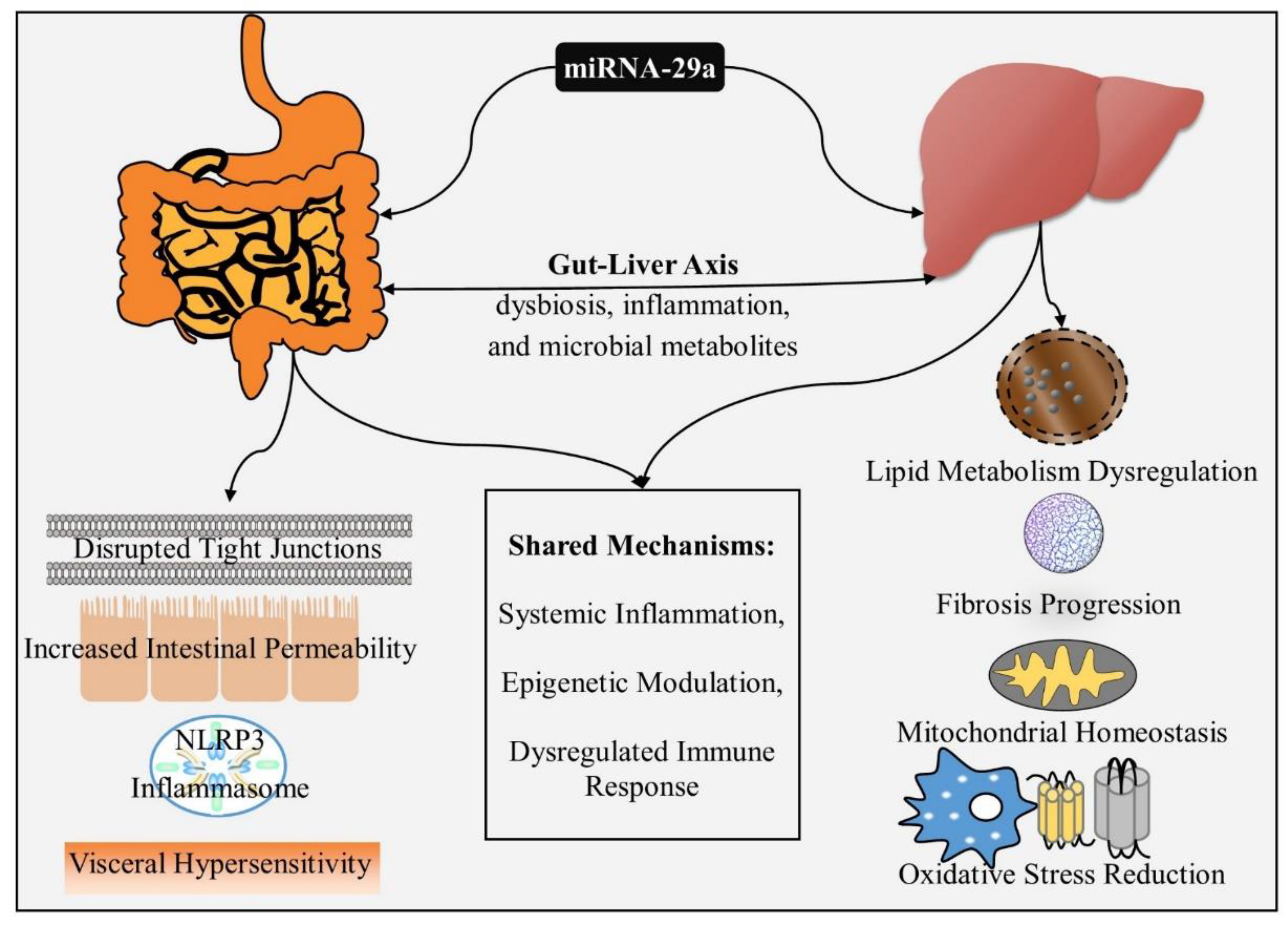

Table 1.
Shared and Specific Effects of miRNA-29a in IBS and MASLD.
| Aspect | IBS | MASLD | Shared Mechanisms |
| Barrier Integrity | Disrupts ZO-1 and Claudin-1, increasing gut permeability. | Preserves barrier function via suppression of CD36. | Impaired epithelial and endothelial barriers. |
| Inflammation | Activates NLRP3 inflammasome, amplifying cytokine release. | Reduces inflammatory pathways (e.g., DNMT3b, MAVS). | Elevated TNF-α, IL-6, and IL-1β promote systemic effects. |
| Metabolism Regulation | Alters intestinal water homeostasis via AQP modulation. | Regulates lipid metabolism and cholesterol via HMGCR. | Dysregulated mitochondrial function. |
| Mitochondrial Stress | Linked to visceral hypersensitivity and pain signaling. | Reduces oxidative stress and proteostatic imbalances. | Compromised mitochondrial health affects both disorders. |
| Microbial Dysbiosis | Gut dysbiosis exacerbates IBS symptoms. | Dysbiosis fuels hepatic inflammation. | Gut-liver crosstalk mediated by microbiota and metabolites. |
| Epigenetic Modulation | Influences pathways like NF-κB and MAPK. | Modulates TGF-β/ SMAD3 and DNMT3b signaling. | Epigenetic regulation drives inflammation and fibrosis. |
Disclaimer/Publisher’s Note: The statements, opinions and data contained in all publications are solely those of the individual author(s) and contributor(s) and not of MDPI and/or the editor(s). MDPI and/or the editor(s) disclaim responsibility for any injury to people or property resulting from any ideas, methods, instructions or products referred to in the content. |
© 2024 by the authors. Licensee MDPI, Basel, Switzerland. This article is an open access article distributed under the terms and conditions of the Creative Commons Attribution (CC BY) license (http://creativecommons.org/licenses/by/4.0/).
Copyright: This open access article is published under a Creative Commons CC BY 4.0 license, which permit the free download, distribution, and reuse, provided that the author and preprint are cited in any reuse.