Preprint
Review

This version is not peer-reviewed.

The Role of Nanos in Germ Granules—Putative Posttranscriptional Regulation Hubs in Germ Cell Specification and Development Across Species

Submitted:

21 November 2024

Posted:

22 November 2024

You are already at the latest version

Abstract

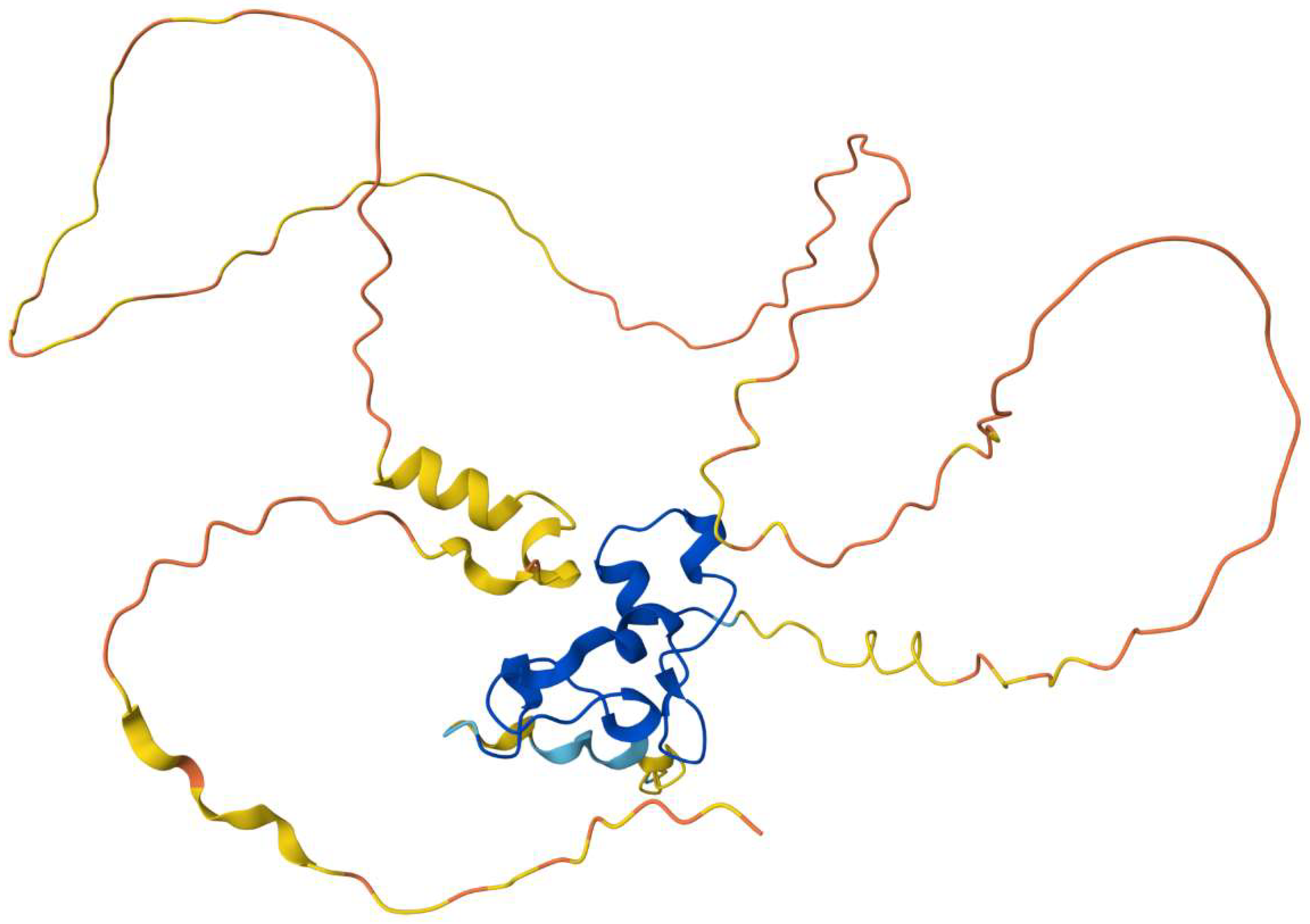
Nanos, initially identified in Drosophila melanogaster (fly) as a morphogen essential for body patterning and germ cell development, is a highly conserved RNA-binding protein critical for germ cell formation across species. Nanos dysfunction leads to infertility from flies to humans. While Drosophila has a single nanos gene, paralogs (nanos1-3) exist in species like Danio rerio (zebrafish), Caenorhabditis elegans (roundworm), Xenopus laevis (frog), and mammals, each with distinct reproductive roles. Animal germ cells contain a characteristic perinuclear structure called nuage built of RNAs and RNA-binding proteins (RBPs), with nanos as one of its most conserved components. Nuage is essential for germ cell specification, development, maintenance, and integrity across species, exhibiting variable cytoplasmic sub-localizations, structure and shape depending on the sex and developmental stage. It gives rise to cytoplasmic germ granules, basic ribonucleoprotein (RNP) condensates unique to germ cells. This review examines nanos' role within nuage and germ granules across model organisms and highlights key questions regarding its biological significance, particularly in human reproduction.

Keywords: 
;  ;  ;  ;  ;  ;  ;  ;  

1. Introduction

Germ cells are the only cell types that are totipotent and link generations. Exploring the mechanisms of their specification and development across different animal groups is crucial for understanding human infertility. Depending on the species, germ cells are formed through either the preformation or induction mechanism. In preformation, germ cells inherit a maternal cytoplasmic structure called germ plasm from the oocyte. The zygote's daughter cells that inherit this germ plasm become germ cells. In contrast, in the inductive mechanism, germ cells are formed later during early embryonic development from somatic cells upon signals from surrounding tissues. In germ plasm-bearing animals, primordial germ cells (PGCs) originate from the endoderm. In contrast, in animals using induction, such as axolotls and mice, PGCs arise from pluripotent epiblast cells during gastrulation induced by mesodermal signals. The totipotency of PGCs, in several animal models, as seen in Xenopus laevis and Caenorhabditis elegans, is partly maintained by the transient suppression of somatic transcription by germ plasm components, mediated by nanos during embryonic patterning [1,2]. In the inductive mode of PGC specification, totipotency requires three concurrent processes: suppression of somatic fate, activation of pluripotency genes, and epigenetic reprogramming for review, see [3].
Research on mammalian PGC specification, including in humans, has largely been derived by studies using pluripotent stem cells for review, see [4]. In vitro, these cells differentiate into PGCs by forming embryoid bodies (EBs). In mice, a cascade of transcription factors (TFs) such as Prdm1, Prdm14, and Tfap2c marks the earliest stages of PGC specification, beginning at embryonic day E6.5 for review, see [5]. Their expression is induced by bone morphogenetic protein 4 (Bmp4) from the extra-embryonic ectoderm, along with Wnt3 signalling from the posterior visceral endoderm of the epiblast [6]. These activated TFs, in turn, remote the expression of germline-specific genes, such as NANOS1 via PRDM14 stimulation [7] and NANOS3 [8], along with other key genes during human PGC specification. Simultaneously, these TFs repress genes involved in somatic differentiation. While several genes critical for PGC specification, such as BMP, WNT, and PRDM14, are shared between mice and humans, their molecular targets and functions are only sometimes conserved across these organisms [7]. In a primate model organism more closely related to humans, the Cynomolgus monkey (Macaca fascicularis), PGCs induced through in vitro specification exhibit upregulation of NANOS1-3 during EB formation. However, unlike in humans, NANOS1-3 proteins are also expressed in somatic tissues at this developmental stage [9]. The expression of NANOS2 and NANOS3 decreases by day 3 of PGC specification. Later, it increases markedly from day 7 onwards in the in vitro-generated EBs, despite both genes being expressed in embryonic stem cells. A similar rise in NANOS1 expression is observed in cynomolgus EBs after day 14 [9].
Notably, despite the differences between preformation and induction mechanisms, both converge on a shared feature: the presence of the nuage, a membraneless cytoplasmic structure composed of RNAs and RNA-binding proteins (RBPs), a defining characteristic of animal germ cells. Best studied in Drosophila, Caenorhabditis, Xenopus, and Danio, the nuage gives rise to germ granules—higher-order cytoplasmic structures formed by ribonucleoproteins (RNPs). Crucially, several conserved components of these granules, such as dazl, nanos, dead-end (Dnd), and the RNA helicase vasa/ddx4, are vital for germ cell specification and development across species, including in humans [10,11]. While germ granules are likely essential for proper germ cell function, their structure and roles remain insufficiently explored, highlighting the need for further study to fully understand their significance.

Nanos’ Structure and Function in Germ Cells

Compared to other tissues in the body, the germline is highly enriched with germline-specific RBPs, many of which are essential for germ cell development [12]. Among these, the nanos protein, a crucial component of nuage and germ granules, was first identified in Drosophila as a morphogen necessary for body patterning and germ cell development [13,14]. Nanos is expressed in both pluripotent and germ cells for review, see [15]. Beyond Drosophila melanogaster, nanos homologs have been extensively studied in various model organisms, including C. elegans [16], Planarians [17], [18], X. laevis [19], Danio rerio [20], Mus musculus [21,22], and Homo sapiens [23]. Nanos proteins have been shown to repress mRNAs, playing several roles in germ cell specification, development, and the maintenance of germ cell fate and survival. Depending on the species, nanos may exist as a single gene and isoform, as in Drosophila, or as multiple paralogs with distinct isoforms for review, see [24].
The Nanos protein contains a highly conserved C-terminal RNA-binding domain composed of two zinc fingers (CCHC)2. In contrast, the N-terminal region is divergent and intrinsically disordered (intrinsically disordered region, IDRs), except for an internal 17-amino-acid fragment in mammals known as the NOT1-interacting motif (NIM) (Figure 1 Alphafold Hs), which is responsible for binding and recruiting the deadenylation complex to target RNAs for repression [25,26]. The length of the nanos N-terminal region varies, with the most extended sequence found in human NANOS1. The interaction of nanos with RNA is often conferred by pumilio (pum), which determines the specific RNA targets for repression [19,23,27,28]. Together, nanos-pum complexes mediate translational repression, specifically in germ cells, resulting in the suppression of somatic gene expression, inhibition of the cell cycle [29], and repression of autophagy for review, see [24,30].
In studies using model organisms, disruption of the nanos gene has consistently demonstrated its critical role in germ cell development. In Xenopus, loss of nanos leads to a significant reduction in the number of PGCs and the absence of germ cells in the mature gonad [19]. Similarly, in C. elegans, depletion of nanos in PGCs triggers apoptosis [16]. In Drosophila embryos, maternal nanos is required to establish and maintain germline identity by preventing apoptosis and adopting somatic cell fate. Pole cells lacking maternal nanos undergo apoptosis during mid to late embryogenesis [31]. In D. rerio, mutations in the nanos3 gene result in the loss of germline stem cells and infertility in females [32]. Similarly, disrupting Nanos2 or Nanos3 in mice leads to infertility [33]. In humans, mutations in the NANOS1 gene are associated with the absence of germ cells in seminiferous tubules [34], while mutations in NANOS3 are linked to premature ovarian failure, causing infertility [35]. Additionally, abnormal NANOS expression in humans has been associated with various types of cancer for review, see [36].

2. Nanos, a Component of Nuage—A Hallmark Structure of Germ Cells Across Animal Species

Nanos is a key component of the nuage in Drosophila and other species. Nuage, a conserved cytoplasmic structure in metazoan germ cells, plays a crucial role in germ cell determination [37]. It is present in both male and female germ cells, regardless of species or the mechanism of germ cell specification. First identified by electron microscopy as an electron-dense, amorphous "cloud" surrounding germ cell nuclei [38] for review, see [39], nuage consists of membraneless condensates that form structured germ granules rich in RNA and RNA-binding proteins. These germ granules, composed of RNP condensates, regulate post-transcriptional gene expression in germ cells, with mRNAs traversing through the perinuclear nuage before reaching the bulk cytoplasm. Many nuage proteins are implicated in RNA-mediated interference pathways, including siRNA and piwi-interacting RNAs, suggesting that small silencing RNAs are produced within the nuage [40].
Nuage is a dynamic structure, varying in shape and content depending on the species, germ cell sex, and developmental stage. For example, in its perinuclear form, nuage interacts with nuclear pores [40], while in male germ cells, a mitochondria-associated form called inter mitochondrial cement (IMC) is present [41]. In oocytes, nuage appears as the Balbiani body (Bb)/ mitochondrial cloud, a large conserved membraneless aggregate found across species [42]. The Bb structure varies among organisms, sometimes encompassing mitochondria or interacting with the Golgi and endoplasmic reticulum, as observed in mice [43]. Finally, nuage is also a key germ cell determinant in mammals, including humans [37].

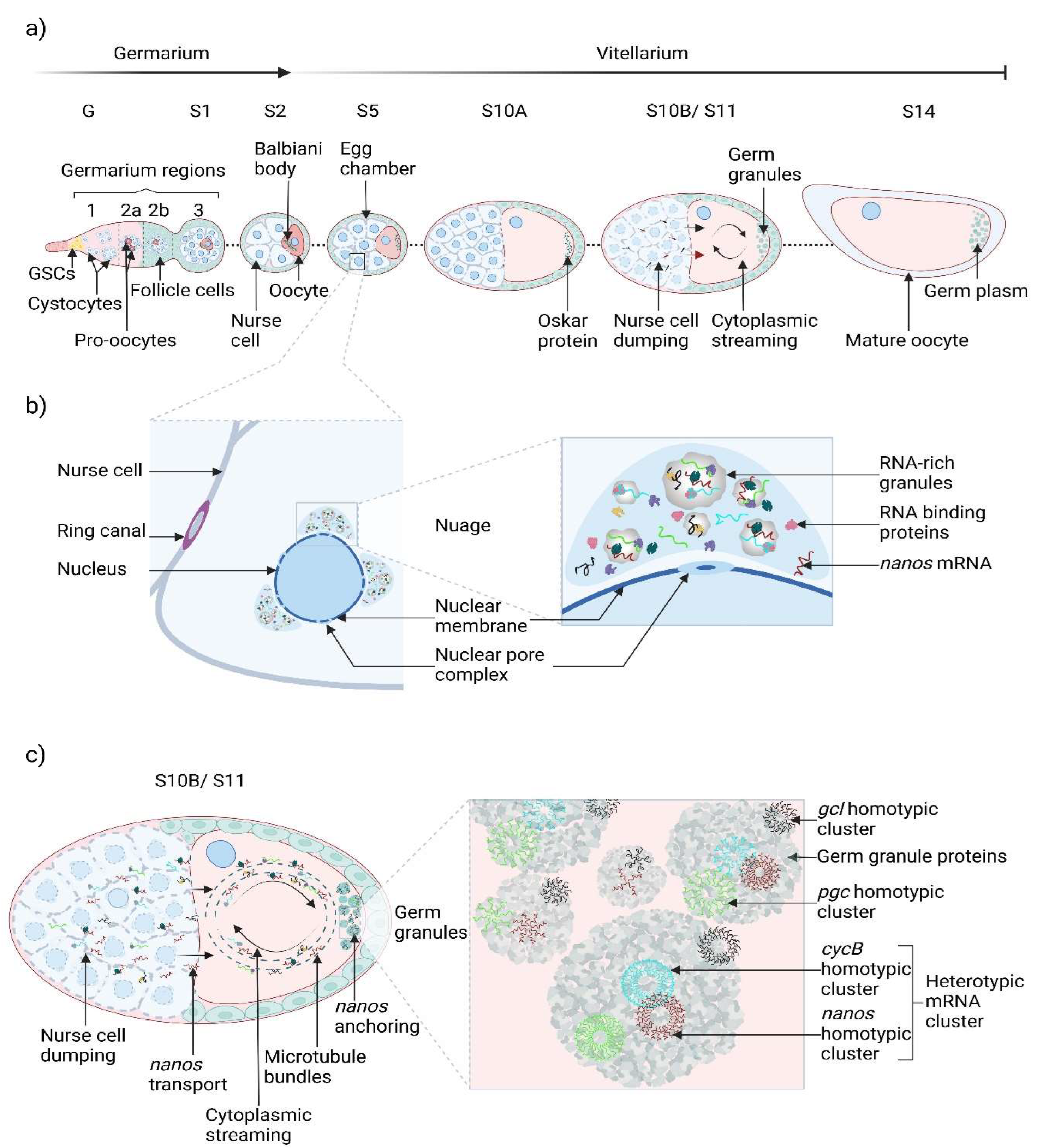
2.1. Nanos Implication in the Nuage Life Cycle in D. melanogaster

In Drosophila, the nuage appears during the early stages of oogenesis and disappears before oocyte maturation (Figure 2) [44]. The precise spatial localization and activity of the nuage component nanos are critical for germline formation in Drosophila. During early germline development, nanos mRNA, along with other maternal mRNAs, is produced by ovarian nurse cells located anteriorly within the egg chamber (Figure 2). As oogenesis progresses, the nurse cells "dump" their contents into the growing oocyte, and nanos mRNA accumulates within the nuage, forming germ granules in the germ plasm at the posterior of the oocyte (Figure 2) [45]. Nanos mRNA, together with other components, suppresses somatic differentiation and promotes totipotency, both essential for germ cell maintenance [46]. The localization of nanos mRNA within Drosophila germ granules is regulated by the RBP oskar, which initiates germ plasm formation and later the pole plasm at the posterior of pole cells (Figure 2). At this site, oskar activates nanos translation [47].
The posterior localization of nanos mRNA within the oocyte is mediated by sequences within its 3’ untranslated region (3’UTR), which are recognized by the RBP oskar [48]. While posteriorly localized nanos mRNA undergoes translation, unlocalized nanos mRNA is repressed through 3’UTR-mediated mechanisms involving the RBP smaug [49]. Thus, the cis-acting elements within the nanos 3’UTR are critical for both translational activation at the posterior by oskar and repression in the bulk cytoplasm by smaug [50,51]. This repression may be partially explained by the ability of the translational control element in the 3’UTR to form a secondary structure regulated by the DEAD-box helicase vasa [50,52]. A prevailing theory suggests that cis-elements within the nanos 3’UTR, along with the RBPs oskar, vasa, and tudor [53], coordinate the translational regulation of nanos mRNA, determining its activation at the posterior or repression in the bulk cytoplasm. These 3’UTR signals are also essential for the storage and degradation of nanos mRNA [30,54]. Additionally, nanos and oskar mRNAs play a significant role in controlling the size and composition of germ granules. The removal of nanos and oskar mRNAs results in a 1.8-fold increase in germ granule size, suggesting that these mRNAs limit germ granule condensation and growth [55]. These findings highlight the critical role of RNA content in regulating germ granule function.
In addition to nanos mRNA, the aforementioned RBPs also regulate mRNAs encoding other RBPs, such as dazl, dnd, polar granule component (pgc), and cyclin B (cycB) [30]. The oskar, nanos, vasa, and tudor are critical components of the germ plasm, governing the post-transcriptional regulation of RNA targets within Drosophila germ granules. These granules are known to contain up to 117 distinct mRNA types [56]. Yet, no common localization pathway has been identified to direct the movement of nanos and other germ-cell mRNAs into the granules. Despite their similar distribution patterns, nanos, pgc, and cycB mRNAs do not co-localize in the nurse cells or the bulk oocyte cytoplasm. However, upon reaching the posterior, single copies of these mRNAs are recruited by vasa into nascent germ granules [57]. Once recruited as single mRNA molecules, nanos, pgc, and cycB mRNAs form homotypic mRNA clusters, with nanos mRNA present in quantities ranging from more than four to over 60 molecules per granule, indicating substantial variability in nanos mRNA content among granules [57]. By late oogenesis instead, fluorescence in situ hybridization experiments revealed significant overlap in the presence of pgc and cycB mRNAs within granules containing nanos, suggesting a final physical association between these transcripts [57].
Thus, germ-cell-destined mRNAs, including nanos, pgc, and cycB, initially travel within the oocyte as single mRNA molecules in ribonucleoprotein particles. They subsequently co-assemble into homotypic clusters within germ granules and further organize into higher-order heterogeneous granules, specifically at the posterior of the oocyte [57]. Additionally, the number and mRNA content of nanos-containing granules increases as the oocyte matures, reaching peak levels by stage 14 (Figure 2) [57]. This co-packaging of germ-cell-destined transcripts ensures their effective segregation into pole cells after fertilization, giving rise to the future primordial germ cells [57].

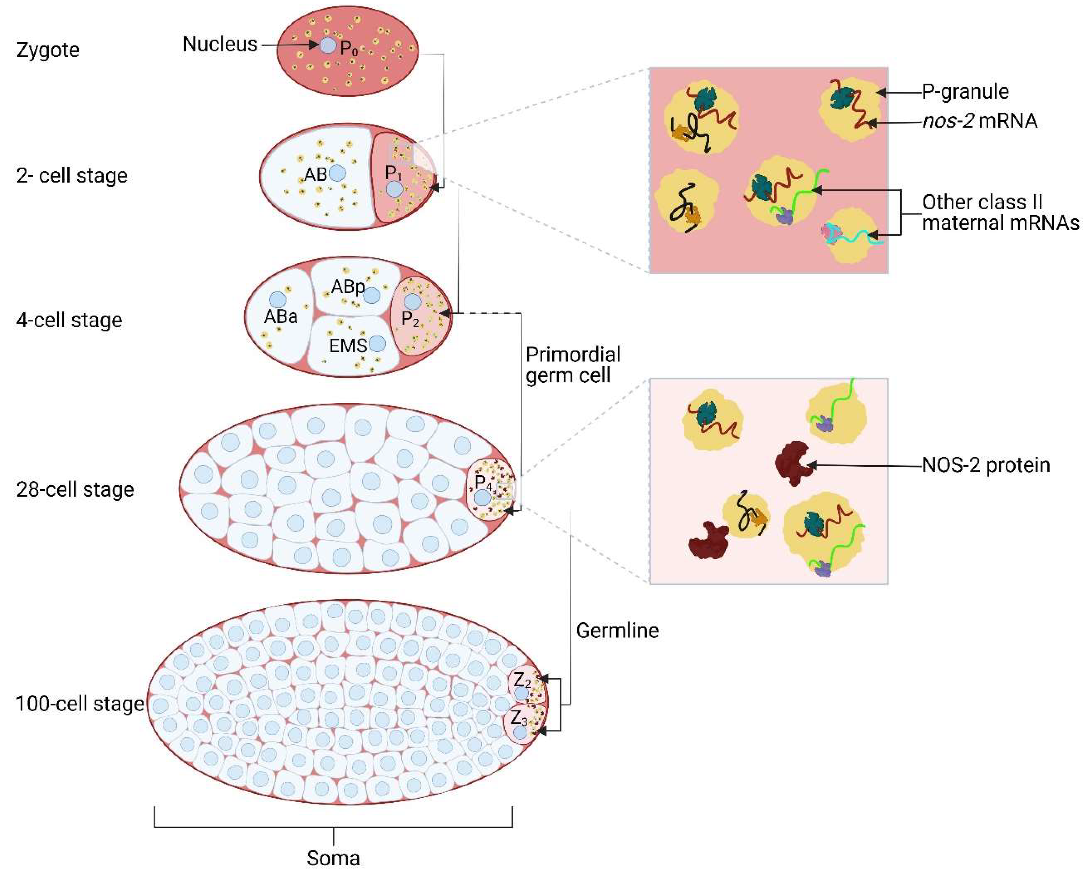
2.2. C. elegans nanos2-Containing Germ Granules Associates with Nuclear Pores

In C. elegans, the germline is segregated from the soma early in embryogenesis. In oocytes, germ granules, known as P granules, are initially distributed uniformly throughout the cytoplasm but become localized to the posterior after fertilization [58]. The separation of the germ lineage from somatic lineages occurs progressively through a series of four asymmetric cell divisions, resulting in the formation of germline blastomeres (P1, P2, P3, and P4), with only the P4 blastomere inheriting the germ plasm and eliminating somatic differentiation factors (Figure 3). As a result, the P4 cell serves as the sole primordial germ cell, which subsequently divides, transferring its contents equally into the daughter Z2 and Z3 germ cells (Figure 3).
In C. elegans, P granules are continuously present in germ cells throughout all stages of development [59]. Comparable to the germ granules of D. melanogaster, P granules remain associated with the nuclear membrane throughout the life cycle [60,61]. The majority of the approximately 40 known P-granule proteins are RBPs [62], suggesting their role in post-transcriptional RNA regulation. As seen in Drosophila, P granules are also sites for mRNA export from the nucleus [63].
In C. elegans, three nanos paralogs (nanos1-3) are expressed in germ cells, but only nanos2 mRNA associates with P granules, while it is degraded in somatic cells [64]. Nanos2 mRNA is first translated in the P4 blastomere and later in its daughter cells, Z2 and Z3 (Figure 3). Notably, the translational activation of maternal nanos2 mRNA is the earliest known molecular event specific to the germline founder cell, P4. This translational rate is regulated by the 3′UTR, bound by four maternal RBPs—OMA-1, OMA-2, MEX-3, and SPN-4—that suppress nanos2 translation. In contrast, the maternal RBP POS-1 binds to the 3′UTR in P4, relieving this suppression and activating nanos2 translation [64].
Furthermore, using in situ hybridization, it was observed that nanos2 mRNA is concentrated in small loci around the nuclei of germline blastomeres [65]. In Caenorhabditis, it was first discovered that P granules not only associate with nuclear pores, as seen in Drosophila but also interact directly with nuclear pore complexes (NPCs) [65]. NPCs are large macromolecular structures that regulate transport between the nucleus and cytoplasm and are composed of approximately 30 conserved proteins known as nucleoporins (Nups), found across species from yeast to humans [66]. Specifically, the nucleoporin CeNup98 is enriched in translationally repressed P granules containing nanos2 mRNA. In individuals lacking CeNup98, P granules dispersed, releasing nanos2 mRNA, and embryos depleted of CeNup98 expressed nanos2 prematurely. This indicates that CeNup98 directly maintains P granule integrity by interacting with P granule-associated mRNAs [65].
Interestingly, Caenorhabditis P granules exhibit liquid-like behaviour, such as dissolution, condensation, and surface wetting on nuclei, indicating that these germ granules rely on low-affinity interactions [67]. This suggests that CeNup98 may promote the ‘liquid phase’ of P granules by increasing RNA-protein interactions within these structures [16,68].

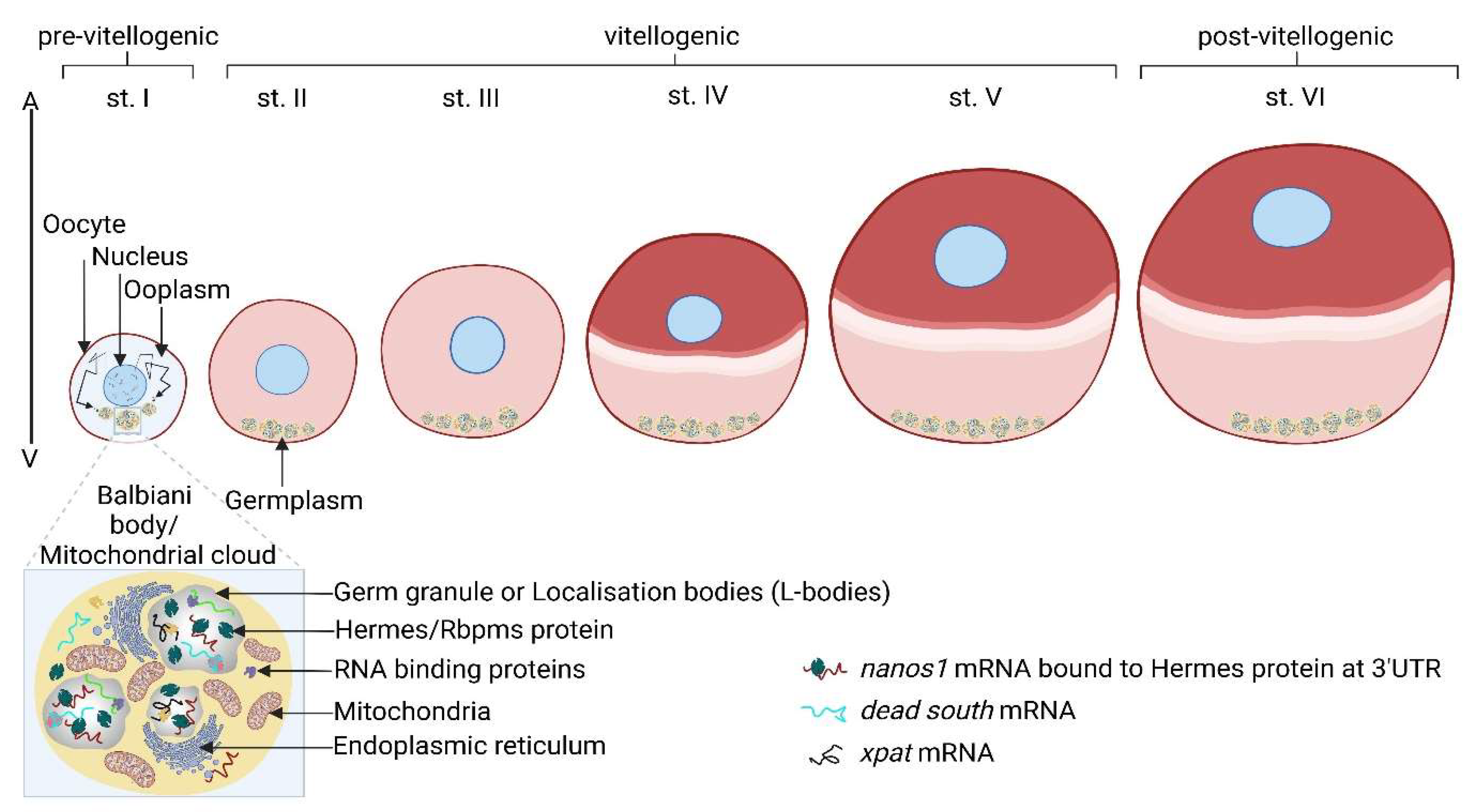
2.3. Xenopus nanos1 Activity within the Oocyte’s Balbiani Body

Xenopus oocytes are highly polarized cells characterized by the animal (future anterior) and vegetal (future posterior) poles. The darkly pigmented animal pole, or upper hemisphere, gives rise to the ectoderm, while the unpigmented vegetal pole, or lower hemisphere, gives rise to the endoderm and mesoderm. Importantly, the germplasm is localized at the vegetal pole (Figure 4).
The Xenopus germplasm consists of tightly bound vegetal cortical germ granules composed of RNPs, which guide future germline development. The Bb (Figure 4) plays a key role in transporting specific mRNAs to the vegetal pole, where germ granules are located. At the previtellogenic stage (Figure 4), the Bb is a dense structure containing mRNA-containing RNPs (mRNPs), cytoskeleton, endoplasmic reticulum, and mitochondria [69]. In addition, the Bb contains RBPs like tudor domain-containing protein 6 (tdrd6), which interacts with germ plasm mRNAs, including nanos1 (formerly Xcat-2) also located within the Bb at this stage [70]. Tdrd6 is crucial for the structural integrity of the Bb and PGC formation, as evidenced by tdrd6 gene knockdown phenotypes [71].
The localization of nanos1 mRNA within the Bb is influenced by temperature and ATP concentration. Higher temperatures or increased ATP levels accelerate the speed of nanos1 mRNA localization, while ATP depletion or lower temperatures reduce its accumulation in the Bb [72]. This process is kinesin II-dependent but does not require an intact cytoskeleton in stage I oocytes [73]. Although the overall polarity of the oocyte relies on the cytoskeleton, nanos1 mRNA localizes to vegetal RNP particles via diffusion into pre-existing particles rather than cytoskeletal transport [69]. Nanos1 mRNA remains anchored to the cortex during oocyte maturation, similar to its behaviour at the posterior pole in Drosophila (Figure 4) [48]. Localization depends on sequences in the 3’UTR, with the initial 250 nucleotides of the 3’UTR and six UGCAC repeats within the region being crucial [74]. Additionally, sequences extending 150 nucleotides near the open reading frame and 120 nucleotides at the 3’UTR’s end are required for efficient localization to the vegetal cortex and germ granules [74,75]. Most localization signals reside in the 3’UTR and consist of multiple elements, exhibiting functional redundancy [76].
In stage II oocytes, nanos1 mRNA follows the movement of the Bb as it fragments, with each fragment containing germ plasm that migrates to a localized area in the vegetal cortex. By stage III and early stage IV, when the animal-vegetal axis is distinguished by reduced pigmentation at the vegetal pole (Figure 4), nanos1 mRNA remains localized within the vegetal cortex, a feature that persists through stage VI oocytes and ovulated eggs [77]. The formation of the vegetal cortex in stage VI Xenopus oocytes, which will become the future posterior, is analogous to the formation of the posterior pole plasm in Drosophila, where localized materials similarly accumulate through a multi-stage process during oogenesis [13]. This conserved mechanism of localized germ plasm formation highlights an evolutionary strategy shared across species to ensure germ cell fate determination.
Hermes is a crucial RBP interacting with nanos1 mRNA in Xenopus, which specifically binds to nanos1 mRNA within the nucleus of stage I oocytes. Notably, the binding requires additional factors, one of which may be heterogeneous nuclear RNP (hnRNP)-I, a known interactor of hermes [78]. Hermes is a constituent of germinal granules and co-localises with nanos1 mRNA within the Bb, particularly in areas where germplasm is formed. This interaction is mediated through sequence-specific RNA localisation signals within the nanos1 3’UTR [79]. For instance, the UGCAC sequences, essential for targeting nanos1 mRNA to the Bb germplasm, are also necessary for its interaction with hermes. However, direct binding between hermes and UGCAC repeats has yet to be confirmed [78]. Hermes's hydrophilic C-terminal 34 amino acids are critical for homodimer formation, a structure required for nanos1 mRNA binding [78]. Deletion of this region, even with the intact RRM domain, disrupts hermes' ability to bind nanos1 mRNA. This region is also 85% conserved between Xenopus hermes and the human homolog RBPMS, underscoring its conserved functional importance [78].
Notably, nanos1 mRNA traverse multiple cellular compartments—nucleus, cytoplasm, Bb, and endoplasmic reticulum—before its final localization within germinal granules in the germplasm. Each stage likely involves modifications such as the addition or removal of proteins. The initial association of hermes with nanos1 mRNA in the nucleus (seen in vivo) suggests that hermes binding may trigger the sorting pathway that leads to the incorporation of nanos1-hermes complexes into germinal granules (Figure 4) (Aguero et al., 2016).
In late-stage oocytes, hermes likely interacts with Rbm42b in the nucleus, a protein co-localizes with nanos1 mRNA [69]. Therefore, hermes may act as a scaffold in the nucleus, recruiting additional proteins to the nanos1 RNP complex. After nuclear export, these protein interactions may help prevent the translation of nanos1 mRNA. The nanos RNP complex then diffuses through the cytoplasm and is eventually captured by an endoplasmic reticulum component within Bb [80], where other RNA-binding proteins will likely join. However, hermes remains the only protein identified in the final germinal granule so far. This dynamic remodelling of the nanos1 RNP particle, progressing toward the germplasm and integrating into germinal granules, highlights the complexity of RNA localisation processes [78]. Further research is needed to fully characterize the composition and behaviour of the endogenous nanos1 RNP particle throughout its localization stages, leading to the formation of the germinal granule. These mechanisms may serve as models for conserved processes in other species, including humans, mainly if conserved sequences or factors are involved.
After fertilization, during the early cleavage stages, the vegetal pole cortex in Xenopus aggregates into larger germplasm accumulations [77]. At this stage, the germplasm is asymmetrically allocated to four cells, designated as presumptive primordial germ cells (pPGCs) (Figure 4). As cleavage progresses, the germplasm shifts from its initial cortical location to a position near the nucleus, accompanied by an increase in the number of pPGCs (Figure 4). Nanos1 mRNA, a key component of the germplasm, undergoes translational repression during oogenesis and remains untranslated until the early embryonic stages. Unlike Drosophila, where nanos mRNA is repressed by smaug binding to its 3’UTR [81], Xenopus nanos1 mRNA repression does not involve the 3’UTR, indicating a more complex regulatory mechanism. In Xenopus, the sequestration of nanos1 in germinal granules is insufficient to fully account for its repression, suggesting additional inhibitory mechanisms [82]. One such mechanism involves a structural element within nanos1 mRNA that sterically hinders ribosome scanning, repressing translation without a specific repressor [82]. However, physical sequestration within germinal granules may also contribute to the repression.
Nanos1 protein accumulates in the germplasm only during a narrow window of early embryogenesis, specifically between the late blastula and gastrula stages, suggesting its role is focused on germ cell determination rather than broader patterning, as seen in Drosophila. Nanos1 mRNA translation is regulated independently of its localization [83], which contrasts with Drosophila and Caenorhabditis, where 3’UTR-mediated repression and activation are crucial for germline expression [64,84]. In Xenopus, a vertebrate-specific RNA-binding protein, dnd1, is essential and sufficient for activating nanos1 mRNA translation in early embryos [85,86,87]. Notably, unlike in Drosophila and Caenorhabditis, the 3’UTR of Xenopus nanos1 mRNA is not required for its translational activation by dnd1. In Xenopus, Dnd1 protein prevents nanos1 mRNA from interacting with the translational repressor eIF3f, ensuring proper translation [87].
Overall, the regulation of nanos in Xenopus highlights a vertebrate-specific complexity in mRNA translation control. This contrasts with the 3’UTR-dependent mechanisms seen in invertebrates like Drosophila and Caenorhabditis, suggesting evolutionary divergence in the molecular pathways governing germline development.

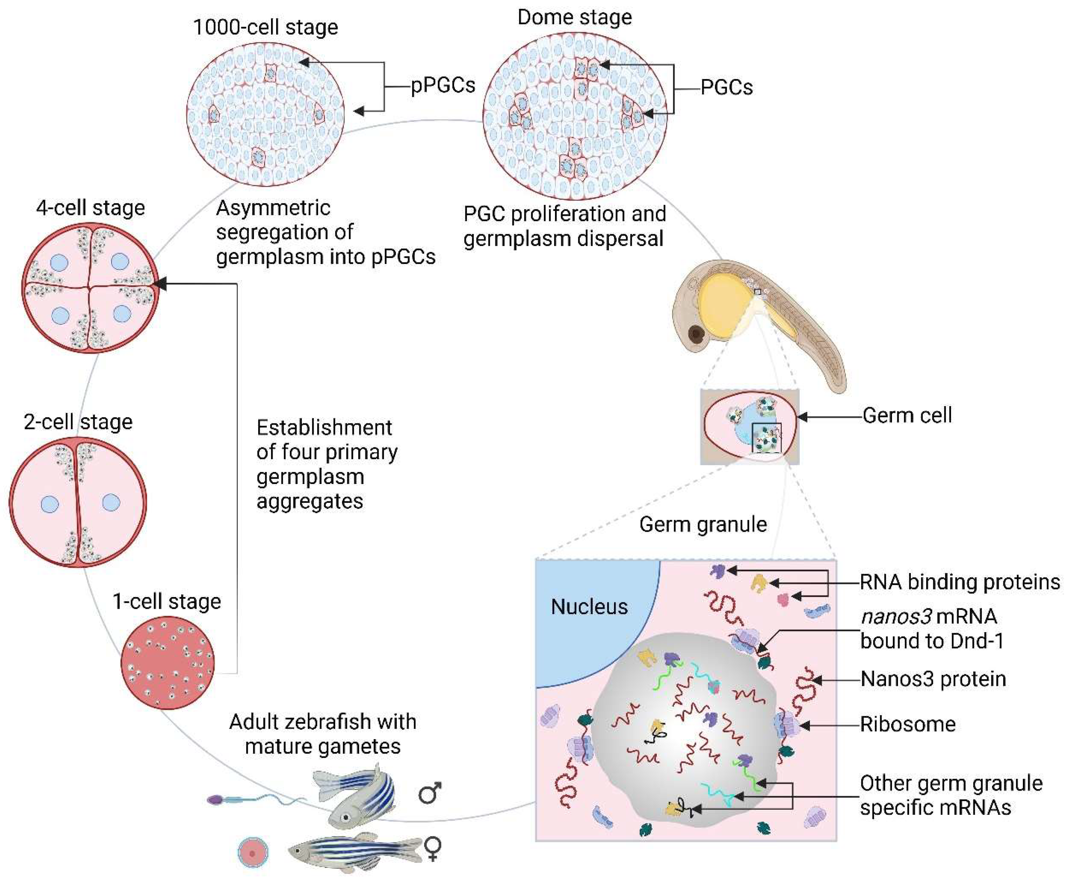
2.4. Nanos1 mRNA Localization and Post-Transcriptional Regulation in D. rerio Germ Cell Development

Fishes, particularly within closely related phylogenetic groups, exhibit strong conservation of germ plasm components, with key mRNAs such as dnd, nanos, vasa, and dazl found across multiple teleost species [88,89,90]. In D. rerio, these mRNAs co-localize with the Bb during early oogenesis and are transported to the vegetal cortex, with their distribution changing as oogenesis progresses (Figure 5) [91]. The nanos1 paralogue is expressed in early-stage germ cells in larval and adult ovaries [92]. Extensive research on D. rerio has demonstrated the presence of maternally derived germ plasm and identified similar localization patterns of maternal determinants in at least a dozen other teleost species, including Oryzias latipes (medaka), highlighting conservation across diverse evolutionary timelines [10,93,94,95].
Upon oocyte fertilization in D. rerio, maternally derived germ plasm RNPs aggregate at the embryonic cleavage furrows with the help of cytoskeletal components (Figure 5) [96,97,98,99]. These interactions enable the germplasm to localize at the distal ends of the first two cleavage furrows. During embryonic cellularization, the emerging four germ plasm aggregates are internalized into PGCs, which are asymmetrically inherited by a single daughter cell during subsequent divisions [100,101]. These germ plasm aggregates remain largely intact through the late cleavage, blastula, and early gastrula stages, ultimately leading to embryos with 4-12 cells containing germ plasm (Figure 5).
In D. rerio, nanos1 protein, though dispersed throughout the cytoplasm of germ cells, accumulates in perinuclear granules with vasa protein, suggesting a role linked to nuclear export [20]. Localisation of vasa within these granules in Danio, was also found in one-day-old embryos [101]. Similarly, in Caenorhabditis and Xenopus, germ plasm components concentrate near nuclear pores (nuage) via perinuclear granules [59].
The specific localization of nanos1 mRNA in zebrafish PGCs is controlled by mechanisms of asymmetry, RNA stability, and translational regulation [47,102] — mechanisms conserved between Drosophila and Danio [20]. For instance, non-localized nanos1 RNA in both species is repressed and degraded in somatic cells [47,102], while miR-430 targets like nanos and tdrd7 mRNAs are protected from repression in germ cells [103]. This protection is attributed to the RBP dnd, which counteracts miR-430-mediated degradation in PGCs through binding to specific regions within the 3’UTR of nanos1 and tdrd7 mRNAs [104,105]. This regulation ensures that the nanos1 protein is expressed exclusively in PGCs despite the widespread distribution of its mRNA in early Danio embryos. Similarly, in O. latipes, the differential stability of vasa and nanos1 mRNA between somatic cells and PGCs, which mirrors that seen in Danio, is dependent on the nanos1 3’UTR, indicating a conserved post-transcriptional regulation mechanism across species [94]. This further emphasizes the evolutionary conservation of nanos1 regulation and highlights the crucial role of 3’UTR-mediated control in maintaining germ cell-specific expression of key mRNAs in teleosts.
In addition to governing regulation, the localization of Danio nanos1 mRNA is also governed by its 3’UTR, which contains specific elements such as the MCLE (mitochondrial cloud localization element, with the mitochondrial cloud also known as the Bb) and the GGLE (germinal granule localization element). This is akin to Xenopus nanos1 3’UTR, which contains the UGCAC motif [75,106]. The role of the UGCAC motif within the nanos1 mRNA MCLE highlights the conservation of this mechanism. However, the 3’UTRs of other Danio germ plasm mRNAs typically lack multiple UGCAC motifs [91].
In Danio, the bucky ball (buc) protein plays a central role in Bb formation by directing the assembly of germ plasm elements, such as nanos, vasa, and dazl mRNAs [107]. Functional similarities between buc in Danio and oskar in Drosophila have been observed, particularly in germ cell development [108]. Both oskar and buc possess IDRs, contributing to protein aggregation. The study by Krishnakumar et al. (2018) also showed that buc interacts with vasa protein and nanos mRNA, like oskar, suggesting conserved biochemical functions across species. These findings provide a molecular basis for how two evolutionarily distinct proteins perform similar roles in organizing germ plasm, indicating they form a conserved core complex essential for germ cell development.
The Bb plays a crucial role in establishing animal-vegetal polarity in the oocyte by managing the aggregation and expression of germ plasm mRNAs [109]. After fertilization, germ plasm RNPs are directed to the cleavage furrows as previously shown in Figure 5 [96,110,111,112,113]. Despite forming densely packed supramolecular aggregates resembling germ granules, individual germ plasm RNPs are homotypic, each containing only one type of RNA, such as nanos3 mRNA [114]. This homotypic clustering is maintained throughout the initial cortex assembly, the recruitment of the germ plasm, its asymmetric division, and ultimately, the dispersal of RNPs into the cytoplasm of PGCs towards the end of the blastula stage [114].
This phenomenon mirrors observations in Drosophila, where homotypic RNA clustering within germ granules suggests a potentially conserved self-aggregation mechanism across species for RNAs targeted to PGCs [115,116]. In contrast, while RNPs in Danio appear as distinct spherical entities [71], in Drosophila, homotypic RNA clusters reside within larger granules containing proteins like vasa, tudor, and oskar [115,116]. However, the exact process of RNP formation remains unclear, though RNA-RNA interactions are believed to play a role, as evidenced in various studies [117,118].
These findings support the idea that germ plasm functions as a membraneless organelle formed by fluid-phase properties, playing a critical role in germ cell development [119,120]. There may be distinct pathways for RNA recruitment into the germ plasm, leading to compartmentalization where different classes of germ plasm RNAs initially occupy overlapping but distinct regions. One hypothesis proposes that a common protein factor binds to different RNAs during early oogenesis, facilitating germ plasm assembly [91]. However, the precise role of these granules in determining germ cell fate remains only partially understood. Germline development relies heavily on the complex regulation of RNA and proteins, which are prominently concentrated within germ granules. These phase-separated, membraneless condensates are crucial for various stages of germline development. The presence of RNA-binding proteins in these granules highlights their significant role in post-transcriptional regulation [39].
One such protein is dnd1, which plays a key role in maintaining vertebrate germ cell fate [121]. Dnd1 enhances nanos expression by promoting its translation and RNA stability, a regulatory function supported by studies in Drosophila, Xenopus, and Caenorhabditis, where nanos inhibit somatic differentiation in the germline. This mirrors the effects observed in Danio PGCs lacking dnd1 [19,87,105].
In Danio, the germplasm contains crucial mRNAs like nanos3, which play a vital role in maintaining a renewable germline stem cell pool by inhibiting their differentiation into oocytes [46,122]. Mutations in nanos3 result in the loss of germline stem cells and infertility. Three-dimensional in vivo studies on the molecular dynamic organisation of germ granules in Danio reveal that nanos3 mRNA is positioned at the edges of germ granules, where ribosomes are found, indicating active translation. Dnd1 is essential for maintaining this localization; when dnd1 is absent or translation is inhibited, nanos3 mRNA shifts toward the granule core, reducing nanos3 protein levels and losing germ cell fate. This highlights the importance of sub-granule compartmentalization in post-transcriptional regulation and germ cell totipotency [123].
Additionally, Dnd1 prevents nanos3 mRNA from migrating to the granule core, ensuring the formation of active translation complexes at the periphery. This peripheral localization mirrors Drosophila polar granules, where nanos protein translation also occurs at the granule edges. However, ribosomes and nanos disappear from these sites before germ cell precursor formation [124].
Recent studies reveal that rapid RNA translation is crucial for distinguishing germ cells from somatic cells during early germ cell development, while a portion of mRNAs is stored for future translation. These studies also suggest that translationally repressed mRNAs gather within germ granules, where they are stored and protected from decay [125,126]. For example, the behaviour of nanos3 mRNA supports the idea that germ granules serve dual roles. Some mRNAs are localized and actively translated at the granule's periphery, while others are stored in the core and protected for future use. This core-periphery organization allows for precise control of mRNA translation, which is critical for regulating germline development at various stages.
This mechanism is seen across species such as Caenorhabditis, Drosophila, and Xenopus, where the localization of maternal mRNAs is essential for PGC formation. In Drosophila, for instance, the localization of oskar and nanos mRNAs at the posterior pole of the oocyte is crucial for germ plasm assembly and germ cell function [13,14]. Similarly, maternal mRNAs like nanos1 and dazl are incorporated into the germ plasm via the messenger transport organizer (METRO) pathway in Xenopus. These mRNAs associate with the Bb and are localized through non-directed movement and entrapment within the Bb [73,75,76].
Overall, these findings highlight the conserved role of germ granules in regulating nanos mRNA localization and translation across species, underscoring their critical function in orchestrating germ cell development and ensuring proper germline formation. Given the evolutionary conservation of these mechanisms, similar processes may play a pivotal role in germline development in humans and other mammals, providing valuable insights into fertility and germ cell regulation.

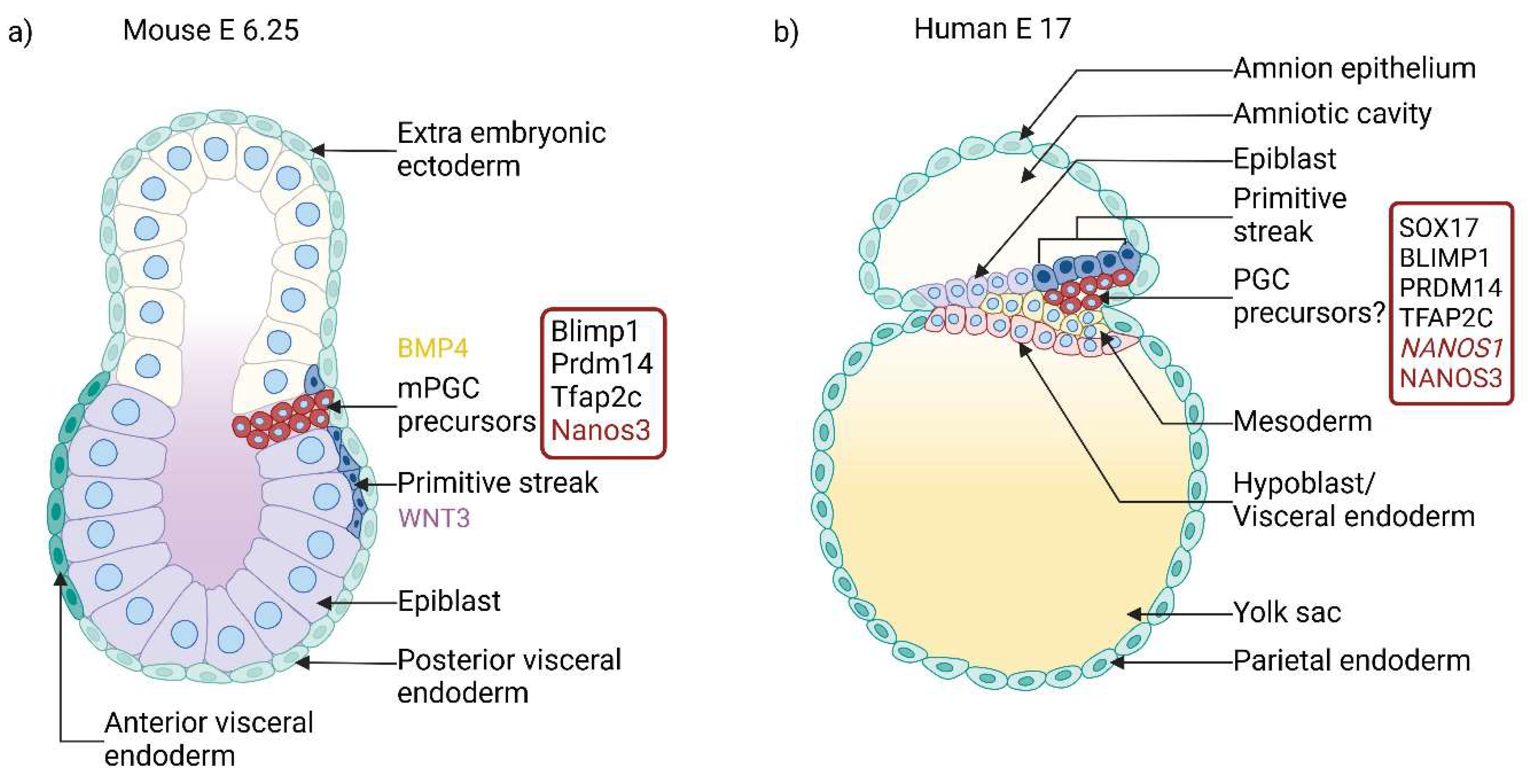
2.5. Mammalian Nanos Homologues Are Also Present in Nuage

In mice undergoing inductive germ cell specification, PGCs arise from a subset of cells located within the proximal epiblast of the developing embryo (Figure 6) for review, see [3]. Notably, the nuage structure has not been observed in epiblast cells at the time of germ cell specification but appears later in PGCs [127,128]. Similarly, in humans, PGCs are thought to originate from the posterior epiblast (Figure 6) [129]. In human development, nuage formation is first detected around week 7 of embryogenesis, coinciding with the migration of PGCs to the primary gonads. From this point onward, nuage undergoes dynamic morphological changes throughout the germ cell [37,130].
In male foetal germ cells, the number of cells containing nuage increases until week 13 of gestation, while in female foetal germ cells, the proportion is even higher. At week 7, nuage is randomly distributed within the cytoplasm of oogonia and spermatogonia. However, by week 13, it becomes predominantly perinuclear in female and male germ cells [37]. This spatial reorganization of nuage parallels similar observations in other model organisms, such as Drosophila, Caenorhabditis, Xenopus, and Danio. Notably, the nuage remains a persistent feature in mammalian germ cells from their initial specification to the formation of mature oocytes and spermatozoa.
In addition to structural changes such as nuage formation, the regulation of key genes like Nanos plays a crucial role in germ cell specification and development in mammals. In mice, studies have shown that Nanos3 mRNA is present in PGCs from their specification at embryonic day (E) 7.0 through E12.5, the stage when PGCs reach the gonads [33]. In contrast, Nanos1 mRNA is not expressed during this early period but is detected later, during the spermatid stage of spermatogenesis [131]. Interestingly, maternally expressed Nanos1 mRNA is abundant in oocytes but rapidly declines after fertilization, with transient expression observed in day 8 embryos and the blastocyst stage. Nanos1 localization in eggs shows no specific spatial sub-localisation and remains homogeneously distributed within the cytoplasm [131]. Disruption of Nanos1 does not lead to infertility or developmental abnormalities in mice, suggesting it is dispensable for mouse germ cell development [131]. In contrast, knockout of Nanos2 and Nanos3 results in infertility, indicating its essential role in germ cell development [33].
In humans, two NANOS proteins, NANOS1 and NANOS3, are expressed during PGC specification, with both upregulated 2–3 weeks after fertilization [7]. NANOS3 is considered a key marker for mammalian PGCs. Unlike in mice, disruption of NANOS1 in humans leads to the absence of germ cells in seminiferous tubules [34]. Instead, NANOS3 disruption is linked to premature ovarian failure [132]. These findings illustrate that although Nanos paralogues are highly conserved across species, their roles in germ cell development can vary significantly in different groups of animals.
Similar to simple organisms, the Bb in mice plays a crucial role in oocyte quality control. In this process, interconnected oocyte cysts transfer cytoplasmic contents, including mitochondria [133], to neighbouring oocytes, ensuring that Bb-containing oocytes survive—a mechanism reminiscent of Drosophila nurse cells, which similarly transfer their contents, including nanos mRNA, to growing oocytes [133].Top of Form
On the other hand, during mammalian spermatogenesis, the nuage undergoes significant structural evolution, eventually forming the chromatoid body (CB). Initially observed as dispersed aggregates, the nuage condenses into a single, spherical body at the round spermatid stage [127] (Figure 7). The CB, which attaches to and moves along the nuclear membrane near nuclear pores, contains over 30 proteins, including RBPs [134] that were initially part of the nuage. Defects in these proteins can lead to spermatogenic failure, as demonstrated in mice. For instance, the vasa protein, a marker of nuage from nematodes to vertebrates and a key component of the CB, is essential for male fertility, though it is expressed in both sexes [135]. Vasa protein, a DEAD-box helicase, likely functions as an RNA chaperone within the nuage and CB.
The CB is described as an RNA processing centre [136], and its origin is thought to stem from the IMC structure in spermatogonia [130]. Unlike oocytes, sperm do not transmit large quantities of mRNAs for embryonic development [137]. Instead, developing spermatids, which cease mRNA transcription during genome compaction, rely heavily on post-transcriptional mechanisms for sperm differentiation. The CB is pivotal as an RNA-rich granule that develops from smaller granules in late pachytene spermatocytes. By the round spermatid stage, when transcriptional activity ceases, these granules condense into the mature CB (Figure 7). For review, see [136]. In humans and mice, NANOS1/Nanos1 proteins localize within the CB, interacting with the RBP PUM2 (Figure 7) [138]. This NANOS1-PUM2 complex forms a critical regulatory hub for RNA processing and localization, conserved across species from Drosophila to humans [23]. Both NANOS1 and PUM2 colocalize with the VASA protein, further highlighting the CB's critical role in RNA regulation. Additionally, NANOS1 and PUM2 interact with GEMIN3, a protein involved in microRNA biogenesis [138], reinforcing the CB’s established function in RNA processing, particularly through microRNA pathways [136].
A pathogenic variant of NANOS1, linked to the absence of germ cells in seminiferous tubules in male patients, exhibits weakened interaction with GEMIN3 compared to the wild-type, as demonstrated by a quantitative yeast two-hybrid assay [34]. This suggests proper protein-protein interactions within the CB are crucial for its function. Moreover, this NANOS1 variant shifts from its regular anti-apoptotic role to a pro-apoptotic function, as shown in the TCam2 cell line, which mimics human primordial germ cells entering the gonads [139]. In conclusion, Nanos proteins, particularly Nanos1, play an essential role within the CB, ensuring the proper regulation of RNA processing in both mice and humans. Their interactions within the CB are crucial for germ cell survival and differentiation, with disruptions leading to severe germ cell defects and infertility. This highlights the conserved and vital role of NANOS1 in the molecular machinery of germ cell development across species.

3. The Ubiquitous Cytoplasmic Granules—Their Role in Germ Cells

Aside from the various forms of nuage, a hallmark of germ cells, these cells also contain other types of membraneless cytoplasmic granules composed of RNPs, such as processing bodies (PBs) and stress granules (SGs). While these granules are found in many cell types, they have a unique composition and specialised functions in germ cells. Nanos proteins are key components of these structures and play critical roles in their regulation and activity.

3.1. Processing Bodies

PBs are membraneless, liquid-liquid phase separation granules responsible for RNA storage and degradation for review, see [140,141]. PBs exhibit dynamic behaviour in germ cells, varying by developmental stage and sex. For example, in mice, PBs gradually disappear by E14.5 in female gonocytes but persist in male germ cells until at least E15.5, while being absent in somatic cells. It is hypothesized that PBs temporarily store translationally repressed but competent mRNAs and their regulators [137]. Male germ cell PBs also contain germline-specific proteins, such as Nanos2, first identified as enriched in PBs in mouse male gonocytes [84].
Nanos2 plays a critical role in male germ cell development by localizing to PBs and interacting with key proteins such as Ddx6 (also known as Rck/p54) and components of the CCR4-NOT deadenylation complex, which mediate mRNA degradation and regulation. In Ddx6-deficient germ cells in mouse chimeric embryos, PBs fail to form properly in gonocytes, resulting in the cytoplasmic dispersion of Nanos2 [142]. The N-terminus of Nanos2 is essential for its localization to PBs and its interaction with Cnot1, a crucial scaffold protein in the CCR4-NOT complex, necessary for proper mRNA degradation and storage [26]. This interaction is functionally essential for Nanos2 in vivo and is RNA-independent, although the mechanisms by which the Nanos2-CCR4-NOT deadenylation complex selects mRNAs have been partially elucidated recently [84]. Additionally, Nanos2 interacts with the RNA-binding protein Dnd1 via its zinc-finger domain, contributing to PB assembly in male gonocytes [143]. This interaction is also necessary for localising the complex in PBs [26,143], a mechanism that may be unique to male mouse gonocytes. The Dnd1-Nanos2 complex plays a key role in recognizing target mRNAs, with Nanos2 enhancing Dnd1's ability to bind target mRNAs and form a ternary complex mediated by Dnd1's RNA recognition motif domains. Thus, Nanos2 functions as a second-layer RBP protein, facilitating Dnd1's target recognition and functional adaptation [144]. mRNAs recruited to PBs by Nanos2 have two possible outcomes: degradation, which prevents premature expression of pathways in germ cells, or storage for future readenylation and translation. For instance, within PBs, Nanos2 has been shown to repress meiotic mRNAs, such as Sycp3, Stra8, Taf71, Dazl, and Meisetz. Notably, these mRNAs were detected only in Nanos2 precipitates despite their low expression in male gonads [84]. Interestingly, both Nanos2 and its paralog Nanos3 localize to PBs during mouse germ cell development. Nanos3 colocalizes with the PB marker Dcp1a, indicating that both proteins play roles in PB assembly and function [145]. Moreover, human NANOS1–3 also interact with human CNOT1, highlighting the conserved role of Nanos proteins in RNA regulation across species [146].
Despite these findings, the localization of Nanos homologs in relation to PBs has not been extensively studied outside mice. However, Nanos2 has also been found to localize to PBs in dairy goats [147]. The emerging view suggests that Nanos activity throughout germ cell development is closely linked to RNA silencing and decay mediated by PB-associated factors [137]. Future research is needed to determine whether Nanos localization to PBs is conserved beyond certain species. Overall, Nanos proteins, particularly Nanos2, play a pivotal role in germ cell development by mediating mRNA regulation through their interactions with PBs. These proteins are essential for both mRNA degradation and storage, with significant implications for controlling gene expression during critical stages of germ cell differentiation. Understanding the conservation of Nanos-PB interactions across species will be crucial for elucidating the broader biological significance of these mechanisms.

3.2. Stress Granules

SGs are cytoplasmic aggregates of mRNPs that form stalled translational preinitiation complexes in response to stress, such as heat shock, oxidative stress, ischemia, or viral infection [148,149]. SGs are essential for the homeostasis of germ cells, as the knockout of the SG marker Tial1 in mice resulted in the failure of PGCs to survive during migration. The formation of SGs, as well as PBs, requires the assembly of untranslated mRNAs with RBPs that contain aggregation domains, such as the aforementioned Ddx6, which has a prion-like domain for condensation and localizes to both PBs and SGs [150]. The Nanos2 mouse paralog interacts with Ddx6, as well as with the PB marker Dcp1a, and colocalizes with Ddx6 in SGs [84]. In addition, Nanos3 has been found in SGs of day E7.5 PGCs, where it colocalizes with Tial1 and phosphorylated eukaryotic initiation factor 2α (eIF2α), both of which are SG markers, while a fraction of Nanos3 also colocalizes with Dcp1a in PBs [145]. Knockout of Nanos3 in mice leads to a phenotype similar to Tial1 knockout, resulting in infertility [33,151]. These data suggest that all three Nanos paralogs may regulate post-transcriptional gene expression in both SGs and PBs in mouse PGCs [145].
The regulatory role of Nanos paralogs has also been studied at later stages of germ cell development, particularly in spermatogonial stem cells (SSCs). An essential role for Nanos2-containing mRNPs in maintaining SSCs in mice was demonstrated [152]. These authors showed that Nanos2-containing mRNPs are present in the most primitive SSCs, with the highest self-renewal activity. The primary function of Nanos2 in these cells is to inhibit SSC differentiation. Nanos2 accomplishes this by directly recruiting and repressing mRNAs that promote differentiation, such as Sohlh2, Dmrt1, and Dazl, and by sequestering and attenuating mTORC1 activity, a critical signalling pathway for both SSC growth and differentiation Phillips CM [152]. Repression of Sohlh2 mRNA by Nanos2 occurs in a 3’UTR-dependent manner, recruiting the mRNA to mRNPs in mouse germline stem cells [152]. Nanos2 achieves this inhibition by condensing its own mRNPs. This dual activity of Nanos2 is essential for SSC homeostasis in the adult testis, as demonstrated in vivo [152].
In conclusion, the Nanos paralogs, particularly Nanos2 and Nanos3, play critical roles in regulating post-transcriptional gene expression within SGs and PBs during germ cell development. Their involvement in mRNA repression, storage, and interaction with key signalling pathways is essential for maintaining germ cell homeostasis, differentiation, and fertility. Understanding these mechanisms further highlights the significance of Nanos-mediated regulation in germline stem cell biology.

4. Significance of 3’UTR Mediated Post-Transcriptional Regulation in Germ Cell Granules

All the previously described cytoplasmic structures in germ cells — different forms of nuage, including PBs, and SGs—are composed of RNP complexes, which consist of mRNAs and RBPs with distinct compositions. These RNPs serve as critical sites for post-transcriptional regulation, modulating specific mRNAs' stability, localization, and translation. The specificity of these interactions, particularly the selection of mRNAs for regulation, is largely determined by elements within the 3’UTRs of target mRNAs. These regions contain specific nucleotide motifs or secondary structures recognized by RBPs, directing the fate of the mRNA—whether it will undergo translation, repression, degradation, or localization to a specific subcellular compartment. The composition of these RNPs is highly dynamic, changing in response to developmental cues or cellular signals, and this temporal regulation ensures that mRNAs are appropriately processed according to the cell’s needs at any given time. Nanos proteins, in cooperation with other RBPs, such as Pumilio, are integral to this process. By interacting with target mRNAs within these complexes, Nanos proteins play critical roles in modulating mRNA fate, particularly in germ cells, where they help preserve germline totipotency and prevent premature differentiation. Through this regulation, Nanos and Pumilio are essential for maintaining the "immortality" of the germline, ensuring the continuity of genetic information across generations [30].
In summary, the dynamic interactions between Nanos, Pumilio, and other RBPs within cytoplasmic RNP structures are fundamental to the post-transcriptional regulation of mRNAs in germ cells. They influence crucial processes such as mRNA stability, translation, and cellular localization and ultimately contribute to the preservation of germline integrity.

5. Germ Granules and Nanos: Dynamic Hubs of Post-Transcriptional Regulation and Germline Integrity

Structurally, germ granules are liquid-liquid phase-separating condensates that exchange components with the cytoplasm. This phenomenon occurs when the interactions between molecules are more thermodynamically favourable than their interactions with the solvent [153,154,155,156]. Based on current literature, phase separation is induced by multivalent low-affinity protein-protein, protein-RNA, and RNA-RNA interactions [157]. Phase separation is essential in various stages of germline development, driving the formation and partitioning of germ granules that establish germ cell identity during early embryogenesis with pioneering work in Caenorhabditis [67].
Germ granules comprise mRNPs that regulate post-transcriptional gene expression in germ cells across species, playing a crucial role in maintaining germline integrity. Their strategic location near nuclear pores allows for the selective processing and reshaping of mRNAs within the mRNPs as they exit the nucleus, ensuring their proper regulation before reaching the cytoplasm. For example, it facilitates the interaction between mRNPs and membrane-bound organelles to form the Bb in Xenopus and Danio oocytes for review, see [158]. The formation of active translation complexes at the periphery of phase-separated germ granules is critical for maintaining mRNA clusters at the condensate's edge, as recent studies have shown [123]. This mechanism is fundamental in species such as Drosophila, Caenorhabditis, Danio, and Xenopus, where germ cells rely extensively on maternal RNA and proteins throughout prolonged stages of development for review, see [159]. In Danio, nanos3 mRNA is actively translated at the edges of germ granules, a localization maintained by the dnd1 protein. When dnd1 is absent, or translation is inhibited, nanos3 mRNA shifts to the granule core, resulting in reduced nanos3 protein levels and a loss of germ cell fate [123]. This arrangement mirrors Drosophila polar granules, where translation occurs at the periphery [124]. Furthermore, in Drosophila, single-molecule imaging has demonstrated that nanos mRNA translation occurs precisely at the surface of germ granules [160]. At this location, the coding region of the mRNA, associated with polysomes for active translation, remains exposed on the granule's surface, while the 3’UTR is anchored internally. In contrast, translation is inhibited when the 5’ untranslated region (5’UTR) is positioned inside the granule. The previously mentioned RBP smaug, which represses nanos mRNA translation in somatic cells, is inactivated in the posterior of the embryo by being sequestered into the interior of germ granules by oskar protein. This study suggests that actively translating mRNAs adopt an extended conformation, while untranslated mRNAs become folded or compacted [160].
Altogether, these findings confirm that germ granules have dual roles: some mRNAs are actively translated at the periphery, while others are stored in the core for future use [125,126]. Further studies should investigate the roles of proteins beyond Dnd1 and oskar in modulating the placement and regulation of various RNA molecules, including nanos. Moreover, while these findings emphasize the significance of translation at the edges of germ granules, the importance of translation on the surface of granules compared to the cytoplasm at different stages of germ cell development has yet to be fully explored.
In Caenorhabditis, the localization of nanos mRNA within P granules is associated with translational repression [125,126]. However, studies in this organism have shown that germline mRNA translation can still occur even in the absence of germ granules [125], and P granules themselves are largely non-essential for fertility. This suggests species-specific differences that govern how maternal transcripts are maintained and utilized [125]. Future research should aim to determine the criticality of each translation site for protein regulation.
Notably, proteins with IDRs are key contributors to condensation, promoting phase separation in germ granules [153,155,156]. Examples include meg3 in Caenorhabditis [125], Buc in Danio [107], xvelo1 in Xenopus [69], and oskar in Drosophila for review, see [10,161]. In humans, the N-terminal IDR of NANOS1 appears functionally important for interactions with proteins like GEMIN3, a factor in microRNA biogenesis [138]. However, the potential role of this IDR in condensation has not yet been demonstrated. Similarly, the N-terminal region of Caenorhabditis nanos-3 is predicted to be intrinsically disordered and functionally significant, containing binding domains for the PUM homolog fbf [68,162].
Interestingly, protein phase separation can be modulated by post-translational modifications, particularly phosphorylation [156,163]. In this context, it is possible that the serine residue missing from the NANOS1 IDR at position 78 in patients lacking germ cells in seminiferous tubules [34] could be sensitive to phosphorylation. Its deletion may contribute to the infertility phenotype by altering the condensate properties of germ granules.
Additionally, RNA itself has been shown to phase-separate in vitro, RNA-RNA interactions playing a pivotal role in phase separation as well as in the formation of membraneless compartments with specific RNAs enriched in various in vivo location for review, see [158,164]
mRNA modifications also play a crucial role in germ granule dynamics. For example, in Danio, over a third of maternal mRNAs, including germ plasm mRNAs, are highly modified with N6-methyladenosine (m6A) residues [165]. The RNA-binding protein Igf2bp3, an m6A reader, plays a key role in germ plasm assembly and germ cell specification by protecting germ plasm mRNAs from decay during development [109,166,167]. Igf2bp3 is recruited by the buc protein, which binds in a KH domain-dependent manner, and their interaction is critical for regulating germ plasm mRNAs. The loss of Igf2bp3 results in defective germ plasm assembly and germ cell specification, mirroring the effects of buc deletion, highlighting Igf2bp3 as a key effector in Buc-regulated gene expression and germ plasm assembly [109].
In summary, germ granules act as hubs for precise post-transcriptional regulation, where sub-granule compartmentalization facilitates active translation at the periphery while storing other mRNAs for future use. This mechanism is fundamental for germ cell specification and development across multiple species. Future research should focus on elucidating the molecular dynamics of mRNA localization, phase separation, and protein interactions within germ granules, with particular focus on RNA modifications, species-specific differences, and the role of germ granules at different stages of germ cell development.

6. Conclusions and Perspectives

As highlighted in the review, nanos is a critical factor for germ cell specification and development across diverse species, serving as a highly conserved structural component of germ granules. Disruption of nanos leads to infertility in a wide range of organisms, from Drosophila to humans. However, the precise mechanism of germ granule formation in mammalian PGCs remains unclear, and no specific germ granule nucleator has been identified in mammals. Several questions arise regarding the role of nanos in this process.
One intriguing possibility is that the IDRs of nanos proteins, such as NANOS1, may contribute to forming phase-separated condensates. If this is the case, is NANOS1 required to initiate germ granule nucleation? Additionally, do mutations in NANOS1 or NANOS3 disrupt the structure or function of germ granules? If so, could these disruptions be linked to post-translational modifications like phosphorylation?
The availability of in vitro systems for deriving human PGCs from stem cells offers significant potential for investigating key aspects of germ cell development, particularly the timing and mechanisms of nuage formation in germ cells following an inductive specification pathway of germ cell specification, including humans. Current observations indicate that nuage formation in human embryonic development occurs around week 7, coinciding with the migration of PGCs to the primary gonads. However, it remains to be determined whether the nucleation of nuage-associated RNAs and proteins begins earlier in development and whether Nanos proteins contribute to this early process. Additionally, Nanos mRNA itself may play a role in nuage formation before being translated, emphasizing its multifaceted role in germ cell development.
Addressing these questions will help clarify the role of NANOS proteins in human reproduction and shed light on their broader significance in germ granule formation and germ cell development. A key challenge for future research will be to understand the mechanistic pathways via which NANOS mRNA and protein works to localize, silence, translate, and degrade specific mRNAs during the germline's life cycle. Advances in single-molecule technologies, which allow for the visualization of RNA processes within cells, offer significant potential to advance this field.

Author Contributions

Bellary Lakshmi prepared all the Figures and wrote part of the manuscript, Matisa Alla wrote part of the manuscript and reviewed the whole paper, and Jadwiga Jaruzelska wrote part of the manuscript and reviewed the whole paper.

Funding

This paper was supported by the project nr 2019/35/B/NZ1/01665 from the National Sciences Centre Poland.

Acknowledgments

We thank our Colleague, dr Kusz-Zamelczyk, for reading the manuscript and providing critical comments.

Conflicts of Interest

The authors declare no conflict of interest.

References

  1. White, J.A.; Heasman, J. , Maternal control of pattern formation in Xenopus laevis. J Exp Zool B Mol Dev Evol 2008, 310, 73–84. [Google Scholar] [CrossRef] [PubMed]
  2. Venkatarama, T.; Lai, F.; Luo, X.; Zhou, Y.; Newman, K.; King, M.L. , Repression of zygotic gene expression in the Xenopus germline. Development 2010, 137, 651–660. [Google Scholar] [CrossRef] [PubMed]
  3. Kumar, D.L.; DeFalco, T. , Of Mice and Men: In Vivo and In Vitro Studies of Primordial Germ Cell Specification. Semin Reprod Med 2017, 35, 139–146. [Google Scholar] [CrossRef] [PubMed]
  4. Kobayashi, T.; Surani, M.A. , On the origin of the human germline. Development 2018, 145. [Google Scholar] [CrossRef]
  5. Kojima, I.; Nakagawa, Y.; Ohtsu, Y.; Hamano, K.; Medina, J.; Nagasawa, M. , Return of the glucoreceptor: Glucose activates the glucose-sensing receptor T1R3 and facilitates metabolism in pancreatic beta-cells. J Diabetes Investig 2015, 6, 256–263. [Google Scholar] [CrossRef]
  6. Magnusdottir, E.; Surani, M.A. , How to make a primordial germ cell. Development 2014, 141, 245–252. [Google Scholar] [CrossRef]
  7. Sybirna, A.; Tang, W.W.C.; Pierson Smela, M.; Dietmann, S.; Gruhn, W.H.; Brosh, R.; Surani, M.A. , A critical role of PRDM14 in human primordial germ cell fate revealed by inducible degrons. Nat Commun 2020, 11, 1282. [Google Scholar] [CrossRef]
  8. Kobayashi, T.; Zhang, H.; Tang, W.W.C.; Irie, N.; Withey, S.; Klisch, D.; Sybirna, A.; Dietmann, S.; Contreras, D.A.; Webb, R.; Allegrucci, C.; Alberio, R.; Surani, M.A. Principles of early human development and germ cell program from conserved model systems. Nature 2017, 546, 416–420. [Google Scholar] [CrossRef]
  9. Yamauchi, K.; Hasegawa, K.; Chuma, S.; Nakatsuji, N.; Suemori, H. , In vitro germ cell differentiation from cynomolgus monkey embryonic stem cells. PLoS One 2009, 4, e5338. [Google Scholar] [CrossRef]
  10. Hansen, C.L.; Pelegri, F. , Primordial Germ Cell Specification in Vertebrate Embryos: Phylogenetic Distribution and Conserved Molecular Features of Preformation and Induction. Front Cell Dev Biol 2021, 9, 730332. [Google Scholar] [CrossRef]
  11. Strome, S.; Updike, D. , Specifying and protecting germ cell fate. Nat Rev Mol Cell Biol 2015, 16, 406–416. [Google Scholar] [CrossRef] [PubMed]
  12. Gerstberger, S.; Hafner, M.; Tuschl, T. , A census of human RNA-binding proteins. Nat Rev Genet 2014, 15, 829–845. [Google Scholar] [CrossRef] [PubMed]
  13. Ephrussi, A.; Dickinson, L.K.; Lehmann, R. , Oskar organizes the germ plasm and directs localization of the posterior determinant nanos. Cell 1991, 66, 37–50. [Google Scholar] [CrossRef] [PubMed]
  14. Wang, C.; Lehmann, R. , Nanos is the localized posterior determinant in Drosophila. Cell 1991, 66, 637–647. [Google Scholar] [CrossRef] [PubMed]
  15. Extavour, C.G.; Pang, K.; Matus, D.Q.; Martindale, M.Q. , vasa and nanos expression patterns in a sea anemone and the evolution of bilaterian germ cell specification mechanisms. Evol Dev 2005, 7, 201–215. [Google Scholar] [CrossRef]
  16. Subramaniam, K.; Seydoux, G. , nos-1 and nos-2, two genes related to Drosophila nanos, regulate primordial germ cell development and survival in Caenorhabditis elegans. Development 1999, 126, 4861–4871. [Google Scholar] [CrossRef]
  17. Wang, Y.; Zayas, R.M.; Guo, T.; Newmark, P.A. , nanos function is essential for development and regeneration of planarian germ cells. Proc Natl Acad Sci U S A 2007, 104, 5901–5906. [Google Scholar] [CrossRef]
  18. Juliano, C.E.; Yajima, M.; Wessel, G.M. , Nanos functions to maintain the fate of the small micromere lineage in the sea urchin embryo. Dev Biol 2010, 337, 220–232. [Google Scholar] [CrossRef]
  19. Lai, F.; Singh, A.; King, M.L. , Xenopus Nanos1 is required to prevent endoderm gene expression and apoptosis in primordial germ cells. Development 2012, 139, 1476–1486. [Google Scholar] [CrossRef]
  20. Koprunner, M.; Thisse, C.; Thisse, B.; Raz, E. , A zebrafish nanos-related gene is essential for the development of primordial germ cells. Genes Dev 2001, 15, 2877–2885. [Google Scholar] [CrossRef]
  21. Saga, Y. , Mouse germ cell development during embryogenesis. Curr Opin Genet Dev 2008, 18, 337–341. [Google Scholar] [CrossRef] [PubMed]
  22. Saga, Y. , Sexual development of mouse germ cells: Nanos2 promotes the male germ cell fate by suppressing the female pathway. Dev Growth Differ 2008, 50 Suppl 1, S141–7. [Google Scholar] [CrossRef]
  23. Jaruzelska, J.; Kotecki, M.; Kusz, K.; Spik, A.; Firpo, M.; Reijo Pera, R.A. , Conservation of a Pumilio-Nanos complex from Drosophila germ plasm to human germ cells. Dev Genes Evol 2003, 213, 120–126. [Google Scholar] [CrossRef] [PubMed]
  24. De Keuckelaere, E.; Hulpiau, P.; Saeys, Y.; Berx, G.; van Roy, F. , Nanos genes and their role in development and beyond. Cell Mol Life Sci 2018, 75, 1929–1946. [Google Scholar] [CrossRef] [PubMed]
  25. Bhandari, D.; Raisch, T.; Weichenrieder, O.; Jonas, S.; Izaurralde, E. , Structural basis for the Nanos-mediated recruitment of the CCR4-NOT complex and translational repression. Genes Dev 2014, 28, 888–901. [Google Scholar] [CrossRef] [PubMed]
  26. Suzuki, A.; Saba, R.; Miyoshi, K.; Morita, Y.; Saga, Y. , Interaction between NANOS2 and the CCR4-NOT deadenylation complex is essential for male germ cell development in mouse. PLoS One 2012, 7, e33558. [Google Scholar] [CrossRef]
  27. White, E.K.; Moore-Jarrett, T.; Ruley, H.E. , PUM2, a novel murine puf protein, and its consensus RNA-binding site. RNA 2001, 7, 1855–1866. [Google Scholar]
  28. Wang, X.; McLachlan, J.; Zamore, P.D.; Hall, T.M. , Modular recognition of RNA by a human pumilio-homology domain. Cell 2002, 110, 501–512. [Google Scholar] [CrossRef]
  29. Ilaslan, E.; Kwiatkowska, K.; Smialek, M.J.; Sajek, M.P.; Lemanska, Z.; Alla, M.; Janecki, D.M.; Jaruzelska, J.; Kusz-Zamelczyk, K. , Distinct Roles of NANOS1 and NANOS3 in the Cell Cycle and NANOS3-PUM1-FOXM1 Axis to Control G2/M Phase in a Human Primordial Germ Cell Model. Int J Mol Sci 2022, 23. [Google Scholar] [CrossRef]
  30. Lai, F.; King, M.L. , Repressive translational control in germ cells. Mol Reprod Dev 2013, 80, 665–676. [Google Scholar] [CrossRef]
  31. Hayashi, Y.; Hayashi, M.; Kobayashi, S. , Nanos suppresses somatic cell fate in Drosophila germ line. Proc Natl Acad Sci U S A 2004, 101, 10338–10342. [Google Scholar] [CrossRef] [PubMed]
  32. Beer, R.L.; Draper, B.W. , nanos3 maintains germline stem cells and expression of the conserved germline stem cell gene nanos2 in the zebrafish ovary. Dev Biol 2013, 374, 308–318. [Google Scholar] [CrossRef] [PubMed]
  33. Tsuda, M.; Sasaoka, Y.; Kiso, M.; Abe, K.; Haraguchi, S.; Kobayashi, S.; Saga, Y. , Conserved role of nanos proteins in germ cell development. Science 2003, 301, 1239–1241. [Google Scholar] [CrossRef] [PubMed]
  34. Kusz-Zamelczyk, K.; Sajek, M.; Spik, A.; Glazar, R.; Jedrzejczak, P.; Latos-Bielenska, A.; Kotecki, M.; Pawelczyk, L.; Jaruzelska, J. , Mutations of NANOS1, a human homologue of the Drosophila morphogen, are associated with a lack of germ cells in testes or severe oligo-astheno-teratozoospermia. J Med Genet 2013, 50, 187–193. [Google Scholar] [CrossRef] [PubMed]
  35. Wu, X.; Wang, B.; Dong, Z.; Zhou, S.; Liu, Z.; Shi, G.; Cao, Y.; Xu, Y. , A NANOS3 mutation linked to protein degradation causes premature ovarian insufficiency. Cell Death Dis 2013, 4, e825. [Google Scholar] [CrossRef]
  36. Ilaslan, E.; Sajek, M.P.; Jaruzelska, J.; Kusz-Zamelczyk, K. , Emerging Roles of NANOS RNA-Binding Proteins in Cancer. Int J Mol Sci 2022, 23. [Google Scholar] [CrossRef]
  37. Kellokumpu-Lehtinen, P.L.; Soderstrom, K.O. , Occurrence of nuage in fetal human germ cells. Cell Tissue Res 1978, 194, 171–177. [Google Scholar] [CrossRef]
  38. Eddy, E.M. , Germ plasm and the differentiation of the germ cell line. Int Rev Cytol 1975, 43, 229–280. [Google Scholar]
  39. Voronina, E.; Seydoux, G.; Sassone-Corsi, P.; Nagamori, I. , RNA granules in germ cells. Cold Spring Harb Perspect Biol 2011, 3. [Google Scholar] [CrossRef]
  40. Ouyang, J.P.T.; Seydoux, G. , Nuage condensates: accelerators or circuit breakers for sRNA silencing pathways? RNA 2022, 28, 58–66. [Google Scholar] [CrossRef]
  41. Amigo, I.; Traba, J.; Satrustegui, J.; del Arco, A. , SCaMC-1Like a member of the mitochondrial carrier (MC) family preferentially expressed in testis and localized in mitochondria and chromatoid body. PLoS One 2012, 7, e40470. [Google Scholar] [CrossRef] [PubMed]
  42. Oh, D.; Houston, D.W. , RNA Localization in the Vertebrate Oocyte: Establishment of Oocyte Polarity and Localized mRNA Assemblages. Results Probl Cell Differ 2017, 63, 189–208. [Google Scholar] [PubMed]
  43. Pepling, M.E.; Wilhelm, J.E.; O'Hara, A.L.; Gephardt, G.W.; Spradling, A.C. , Mouse oocytes within germ cell cysts and primordial follicles contain a Balbiani body. Proc Natl Acad Sci U S A 2007, 104, 187–192. [Google Scholar] [CrossRef] [PubMed]
  44. Wallace, R.A.; Selman, K. , Ultrastructural aspects of oogenesis and oocyte growth in fish and amphibians. J Electron Microsc Tech 1990, 16, 175–201. [Google Scholar] [CrossRef] [PubMed]
  45. Forrest, K.M.; Gavis, E.R. , Live imaging of endogenous RNA reveals a diffusion and entrapment mechanism for nanos mRNA localization in Drosophila. Curr Biol 2003, 13, 1159–1168. [Google Scholar] [CrossRef]
  46. Wang, Z.; Lin, H. , Nanos maintains germline stem cell self-renewal by preventing differentiation. Science 2004, 303, 2016–2019. [Google Scholar] [CrossRef]
  47. Gavis, E.R.; Lehmann, R. , Translational regulation of nanos by RNA localization. Nature 1994, 369, 315–318. [Google Scholar] [CrossRef]
  48. Ephrussi, A.; Lehmann, R. , Induction of germ cell formation by oskar. Nature 1992, 358, 387–392. [Google Scholar] [CrossRef]
  49. Pekovic, F.; Rammelt, C.; Kubikova, J.; Metz, J.; Jeske, M.; Wahle, E. , RNA binding proteins Smaug and Cup induce CCR4-NOT-dependent deadenylation of the nanos mRNA in a reconstituted system. Nucleic Acids Res 2023, 51, 3950–3970. [Google Scholar] [CrossRef]
  50. Gavis, E.R.; Lunsford, L.; Bergsten, S.E.; Lehmann, R. , A conserved 90 nucleotide element mediates translational repression of nanos RNA. Development 1996, 122, 2791–2800. [Google Scholar] [CrossRef]
  51. Ramat, A.; Garcia-Silva, M.R.; Jahan, C.; Nait-Saidi, R.; Dufourt, J.; Garret, C.; Chartier, A.; Cremaschi, J.; Patel, V.; Decourcelle, M.; Bastide, A.; Juge, F.; Simonelig, M. , The PIWI protein Aubergine recruits eIF3 to activate translation in the germ plasm. Cell Res 2020, 30, 421–435. [Google Scholar] [CrossRef] [PubMed]
  52. Johnstone, O.; Lasko, P. , Translational regulation and RNA localization in Drosophila oocytes and embryos. Annu Rev Genet 2001, 35, 365–406. [Google Scholar] [CrossRef] [PubMed]
  53. Wang, C.; Dickinson, L.K.; Lehmann, R. , Genetics of nanos localization in Drosophila. Dev Dyn 1994, 199, 103–115. [Google Scholar] [CrossRef] [PubMed]
  54. Gavis, E.R.; Curtis, D.; Lehmann, R. , Identification of cis-acting sequences that control nanos RNA localization. Dev Biol 1996, 176, 36–50. [Google Scholar] [CrossRef] [PubMed]
  55. Curnutte, H.A.; Lan, X.; Sargen, M.; Ao Ieong, S.M.; Campbell, D.; Kim, H.; Liao, Y.; Lazar, S.B.; Trcek, T. , Proteins rather than mRNAs regulate nucleation and persistence of Oskar germ granules in Drosophila. Cell Rep 2023, 42, 112723. [Google Scholar] [CrossRef]
  56. Chiappetta, A.; Liao, J.; Tian, S.; Trcek, T. , Structural and functional organization of germ plasm condensates. Biochem J 2022, 479, 2477–2495. [Google Scholar] [CrossRef]
  57. Little, S.C.; Sinsimer, K.S.; Lee, J.J.; Wieschaus, E.F.; Gavis, E.R. , Independent and coordinate trafficking of single Drosophila germ plasm mRNAs. Nat Cell Biol 2015, 17, 558–568. [Google Scholar] [CrossRef]
  58. Phillips, C.M.; Updike, D.L. , Germ granules and gene regulation in the Caenorhabditis elegans germline. Genetics 2022, 220. [Google Scholar]
  59. Pitt, J.N.; Schisa, J.A.; Priess, J.R. , P granules in the germ cells of Caenorhabditis elegans adults are associated with clusters of nuclear pores and contain RNA. Dev Biol 2000, 219, 315–333. [Google Scholar] [CrossRef]
  60. Eddy, E.M.; Ito, S. , Fine structural and radioautographic observations on dense perinuclear cytoplasmic material in tadpole oocytes. J Cell Biol 1971, 49, 90–108. [Google Scholar] [CrossRef]
  61. Hay, B.; Ackerman, L.; Barbel, S.; Jan, L.Y.; Jan, Y.N. , Identification of a component of Drosophila polar granules. Development 1988, 103, 625–640. [Google Scholar] [CrossRef] [PubMed]
  62. Updike, D.; Strome, S. , P granule assembly and function in Caenorhabditis elegans germ cells. J Androl 2010, 31, 53–60. [Google Scholar] [CrossRef] [PubMed]
  63. Sheth, U.; Pitt, J.; Dennis, S.; Priess, J.R. , Perinuclear P granules are the principal sites of mRNA export in adult C. elegans germ cells. Development 2010, 137, 1305–1314. [Google Scholar] [CrossRef] [PubMed]
  64. Jadhav, S.; Rana, M.; Subramaniam, K. , Multiple maternal proteins coordinate to restrict the translation of C. elegans nanos-2 to primordial germ cells. Development 2008, 135, 1803–1812. [Google Scholar] [CrossRef] [PubMed]
  65. Voronina, E.; Seydoux, G. , The C. elegans homolog of nucleoporin Nup98 is required for the integrity and function of germline P granules. Development 2010, 137, 1441–1450. [Google Scholar] [CrossRef]
  66. Capelson, M.; Hetzer, M.W. , The role of nuclear pores in gene regulation, development and disease. EMBO Rep 2009, 10, 697–705. [Google Scholar] [CrossRef]
  67. Brangwynne, C.P.; Eckmann, C.R.; Courson, D.S.; Rybarska, A.; Hoege, C.; Gharakhani, J.; Julicher, F.; Hyman, A.A. , Germline P granules are liquid droplets that localize by controlled dissolution/condensation. Science 2009, 324, 1729–1732. [Google Scholar] [CrossRef]
  68. Kraemer, B.; Crittenden, S.; Gallegos, M.; Moulder, G.; Barstead, R.; Kimble, J.; Wickens, M. , NANOS-3 and FBF proteins physically interact to control the sperm-oocyte switch in Caenorhabditis elegans. Curr Biol 1999, 9, 1009–1018. [Google Scholar] [CrossRef]
  69. Nijjar, S.; Woodland, H.R. , Protein interactions in Xenopus germ plasm RNP particles. PLoS One 2013, 8, e80077. [Google Scholar] [CrossRef]
  70. Mosquera, L.; Forristall, C.; Zhou, Y.; King, M.L. , A mRNA localized to the vegetal cortex of Xenopus oocytes encodes a protein with a nanos-like zinc finger domain. Development 1993, 117, 377–386. [Google Scholar] [CrossRef]
  71. Roovers, E.F.; Kaaij, L.J.T.; Redl, S.; Bronkhorst, A.W.; Wiebrands, K.; de Jesus Domingues, A.M.; Huang, H.Y.; Han, C.T.; Riemer, S.; Dosch, R.; Salvenmoser, W.; Grun, D.; Butter, F.; van Oudenaarden, A.; Ketting, R.F. , Tdrd6a Regulates the Aggregation of Buc into Functional Subcellular Compartments that Drive Germ Cell Specification. Dev Cell 2018, 46, 285–301. [Google Scholar] [CrossRef] [PubMed]
  72. Heinrich, B.; Deshler, J.O. , RNA localization to the Balbiani body in Xenopus oocytes is regulated by the energy state of the cell and is facilitated by kinesin II. RNA 2009, 15, 524–536. [Google Scholar] [CrossRef] [PubMed]
  73. Chang, P.; Torres, J.; Lewis, R.A.; Mowry, K.L.; Houliston, E.; King, M.L. , Localization of RNAs to the mitochondrial cloud in Xenopus oocytes through entrapment and association with endoplasmic reticulum. Mol Biol Cell 2004, 15, 4669–4681. [Google Scholar] [CrossRef] [PubMed]
  74. Kloc, M.; Bilinski, S.; Pui-Yee Chan, A.; Etkin, L.D. , The targeting of Xcat2 mRNA to the germinal granules depends on a cis-acting germinal granule localization element within the 3'UTR. Dev Biol 2000, 217, 221–229. [Google Scholar] [CrossRef]
  75. Zhou, Y.; King, M.L. , RNA transport to the vegetal cortex of Xenopus oocytes. Dev Biol 1996, 179, 173–183. [Google Scholar] [CrossRef]
  76. King, M.L.; Messitt, T.J.; Mowry, K.L. , Putting RNAs in the right place at the right time: RNA localization in the frog oocyte. Biol Cell 2005, 97, 19–33. [Google Scholar] [CrossRef]
  77. Forristall, C.; Pondel, M.; Chen, L.; King, M.L. , Patterns of localization and cytoskeletal association of two vegetally localized RNAs, Vg1 and Xcat-2. Development 1995, 121, 201–208. [Google Scholar] [CrossRef]
  78. Aguero, T.; Zhou, Y.; Kloc, M.; Chang, P.; Houliston, E.; King, M.L. , Hermes (Rbpms) is a Critical Component of RNP Complexes that Sequester Germline RNAs during Oogenesis. J Dev Biol 2016, 4. [Google Scholar] [CrossRef]
  79. Song, H.W.; Cauffman, K.; Chan, A.P.; Zhou, Y.; King, M.L.; Etkin, L.D.; Kloc, M. , Hermes RNA-binding protein targets RNAs-encoding proteins involved in meiotic maturation, early cleavage, and germline development. Differentiation 2007, 75, 519–528. [Google Scholar] [CrossRef]
  80. Machado, R.J.; Moore, W.; Hames, R.; Houliston, E.; Chang, P.; King, M.L.; Woodland, H.R. , Xenopus Xpat protein is a major component of germ plasm and may function in its organisation and positioning. Dev Biol 2005, 287, 289–300. [Google Scholar] [CrossRef]
  81. Dahanukar, A.; Wharton, R.P. , The Nanos gradient in Drosophila embryos is generated by translational regulation. Genes Dev 1996, 10, 2610–2620. [Google Scholar] [CrossRef] [PubMed]
  82. Luo, X.; Nerlick, S.; An, W.; King, M.L. , Xenopus germline nanos1 is translationally repressed by a novel structure-based mechanism. Development 2011, 138, 589–598. [Google Scholar] [CrossRef] [PubMed]
  83. MacArthur, H.; Bubunenko, M.; Houston, D.W.; King, M.L. , Xcat2 RNA is a translationally sequestered germ plasm component in Xenopus. Mech Dev 1999, 84, 75–88. [Google Scholar] [CrossRef] [PubMed]
  84. Suzuki, A.; Igarashi, K.; Aisaki, K.; Kanno, J.; Saga, Y. , NANOS2 interacts with the CCR4-NOT deadenylation complex and leads to suppression of specific RNAs. Proc Natl Acad Sci U S A 2010, 107, 3594–3599. [Google Scholar] [CrossRef] [PubMed]
  85. Horvay, K.; Claussen, M.; Katzer, M.; Landgrebe, J.; Pieler, T. , Xenopus Dead end mRNA is a localized maternal determinant that serves a conserved function in germ cell development. Dev Biol 2006, 291, 1–11. [Google Scholar] [CrossRef]
  86. Weidinger, G.; Stebler, J.; Slanchev, K.; Dumstrei, K.; Wise, C.; Lovell-Badge, R.; Thisse, C.; Thisse, B.; Raz, E. , dead end, a novel vertebrate germ plasm component, is required for zebrafish primordial germ cell migration and survival. Curr Biol 2003, 13, 1429–1434. [Google Scholar] [CrossRef]
  87. Aguero, T.; Jin, Z.; Chorghade, S.; Kalsotra, A.; King, M.L.; Yang, J. , Maternal Dead-end 1 promotes translation of nanos1 by binding the eIF3 complex. Development 2017, 144, 3755–3765. [Google Scholar] [CrossRef]
  88. Lin, F.; Xu, S.; Ma, D.; Xiao, Z.; Zhao, C.; Xiao, Y.; Chi, L.; Liu, Q.; Li, J. , Germ line specific expression of a vasa homologue gene in turbot (Scophthalmus maximus): evidence for vasa localization at cleavage furrows in euteleostei. Mol Reprod Dev 2012, 79, 803–813. [Google Scholar] [CrossRef]
  89. Nagasawa, K.; Fernandes, J.M.; Yoshizaki, G.; Miwa, M.; Babiak, I. , Identification and migration of primordial germ cells in Atlantic salmon, Salmo salar: characterization of vasa, dead end, and lymphocyte antigen 75 genes. Mol Reprod Dev 2013, 80, 118–131. [Google Scholar] [CrossRef]
  90. Presslauer, C.; Nagasawa, K.; Fernandes, J.M.; Babiak, I. , Expression of vasa and nanos3 during primordial germ cell formation and migration in Atlantic cod (Gadus morhua L.). Theriogenology 2012, 78, 1262–1277. [Google Scholar] [CrossRef]
  91. Kosaka, K.; Kawakami, K.; Sakamoto, H.; Inoue, K. , Spatiotemporal localization of germ plasm RNAs during zebrafish oogenesis. Mech Dev 2007, 124, 279–289. [Google Scholar] [CrossRef] [PubMed]
  92. Draper, B.W.; McCallum, C.M.; Moens, C.B. , nanos1 is required to maintain oocyte production in adult zebrafish. Dev Biol 2007, 305, 589–598. [Google Scholar] [CrossRef] [PubMed]
  93. Saito, T.; Otani, S.; Fujimoto, T.; Suzuki, T.; Nakatsuji, T.; Arai, K.; Yamaha, E. , The germ line lineage in ukigori, Gymnogobius species (Teleostei: Gobiidae) during embryonic development. Int J Dev Biol 2004, 48, 1079–1085. [Google Scholar] [CrossRef] [PubMed]
  94. Herpin, A.; Rohr, S.; Riedel, D.; Kluever, N.; Raz, E.; Schartl, M. , Specification of primordial germ cells in medaka (Oryzias latipes). BMC Dev Biol 2007, 7, 3. [Google Scholar] [CrossRef]
  95. Skugor, A.; Tveiten, H.; Johnsen, H.; Andersen, O. , Multiplicity of Buc copies in Atlantic salmon contrasts with loss of the germ cell determinant in primates, rodents and axolotl. BMC Evol Biol 2016, 16, 232. [Google Scholar] [CrossRef]
  96. Eno, C.; Gomez, T.; Slusarski, D.C.; Pelegri, F. , Slow calcium waves mediate furrow microtubule reorganization and germ plasm compaction in the early zebrafish embryo. Development 2018, 145. [Google Scholar] [CrossRef]
  97. Miranda-Rodriguez, J.R.; Salas-Vidal, E.; Lomeli, H.; Zurita, M.; Schnabel, D. , RhoA/ROCK pathway activity is essential for the correct localization of the germ plasm mRNAs in zebrafish embryos. Dev Biol 2017, 421, 27–42. [Google Scholar] [CrossRef]
  98. Pelegri, F.; Knaut, H.; Maischein, H.M.; Schulte-Merker, S.; Nusslein-Volhard, C. , A mutation in the zebrafish maternal-effect gene nebel affects furrow formation and vasa RNA localization. Curr Biol 1999, 9, 1431–1440. [Google Scholar] [CrossRef]
  99. Moravec, C.E.; Pelegri, F. , The role of the cytoskeleton in germ plasm aggregation and compaction in the zebrafish embryo. Curr Top Dev Biol 2020, 140, 145–179. [Google Scholar]
  100. Braat, A.K.; Speksnijder, J.E.; Zivkovic, D. , Germ line development in fishes. Int J Dev Biol 1999, 43, 745–760. [Google Scholar]
  101. Knaut, H.; Pelegri, F.; Bohmann, K.; Schwarz, H.; Nusslein-Volhard, C. , Zebrafish vasa RNA but not its protein is a component of the germ plasm and segregates asymmetrically before germline specification. J Cell Biol 2000, 149, 875–888. [Google Scholar] [CrossRef] [PubMed]
  102. Bashirullah, A.; Halsell, S.R.; Cooperstock, R.L.; Kloc, M.; Karaiskakis, A.; Fisher, W.W.; Fu, W.; Hamilton, J.K.; Etkin, L.D.; Lipshitz, H.D. , Joint action of two RNA degradation pathways controls the timing of maternal transcript elimination at the midblastula transition in Drosophila melanogaster. EMBO J 1999, 18, 2610–2620. [Google Scholar] [CrossRef] [PubMed]
  103. Mishima, Y.; Giraldez, A.J.; Takeda, Y.; Fujiwara, T.; Sakamoto, H.; Schier, A.F.; Inoue, K. , Differential regulation of germline mRNAs in soma and germ cells by zebrafish miR-430. Curr Biol 2006, 16, 2135–2142. [Google Scholar] [CrossRef] [PubMed]
  104. Kobayashi, M.; Tani-Matsuhana, S.; Ohkawa, Y.; Sakamoto, H.; Inoue, K. , DND protein functions as a translation repressor during zebrafish embryogenesis. Biochem Biophys Res Commun 2017, 484, 235–240. [Google Scholar] [CrossRef] [PubMed]
  105. Kedde, M.; Strasser, M.J.; Boldajipour, B.; Oude Vrielink, J.A.; Slanchev, K.; le Sage, C.; Nagel, R.; Voorhoeve, P.M.; van Duijse, J.; Orom, U.A.; Lund, A.H.; Perrakis, A.; Raz, E.; Agami, R. , RNA-binding protein Dnd1 inhibits microRNA access to target mRNA. Cell 2007, 131, 1273–1286. [Google Scholar] [CrossRef] [PubMed]
  106. Betley, J.N.; Frith, M.C.; Graber, J.H.; Choo, S.; Deshler, J.O. , A ubiquitous and conserved signal for RNA localization in chordates. Curr Biol 2002, 12, 1756–1761. [Google Scholar] [CrossRef]
  107. Bontems, F.; Stein, A.; Marlow, F.; Lyautey, J.; Gupta, T.; Mullins, M.C.; Dosch, R. , Bucky ball organizes germ plasm assembly in zebrafish. Curr Biol 2009, 19, 414–422. [Google Scholar] [CrossRef]
  108. Krishnakumar, P.; Riemer, S.; Perera, R.; Lingner, T.; Goloborodko, A.; Khalifa, H.; Bontems, F.; Kaufholz, F.; El-Brolosy, M.A.; Dosch, R. , Functional equivalence of germ plasm organizers. PLoS Genet 2018, 14, e1007696. [Google Scholar] [CrossRef]
  109. Ren, F.; Miao, R.; Xiao, R.; Mei, J. , m(6)A reader Igf2bp3 enables germ plasm assembly by m(6)A-dependent regulation of gene expression in zebrafish. Sci Bull (Beijing) 2021, 66, 1119–1128. [Google Scholar] [CrossRef]
  110. Yoon, C.; Kawakami, K.; Hopkins, N. , Zebrafish vasa homologue RNA is localized to the cleavage planes of 2- and 4-cell-stage embryos and is expressed in the primordial germ cells. Development 1997, 124, 3157–3165. [Google Scholar] [CrossRef]
  111. Howley, C.; Ho, R.K. , mRNA localization patterns in zebrafish oocytes. Mech Dev 2000, 92, 305–309. [Google Scholar] [CrossRef] [PubMed]
  112. Hashimoto, Y.; Maegawa, S.; Nagai, T.; Yamaha, E.; Suzuki, H.; Yasuda, K.; Inoue, K. , Localized maternal factors are required for zebrafish germ cell formation. Dev Biol 2004, 268, 152–161. [Google Scholar] [CrossRef] [PubMed]
  113. Theusch, E.V.; Brown, K.J.; Pelegri, F. , Separate pathways of RNA recruitment lead to the compartmentalization of the zebrafish germ plasm. Dev Biol 2006, 292, 129–141. [Google Scholar] [CrossRef] [PubMed]
  114. Eno, C.; Hansen, C.L.; Pelegri, F. , Aggregation, segregation, and dispersal of homotypic germ plasm RNPs in the early zebrafish embryo. Dev Dyn 2019, 248, 306–318. [Google Scholar] [CrossRef] [PubMed]
  115. Trcek, T.; Grosch, M.; York, A.; Shroff, H.; Lionnet, T.; Lehmann, R. , Drosophila germ granules are structured and contain homotypic mRNA clusters. Nat Commun 2015, 6, 7962. [Google Scholar] [CrossRef]
  116. Niepielko, M.G.; Eagle, W.V.I.; Gavis, E.R. , Stochastic Seeding Coupled with mRNA Self-Recruitment Generates Heterogeneous Drosophila Germ Granules. Curr Biol 2018, 28, 1872–1881. [Google Scholar] [CrossRef]
  117. Jain, A.; Vale, R.D. , RNA phase transitions in repeat expansion disorders. Nature 2017, 546, 243–247. [Google Scholar] [CrossRef]
  118. Van Treeck, B.; Protter, D.S.W.; Matheny, T.; Khong, A.; Link, C.D.; Parker, R. , RNA self-assembly contributes to stress granule formation and defining the stress granule transcriptome. Proc Natl Acad Sci U S A 2018, 115, 2734–2739. [Google Scholar] [CrossRef]
  119. Riemer, S.; Bontems, F.; Krishnakumar, P.; Gomann, J.; Dosch, R. , A functional Bucky ball-GFP transgene visualizes germ plasm in living zebrafish. Gene Expr Patterns 2015, 18, 44–52. [Google Scholar] [CrossRef]
  120. Boke, E.; Ruer, M.; Wuhr, M.; Coughlin, M.; Lemaitre, R.; Gygi, S.P.; Alberti, S.; Drechsel, D.; Hyman, A.A.; Mitchison, T.J. , Amyloid-like Self-Assembly of a Cellular Compartment. Cell 2016, 166, 637–650. [Google Scholar] [CrossRef]
  121. Gross-Thebing, T.; Yigit, S.; Pfeiffer, J.; Reichman-Fried, M.; Bandemer, J.; Ruckert, C.; Rathmer, C.; Goudarzi, M.; Stehling, M.; Tarbashevich, K.; Seggewiss, J.; Raz, E. , The Vertebrate Protein Dead End Maintains Primordial Germ Cell Fate by Inhibiting Somatic Differentiation. Dev Cell 2017, 43, 704–715. [Google Scholar] [CrossRef] [PubMed]
  122. Gilboa, L.; Lehmann, R. , Repression of primordial germ cell differentiation parallels germ line stem cell maintenance. Curr Biol 2004, 14, 981–986. [Google Scholar] [CrossRef] [PubMed]
  123. Westerich, K.J.; Tarbashevich, K.; Schick, J.; Gupta, A.; Zhu, M.; Hull, K.; Romo, D.; Zeuschner, D.; Goudarzi, M.; Gross-Thebing, T.; Raz, E. , Spatial organization and function of RNA molecules within phase-separated condensates in zebrafish are controlled by Dnd1. Dev Cell 2023, 58, 1578–1592. [Google Scholar] [CrossRef] [PubMed]
  124. Amikura, R.; Kashikawa, M.; Nakamura, A.; Kobayashi, S. , Presence of mitochondria-type ribosomes outside mitochondria in germ plasm of Drosophila embryos. Proc Natl Acad Sci U S A 2001, 98, 9133–9138. [Google Scholar] [CrossRef] [PubMed]
  125. Lee, C.S.; Putnam, A.; Lu, T.; He, S.; Ouyang, J.P.T.; Seydoux, G. , Recruitment of mRNAs to P granules by condensation with intrinsically-disordered proteins. Elife 2020, 9. [Google Scholar] [CrossRef]
  126. Parker, D.M.; Winkenbach, L.P.; Boyson, S.; Saxton, M.N.; Daidone, C.; Al-Mazaydeh, Z.A.; Nishimura, M.T.; Mueller, F.; Osborne Nishimura, E. , mRNA localization is linked to translation regulation in the Caenorhabditis elegans germ lineage. Development 2020, 147. [Google Scholar]
  127. Eddy, E.M. , Fine structural observations on the form and distribution of nuage in germ cells of the rat. Anat Rec 1974, 178, 731–757. [Google Scholar] [CrossRef]
  128. Morroni, M.; Cangiotti, A.M.; Marzioni, D.; D'Angelo, A.; Gesuita, R.; De Nictolis, M. , Intermitochondrial cement (nuage) in a spermatocytic seminoma: comparison with classical seminoma and normal testis. Virchows Arch 2008, 453, 189–196. [Google Scholar] [CrossRef]
  129. Castillo-Venzor, A.; Penfold, C.A.; Morgan, M.D.; Tang, W.W.; Kobayashi, T.; Wong, F.C.; Bergmann, S.; Slatery, E.; Boroviak, T.E.; Marioni, J.C.; Surani, M.A. , Origin and segregation of the human germline. Life Sci Alliance 2023, 6. [Google Scholar] [CrossRef]
  130. Paniagua, R.; Nistal, M.; Amat, P.; Rodriguez, M.C. , Presence of ribonucleoproteins and basic proteins in the nuage and intermitochondrial bars of human spermatogonia. J Anat 1985, 143, 201–206. [Google Scholar]
  131. Haraguchi, S.; Tsuda, M.; Kitajima, S.; Sasaoka, Y.; Nomura-Kitabayashid, A.; Kurokawa, K.; Saga, Y. , nanos1: a mouse nanos gene expressed in the central nervous system is dispensable for normal development. Mech Dev 2003, 120, 721–731. [Google Scholar] [CrossRef] [PubMed]
  132. Sousa, B.L.; Nishi, M.Y.; Santos, M.G.; Brito, V.N.; Domenice, S.; Mendonca, B.B. , Mutation analysis of NANOS3 in Brazilian women with primary ovarian failure. Clinics (Sao Paulo) 2016, 71, 695–698. [Google Scholar] [CrossRef]
  133. Elkouby, Y.M.; Mullins, M.C. , Coordination of cellular differentiation, polarity, mitosis and meiosis - New findings from early vertebrate oogenesis. Dev Biol 2017, 430, 275–287. [Google Scholar] [CrossRef] [PubMed]
  134. Meikar, O.; Vagin, V.V.; Chalmel, F.; Sostar, K.; Lardenois, A.; Hammell, M.; Jin, Y.; Da Ros, M.; Wasik, K.A.; Toppari, J.; Hannon, G.J.; Kotaja, N. , An atlas of chromatoid body components. RNA 2014, 20, 483–495. [Google Scholar] [CrossRef] [PubMed]
  135. Tanaka, S.S.; Toyooka, Y.; Akasu, R.; Katoh-Fukui, Y.; Nakahara, Y.; Suzuki, R.; Yokoyama, M.; Noce, T. , The mouse homolog of Drosophila Vasa is required for the development of male germ cells. Genes Dev 2000, 14, 841–853. [Google Scholar] [CrossRef]
  136. Kotaja, N.; Sassone-Corsi, P. , The chromatoid body: a germ-cell-specific RNA-processing centre. Nat Rev Mol Cell Biol 2007, 8, 85–90. [Google Scholar] [CrossRef]
  137. Cassani, M.; Seydoux, G. , P-body-like condensates in the germline. Semin Cell Dev Biol 2024, 157, 24–32. [Google Scholar] [CrossRef]
  138. Ginter-Matuszewska, B.; Kusz, K.; Spik, A.; Grzeszkowiak, D.; Rembiszewska, A.; Kupryjanczyk, J.; Jaruzelska, J. , NANOS1 and PUMILIO2 bind microRNA biogenesis factor GEMIN3, within chromatoid body in human germ cells. Histochem Cell Biol 2011, 136, 279–287. [Google Scholar] [CrossRef]
  139. Janecki, D.M.; Ilaslan, E.; Smialek, M.J.; Sajek, M.P.; Kotecki, M.; Ginter-Matuszewska, B.; Krainski, P.; Jaruzelska, J.; Kusz-Zamelczyk, K. , Human NANOS1 Represses Apoptosis by Downregulating Pro-Apoptotic Genes in the Male Germ Cell Line. Int J Mol Sci 2020, 21. [Google Scholar] [CrossRef]
  140. Hubstenberger, A.; Noble, S.L.; Cameron, C.; Evans, T.C. , Translation repressors, an RNA helicase, and developmental cues control RNP phase transitions during early development. Dev Cell 2013, 27, 161–173. [Google Scholar] [CrossRef]
  141. Hirose, T.; Ninomiya, K.; Nakagawa, S.; Yamazaki, T. , A guide to membraneless organelles and their various roles in gene regulation. Nat Rev Mol Cell Biol 2023, 24, 288–304. [Google Scholar] [CrossRef] [PubMed]
  142. Shimada, R.; Kiso, M.; Saga, Y. , ES-mediated chimera analysis revealed requirement of DDX6 for NANOS2 localization and function in mouse germ cells. Sci Rep 2019, 9, 515. [Google Scholar] [CrossRef] [PubMed]
  143. Suzuki, A.; Niimi, Y.; Shinmyozu, K.; Zhou, Z.; Kiso, M.; Saga, Y. , Dead end1 is an essential partner of NANOS2 for selective binding of target RNAs in male germ cell development. EMBO Rep 2016, 17, 37–46. [Google Scholar] [CrossRef] [PubMed]
  144. Hirano, T.; Wright, D.; Suzuki, A.; Saga, Y. , A cooperative mechanism of target RNA selection via germ-cell-specific RNA-binding proteins NANOS2 and DND1. Cell Rep 2022, 39, 110894. [Google Scholar] [CrossRef]
  145. Yamaji, M.; Tanaka, T.; Shigeta, M.; Chuma, S.; Saga, Y.; Saitou, M. , Functional reconstruction of NANOS3 expression in the germ cell lineage by a novel transgenic reporter reveals distinct subcellular localizations of NANOS3. Reproduction 2010, 139, 381–393. [Google Scholar] [CrossRef]
  146. Suzuki, A.; Niimi, Y.; Saga, Y. , Interaction of NANOS2 and NANOS3 with different components of the CNOT complex may contribute to the functional differences in mouse male germ cells. Biol Open 2014, 3, 1207–1216. [Google Scholar] [CrossRef]
  147. Yao, X.; Tang, F.; Yu, M.; Zhu, H.; Chu, Z.; Li, M.; Liu, W.; Hua, J.; Peng, S. , Expression profile of Nanos2 gene in dairy goat and its inhibitory effect on Stra8 during meiosis. Cell Prolif 2014, 47, 396–405. [Google Scholar] [CrossRef]
  148. Anderson, P.; Kedersha, N. , RNA granules. J Cell Biol 2006, 172, 803–808. [Google Scholar] [CrossRef]
  149. Anderson, P.; Kedersha, N. , Stress granules: the Tao of RNA triage. Trends Biochem Sci 2008, 33, 141–150. [Google Scholar] [CrossRef]
  150. Bish, R.; Cuevas-Polo, N.; Cheng, Z.; Hambardzumyan, D.; Munschauer, M.; Landthaler, M.; Vogel, C. , Comprehensive Protein Interactome Analysis of a Key RNA Helicase: Detection of Novel Stress Granule Proteins. Biomolecules 2015, 5, 1441–1466. [Google Scholar] [CrossRef]
  151. Beck, A.R.; Miller, I.J.; Anderson, P.; Streuli, M. , RNA-binding protein TIAR is essential for primordial germ cell development. Proc Natl Acad Sci U S A 1998, 95, 2331–2336. [Google Scholar] [CrossRef]
  152. Zhou, Z.; Shirakawa, T.; Ohbo, K.; Sada, A.; Wu, Q.; Hasegawa, K.; Saba, R.; Saga, Y. , RNA Binding Protein Nanos2 Organizes Post-transcriptional Buffering System to Retain Primitive State of Mouse Spermatogonial Stem Cells. Dev Cell 2015, 34, 96–107. [Google Scholar] [CrossRef] [PubMed]
  153. Shin, Y.; Brangwynne, C.P. , Liquid phase condensation in cell physiology and disease. Science 2017, 357. [Google Scholar] [CrossRef] [PubMed]
  154. Banani, S.F.; Lee, H.O.; Hyman, A.A.; Rosen, M.K. , Biomolecular condensates: organizers of cellular biochemistry. Nat Rev Mol Cell Biol 2017, 18, 285–298. [Google Scholar] [CrossRef] [PubMed]
  155. Struhl, K. , Intrinsically disordered regions (IDRs): A vague and confusing concept for protein function. Mol Cell 2024, 84, 1186–1187. [Google Scholar] [CrossRef] [PubMed]
  156. Lemke, E.A.; Babu, M.M.; Kriwacki, R.W.; Mittag, T.; Pappu, R.V.; Wright, P.E.; Forman-Kay, J.D. , Intrinsic disorder: A term to define the specific physicochemical characteristic of protein conformational heterogeneity. Mol Cell 2024, 84, 1188–1190. [Google Scholar] [CrossRef]
  157. Banani, S.F.; Rice, A.M.; Peeples, W.B.; Lin, Y.; Jain, S.; Parker, R.; Rosen, M.K. , Compositional Control of Phase-Separated Cellular Bodies. Cell 2016, 166, 651–663. [Google Scholar] [CrossRef]
  158. So, C.; Cheng, S.; Schuh, M. , Phase Separation during Germline Development. Trends Cell Biol 2021, 31, 254–268. [Google Scholar] [CrossRef]
  159. Houston, D.W.; King, M.L. , Germ plasm and molecular determinants of germ cell fate. Curr Top Dev Biol 2000, 50, 155–181. [Google Scholar]
  160. Chen, R.; Stainier, W.; Dufourt, J.; Lagha, M.; Lehmann, R. , Direct observation of translational activation by a ribonucleoprotein granule. Nat Cell Biol 2024, 26, 1322–1335. [Google Scholar] [CrossRef]
  161. Lehmann, R. , Germ Plasm Biogenesis--An Oskar-Centric Perspective. Curr Top Dev Biol 2016, 116, 679–707. [Google Scholar] [PubMed]
  162. Doenier, J.; Lynch, T.R.; Kimble, J.; Aoki, S.T. , An improved in vivo tethering assay with single molecule FISH reveals that a nematode Nanos enhances reporter expression and mRNA stability. RNA 2021, 27, 643–652. [Google Scholar] [CrossRef] [PubMed]
  163. Li, J.; Zhang, M.; Ma, W.; Yang, B.; Lu, H.; Zhou, F.; Zhang, L. , Post-translational modifications in liquid-liquid phase separation: a comprehensive review. Mol Biomed 2022, 3, 13. [Google Scholar] [CrossRef] [PubMed]
  164. Cech, T.R. , RNA in biological condensates. RNA 2022, 28, 1–2. [Google Scholar] [CrossRef] [PubMed]
  165. Zhao, B.S.; Wang, X.; Beadell, A.V.; Lu, Z.; Shi, H.; Kuuspalu, A.; Ho, R.K.; He, C. , m(6)A-dependent maternal mRNA clearance facilitates zebrafish maternal-to-zygotic transition. Nature 2017, 542, 475–478. [Google Scholar] [CrossRef]
  166. Huang, H.; Weng, H.; Sun, W.; Qin, X.; Shi, H.; Wu, H.; Zhao, B.S.; Mesquita, A.; Liu, C.; Yuan, C.L.; Hu, Y.C.; Huttelmaier, S.; Skibbe, J.R.; Su, R.; Deng, X.; Dong, L.; Sun, M.; Li, C.; Nachtergaele, S.; Wang, Y.; Hu, C.; Ferchen, K.; Greis, K.D.; Jiang, X.; Wei, M.; Qu, L.; Guan, J.L.; He, C.; Yang, J.; Chen, J. , Author Correction: Recognition of RNA N(6)-methyladenosine by IGF2BP proteins enhances mRNA stability and translation. Nat Cell Biol 2018, 20, 1098. [Google Scholar] [CrossRef]
  167. Huang, H.; Weng, H.; Sun, W.; Qin, X.; Shi, H.; Wu, H.; Zhao, B.S.; Mesquita, A.; Liu, C.; Yuan, C.L.; Hu, Y.C.; Huttelmaier, S.; Skibbe, J.R.; Su, R.; Deng, X.; Dong, L.; Sun, M.; Li, C.; Nachtergaele, S.; Wang, Y.; Hu, C.; Ferchen, K.; Greis, K.D.; Jiang, X.; Wei, M.; Qu, L.; Guan, J.L.; He, C.; Yang, J.; Chen, J. , Recognition of RNA N(6)-methyladenosine by IGF2BP proteins enhances mRNA stability and translation. Nat Cell Biol 2018, 20, 285–295. [Google Scholar] [CrossRef]
Figure 1. AlphaFold structural prediction of the human NANOS homolog-1 protein. The structure includes the NIM at the N-terminal (yellow helical structure) and two conserved zinc finger domains (CCHC)2 at the C-terminal (shown in dark blue). IDRs are represented in orange or yellow, indicating variable structural flexibility. The pLDDT values, ranging from very high to very low stability, are colour-coded as follows: dark blue (very high confidence, pLDDT > 90), light blue (high confidence, 90 > pLDDT > 70), yellow (low confidence, 70 > pLDDT > 50), and orange (very low confidence, pLDDT < 50). Abbreviations: NIM, NOT1-interacting motif; IDRs, intrinsically disordered region; pLDDT, per-residue local confidence.
Preprints 140455 g001
Figure 2. Localisation of nanos mRNA into germ granules during Drosophila oogenesis. (a) Drosophila oogenesis spans 14 stages in total. S1 begins in the germarium, where 2–3 GSCs reside. One GSC divides asymmetrically, producing a procystoblast, which then undergoes four incomplete mitotic divisions to form a 16-cell cyst, with cells interconnected by ring canals, creating a shared cytoplasm. Among these, the first two dividing cells become pro-oocytes, with one eventually entering meiosis, while the remaining differentiate into nurse cells (S1–S5). In S2, a Balbiani body—composed of densely packed mitochondria (shown in green) and RNA—appears in the oocyte. At S10A, anterior-posterior polarity is established within the oocyte, and oskar RNA is transported into it, initiating germ granule formation with oskar protein nucleating germ granules at the posterior. Subsequently, Vasa, Tud, and Aub proteins become essential components of these germ granules (S10B/S11). In the mature egg (S14), germ granules containing germline-specific RNAs localize to the posterior pole, forming the germplasm. (b) Nuage: RNA-rich perinuclear granules surrounding the nurse cell nucleus, often near the nuclear pore complex, where nanos mRNA and protein localize in early oogenesis. (c) S10B/S11: Nurse cell dumping occurs during this stage. Simultaneous disassembly of the actin meshwork and the alignment of microtubules into circular bundles within the oocyte enable unidirectional cytoplasmic streaming. This streaming action draws the dumped contents from nurse cells into the oocyte. The nanos mRNPs get transported and anchored at the posterior pole during this process. The enlarged view shows the organization of homotypic and heterotypic clusters of nanos mRNA alongside other germ granule-specific mRNAs, including pgc, cycB, and gcl. Abbreviations: S, Stage; G, germarium; GSCs, germline stem cells; mRNPs, mRNA-containing RNPs; pgc, polar granule component; cycB, cyclin B; gcl, germ cell-less.
Preprints 140455 g002
Figure 3. Localization of nanos mRNA and NOS-2 protein in Caenorhabditis elegans P granules and germ cells. In the zygote (P0), P granules containing nos-2 mRNA are uniformly distributed throughout the cytoplasm. During successive asymmetric divisions, P0 gives rise to germline blastomeres (P1, P2, and P3) and somatic blastomeres (AB and EMS; C and D not shown). Most P granules containing Class II maternal mRNAs segregate into germline blastomeres where they persist, while maternal mRNAs in somatic blastomeres are degraded. Consequently, somatic blastomeres become transcriptionally active, whereas germline blastomeres remain transcriptionally inactive. At the 28-cell stage, the primordial germ cell (P4) is formed and begins expressing NOS-2 protein. By the ~100-cell stage, P4 divides equally into two germline cells, Z2 and Z3, expressing NOS-2 protein.
Preprints 140455 g003
Figure 4. nanos1 mRNA localization in germ granules during Xenopus oocyte development. In Xenopus oocytes, nanos1 mRNA (previously known as Xcat-2) increases in abundance during the pre-vitellogenic stage I. Lacking active, cytoskeleton-mediated transport, nanos1 mRNA diffuses randomly through the ooplasm until it localizes to the perinuclear mitochondrial cloud (also called Balbiani body) (path indicated by arrow line). Within this mitochondrial cloud, nanos1 mRNA associates with germ granules (also called localization bodies or L-bodies), binding to Hermes protein via the 3’ UTR region. Other germ granule-specific mRNAs, such as xpat and dead south, also localize within these granules. The enlarged view highlights the components within the Balbiani body, including germ granules with nanos1 mRNA bound to Hermes, as well as xpat and dead south mRNAs. In stage II, as vitellogenesis begins, the mitochondrial cloud—now containing nanos1 mRNA—migrates to the vegetal cortex of the oocyte, where it integrates into the germplasm. By stage III and early stage IV, the animal-vegetal axis of the oocyte becomes marked by reduced pigmentation at the vegetal pole. The nanos1 mRNA remains translationally repressed throughout these stages and strictly localized to the vegetal cortex. This localization is maintained until stage VI, mature oocytes and persists in ovulated eggs, ensuring that nanos1 is positioned for its role in early embryonic development.
Preprints 140455 g004
Figure 5. Translation of nanos3 mRNA in germ granules during zebrafish (Danio rerio) embryonic development. In zebrafish embryos, germplasm is distributed symmetrically across cells through the 4-cell stage, forming four primary germplasm aggregates. By the 1000-cell stage, germplasm is asymmetrically segregated into specific cells to establish presumptive primordial germ cells (pPGCs), which proliferate and migrate during the dome stage. Within germ cells, nanos3 mRNA is localized to germ granules positioned near the nucleus. The translation of the Nanos3 protein occurs at the periphery of these germ granules and is regulated by the binding of Dnd-1 to nanos3 mRNA and ribosome availability. The inset highlights germ granule components, showing nanos3 mRNA bound to Dnd-1, nascent Nanos3 protein synthesised, ribosomes, RNA-binding proteins, and other mRNAs specific to germ granules.
Preprints 140455 g005
Figure 6. Nanos3/NANOS3 and associated transcription factors in primordial germ cell (PGC) precursor specification during mouse and human gastrulation. (a) In mouse gastrulation at stage E6.25, the Nanos3 protein is expressed in PGC precursors near the primitive streak. The expression of BMP4 and WNT3 signals supports PGC specification, while transcription factors Blimp1, Prdm14, and Tfap2c are co-expressed with Nanos3. (b) In human gastrulation at stage E17, NANOS1 mRNA and NANOS3 protein are expressed in assumed PGC precursors at the posterior of the primitive streak. Key transcription factors SOX17, BLIMP1, PRDM14, and TFAP2C are also expressed, contributing to PGC lineage specification. Abbreviations: BMP4, bone morphogenetic protein 4; WNT3, Wnt/β-catenin signaling pathway member 3; Blimp1, B-lymphocyte-induced maturation protein 1; Prdm14, PR-domain containing protein 14; Tfap2c, transcription factor AP-2 gamma; SOX17, SRY-box transcription factor 17.
Preprints 140455 g006
Figure 7. NANOS2 protein localization and associated components in the CB of round spermatids. This diagram depicts the round spermatid stage, highlighting the CB as a critical structure adjacent to the nucleus. The CB, a dense, membraneless structure, is involved in RNA processing and storage during spermatogenesis. The enlarged view of the CB reveals a network of fibrous filaments interwoven with various molecular components, including NANOS2 protein, snRNPs, mRNAs, DEAD-box, and AGO/PIWI family proteins. The presence of NANOS2 protein within the CB is crucial for regulating mRNA stability and translation, supporting germ cell development. Abbreviations: CB, chromatoid body; snRNP, small nuclear RNPs; DEAD-box protein, RNA helicase; AGO/PIWI protein, Argonaute/PIWI family RNA-binding proteins.
Preprints 140455 g007
Disclaimer/Publisher’s Note: The statements, opinions and data contained in all publications are solely those of the individual author(s) and contributor(s) and not of MDPI and/or the editor(s). MDPI and/or the editor(s) disclaim responsibility for any injury to people or property resulting from any ideas, methods, instructions or products referred to in the content.
Copyright: This open access article is published under a Creative Commons CC BY 4.0 license, which permit the free download, distribution, and reuse, provided that the author and preprint are cited in any reuse.
Prerpints.org logo

Preprints.org is a free preprint server supported by MDPI in Basel, Switzerland.

Subscribe

Disclaimer

Terms of Use

Privacy Policy

Privacy Settings

© 2026 MDPI (Basel, Switzerland) unless otherwise stated