Preprint
Case Report

This version is not peer-reviewed.

Benign Metastasizing Leiomyomas (BML) an Imaging and Histological Challenge: A Case Report of Pelvic Extended Localization

Submitted:

15 November 2024

Posted:

18 November 2024

You are already at the latest version

Abstract

Leiomyomas are the most common gynecological tumors; they are benign and originate from smooth muscle cells together with fibrous connective tissue. The most common location is the uterus although rare cases of extra-uterine locations with benign histological features have been described including intravenous leiomyomatosis, disseminated peritoneal leiomyomatosis, benign metastasizing leiomyomas, parasitic and retroperitoneal leiomyomatosis, which can present as single or multiple myomatous nodules. These entities must be differentiated from malignant lesions such as leiomyosarcoma. Imaging approach, mainly magnetic resonance imaging, plays an important but not always diriment role in the diagnosis of leiomyomas. Therefore, histological examination is necessary for a definitive diagnosis. In this report, we describe the case of a 36-year-old woman with histologic examination suggestive of myoma, underwent an MRI examination at our department for further diagnostic evaluation. MRI revealed the presence of two inhomogeneous and highly vascularized nodular lesions at the ultrasound checkup performed six months after surgery. MR examination was performed using multiplanar T2 weighted sequences with and without fat suppression, T1 weighted fat-suppressed sequences pre- and post-gadolinium administration and diffusion weighted imaging. This approach allowed to highlight the presence of a major myomatous lesion at the level of the isthmus surrounded by many confluent nodules in the pelvis with the same benign features. The MRI findings suggested the diagnosis of benign metastasizing leiomyomatosis, then confirmed by the definitive histological examination which found no malignant features of the lesions.

Keywords: 
;  ;  ;  ;  

Introduction

Classification of extra-uterine Leiomyomas. Leiomyomas (LM) are benign tumors originating from smooth muscle cells with varying amounts of fibrous connective tissue [1]. LM are the most common gynecological tumors occurring in 20% to 30% of women beyond the age of 35 years [2].
They usually appear as well circumscribed masses and have a pseudo-capsule formed by areolar tissue and muscle fibers separating the tumor from the local surrounding structures [3].
LMs are usually confined to the uterus, although LM can appear anywhere there are smooth muscle cells and stroma. Rare cases of extra-uterine locations with histological features of benignity have been described in the literature [3,4]. The most common site of extra-uterine LMs are the broad ligaments, cervix and vagina [4]. Other possible but unusual extra-uterine locations of LM are: intravenous leiomyomatosis characterized by the growth of tumor cells within the inferior vena cava or other systemic veins and that may reach the heart; disseminated peritoneal leiomyomatosis (DPL) manifested usually as many peritoneal nodules similar to those of peritoneal carcinomatosis; benign metastasizing leiomyomas (BML): smooth muscle tumour deposits derived from a benign leiomyoma in extrauterine locations or other organs, such as the heart or spinal cord; parasitic leiomyomatosis and retroperitoneal leiomyomas. They are often present as a single or numerous pelvic or retroperitoneal tumors [3,5,6].
Extra-uterine Leiomyomas pathogenesis. It is known that the growth of LM is related to several factors including vascularization, hormones and patient age. LM cells surface is indeed rich in receptors for estrogen and progesterone, especially in those cases of diffuse or extra-uterine LM [7].
As regards the etiopathogenesis of DPL and the other forms of extra-uterine LM, there are different theories, but it remains still unclear due to the rarity of these conditions. DPL can be primary or parasitic [8]; some authors suggest a potential role of endogenous or exogenous hormones in inducing implantation and proliferation of smooth muscle cells or metaplasia of undifferentiated mesenchymal cells [3]; other evidence suggest that in some cases primary uterine LM may actually be low-grade leiomyosarcomas (LMS) with a low malignant potential [9].
Diagnosis: Imaging and Histological features. The diagnosis of DPL represents a clinical and radiological challenge, LMs are commonly encountered as incidental findings since patients are generally asymptomatic and the disease evolution is indolent [10]. Clinical presentation depends on extra-abdominal location, size and number of nodules [11]. Patients could be asymptomatic or present with abdominal pain, dysmenorrhoea, menorrhagia, ascites, infertility, abdominal compression [12].
Despite often being diagnosed incidentally in asymptomatic patients, uterine LM can be assessed by many imaging modalities. Ultrasound (US) with both trans-abdominal and trans-vaginal routes is the first line technique due to its accessibility, relatively low costs and reliability. It provides information on the size, site and number of uterine myomas, the characteristics of which can be uniformly assessed using the terminology and measurements proposed in 2015 by the Morphological Uterus Sonographic Assessment (MUSA) consensus paper [13].
As already mentioned above, US imaging is often the first line technique to evaluate gynecological patients. A typical uterine LM appears as a well-defined round nodule on US that is either inside or attached to the myometrium. Its echogenicity varies, frequently exhibiting internal hyperechogenicity, internal fan-shaped shadows and/or shadows at the lesion's periphery. Color or power-Doppler can show circumferential flow around the formation. A degenerating LM may have minimal echogenicity, a hyperechogenic rim with no internal vascularity, mixed echogenicity, or hypoechogenic cystic portions [14].
Despite being more expensive and less available than US, magnetic resonance imaging (MRI) offers a reproducible evaluation of the leiomyoma’s localization and information on their relationships to anatomical structures and organs in the pelvis, which are crucial in pre-operative planning [15]. MRI also provides a better characterization of myomas’ components. In MR exams, LMs typically present as masses which are hypo-isointense on T2-WI and T1-WI and have good contrast-enhancement; LMS often have the same characteristics of simple LM, making their imaging differential diagnosis very difficult. The amount of tumor necrosis and the presence of a peripheral rim are features that may help distinguish LM from LMS [16]. Low signal intensity on diffusion weighted imaging (DWI) may indicate uterine LM, while moderate to high signal intensity may signify uterine LMS [17].
Comparing the use of dynamic contrast-enhanced MRI (DCE-MRI) to conventional MRI in the evaluation of degenerated LM and LMS, Goto et al. [18] found that DCE-MRI has a higher specificity, accuracy, and positive predictive value (PPV) than conventional MRI with values of 87.5%, 90.5%, and 71.4%, respectively, while its negative predictive value and sensitivity were both 100%. They emphasized the advice to acquiring DCE images 40–80 seconds after contrast administration since degenerated LM show little enhancement in the early phase at nearly 60 seconds while LMS quickly enhance at 20–90 seconds, although other additional investigations did not support these findings.
According to the currently available data, uterine LM and LMS cannot even be distinguished with computed tomography (CT) or positron emission tomography (PET/CT) with fluorodeoxyglucose (FDG). Even though sarcomas have higher average FDG uptake than benign nodules, there are substantial differences between individual tumors preventing a reliable cut-off value from being established [19]. However, Umesaki et al.[20] proposed a standardized uptake value (SUV) cut-off value of 2.5 in differentiating some malignant tumors from DPL.
Up to date imaging techniques are not reliable in differentiating benign versus malignant LM, prerogatives of histology, recognizing early morphologic criteria for malignancy.
In LM with extrauterine locations, even histological differentiation between benign and malignant entities can be challenging. The only features suggesting malignancy in these cases can be an increase in the grade of cell atypia, mitotic activity and necrosis, since benign nodules of DPL do not exhibit atypia or mitotic activity, which are typical of LMS [21].
Treatment of extra-uterine Leyomiomas. Currently there is no unified treatment standard due to the rarity of extra-uterine LM. Surgical treatment is still the first choice for single or localized lesions; moreover, patients must always be properly informed about the risks of dissemination [14].
In case of imaging extra-uterine LM for young women not accepting surgery or with desire of pregnancy, biopsy should be performed to exclude malignancy before conservative treatment is undertaken.
These lesions usually express receptors for estrogen and progesterone, for this reason pharmacological suppression of hormone production is one of the main goals to avoid disease progression or recurrence [22]. Medical or surgical castration with or without removal of leiomyomatous implants is an option of treatment [3].
Most authors suggest after surgery close clinical and imaging follow-up and endocrine therapy since the risk of recurrence is variable but always significant [7,10].

Case Study

In December 2022, a 36-year-old woman came to our diagnostic imaging department to have an in-depth MRI examination after the ultrasound evidence of rapidly growing and highly vascularized myometrial lesions. The patient had already undergone laparotomic myomectomy eight months earlier, in March 2022, after which the histological examination reported the diagnosis of uterine LM. She was asymptomatic, and her CA-125 levels were within normal limits. Gynecological examination performed a post-operative ultrasound check-up approximately six months after surgery revealed the presence of at least two myometrial tumors with inhomogeneous ecostructure, richly vascularized on color-Doppler evaluation. To better evaluate the nature of the lesions, a pelvic MRI using a 3-T magnet was performed (Philips Achieva, Best, the Netherlands) with the patient in supine position and using a pelvic phased-array coil. Pelvic MRI protocol included axial T1-weighted imaging, also with fat saturation, sagittal, axial and coronal T2-weighted imaging, Diffusion-weighted imaging (DWI) and dynamic contrast-enhanced (DCE) imaging.
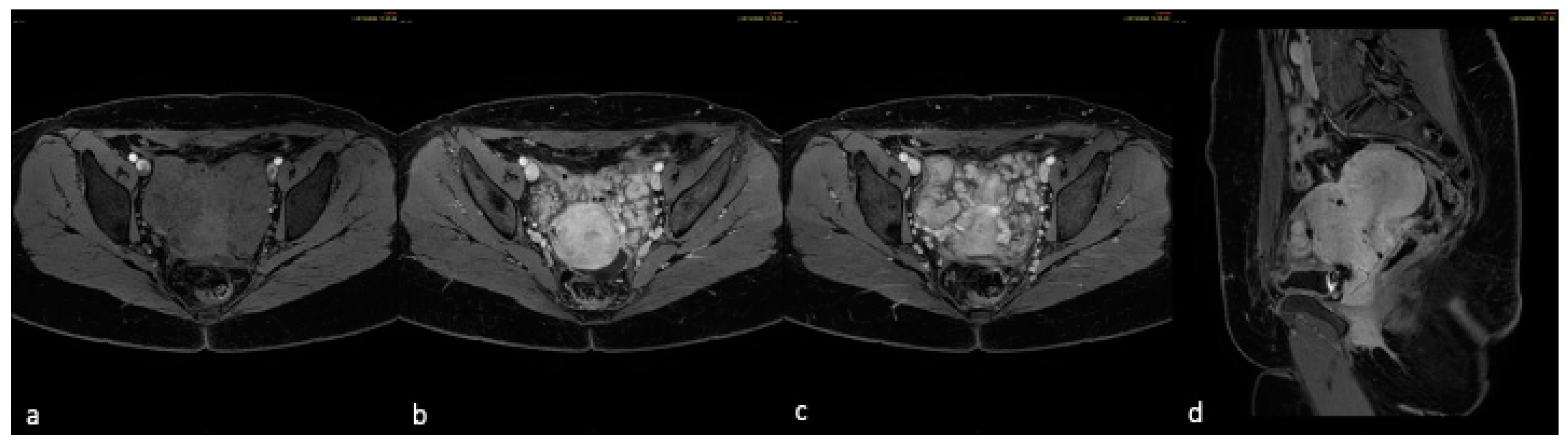
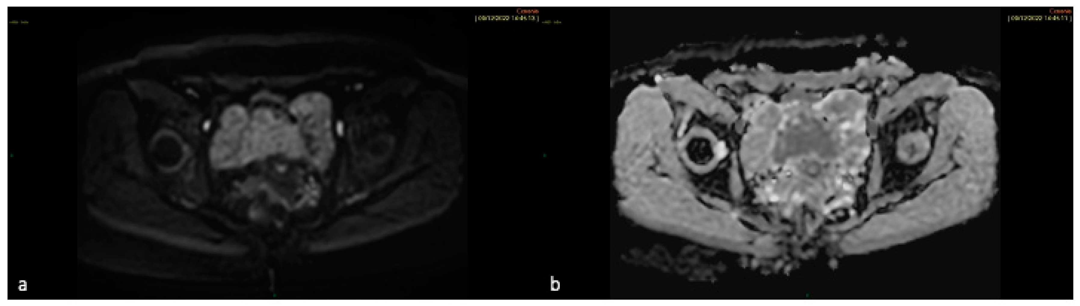
We described a 57 mm myomatous formation with subserosal development localized at the level of the isthmus/cervix with epicenter of origin at the level of the cervical stroma. This lesion showed intermediate signal intensity on T2-WI (Figure 1), intense contrast-enhancement synchronous to the myometrium (Figure 2) and modest restriction on DWI, confirmed on ADC (apparent diffusion coefficient) map (Figure 3). Additional multiple tissue nodules, some of which with confluent appearance, were present in the pelvis occupying the vesico-uterine plica and parameters bilaterally; they showed no definite plane of cleavage with the main lesion, of which they had the same signal characteristics. Considering the patient history, who had already undergone laparotomic myomectomy, we hypothesized a case of pelvic extra-uterine dissemination of LM.
Two nodular neoformations measuring 9x5.5x2.5 cm and 3x2.5x1 cm respectively, were examined; they are well circumscribed but unencapsulated and have a bulging, firm, whorled, white cut surface. Extensive sampling was performed.
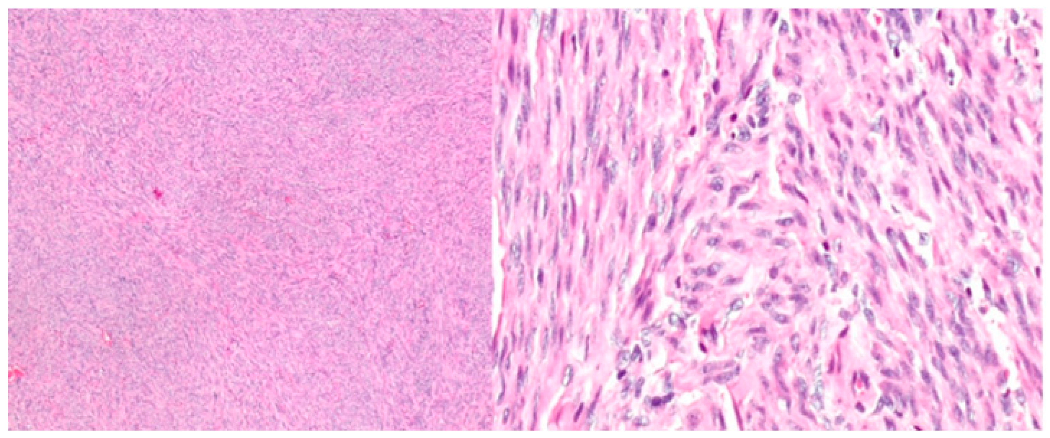
Histology of the neoplasm was characterized by spindle cells arranged in intersecting fascicles, with no significant atypia and only rare mitoses (Figure 1). Neither necrosis nor hemorrhage was found. The two neoplasms are also positive for ER, PR and negative for p53. Proliferative index KI67 was less than 1%.

Discussion

According to a systematic review by Barnas et al. [23], only a minority of cases of extrauterine LM, particularly DPL and BML, develop without a history of gynecological surgery (<20%). Such surgeries include myomectomy or hysterectomy via laparotomy or laparoscopic morcellation of the uterus which may lead to peritoneal seeding. This observation also supports the hypothesis of iatrogenic lymphatic dissemination held by Awonuga et al. [24]. The differential diagnosis of benign LM versus LMS can only be assessed by histological examination, even though imaging plays an important role in pre-operative evaluation and follow-up. This also applies to extra-uterine localization forms, as in our case. Since no pathognomonic characteristic of malignant LMS can be detected by any of the available imaging modalities, including US, CT, MRI and PET-FDG, and because of the many shared imaging features of benign and malignant smooth muscle cells tumors, the discriminating capacity of pelvic imaging is constrained [25]. After clinical evaluation, as in our case, pelvic ultrasonography should be the first imaging modality considered for follow-up in women that underwent surgery for treatment of uterine lesions; MRI should be considered when the patient's clinical features and/or ultrasound findings raise questions about the nature of the lesion and malignancy is suspected. Considering that LM can be treated with minimally invasive techniques, such as laparoscopic morcellation or uterine artery embolization, it is crucial to differentiate benign from malignant uterine smooth muscle cells tumors prior to surgery to avoid a delayed diagnosis and prevent unintentional dissemination.

Conclusion

Extra-uterine leiomyomatosis is a rare pathology, encompassing several entities all defined by the presence of benign tumors arising from uterine smooth muscle cells, which mainly affect pre-menopausal women with a history of either operated or unoperated uterine myoma. Since the differential diagnosis with peritoneal carcinomatosis or metastatic LMS is not always possible by imaging and clinical findings, the diagnosis is histological and can be obtained by biopsies or histological analysis of the operative specimen.
Treatment is typically tailored based on factors such as the patient's age, symptoms and comorbidities of the patients and location, size and extension of the tumors. Even though there is no consensus, it is based on surgical resection and surgical or medical castration, respectively with bilateral adnexectomy and hormonotherapy. Patients need close and prolonged follow-up due to the high risk of recurrence.

Author Contributions

Conceptualization, R.M, G.M. and F.G.; methodology, E.G., M.S., C.C., F.M., F.R., C.G., M.V., L.P., and B.B.; formal analysis, E.G., M.S., C.C., F.M., F.R., C.G., M.V., L.P., and B.B.; writing original draft preparation, R.M, G.M. and F.G.; writing—review and editing, E.G., M.S., C.C., F.M., F.R., C.G., M.V., L.P., and B.B. All authors have read and agreed to the published version of the manuscript.

Funding

This research received no external funding.

Data Availability Statement

The original contributions presented in the study are included in the article, further inquiries can be directed to the corresponding authors.

Conflicts of Interest

The authors declare no conflicts of interest.

References

  1. E. Murase, E.S. Siegelman, E.K. Outwater, L.A. Perez-Jaffe, R.W. Tureck, Uterine Leiomyomas: Histopathologic Features, MR Imaging Findings, Differential Diagnosis, and Treatment, RadioGraphics 19 (1999) 1179–1197. [CrossRef]
  2. E.E. Wallach, V.C. Buttram, R.C. Reiter, Uterine leiomyomata: etiology, symptomatology, and management, Fertil Steril 36 (1981) 433–445. [CrossRef]
  3. N. Fasih, A.K. Prasad Shanbhogue, D.B. Macdonald, et al., Leiomyomas beyond the Uterus: Unusual Locations, Rare Manifestations, RadioGraphics 28 (2008) 1931–1948. [CrossRef]
  4. Y. Li, T. Xu, M. Wang, L. Jiang, Q. Liu, K. Liu, Concurrent Benign Metastasizing Leiomyoma in the Abdominal Wall and Pelvic Cavity: A Case Report and Review of the Literature, Front Surg 9 (2022). [CrossRef]
  5. H.-Y. Low, Y. Zhao, K.-S. Huang, H.-P. Shen, P.-J. Wu, C.-J. Tseng, Intravenous leiomyomatosis of the uterus: A clinicopathological analysis of nine cases and literature review, Taiwan J Obstet Gynecol 56 (2017) 362–365. [CrossRef]
  6. J. Boavida Ferreira, R. Cabrera, F. Santos, et al., Benign Metastasizing Leiomyomatosis to the Skin and Lungs, Intravenous Leiomyomatosis, and Leiomyomatosis Peritonealis Disseminata: A Series of Five Cases, Oncologist 27 (2022) e89–e98. [CrossRef]
  7. M. Ciebiera, A. Słabuszewska-Jóźwiak, K. Zaręba, G. Jakiel, A case of disseminated peritoneal leiomyomatosis after two laparoscopic procedures due to uterine fibroids, Videosurgery and Other Miniinvasive Techniques 1 (2017) 110–114. [CrossRef]
  8. C.W. Ernest Ong, S.L. Siow, Case report: Leiomyoma of the anterior abdominal wall., Med J Malaysia 71 (2016) 81–2.
  9. T. Miyake, T. Enomoto, Y. Ueda, et al., A Case of Disseminated Peritoneal Leiomyomatosis Developing after Laparoscope-Assisted Myomectomy, Gynecol Obstet Invest 67 (2009) 96–102. [CrossRef]
  10. E. Declas, J.P. Lucot, La léiomyomatose extra-utérine : revue de la littérature, Gynécologie Obstétrique Fertilité & Sénologie 47 (2019) 582–590. [CrossRef]
  11. I. Ziouziou, H. Bennani, F. Zouaidia, et al., Léiomyome rétro-péritonéal : à propos d’un cas, Progrès En Urologie 24 (2014) 262–265. [CrossRef]
  12. I. Prasad, S. Sinha, U. Sinha, T. Kumar, J. Singh, Diffuse Leiomyomatosis of the Uterus: A Diagnostic Enigma, Cureus (2022). [CrossRef]
  13. T. Van den Bosch, L. Ameye, D. Van Schoubroeck, T. Bourne, D. Timmerman, Intra-cavitary uterine pathology in women with abnormal uterine bleeding: a prospective study of 1220 women., Facts Views Vis Obgyn 7 (2015) 17–24.
  14. O. Sizzi, L. Manganaro, A. Rossetti, et al., Assessing the risk of laparoscopic morcellation of occult uterine sarcomas during hysterectomy and myomectomy: Literature review and the ISGE recommendations, European Journal of Obstetrics & Gynecology and Reproductive Biology 220 (2018) 30–38. [CrossRef]
  15. O. AlShalabi, F.O. Alahmar, H. Aljasem, B. Alsaid, A. AlShalabi, Pelvic Myxoid Leiomyoma Mass between Vagina and Rectum, Case Rep Surg 2016 (2016) 1–3. [CrossRef]
  16. Y.O. Tanaka, M. Nishida, H. Tsunoda, Y. Okamoto, H. Yoshikawa, Smooth muscle tumors of uncertain malignant potential and leiomyosarcomas of the uterus: MR findings, Journal of Magnetic Resonance Imaging 20 (2004) 998–1007. [CrossRef]
  17. K. Sato, N. Yuasa, M. Fujita, Y. Fukushima, Clinical application of diffusion-weighted imaging for preoperative differentiation between uterine leiomyoma and leiomyosarcoma, Am J Obstet Gynecol 210 (2014) 368.e1-368.e8. [CrossRef]
  18. A. Goto, S. Takeuchi, K. Sugimura, T. Maruo, Usefulness of Gd-DTPA contrast-enhanced dynamic MRI and serum determination of LDH and its isozymes in the differential diagnosis of leiomyosarcoma from degenerated leiomyoma of the uterus, International Journal of Gynecological Cancer 12 (2002) 354–361. [CrossRef]
  19. K. Kitajima, K. Murakami, Y. Kaji, K. Sugimura, Spectrum of FDG PET/CT Findings of Uterine Tumors, American Journal of Roentgenology 195 (2010) 737–743. [CrossRef]
  20. N. Umesaki, T. Tanaka, M. Miyama, et al., Positron Emission Tomography with 18F-Fluorodeoxyglucose of Uterine Sarcoma: A Comparison with Magnetic Resonance Imaging and Power Doppler Imaging, Gynecol Oncol 80 (2001) 372–377. [CrossRef]
  21. A. Roue, C. Laboisse, N. Winer, et al., Léiomyome pelvien extra-utérin: diagnostic et prise en charge, J Gynecol Obstet Biol Reprod (Paris) 36 (2007) 403–408. [CrossRef]
  22. T. Takeda, K. Masuhara, S. Kamiura, Successful Management of a Leiomyomatosis Peritonealis Disseminata With an Aromatase Inhibitor, Obstetrics & Gynecology 112 (2008) 491–493. [CrossRef]
  23. E. Barnaś, M. Książek, R. Raś, A. Skręt, J. Skręt- Magierło, E. Dmoch- Gajzlerska, Benign metastasizing leiomyoma: A review of current literature in respect to the time and type of previous gynecological surgery, PLoS One 12 (2017) e0175875. [CrossRef]
  24. A.O. Awonuga, V.I. Shavell, A.N. Imudia, M. Rotas, M.P. Diamond, E.E. Puscheck, Pathogenesis of Benign Metastasizing Leiomyoma, Obstet Gynecol Surv 65 (2010) 189–195. [CrossRef]
  25. S. Sun, P.A. Bonaffini, S. Nougaret, et al., How to differentiate uterine leiomyosarcoma from leiomyoma with imaging, Diagn Interv Imaging 100 (2019) 619–634. [CrossRef]
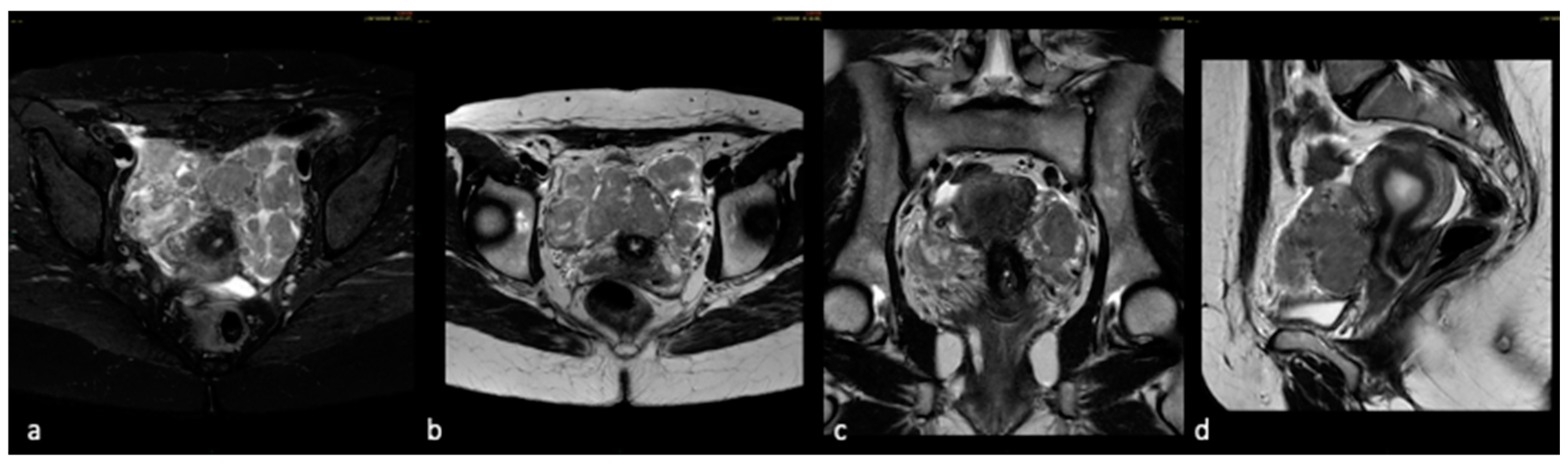
Figure 1. T2 weighted imaging. Axial T2-WI with (a) and without (b) fat suppression show the main myomatous lesion with exophytic and sub serosal development localized at the level of the histmus/cervix and multiple additional nodules with confluent appearance and no secure cleavage plane with the main lesion. All lesions shows similar MR features with intermediate signal intensity in T2 WI and FS T2 WI images like tissutal component; coronal (c) and sagittal (d) T2 WI show the extension of these nodules into the vesico-uterine plica and the parameters.
Preprints 139744 g001
Figure 2. T1 weighted imaging with fat-suppression pre- and post-contrast administration. On T1 WI before contrast-medium administration (a), the main lesion and the nodules disseminated in the pelvis are iso-ipontense to the adjacent myometrium; after contrast-medium administration (b and c – axial plane; d – sagittal plane) they show intense homogeneous contrast-enhancement synchronous to the myometrium.
Preprints 139744 g002
Figure 3. Diffusion weighted imaging (DWI) and ADC (apparent diffusion coefficient) map. The intermediate signal intensity at high–b value DWI (a) without significantly low signal intensity on ADC map (b) along with the other features helped us direct our diagnostic hypothesis toward lesions of benign character not characterized by iper-cellular component.
Preprints 139744 g003
Figure 4. Histological section of the lesion. Leiomyoma characterized by spindle cells arranged in intersecting fascicles, with no significant atypia and only rare mitoses. Neither necrosis nor hemorrhage was found.
Preprints 139744 g004
Disclaimer/Publisher’s Note: The statements, opinions and data contained in all publications are solely those of the individual author(s) and contributor(s) and not of MDPI and/or the editor(s). MDPI and/or the editor(s) disclaim responsibility for any injury to people or property resulting from any ideas, methods, instructions or products referred to in the content.
Copyright: This open access article is published under a Creative Commons CC BY 4.0 license, which permit the free download, distribution, and reuse, provided that the author and preprint are cited in any reuse.
Prerpints.org logo

Preprints.org is a free preprint server supported by MDPI in Basel, Switzerland.

Subscribe

© 2026 MDPI (Basel, Switzerland) unless otherwise stated

Accessibility

Disclaimer

Terms of Use

Privacy Policy

Privacy Settings