

Submitted:
15 November 2024
Posted:
18 November 2024
You are already at the latest version
Abstract
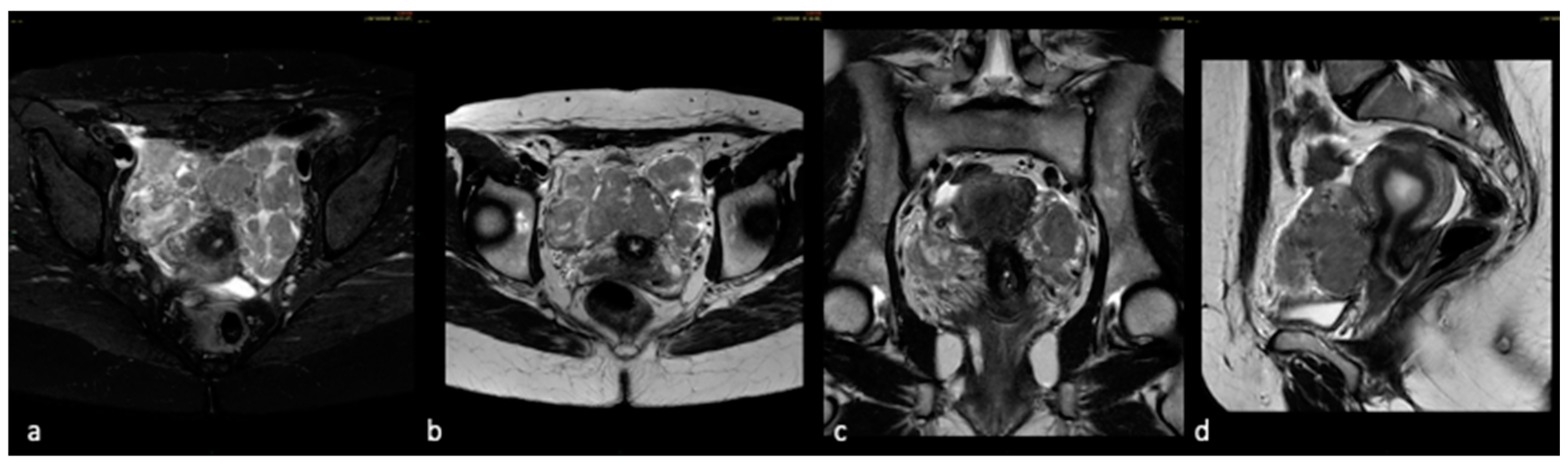
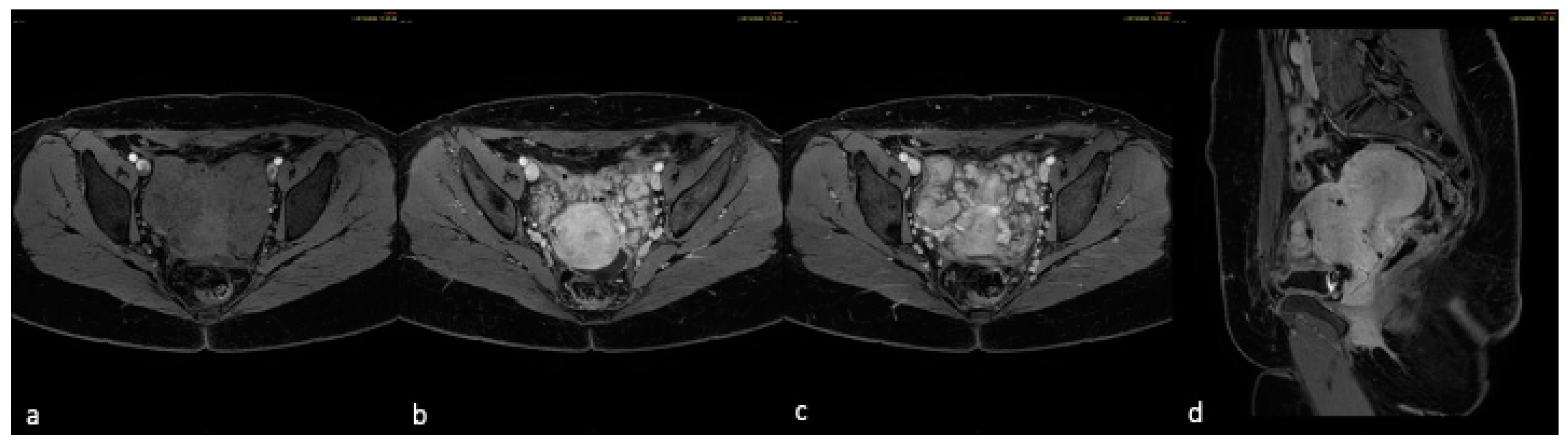
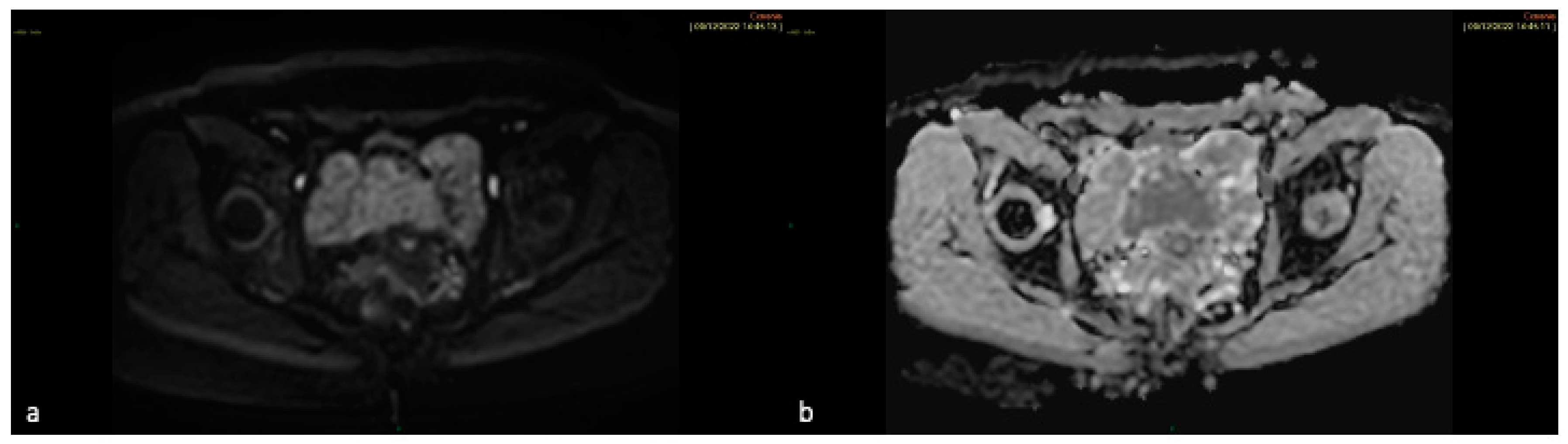
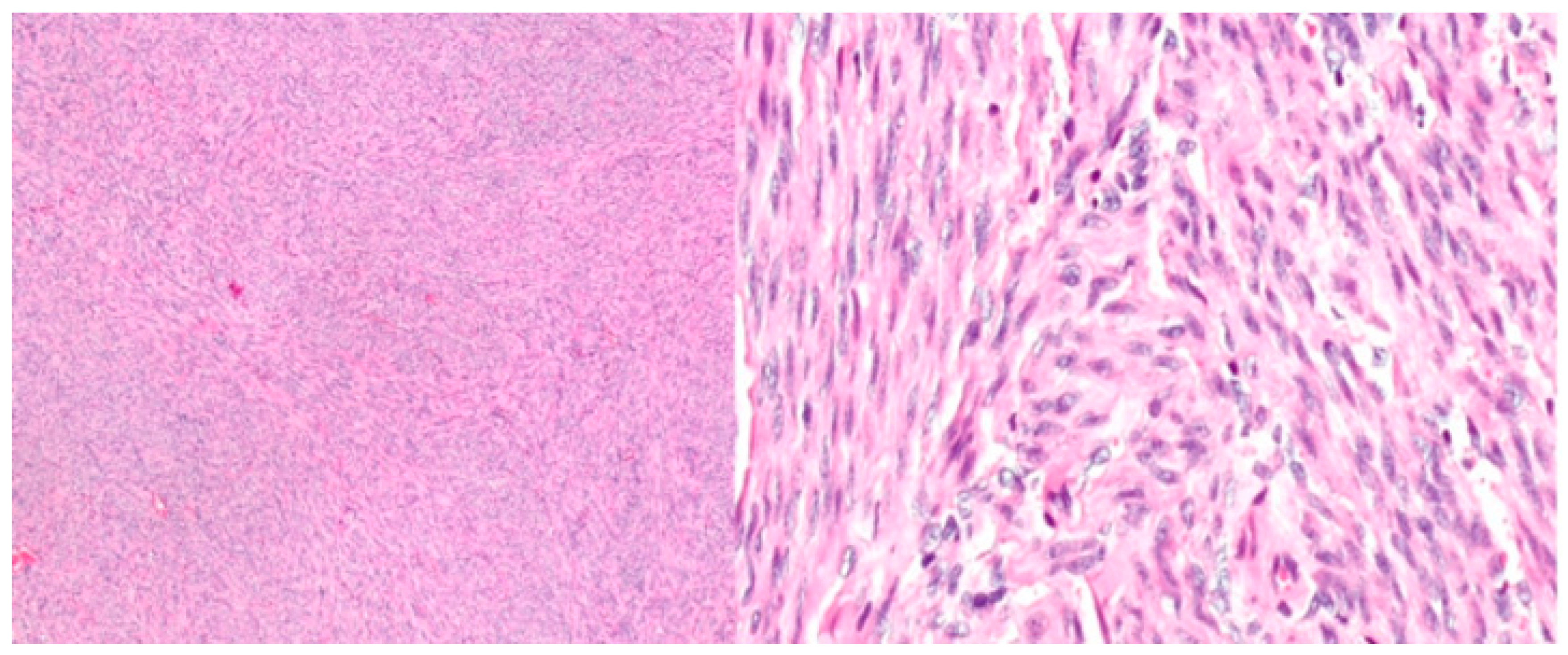
Leiomyomas are the most common gynecological tumors; they are benign and originate from smooth muscle cells together with fibrous connective tissue. The most common location is the uterus although rare cases of extra-uterine locations with benign histological features have been described including intravenous leiomyomatosis, disseminated peritoneal leiomyomatosis, benign metastasizing leiomyomas, parasitic and retroperitoneal leiomyomatosis, which can present as single or multiple myomatous nodules. These entities must be differentiated from malignant lesions such as leiomyosarcoma. Imaging approach, mainly magnetic resonance imaging, plays an important but not always diriment role in the diagnosis of leiomyomas. Therefore, histological examination is necessary for a definitive diagnosis. In this report, we describe the case of a 36-year-old woman with histologic examination suggestive of myoma, underwent an MRI examination at our department for further diagnostic evaluation. MRI revealed the presence of two inhomogeneous and highly vascularized nodular lesions at the ultrasound checkup performed six months after surgery. MR examination was performed using multiplanar T2 weighted sequences with and without fat suppression, T1 weighted fat-suppressed sequences pre- and post-gadolinium administration and diffusion weighted imaging. This approach allowed to highlight the presence of a major myomatous lesion at the level of the isthmus surrounded by many confluent nodules in the pelvis with the same benign features. The MRI findings suggested the diagnosis of benign metastasizing leiomyomatosis, then confirmed by the definitive histological examination which found no malignant features of the lesions.
Keywords:
Introduction
Case Study
Discussion
Conclusion
Author Contributions
Funding
Informed Consent Statement
Data Availability Statement
Conflicts of Interest
References
- E. Murase, E.S. Siegelman, E.K. Outwater, L.A. Perez-Jaffe, R.W. Tureck, Uterine Leiomyomas: Histopathologic Features, MR Imaging Findings, Differential Diagnosis, and Treatment, RadioGraphics 19 (1999) 1179–1197. [CrossRef]
- E.E. Wallach, V.C. Buttram, R.C. Reiter, Uterine leiomyomata: etiology, symptomatology, and management, Fertil Steril 36 (1981) 433–445. [CrossRef]
- N. Fasih, A.K. Prasad Shanbhogue, D.B. Macdonald, et al., Leiomyomas beyond the Uterus: Unusual Locations, Rare Manifestations, RadioGraphics 28 (2008) 1931–1948. [CrossRef]
- Y. Li, T. Xu, M. Wang, L. Jiang, Q. Liu, K. Liu, Concurrent Benign Metastasizing Leiomyoma in the Abdominal Wall and Pelvic Cavity: A Case Report and Review of the Literature, Front Surg 9 (2022). [CrossRef]
- H.-Y. Low, Y. Zhao, K.-S. Huang, H.-P. Shen, P.-J. Wu, C.-J. Tseng, Intravenous leiomyomatosis of the uterus: A clinicopathological analysis of nine cases and literature review, Taiwan J Obstet Gynecol 56 (2017) 362–365. [CrossRef]
- J. Boavida Ferreira, R. Cabrera, F. Santos, et al., Benign Metastasizing Leiomyomatosis to the Skin and Lungs, Intravenous Leiomyomatosis, and Leiomyomatosis Peritonealis Disseminata: A Series of Five Cases, Oncologist 27 (2022) e89–e98. [CrossRef]
- M. Ciebiera, A. Słabuszewska-Jóźwiak, K. Zaręba, G. Jakiel, A case of disseminated peritoneal leiomyomatosis after two laparoscopic procedures due to uterine fibroids, Videosurgery and Other Miniinvasive Techniques 1 (2017) 110–114. [CrossRef]
- C.W. Ernest Ong, S.L. Siow, Case report: Leiomyoma of the anterior abdominal wall., Med J Malaysia 71 (2016) 81–2.
- T. Miyake, T. Enomoto, Y. Ueda, et al., A Case of Disseminated Peritoneal Leiomyomatosis Developing after Laparoscope-Assisted Myomectomy, Gynecol Obstet Invest 67 (2009) 96–102. [CrossRef]
- E. Declas, J.P. Lucot, La léiomyomatose extra-utérine : revue de la littérature, Gynécologie Obstétrique Fertilité & Sénologie 47 (2019) 582–590. [CrossRef]
- I. Ziouziou, H. Bennani, F. Zouaidia, et al., Léiomyome rétro-péritonéal : à propos d’un cas, Progrès En Urologie 24 (2014) 262–265. [CrossRef]
- I. Prasad, S. Sinha, U. Sinha, T. Kumar, J. Singh, Diffuse Leiomyomatosis of the Uterus: A Diagnostic Enigma, Cureus (2022). [CrossRef]
- T. Van den Bosch, L. Ameye, D. Van Schoubroeck, T. Bourne, D. Timmerman, Intra-cavitary uterine pathology in women with abnormal uterine bleeding: a prospective study of 1220 women., Facts Views Vis Obgyn 7 (2015) 17–24.
- O. Sizzi, L. Manganaro, A. Rossetti, et al., Assessing the risk of laparoscopic morcellation of occult uterine sarcomas during hysterectomy and myomectomy: Literature review and the ISGE recommendations, European Journal of Obstetrics & Gynecology and Reproductive Biology 220 (2018) 30–38. [CrossRef]
- O. AlShalabi, F.O. Alahmar, H. Aljasem, B. Alsaid, A. AlShalabi, Pelvic Myxoid Leiomyoma Mass between Vagina and Rectum, Case Rep Surg 2016 (2016) 1–3. [CrossRef]
- Y.O. Tanaka, M. Nishida, H. Tsunoda, Y. Okamoto, H. Yoshikawa, Smooth muscle tumors of uncertain malignant potential and leiomyosarcomas of the uterus: MR findings, Journal of Magnetic Resonance Imaging 20 (2004) 998–1007. [CrossRef]
- K. Sato, N. Yuasa, M. Fujita, Y. Fukushima, Clinical application of diffusion-weighted imaging for preoperative differentiation between uterine leiomyoma and leiomyosarcoma, Am J Obstet Gynecol 210 (2014) 368.e1-368.e8. [CrossRef]
- A. Goto, S. Takeuchi, K. Sugimura, T. Maruo, Usefulness of Gd-DTPA contrast-enhanced dynamic MRI and serum determination of LDH and its isozymes in the differential diagnosis of leiomyosarcoma from degenerated leiomyoma of the uterus, International Journal of Gynecological Cancer 12 (2002) 354–361. [CrossRef]
- K. Kitajima, K. Murakami, Y. Kaji, K. Sugimura, Spectrum of FDG PET/CT Findings of Uterine Tumors, American Journal of Roentgenology 195 (2010) 737–743. [CrossRef]
- N. Umesaki, T. Tanaka, M. Miyama, et al., Positron Emission Tomography with 18F-Fluorodeoxyglucose of Uterine Sarcoma: A Comparison with Magnetic Resonance Imaging and Power Doppler Imaging, Gynecol Oncol 80 (2001) 372–377. [CrossRef]
- A. Roue, C. Laboisse, N. Winer, et al., Léiomyome pelvien extra-utérin: diagnostic et prise en charge, J Gynecol Obstet Biol Reprod (Paris) 36 (2007) 403–408. [CrossRef]
- T. Takeda, K. Masuhara, S. Kamiura, Successful Management of a Leiomyomatosis Peritonealis Disseminata With an Aromatase Inhibitor, Obstetrics & Gynecology 112 (2008) 491–493. [CrossRef]
- E. Barnaś, M. Książek, R. Raś, A. Skręt, J. Skręt- Magierło, E. Dmoch- Gajzlerska, Benign metastasizing leiomyoma: A review of current literature in respect to the time and type of previous gynecological surgery, PLoS One 12 (2017) e0175875. [CrossRef]
- A.O. Awonuga, V.I. Shavell, A.N. Imudia, M. Rotas, M.P. Diamond, E.E. Puscheck, Pathogenesis of Benign Metastasizing Leiomyoma, Obstet Gynecol Surv 65 (2010) 189–195. [CrossRef]
- S. Sun, P.A. Bonaffini, S. Nougaret, et al., How to differentiate uterine leiomyosarcoma from leiomyoma with imaging, Diagn Interv Imaging 100 (2019) 619–634. [CrossRef]
Disclaimer/Publisher’s Note: The statements, opinions and data contained in all publications are solely those of the individual author(s) and contributor(s) and not of MDPI and/or the editor(s). MDPI and/or the editor(s) disclaim responsibility for any injury to people or property resulting from any ideas, methods, instructions or products referred to in the content. |
© 2024 by the authors. Licensee MDPI, Basel, Switzerland. This article is an open access article distributed under the terms and conditions of the Creative Commons Attribution (CC BY) license (http://creativecommons.org/licenses/by/4.0/).


