Submitted:
13 November 2024
Posted:
14 November 2024
You are already at the latest version
Abstract
Bats (Chiroptera) are among the most diverse and geographically dispersed mammals. They are of great importance to the ecosystem, as pollinators, seed dispersers and pest controllers, in addition to being hosts to several parasitic arthropods, including ticks, mites, lice, fleas and flies. Their diet includes tissues and blood or other body fluids of bats. Bats are reservoirs of several disease-causing agents, many of them pathogenic to humans, such as bacteria, as well as protozoa, viruses and fungi. This study was conducted in Monte Negro, Rondônia, Brazil and the occurrence of parasitic arthropods in bats was evaluated, as well as a screening of bacteria that these ectoparasites can carry. Through a total of 69 nocturnal captures, 217 chiropterans were sampled, representing 23 species and six families. A total of 592 specimens of parasitic arthropods (ticks, mites and flies) were collected from these bats (9% dipterans, 59% ticks and 32% mites). Bartonella sp. were found in two species of bat flies (Trichobius joblingi and Strebla mirabilis) in peri-urban and forest areas with an infection rate of 62% and 38%, respectively. We report for the first time in Rondônia the argasid tick Ornithodoros hasei and its infection by a spotted fever group bacterium ‘Candidatus Rickettsia wissemanii’ in a peri-urban area.
Keywords:
Amazon
; Chiroptera
; Ectoparasites
; Ticks
; Mites
; Flies
; Vector-borne Bacteria
1. Background
Among living mammals, Chiroptera is one of the most diverse orders, representing about 20% of all mammals in the world [1]. According to the most recent estimate, this order comprises 20 families and more than 1,400 species [1,2,3]. Despite the recurring disturbances caused by human actions in nature [4], factors such as the development and popularization of new sampling methods, combined with the improvement of systematic reviews and the description of new species, have contributed to an exponential increase in the richness of known bats in Brazil. This keeps the country among the three with the greatest diversity of bats in the world [5], being surpassed only by Indonesia with 230 species [6] and by Colombia with 201 species [7]. In Brazil, nine families, 64 genera and 182 species are known, of which 150 species are found in the Amazon region of Brazil and 86 species with 16 genera in the state of Rondônia [8,9,10].
Bats have been reported as hosts of various arthropod ectoparasites, such as ticks (Argasidae, Ixodidae) [11,12], mites (Mesostigmata [13], Sarcoptiformes [14], and Trombidiformes [15]), lice (Phthiraptera) [16], fleas (Siphonaptera) [17], and flies (Diptera) [18]. In the Amazon, arthropods of the class Arachnida, three families, five genera, and 60 species have been reported in bats and, in the state of Rondônia, three families, four genera and 16 species have been reported so far [19,20,21]. Regarding the class Insecta, 43 species in 16 genera of the Streblidae family and four species in two genera of the Nycteribiidae family have been reported [22,23].
The diet of these ectoparasites includes blood or other bodily fluids and tissues of the bats [13]. The presence of ectoparasites can cause irritation and stress for bats, which can affect their health and behavior [24]. However, the prevalence and diversity of these ectoparasites vary between bat species and their habitats [18].Chiroptera and their ectoparasites are reservoirs of etiological agents of various diseases, and many of them are pathogenic to humans, such as bacteria of the genera Rickettsia [25], Coxiella [26], Borrelia [27], and Bartonella [28], as well as protozoa [29], viruses [30] and fungi [31].
According to Hayman [32] and Subudhi et al. [33], in recent years, the interest in bat research has increased due to the occurrence of what is known as the spillover phenomenon, in other words, the transmission of a pathogen from its natural reservoir or host species to a new host species, thus enhancing the possibility of spreading diseases to humans and other mammals.
Despite several studies over the past years, the study of ectoparasites in bats have been unevenly focused within Brazil’s large geographical area. The study by Graciolli and Bernard [18] formally recommended the exploration of areas such as south-central to carry out new inventories in the Amazon. Thus, in this context, the present study aimed to record the pathogens infecting ectoparasite fauna of bats in the south-central area in Rondônia State, in the western Brazilian Amazon.
2. Materials and Methods
2.1. Study Site
The study was carried out between November 2020 and November 2022 in Monte Negro, a central region in the state of Rondônia, 250 km southwest of the capital Porto Velho. It is located at a latitude of 10º 17’ 40” S and longitude of 63º 19’ 31” W, at an altitude of 123 meters, and occupies a territorial area of 1,931.378 km² with an estimated population of 11,548 inhabitants, according to data from the 2022 IBGE census (Figure 1). The region is characterized by mixed soils and equatorial vegetation and a hot and humid climate, with high rainfall with an annual average varying between 1,440 mm in November to April and 557 mm in the dry period from May to October, with average temperatures of 25 to 29 ºC and relative humidity between 70 and 80% throughout the year [34]. The sites of bat capture were forest (dense vegetation and low human density), Peri-Urban area (characterized by having small sites and patches of forests with dense vegetation and low human density), and urban area, with a diameter of 4 km2 per area, with no forest and with high human density, and 12% of empty houses and empty offices.
2.2. Bat Capture and Sample Collection
To capture the bats, we used ten mist nets measuring five meters long and two meters wide, suspended at a height of 1.5 meters from the ground. Additionally, we conducted active searches in artificial shelters in urban areas, resulting in the capture of 18% of the bats in the roof spaces of homes.
The mist nets were strategically positioned close to fruit trees and animal breeding areas, previously analyzed in terms of the location and behavior of bats in the environment. As necessary, these nets were occasionally relocated to meet the specific demands of the situation. To prevent escape or predation of captured specimens, the nets were inspected at 15-minute intervals.
This procedure was carried out with extreme caution, aiming to avoid any damage or injuries to the animals. After carrying out the procedures (biometric data collection processes, external morphology analysis, and photographing), the bats were promptly sent to the fields to be released. Taxonomic identification of the bats followed specialized scientific literature [35,36,37]. The nomenclature of the species and the taxonomic arrangement followed Abreu Jr et al. [9].
Captures took place from 6:00 pm to 11:00 pm, for three consecutive nights, each month, during the waning moon (as suggested by the Butantan Institute protocol), for 25 months, totaling 345 hours. According to Straube and Bianconi (2002), the capture effort, measured as the total area (m²) of the nets multiplied by the exposure time (hours) was 157,500 m2/h. [37]
The bats were initially kept in individual cloth bags and subsequently handled and thoroughly examined for the presence of ectoparasites. These were removed using tweezers and placed in DNA-free microtubes containing 100% ethanol. Each microtube housed the ectoparasites collected from a single bat and was identified by a tracing paper label containing the animal’s code written in graphite pencil and inserted inside the tube. The molecular analysis of the bacteria in the ectoparasites studied is described below. No blood was taken from the bats.
2.3. Ticks, Mites and Flies
The ticks collected from the bats were morphologically identified at the species level following Jones and Clifford [38] and Labruna et al. [39]. In addition, 25 larval ticks were identified at the species level by molecular analysis. For this purpose, larvae were individually submitted to DNA extraction using the guanidine isothiocyanate phenol technique [40] and tested via a polymerase chain reaction (PCR) assay targeting a ≈460-bp fragment of the ticks’ 16S rRNA mitochondrial gene, as described by Mangold et al. [41]. The PCR products were purified and sequenced with the Big Dye Terminator Cycle Sequencing kit (Applied Biosystems, Foster City, CA, USA) in an automatic sequencer (ABI 3500 Genetic Analyzer, Applied Biosystems) according to the manufacturer’s protocol. The generated sequences were submitted to BLAST analysis (www.ncbi.nlm.nih.gov/blast) to infer the closest identities to the tick DNA sequences available in GenBank.
The mites were morphologically identified to species level following the keys available in Rudnick (1960) [42], Machado-Allison (1965a, b) [43,44] and Herrin and Tipton (1975) [45]. All the other mites were under identification at the time of writing and will be identified to species level in future studies.
2.4. Molecular Detection of Vector-Borne Bacteria
A sample of 87 ticks was selected for testing for molecular detection of bacteria of the genera Rickettsia, Borrelia, and Coxiella. This sample included the 25 individual larvae mentioned above, plus 62 larvae that were processed individually (8 larvae) or in pools, each containing 2 to 3 individuals (a total of 19 pools containing 54 larvae), using the same DNA extraction protocol mentioned above. This procedure resulted in a total of 52 extracted DNA samples, which were initially tested using a TaqMan real-time PCR assay targeting the rickettsial citrate synthase gene (gltA), as described elsewhere [48,49]. Samples that were positive according to the real-time PCR (cycle threshold ≤ 35) were tested via a conventional PCR that targeted a 401-bp fragment of the Rickettsia gltA gene [48]. In each set of reactions, negative control tubes containing water and a positive control tube containing Rickettsia vini DNA were included. The PCR products were DNA-sequenced and submitted to BLAST analyses as described above.
For the detection of Borrelia DNA, all 52 tick samples were tested via a TaqMan real-time PCR assay that targeted the 16S rRNA gene of bacteria of the genus Borrelia, as described elsewhere [50]. Negative control tubes containing water and a positive control tube containing Borrelia anserina DNA were included. For the detection of Coxiella DNA, the 52 tick samples were tested via a conventional PCR that targeted a 687-bp fragment of the transposase elements gene (IS1111) of organisms of the genus Coxiella, as described elsewhere [51]. Negative control tubes containing water and a positive control tube containing C. burnetii DNA were included.
For the molecular analysis, the DNA extraction of the mites was performed using the DNeasy Blood & Tissue kit (Qiagen, Hilden, Germany), following the manufacturer’s instructions. Each mite DNA sample was subjected to a conventional PCR targeting a fragment of ~800 bp of the 18S rRNA gene (endogenous control) [52] to verify the success of the DNA extraction procedure. Negative (Milli-Q water free of DNA) and positive (pool of dust mites) controls were included in each reaction.
One hundred and fifteen mites were selected for the detection of organisms of the genera Bartonella, Rickettsia, Borrelia and Coxiella. The protocols for the last three bacterial genera were the same used for the ticks. For Bartonella, we used a conventional PCR targeting a fragment of the nuoG gene of Bartonella spp. according to Colborn et al. [53].
All the PCR products with concentrations over 20 ng/µL were selected and purified with ExoSap-IT (GE Healthcare Pittsburgh, PA). Sanger sequencing was performed at the Centro de Pesquisa sobre Genoma Humano e Células Tronco do Instituto de Biociências da USP, São Paulo, SP, Brazil. The obtained sequences were assembled with Sequencing Analysis 5.3.1 and submitted to BLAST analysis (Altschul et al. [54]) to infer similarities with Bartonella sequences available in GenBank. Different haplotypes were visually discriminated after an alignment using the CLUSTAL W algorithm (Thompson et al. [55]) implemented in Geneious R11 [56]. The molecular analysis for the dipteran was performed in the same way that the mites’ DNA were extracted. Additionally, the material was tested for the same bacteria using the protocols used for ticks and mites, as described above.
2.5. Ethical Aspects
The study was approved by the Biodiversity Authorization and Information System (SISBio) of the Instituto Chico Mendes de Conservação da Biodiversidade - ICMBio (No. 77013) and the capture procedures were in accordance with the resolutions of the Animal Experimentation Ethics Committee of ICB/USP (approved under CEUA No. 7946291123).
3. Results
After 75 nocturnal samplings, with a total sampling effort of 157,500 m²/h, 217 individual bats were captured, belonging to 23 species and six families, as shown in Table 1.
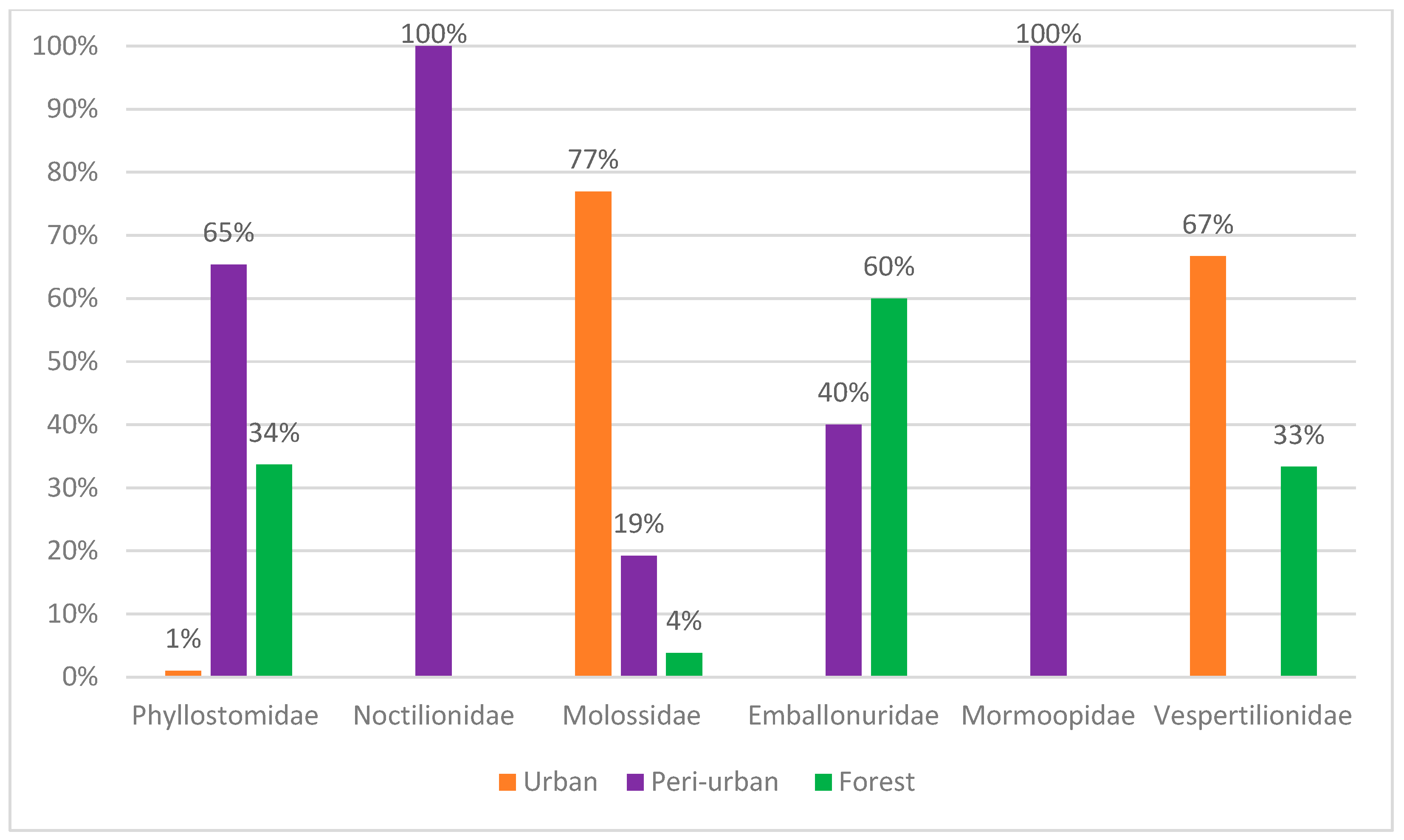
Ten percent of the total number of bats were captured in urban areas. Of these, 4% belonged to the Phyllostomidae family, 9% to the Vespertilionidae family and 87% to the Molossidae family. These bats were captured in residential penthouses through active searches of empty homes and offices.
Sixt seven percent of specimens were captured in peri-urban areas. Of these, 46% belonged to the Phyllostomidae family and 37% to the Noctilionidae family. The families Emballonuridae, Mormoopidae and Molossidae were responsible for 7%, 6% and 4% of captures, respectively.
Twenty-three percent of the specimens were captured in forest fragments. The Phyllostomidae family represented 66% of the captures, followed by the Emballonuridae family with 30%. The Molossidae and Vespertilionidae families represented only 2% each, considering the total sampling efforts.
Figure 2.
Relative abundance of chiropteran families captured in the sampled areas.
A total of 592 specimens of ectoparasites were collected from 14 bats and identified into seven families, as shown in Table 1. Overall, 37% of the bats were infested by at least one type of ectoparasite. Regarding the capture area, 67% of the bats were captured in peri-urban areas, 23% in forests and 10% in urban areas.
Among the 217 captured bats, 23 (10.6%) were found to be infested by ticks. A total of 308 ticks were collected, giving a mean intensity of infestation of 13.4 ticks/infested bat. In most of the infested bats, less than 10 ticks were collected; however, in two bats, a total of 87 and 100 ticks were collected. The 308 ticks were morphologically identified as Ornithodoros hasei (302 larvae collected from 20 Noctilio leporinus and one Pteronotus rubiginosus) and Ornithodoros marinkellei (six larvae collected from three P. rubiginosus). One P. rubiginosus was co-infested by the two tick species. Overall, O. hasei was collected from 20 (37.7%) out of 53 N. leporinus and one (11.1%) out of nine P. rubiginosus, whereas O. marinkellei was collected from 33.3% (3/9) of P. rubiginosus. No ticks were found in the remaining 21 bat specimens that were captured in the present. The most infested bats were N. leporinus, with 52% of captures in peri-urban environments, followed by Peropteryx kappleri, with 9% also in peri-urban areas, and Eumops perotis, with 4% of specimens captured in urban environments. These differences suggest that infestation levels may be related to the type of environment, with a higher infestation observed in bats from peri-urban areas.
The molecular identification of the ticks was confirmed via molecular analyses in 23 O. hasei larvae, which generated a single 16S rRNA haplotype (426 bp). Through the BLAST analysis, this haplotype was 100% identical to a sequence of O. hasei from southeastern Brazil (KX099896). Two specimens of O. marinkellei generated a 16S rRNA haplotype (426 bp) that was 100% identical to a sequence of O. marinkellei from Porto Velho, Rondônia (HM582438).
A total of 52 tick samples containing 87 larvae (84 O. hasei, 3 O. marinkellei) were tested for the presence of DNA of organisms of the genera Rickettsia, Borrelia and Coxiella. Only one larva of O. hasei yielded amplicons for the genus Rickettsia, via both the real-time PCR and the conventional PCR assays. For the latter assay, a 350-bp fragment of the gltA gene was generated, which was 100% identical to GenBank available sequences of “Candidatus Rickettsia wissemanii” from O. hasei from French Guiana (MH614266) and the state of Amapá, Brazil (MH614266).
In this study, 17 species of captured bats were infested with parasitic flies (Table 1). A total of 48 bat fly samples were tested for the presence of DNA of Rickettsia, Bartonella, Borrelia and Coxiella. All samples were positive for the endogenous control (18S rRNA), validating the DNA extraction protocol. When tested for detection of DNA of pathogens, two of the 48 bat fly samples were positive for the nuoG gene of Bartonella spp., while all the samples were negative for the other tested bacterial genera. One sequence for the nuoG gene was detected in Trichobius joblingi collected on Carollia brevicauda, and another in Strebla mirabilis collected on Trachops cirrhosis. When compared with the sequences available in GenBank, these sequences were respectively 93.44 and 90.57% (e-value: 3 x 10-149, 1 x 10-132; query cover: 100%) identical to Bartonella sp. from Diphylla ecaudata blood collected in São Paulo, Brazil [53] (GenBank accession numbers PP445025 and PP445026).
In addition, 27 individual bat-associated mites (4 Trombiculidae, 1 Macronyssidae and 22 Spinturnicidae) were tested. All the samples were positive for the endogenous control (18S rRNA). The results show differences in dipteran infestation between peri-urban and forest environments. In the peri-urban area, Carollia brevicauda presented a significantly higher infestation (77%) by Trichobius joblingi and Strebla guajiro. In the forest area, Trachops cirrhosus had a lower infestation (11%) by Strebla mirabilis. No significant differences were found in dipteran species between forest and peri-urban areas.
4. Discussion
After 75 nocturnal samplings, with a total sampling effort of 157,500 m²/h, 217 individual bats were captured, belonging to 23 species and six families, as shown in Table 1.
Ten percent of the total number of bats were captured in urban areas. Of these, 4% belonged to the Phyllostomidae family, 9% to the Vespertilionidae family and 87% to the Molossidae family. These bats were captured in residential penthouses through active searches of empty homes and offices.
Sixt seven percent of specimens were captured in peri-urban areas. Of these, 46% belonged to the Phyllostomidae family and 37% to the Noctilionidae family. The families Emballonuridae, Mormoopidae and Molossidae were responsible for 7%, 6% and 4% of captures, respectively.
Twenty-three percent of the specimens were captured in forest fragments. The Phyllostomidae family represented 66% of the captures, followed by the Emballonuridae family with 30%. The Molossidae and Vespertilionidae families represented only 2% each, considering the total sampling efforts.
We provide the first report of O. hasei on P. rubiginosus, which has been reported as the main host for O. marinkellei [39]. We detected the presence of ‘Candidatus R. wissemanii’ in only one specimen of O. hasei, giving a minimum infection rate of 1.2% (1/84). Previous studies reported this rickettsial agent in one out of three pools of O. hasei larvae from the state of Amapá, eastern Brazilian Amazon [57], in 28.9% (31/107) of O. hasei larvae from French Guiana [58], and in three O. hasei larvae from Argentina [59]. Our report is the first for the western Amazon. Although ‘Candidatus R. wissemanii’ is a member of the spotted fever group of Rickettsia species, its pathogenic role in humans or animals remains to be evaluated [57,58,59,60,61,62]. Finally, Tahir et al. [58] in French Guyana, also tested t O. hasei ticks for Borrelia and Coxiella DNA; similarly to the present study, no ticks were infected by these agents.
Some Rickettsia species are the etiological agents of spotted fever in humans, who acquire the infection through the bite of infected ticks; in Brazil, this is chiefly through ticks of the genus Amblyomma. Despite ‘Candidatus R. wissemanii’ never having been associated with disease in humans or animals, it is a novel tick-borne agent that was only recently described [57,58,59]. Therefore, this result should be better investigated in further studies.
Coxiella burnetii and Borrelia spp. we’re not detected in this study. There are a few cases of the disease identified in Brazil [63,64,65]. Muñoz-Leal et al. [63], Oliveira et al. [66]. Pacheco et al. [67] Further studies are warranted to verify the circulation of relapsing fever in the state of Rondônia, where the argasid fauna is the richest in Brazil. This indicates the need for more intensive epidemiological surveillance by the governments of Brazil. Serological studies should also be carried out in humans and animals to estimate the burden of the disease.
The role of ticks in the transmission of Bartonella spp. is controversial, even though they have been found infected in nature (which does not necessarily class it as a vector). Other arthropods (lice and sandflies) are confirmed vectors. A cat’s scratch and/or bite can transmit the bacteria, as can the saliva, urine, and feces of bats [68].The occurrence of Bartonella sp. has been found in five families of chiropterans [66] and in bat flies of the families Nycteribiidae and Streblidae. Regarding bats from the family Phyllostomidae, Ferreira [69] provided evidence on Bartonella, which is also prevalent in populations of bats and their ectoparasites in Brazil, helping to clarify the distribution of Bartonella sp. related to bat ectoparasites in South America [70]. In this study, infection by Bartonella is recorded for the first time in bat flies (Trichobius joblingi and Strebla mirabilis) in the Brazilian Amazon. Previously in the Amazon, Morse et al. [71] reported Bartonella in parasite flies in French Guyana. In Brazil, the occurrence of infections by Bartonella in parasitic bat flies was reported by Braga et al. [70] in the state of Maranhão, and by Amaral [72] in the state of Rio de Janeiro.
Hayman [32] and Subudhi et al. [33] highlight that, in recent years, interest in bat research has increased due to the occurrence of the spillover phenomenon, in other words, the transmission of a pathogen from its natural reservoir or host species to a new host species, thus enhancing the possibility of spreading diseases to humans and other mammals. In Brazil, the number of complaints of human infestations by bat ticks inside urban and rural households has increased substantially in recent years [73,74,75]. Indeed, health authorities should be aware of the possibility of emerging vector-borne diseases linked to bats in Brazil.
Bats of the family Phyllostomidae showed a great richness of species, being captured in the 3 different areas, with a greater abundance of species observed in peri-urban areas. In total, 13 species were captured. Considering the most-used capture method in this study (mist nets installed at 1.5 meters above ground level), the high richness of phyllostomids was predictable, given the greater diversity of these bats in the tropics, as well as their foraging characteristics [37]. However, it is crucial to point out that using only this method may underestimate other families, such as Vespertilionidae and Molossidae [76], and can thus be considered a selective method. Therefore, the importance of adopting a combination of methods, such as mist nets (canopy), active searches and harp traps is recommended to ensure a comprehensive and representative sampling of bats [77,78,79].
Members of the family Molossidae, were identified in 52% (20) of the sampling effort carried out in urban areas, establishing direct contact with human beings when they lodge in the roof spaces of homes. Harboring a diverse microbiota, made up of pathogenic and non-pathogenic agents, the intensification of contact between these bats and humans can result in highly pathogenic zoonotic however, the tests carried out by PCR assays gave negative results for pathogens. Factors such as the reduction in food supply and the loss of natural habitat for these bats may lead them to move even closer to human settlements, thus contributing to a higher risk of vector-borne bacteria transmission to humans [33,80].
Two tick species, O. hasei and O. marinkellei, were found infesting bats in the present study. Previous reports of O. hasei in Brazil were in the eastern Brazilian Amazon, in the states of Pará [81] and Amapá [57], and in the Pantanal and Caatinga biomes [60,61]. Therefore, our reports are the first for the western Brazilian Amazon. On the other hand, the present report of O. marinkellei just expand its geographic range in the state of Rondônia, since previous records of this tick species were restricted to the northern part of the state [62].
Despite a richness of 17 bat species, ticks were found on only two species, N. leporinus and P. rubiginosus. A previous study reported N. leporinus as a host for O. hasei in the Pantanal biome [61]; thus, this is the first report for the Brazilian Amazon. Interestingly, the relatively high prevalence (37.7%) of O. hasei on N. leporinus in the present study is similar to a previous study that reported this tick species infesting 40% of the bat Artibeus planirostris in the Caatinga [46]. In the Pantanal biomes, where O. hasei was the only tick species associated with bats, Muñoz-Leal et al. [61] proposed that A. planirostris was the most important host for O. hasei, despite several other infested bat species, including N. lepori nus, being also found. Our results suggest that N. leporinus is the most important host species for the studied are, in the municipality of Monte Negro, western Brazilian Amazon. The results show that bats in fragmented ecosystems may be exposed to an increase in the quantity and variety of ectoparasites due to the reduction in the diversity of natural predators and the fragmentation of shelters, which increases the proximity between hosts and, consequently, the spread of ectoparasites and pathogens, and in forests they may present a more limited infestation of ectoparasites, as observed [82]. Regarding mites, none of the specimens tested were positive for pathogens, which may indicate that, despite being associated with bats, these ectoparasites may not be involved in the transmission of the pathogens tested or that the prevalence of these pathogens in mites is low [83].
5. Conclusions
The Amazon has a great diversity of animals, much more than any other Brazilian biome; however, the state of Rondônia has little information regarding the diversity of bats and ectoparasites. In this study, it was possible to observe the diversity of bats of the Phyllostomidae family with 13 species being found. This is a diverse and versatile family in terms of its exploration of food. Furthermore, we can highlight the first report of O. hasei and ‘Candidatus R. wissemanii’ for Rondônia. Bacteria of the genus Bartonella with zoonotic potential were found in the bats Carollia brevicauda and Trachops cirrhosus in rural locations and forest fragments. The presence of this bacteria in bats in the peri-urban area, where 67% of bats were captured, highlights the potential role of ectoparasites in transmission between animals and humans.
Acknowledgments
The authors are grateful to the Brazilian Government CAPES Coordenação de Aperfeiçoamento de Pessoal de Nível Superior – CAPES (Finance Code 001) FAPEAM - Amazonas State Research Support Foundation for the funding (POSGRAD Program/FAPEAM) and Camila Maiara Silva Bonassi for her valuable help in the field. FACP, GG, MBL, also acknowledge the productivity fellowships they receive from CNPq
Author Contributions
LMAC: Conception, design of the work; interpretation of data; drafting of the work or substantial revision of it. LFDJ: Capture and identification of bats, collection, and identification of ectoparasites, molecular analyses of ectoparasites and pathogens, and preparation of the manuscript. INC: Identification of bats and preparation of the manuscript. FRJ: Molecular analyses of ectoparasites and pathogens. GG: Identification of Diptera and preparation of the manuscript. RB-S: Molecular analyses of ectoparasites and pathogens and preparation of the manuscript. FCJ: Molecular analyses of ectoparasites and pathogens and preparation of the manuscript. MCAS: Molecular analyses of ectoparasites and pathogens. MBL: Molecular analyses of ectoparasites and pathogens and preparation of the manuscript. FACP: Preparation of the manuscript.
Funding
This study was funded by the National Institute of Science and Technology (EpiAmo), Brazilian Government.
Availability of data and materials
Data is available from the National Institute of Science and Technology (EpiAmo), Brazilian Government.
Ethical Approval
This study was submitted to and approved by Animal Ethics Committee of the University of São Paulo and licensed by SISBio Ministry of the Environment (MMA) of the Brazilian Government.
Consent to publication
The following authors consent to the publication of the article “Ectoparasites that parasitizes bats (Mammalia, Chiroptera) and their pathogens in the central area of Rondônia, western Brazilian Amazon”.
Luís Marcelo Aranha Camargo____________________________________
Leormando Fortunato Dornelas Júnior______________________________
Irineu Norberto Cunha __________________________________________
Felipe Rodrigues Jorge__________________________________________
Gustavo Graciolli______________________________________________
Ricardo Bassini-Silva___________________________________________
Fernando de Castro Jacinavicius___________________________________
Maria Carolina A. Serpa_________________________________________
Marcelo Bahia Labruna__________________________________________
Felipe Arley Costa Pessoa _______________________________________
Competing interests
The authors declare that there are no competing interests.
References
- Burgin CJ, Colella JP, Kahn PL, Upham NS. How many species of mammals are there? Journal of Mammalogy. 2018 Feb 1;99(1):1–14. [CrossRef]
- Simmons NB. Order chiroptera. Mammal species of the world: a taxonomic and geographic reference / 2. Baltimore: Johns Hopkins Univ. Press; 2005.
- Fenton MB, Simmons NB. Bats : a world of science and mystery. Chicago: The University of Chicago Press; 2014.
- Tavares VC, Gregorin R, Peracchi AL. Diversidade de Morcegos no Brasil: lista atualizada com comentários sobre distribuição e taxonomia. In: Pacheco, S. M.; Marques, R. V.; Esbérard, C. E. L. (Eds) Morcegos no Brasil: Biologia, Sistemática, Ecologia e Conservação. Armazém Digital, Porto Alegre, 2008. pp. 25–58.
- Bernard E, Tavares V da C, Sampaio E. Compilação atualizada das espécies de morcegos (Chiroptera) para a Amazônia Brasileira. Biota Neotropica [Internet]. 2011 Mar 1 [cited 2023 Jun 27];11:35–46. Available online: https://www.scielo.br/j/bn/a/LHH9cL5GBnvTb6pZ3LLL6gC/?lang=pt.
- Kelake M, Yanri Rizky Natanael Simangunsong, Susi Soviana, Upik Kesumawati Hadi, Supriyono Supriyono. Diversity of ectoparasites on bats in dramaga, Bogor, Indonesia. Biotropia: The Southeast Asian Journal of Tropical Biology. 2023 Dec 7;30(3):365–73. [CrossRef]
- Díaz MM, Solari S, Aguirre LF, Aguiar L, Barquez RM. Clave de Identificación de los murciélagos de Sudamérica – Chave de identificação dos morcegos da América do Sul [E-book]. 2. 2nd ed. Tucumãn Argentina: [publisher unknown]; 2016. ISBN: 9789874201102. 160p.
- Reis NR. Morcegos do Brasil. Londrina: Edição Dos Editores; 2007.
- Abreu-JR EF, Casali D, Costa-Araújo R, Garbino GST, Libardi GS, Loretto D, et al. Lista de Mamíferos do Brasil. In: Comitê de Taxonomia da Sociedade Brasileira de Mastozoologia (CT-SBMz). Available online: http://www.sbmz.org/mamiferos-do-brasil (accessed on 15 January 2024).
- Luna EJTP, Silva JBA, Pereira G da R, Cunha LP, Souza JLF de. Fauna Phylostomidae da região central do Estado de Rondônia, Brasil. Research, Society and Development. 2022 Jul 1;11(8):e44911830466. [CrossRef]
- Mendez, E. Parasites f Vampire Bats. In: Natural history of vampire bats. CRC Press, 2018.
- Muñoz-Leal S, Barbier E, Soares FAM, Bernard E, Labruna MB, Dantas-Torres F. New records of ticks infesting bats in Brazil, with observations on the first nymphal stage of Ornithodoros hasei. Experimental and Applied Acarology. 2018 Nov 24;76(4):537–49. [CrossRef]
- Almeida JC de. Estudo da preferência dos ácaros (Acari: Spinturnicidae e Macronyssidae) ectoparasitos por regiões anatômicas em morcegos de área de Mata Atlântica, Rio de Janeiro, Brasil. tedeufrrjbr [Internet]. 2012 Feb 8 [cited 2024 Mar 6]; Available online: https://tede.ufrrj.br/jspui/handle/jspui/3603.
- Lourenço EC, Pinheiro MC, Faccini JLH, Famadas KM. New record, host and localities of bat mite of genus Chirnyssoides (Acari, Sarcoptiformes, Sarcoptidae). Revista Brasileira De Parasitologia Veterinaria. 2013 Jun 1;22(2):260–4.
- De Castro-Jacinavicius F, Bassini-Silva R, Mendoza-Roldan JA, Pepato AR, Ochoa R, Welbourn C, et al. A checklist of chiggers from Brazil, including new records (Acari: Trombidiformes: Trombiculidae and Leeuwenhoekiidae). ZooKeys. 2018 Mar 14;743:1–41. [CrossRef]
- Minaya D, Mendoza J, Iannacone J. Fauna de ectoparásitos en el vampiro común Desmodus rotundus (Geoffroy, 1810) (Chiroptera: Phyllostomidae) de Huarochiri, Lima, y una lista de los ectoparásitos en murciélagos del Perú. Graellsia. 2021 May 26;77(1):e135.
- Cepeda-Duque JC, Ruiz-Correa LF, Cardona-Giraldo A, Ossa-López PA, Rivera-Páez FA, Ramírez-Chaves HE. Hectopsylla pulex (Haller, 1880) (Siphonaptera: Tungidae) infestation on Eptesicus furinalis (Chiroptera: Vespertilionidae) in the Central Andes of Colombia. Papéis Avulsos de Zoologia. 2021 Mar 31;61:e20216138. [CrossRef]
- Graciolli G, Bernard E. Novos registros de moscas ectoparasitas (Diptera, Streblidae e Nycteribiidae) em morcegos (Mammalia, Chiroptera) do Amazonas e Pará, Brasil. Revista Brasileira de Zoologia. 2002 Jul 1;19(suppl 1):77–86. [CrossRef]
- Guerrero R. Streblidae (Diptera: Pupipara) de Venezuela: Sistemática, Ecología y Evolución. Editorial Académica Española, 2019.
- De Castro-Jacinavicius F, Bassini-Silva R, Mendoza-Roldan JA, Pepato AR, Ochoa R, Welbourn C, et al. A checklist of chiggers from Brazil, including new records (Acari: Trombidiformes: Trombiculidae and Leeuwenhoekiidae). ZooKeys. 2018 Mar 14;743:1–41. [CrossRef]
- Muñoz-Leal S, Barbier E, Soares FAM, Bernard E, Labruna MB, Dantas-Torres F. New records of ticks infesting bats in Brazil, with observations on the first nymphal stage of Ornithodoros hasei. Experimental and Applied Acarology. 2018 Nov 24;76(4):537–49. [CrossRef]
- Bassini-Silva R, Jacinavicius FC, Welbourn C, Barros-Battesti DM, Ochoa R. Complete Type Catalog of Trombiculidae sensu lato (Acari: Trombidiformes) of the U.S. National Entomology Collection, Smithsonian Institution. Smithsonian Contributions to Zoology. 2021 Jan 8;(652):2–141. [CrossRef]
- Graciolli G, Linardi PM. Some Streblidae and Nycteribiidae (Diptera: Hippoboscoidea) from Maracá Island, Roraima, Brazil. Memórias do Instituto Oswaldo Cruz. 2002 Jan;97(1):139–41.
- Falcão LAD. Morcegos em florestas tropicais secas brasileiras. repositorioufmgbr [Internet]. 2015 Dec 18 [cited 2024 Mar 6]; Available online: http://hdl.handle.net/1843/BUBD-A9MGTR.
- Matei IA, Corduneanu A, Sándor AD, IonicăAM, Panait L, Kalmár Z, et al. Rickettsia spp. in bats of Romania: high prevalence of Rickettsia monacensis in two insectivorous bat species. Parasites & Vectors. 2021 Feb 10;14(1). [CrossRef]
- Silva-Ramos CR, Faccini-Martínez ÁA, Pérez-Torres J, Hidalgo M, Cuervo C. First molecular evidence of Coxiella burnetii in bats from Colombia. Research in Veterinary Science. 2022 Dec;150:33–5. [CrossRef]
- Jorge FR, Sebastián Muñoz-Leal, Glauber, Carolina M, Meylling M L Magalhães, Lorena, et al. Novel Borrelia Genotypes from Brazil Indicate a New Group of Borrelia spp. Associated with South American Bats. Journal of Medical Entomology [Internet]. 2022 Oct 21 [cited 2024 Sep 7];60(1):213–7. Available online: https://academic.oup.com/jme/article/60/1/213/6767844.
- De Salvo MN, Palmerio A, La Rosa I, Rodriguez A, Beltrán FJ, Gury Dohmen FE, Cicuttin GL. Bartonella spp. in different species of bats from Misiones (Argentina). Rev Argent Microbiol. 2024 Jun 12:S0325-7541(24)00045-2. [CrossRef]
- Dos Santos FCB, Lisboa CV, Xavier SCC, Dario MA, Verde R de S, Calouro AM, et al. Trypanosoma sp. diversity in Amazonian bats (Chiroptera; Mammalia) from Acre State, Brazil. Parasitology. 2017 Nov 16;145(6):828–37.
- Weber MN, Soares M. Corona- and Paramyxoviruses in bats from Brazil: a matter of concern? Animals [Internet]. 2023 Dec 26;14(1):88. Available online: https://www.mdpi.com/2076-2615/14/1/88.
- Ludwig L, Muraoka JY, Bonacorsi C, Donofrio FC. Diversity of fungi obtained from bats captured in urban forest fragments in Sinop, Mato Grosso, Brazil. Brazilian Journal of Biology. 2023;83. [CrossRef]
- Hayman DTS. Bats as Viral Reservoirs. Annual Review of Virology. 2016 Sep 29;3(1):77–99.
- Subudhi S, Rapin N, Misra V. Immune System Modulation and Viral Persistence in Bats: Understanding Viral Spillover. Viruses [Internet]. 2019 Feb 23;11(2). Available online: https://www.ncbi.nlm.nih.gov/pmc/articles/PMC6410205/.
- Aguiar DM de, Cavalcante GT, Lara M do CC de SH, Villalobos EMC, Cunha EMS, Okuda LH, Stéfano E, et al. Prevalência de anticorpos contra agentes virais e bacterianos em eqüídeos do Município de Monte Negro, Rondônia, Amazônia Ocidental Brasileira [Internet]. Brazilian Journal of Veterinary Research and Animal Science. 2008 ; 45( 4): 269-276.[citado 2024 mar. 06] Available online: http://www.revistas.usp.br/bjvras/article/view/26685/28468.
- Gardner AL. Mammals of South America, volume 1: marsupials, xenarthrans, shrews, and bats. University of Chicago Press, 2008.
- Reis NR, Peracchi AL, Batista CB, de Lima IP, Pereira AD. História Natural dos morcegos brasileiros: chave de identificação de espécies. 2017.
- Straube FC, Bianconi GV. Sobre a grandeza e a unidade utilizada para estimar esforço de captura com utilização de redes-de-neblina. Chiroptera Neotropical. 2002.
- Jones EK, Clifford CM. The Systematics of the Subfamily Ornithodorinae (Acarina: Argasidae). V. a Revised Key to Larval Argasidae of the Western Hemisphere and Description of Seven New Species of Ornithodoros. Annals of the Entomological Society of America. 1972 May 15;65(3):730–40. [CrossRef]
- Labruna MB, Nava S, Terassini FA, Onofrio VC, Barros-Battesti DM, Camargo LM et al. Description of adults and nymph, and redescription of the larva, of Ornithodoros marinkellei (Acari:Argasidae), with data on its phylogenetic position. J. Parasitol. 2011 Apr;97(2):207-17. [CrossRef]
- Sangioni LA, Horta MC, Barreto C, Gennari SM, Soares RM, Galvão MAM, et al. Rickettsial Infection in Animals and Brazilian Spotted Fever Endemicity. Emerging Infectious Diseases. 2005 Feb 1;11(2):265–70. [CrossRef]
- Mangold AJ, Bargues MD, Mas-Coma S. Mitochondrial 16S rDNA sequences and phylogenetic relationships of species of Rhipicephalus and other tick genera among Metastriata (Acari: Ixodidae). Parasitology Research. 1998 Apr 6;84(6):478–84. [CrossRef]
- Rudnick A. A revision of the mites of the family of Spinturnicidae (Acarina). (No Title) [Internet]. [cited 2024 Mar 6]; Available online: https://cir.nii.ac.jp/crid/1130000798318231424.
- Machado-Allison CE. Las especies Venezolanas del género Periglischrus Kolenati, 1857 (Acarina, Mesostigmata, Spinturnicidae). 1965a.
- Machado-Allison CE. Notas sobre Mesostigmata Neotropicales.III. Cameronieta thomasi; nuevo género y nueva especie parasita de Chiroptera (Acarina, Spinturnicidae). 1965b.
- Herrin CS, Tipton VJ. Spinturnicid mites of Venezuela (Acarina: Spinturnicidae) [Internet]. BYU ScholarsArchive. 2016. Available online: https://scholarsarchive.byu.edu/byuscib/vol20/iss2/1/.
- Carvalho CJB de, Rafael JA, Couri MS, Riccardi PR, Silva VC, Oliveira SS de, et al. Capítulo 36: Diptera Linnaeus, 1758 [Internet]. repositorio.inpa.gov.br. Editora INPA; 2024 [cited 2024 Mar 6]. Available online: https://repositorio.inpa.gov.br/handle/1/40264.
- Guerrero R. Streblidae (Diptera: Pupipara) de Venezuela: Sistemática, Ecología y Evolución. Editorial Académica Española, 2019.
- Labruna MB, Whitworth T, Bouyer DH, McBride J, Camargo LMA, Camargo EP, et al. Rickettsia bellii and Rickettsia amblyommii in Amblyomma Ticks from the State of Rondônia, Western Amazon, Brazil. Journal of Medical Entomology. 2004 Nov 1;41(6):1073–81.
- Soares JF, Soares HS, Barbieri AM, Labruna MB. Experimental infection of the tick Amblyomma cajennense, Cayenne tick, with Rickettsia rickettsii, the agent of Rocky Mountain spotted fever. Medical and Veterinary Entomology. 2011 Oct 19;26(2):139–51.
- Parola P, Diatta G, Socolovschi C, Mediannikov O, Tall A, Bassene H, et al. Tick-Borne Relapsing Fever Borreliosis, Rural Senegal. Emerging Infectious Diseases. 2011 May;17(5):883–5. [CrossRef]
- Willems H, Thiele D, Frölich-Ritter R, Krauss H. Detection of Coxiella burnetii in Cow’s Milk using the Polymerase Chain Reaction (PCR). Journal of Veterinary Medicine, Series B. 1994 Jan 12;41(1-10):580–7. [CrossRef]
- Otto JC, Wilson KJ. Assessment of the usefulness of ribosomal 18S and mitochondrial COI sequences in Prostigmata phylogeny. In: Proctor, H.C., Norton, R.A., Colloff, M.J. 2001.
- Colborn JM, et al. Improved detection of Bartonella DNA in mammalian hosts and arthropod vectors by real-time PCR using the NADH dehydrogenase gamma subunit (nuoG). Journal of clinical microbiology, v. 48, n. 12, p. 4630-4633, 2010. [CrossRef]
- Altschul SF, et al. Basic local alignment search tool. Journal of molecular biology, v. 215, n. 3, p. 403-410, 1990.
- Thompson JD, Higgins DG, Gibson TJ. CLUSTAL W: improving the sensitivity of progressive multiple sequence alignment through sequence weighting, position-specific gap penalties and weight matrix choice. Nucleic acids research, v. 22, n. 22, p. 4673-4680, 1994. [CrossRef]
- Kearse M, Moir R, Wilson A, Stones-Havas S, Cheung M, Sturrock S, et al. Geneious Basic: An integrated and extendable desktop software platform for the organization and analysis of sequence data. Bioinformatics. 2012 Apr 27;28(12):1647–9. [CrossRef]
- Luz HR, Muñoz-Leal S, Carvalho WD, Castro IJ, Xavier BS, Toledo JJ, et al. Detection of “Candidatus Rickettsia wissemanii” in ticks parasitizing bats (Mammalia: Chiroptera) in the northern Brazilian Amazon. 2019 Aug 31;118(11):3185–9. [CrossRef]
- Tahir D, Socolovschi C, Marié JL, Ganay G, Berenger JM, Bompar JM, et al. New Rickettsia species in argasid ticks Ornithodoros hasei collected from bats in French Guiana. Ticks and Tick-borne Diseases. 2016 Oct;7(6):1089–96.
- Colombo VC, Montani ME, Romina Pavé, Antoniazzi LR, Gamboa MD, Fasano AA, et al. First detection of “Candidatus Rickettsia wissemanii” in Ornithodoros hasei (Schulze, 1935) (Acari: Argasidae) from Argentina. Ticks and Tick-borne Diseases. 2020 Jul 1;11(4):101442–2.
- Luz HR, Muñoz-Leal S, Almeida JC, Faccini JLH, Labruna MB. Ticks parasitizing bats (Mammalia: Chiroptera) in the Caatinga Biome, Brazil. Revista Brasileira De Parasitologia Veterinaria. 2016 Dec 8;25(4):484–91. [CrossRef]
- Muñoz-Leal S, Eriksson A, Santos CF, Fischer E, Almeida JC, Luz HR, Labruna MB. Ticks infesting bats (Mammalia: Chiroptera) in the Brazilian Pantanal. Experimental and Applied Acarology. 2016 Feb 24;69(1):73–85. [CrossRef]
- Labruna MB, Nava S, Terassini FA, Onofrio VC, Barros-Battesti DM, Camargo LMA, et al. Description of Adults and Nymph, and Redescription of the Larva, of Ornithodoros marinkellei (Acari: Argasidae), with Data on Its Phylogenetic Position. Journal of Parasitology. 2011 Apr 1;97(2):207–17. [CrossRef]
- Muñoz-Leal S, Faccini-Martínez ÁA, Teixeira BM, Martins MM, Serpa MCA, Oliveira GMB, Jorge FR, Pacheco RC, Costa FB, Luz HR, Labruna MB. Relapsing Fever Group Borreliae in Human-Biting Argasid ticks, Brazil. Emerg Infect Dis. 2021 Jan;27(1):322-324. [CrossRef]
- Martins TF, Venzal JM, Terassini FA, Costa FB, Marcili A, Camargo LM, Barros-Battesti DM, Labruna MB. New tick records from the state of Rondônia, western Amazon, Brazil. Exp Appl Acarol. 2014 Jan;62(1):121-8. [CrossRef]
- Damasceno IAM, Guerra RC. Revisão review 4231. Coxiella burnetii e a febre Q no Brasil, uma questão de saúde pública Available online: https://www.scielo.br/pdf/csc/v23n12/1413-8123-csc-23-12-4231.pdf.
- Oliveira JMB, Rozental T, de Lemos ERS, Forneas D, Ortega-Mora LM, Porto WJN, et al. Coxiella burnetii in dairy goats with a history of reproductive disorders in Brazil. Acta Tropica [Internet]. 2018 Jul 1 [cited 2022 Dec 9];183:19–22. Available online: https://pubmed.ncbi.nlm.nih.gov/29621535/.
- Pacheco RC, Echaide IE, Alves RN, Beletti ME, Nava S, Labruna MB. Coxiella burnetii in ticks, Argentina. Emerg Infect Dis. 2013 Feb;19(2):344-6. [CrossRef]
- Gomes LGO, Gomes GO, Fodra JD. Massabni AC. Zoonoses: as doenças transmitidas por animais | Revista Brasileira Multidisciplinar. revistarebramcom [Internet]. 2022 Dec 1; Available online: https://revistarebram.com/index.php/revistauniara/article/view/1261.
- Ferreira MS. Estudo de Rickettsias lato sensu em amostras de quirópteros de diferentes regiões do Brasil. wwwarcafiocruzbr [Internet]. 2016 [cited 2024 Mar 6]; Available online: https://www.arca.fiocruz.br/handle/icict/16713.
- Braga MSCO, Gonçalves LR, Silva TMVD, Costa FB, Pereira JG, Santos LSD, et al. Occurrence of Bartonella genotypes in bats and associated Streblidae flies from Maranhão state, northeastern Brazil. Revista Brasileira De Parasitologia Veterinaria. 2020 Jan 1;29(4).
- Morse SF, Daszak P, Kosoy M, Billeter SA, Patterson BW, Dick CW, et al. Global distribution and genetic diversity of Bartonella in bat flies (Hippoboscoidea, Streblidae, Nycteribiidae). 2012. [CrossRef]
- Amaral RB. Universidade Estadual Paulista - UNESP campus de Jaboticabal. Detecção e caracterização molecular de Bartonella spp. em moscas Streblidae e ácaros Macronyssidae e Spinturnicidae parasitas de quirópteros [Internet]. 2018 [cited 2024 Mar 6]. Available online: https://repositorio.unesp.br/server/api/core/bitstreams/f24913fd-3635-4051-93f3-6a7ba3036ec9/content.
- Oliveira SV, Bitencourth K, Borsoi ABP, de Freitas FSS, Castelo Branco Coelho G, Amorim M, Gazeta GS. Human parasitism and toxicosis by Ornithodoros rietcorreai (Acari: Argasidae) in an urban area of Northeastern Brazil. Ticks Tick Borne Dis. 2018 Sep;9(6):1494-1498. [CrossRef]
- Labruna MB, Marcili A, Ogrzewalska M, Barros-Battesti DM, Dantas-Torres F, Fernandes AA, Leite RC, Venzal JM. New records and human parasitism by Ornithodoros mimon (Acari: Argasidae) in Brazil. J Med Entomol. 2014 Jan;51(1):283-7. [CrossRef]
- Nogueira BCF, Campos AK, Muñoz-Leal S, Pinter A, Martins TF. Soft and hard ticks (Parasitiformes: Ixodida) on humans: A review of Brazilian biomes and the impact of environmental change. Acta Trop. 2022 Oct;234:106598. [CrossRef]
- Simmons NB. The Mammals of Paracou, French Guiana. 1998.
- Bergallo HG, Esbérard CE, Mello MAR, Lins V, Mangolin R, Melo GG, Baptista M, et al. Bat Species Richness in Atlantic Forest: What Is the Minimum Sampling Effort? Biotropica. 2003 Jun 1;35(2):278–88.
- Dixon M, Rodriguez R, Ammerman Source L, Hoyt C, Karges J. Chihuahuan Desert resear Ch institute Comparison of Two Survey Methods Used for Bats Along the Lower Canyons of the Rio Grande and in Big Bend National Park. [Internet]. 2004 [cited 2024 Mar 6] p. 241–249. Available online: http://www.cdri.org/publications/proceedings-of-the-symposium-on-the-natural-resources-of-the-chihuahuan-desert-region/#sympterms.
- Flaquer C, Torre I, Arrizabalaga A. Comparison of Sampling Methods for Inventory of Bat Communities. Journal of Mammalogy [Internet]. 2007 Apr 20;88(2):526–33. Available online: https://academic.oup.com/jmammal/article/88/2/526/840471.
- Dornelas Júnior LF, Cunha IN, Camargo LMA. Levantamento bibliográfico: atualização sobre a biodiversidade de morcegos (mammalia; chiroptera) na região central de Rondônia. Revista Científica da Faculdade de Educação e Meio Ambiente [Internet]. 2022 Nov 26 [cited 2024 Mar 6];13(edespccs). Available online: https://revista.unifaema.edu.br/index.php/Revista-FAEMA/article/view/1162/1074.
- Lourenço EC, Famadas KM, Costa A, Bergallo HG. Ticks (Ixodida) associated with bats (Chiroptera): an updated list with new records for Brazil. Parasitology Research. 2023 Aug 19;122(10):2335–52.
- Eriksson A, Filion A, Labruna MB, Muñoz-Leal S, Poulin R, Fischer E, Graciolli G. Effects of forest loss and fragmentation on bat-ectoparasite interactions. Parasitol Res. 2023;122(6):1391-1402. [CrossRef]
- Burazerović J, Orlova M, Obradović M, Ćirović D, Tomanović S. Patterns of abundance and host specificity of bat ectoparasites in the Central Balkans. J Med Entomol. 2018;55(1):20-8. [CrossRef]
Figure 1.
Sites where bats were captured in forest, peri-urban and urban areas in the municipality of Monte Negro, state of Rondônia, Western Brazilian Amazon.
Figure 1.
Sites where bats were captured in forest, peri-urban and urban areas in the municipality of Monte Negro, state of Rondônia, Western Brazilian Amazon.
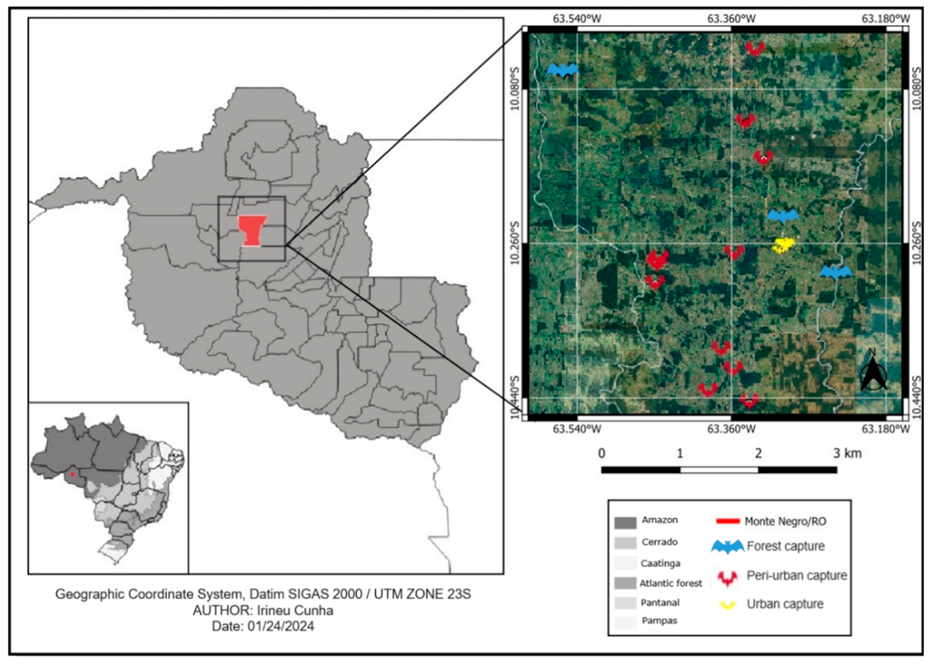

Table 1.
Bacteria identified in ticks, mites and flies from bats in Monte Negro, Rondônia, Brazil.
| BATS | Number | ECTOPARASITES | Number | Detected bacteria | ||||
|---|---|---|---|---|---|---|---|---|
| M | F | M | F | L | ||||
| Emballonuridae | ||||||||
| Emballonurinae | ||||||||
| Peropteryx kappleri (Peters, 1867) | 3 | 3 | Hooperella sp. | - | - | 56 | - | |
| Rhynchonycteris naso (Wied-Neuwied, 1820) | 7 | 12 | Basilia sp. | 1 | 2 | - | - | |
| Spinturnix sp. | 7 | - | - | - | ||||
| Saccopteryx bilineata (Temminck, 1838) | - | 1 | - | - | - | - | - | |
| Molossidae | ||||||||
| Molossinae | ||||||||
| Molossus Molossus (Pallas, 1766) | 7 | 12 | - | - | - | - | - | |
| Eumops perotis (Schinz, 1821) | 2 | 5 | Steatonyssus sp. | - | - | 25 | - | |
| Mormoopidae | ||||||||
| Pteronotus rubiginosus (Wagner, 1843) | 8 | 1 | Trichobius sp. | 9 | 9 | - | - | |
| Paradyschira sp. | - | 1 | - | - | ||||
| Ornithodoros marinkellei | - | - | 6 | - | ||||
| Ornithodoros hasei* | - | - | 10 | - | ||||
| Cameronieta almaensis | 23 | 2 | - | - | ||||
| Noctilionidae | ||||||||
| Noctilio leporinus (Linnaeus, 1758) | 19 | 34 | Ornithodoros hasei* | - | - | 308 | (GenBank No. MH614266) Candidatus Rickettsia wissemanii* | |
| Phyllostomidae | ||||||||
| Carollinae | ||||||||
| Carollia brevicauda (Schinz, 1821) | 13 | 21 | Trichobius joblingi | 11 | 12 | - | (GenBank No. PP445025) Bartonella spp. | |
| Strebla guajiro | 6 | 8 | - | |||||
| Macronyssus sp. | 6 | - | - | - | ||||
| Carollia perspicillata (Linnaeus, 1758) | 1 | 3 | - | - | - | - | - | |
| Desmodontinae | ||||||||
| Desmodus rotundus (Geoffroy, 1810) | 2 | - | Trombiculidae | - | - | 8 | - | |
| Diphylla ecaudata (Spix, 1823) | 3 | 1 | - | - | - | - | ||
| Glossophaginae | ||||||||
| Glossophaga soricina (Pallas, 1766) | 2 | - | Trichobius dugesii | - | 3 | - | - | |
| Periglischrus caligus | 1 | - | - | |||||
| Phyllostominae | ||||||||
| Lophostoma silvícola (d’Orbigny, 1836) | - | 2 | - | - | - | - | - | |
| Phylloderma stenops (Peters, 1865) | 1 | - | Periglischrus torrealbai | 17 | 4 | - | - | |
| Phyllostomus hastatus (Pallas, 1767) | 2 | - | Periglischrus acutisternus | 19 | 4 | - | - | |
| Phyllostomus latifolius (Thomas, 1901) | 1 | - | - | - | - | - | - | |
| Trachops cirrhosus (Spix, 1823) | - | 1 | Strebla mirabilis | 4 | 1 | - | (GenBank No. PP445026) Bartonella sp. | |
| Stenodermatinae | - | |||||||
| Artibeus obscurus (Schinz, 1821) | 12 | 33 | Periglischrus iheringi | 13 | 11 | - | - | |
| Vampyrodes caraccioli (Thomas, 1889) | 1 | - | P. iheringi | 2 | 1 | - | - | |
| Sturnira lilium (Geoffroy Saint-Hilaire, 1810) | 1 | - | Periglischrus sp. | 1 | - | - | - | |
| Vespertilionidae | ||||||||
| Myotinae | ||||||||
| Myotis riparius (Handley, 1960) | 1 | - | Macronyssus sp. | 1 | - | - | - | |
| Vespertilioninae | - | |||||||
| Lasiurus villosissimus (Geoffroy St.-Hilaire, 1806) | - | 1 | - | - | - | - | - | |
| Lasiurus sp. (Gray, 1831) | - | 1 | - | - | - | - | - | |
| TOTAL | 86 | 131 | 121 | 58 | 413 | - | ||
* New records for Rondônia. M – male, F – female, L – larvae.
Disclaimer/Publisher’s Note: The statements, opinions and data contained in all publications are solely those of the individual author(s) and contributor(s) and not of MDPI and/or the editor(s). MDPI and/or the editor(s) disclaim responsibility for any injury to people or property resulting from any ideas, methods, instructions or products referred to in the content. |
© 2024 by the authors. Licensee MDPI, Basel, Switzerland. This article is an open access article distributed under the terms and conditions of the Creative Commons Attribution (CC BY) license (http://creativecommons.org/licenses/by/4.0/).
Copyright: This open access article is published under a Creative Commons CC BY 4.0 license, which permit the free download, distribution, and reuse, provided that the author and preprint are cited in any reuse.


