Submitted:
07 November 2024
Posted:
07 November 2024
You are already at the latest version
Abstract
Objective: This review is aimed at evaluating the role of imaging evaluation in the diagnosis of peripheral vestibular disorders. Methods: The study is a literature review conducted on PubMed and the Cochrane Library. Results: Imaging is not usually recommended in initial consultations for vestibular disorders because only 5-10% of MRI scans reveal findings directly related to the disease. The study is a review of the literature that highlights the utility and limitations of imaging such as computed tomography (CT) and magnetic resonance imaging (MRI). It follows the diagnostic approach from history and physical examination to laboratory tests and imaging. Some conditions like vestibular neuritis and BPPV have limited imaging utility due to the fine details required. Conversely, high-resolution CT and MRI are important for diagnosing Meniere's disease, acoustic neuroma, and superior canal dehiscence. Conclusion: The imaging role varies a lot among specific conditions. Advances in imaging technology, particularly high-resolution MRI, promise enhanced diagnostic capabilities.
Keywords:
1. Introduction
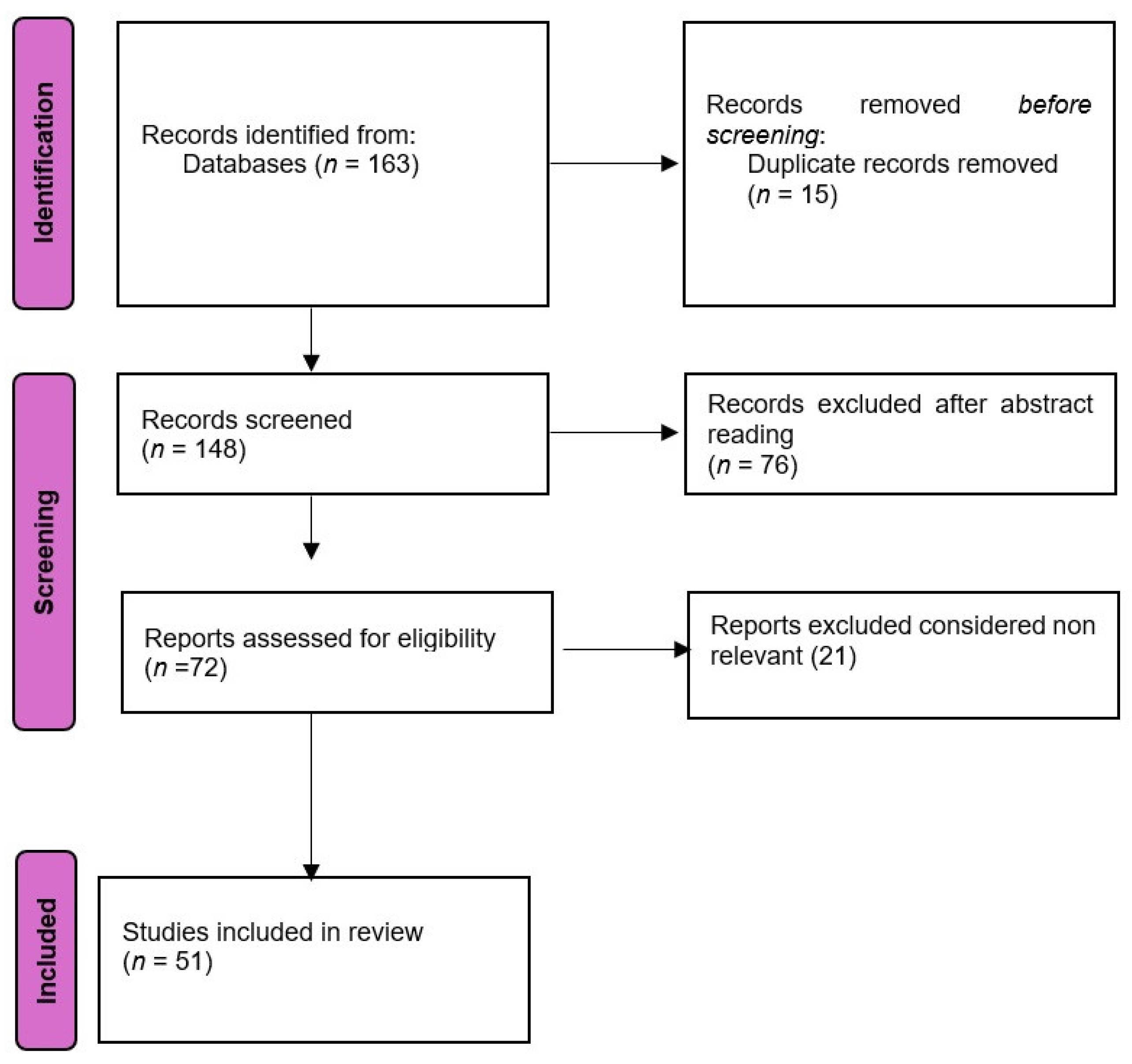
2. Materials and Methods
3. Imaging Modalities in Vestibular Disorder Diagnosis
3.1. Specific Vestibular Disorders and Imaging Utility
3.1.1. Vestibular Neuritis
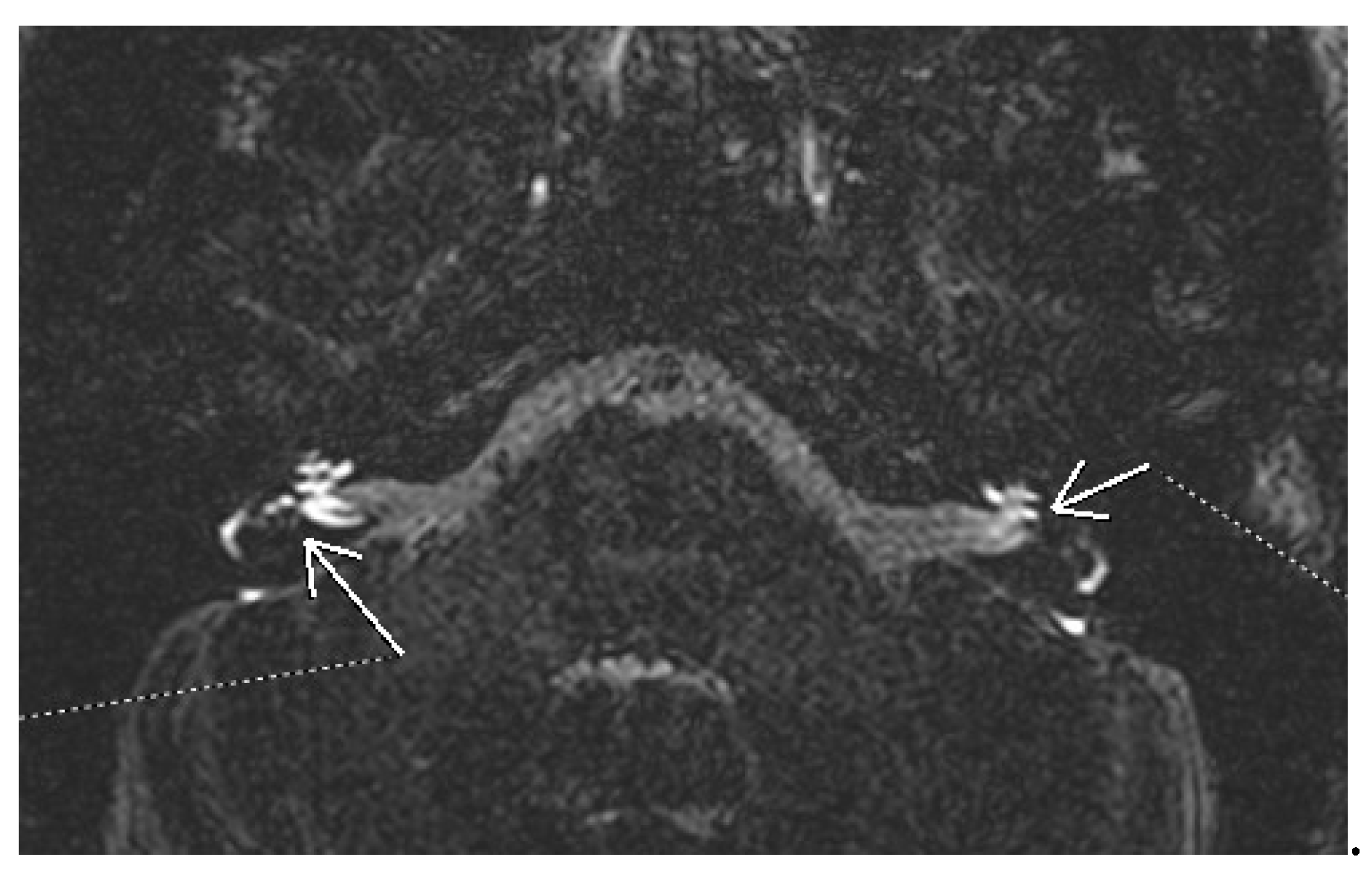
3.1.2. Meniere’s Disease
3.1.3. Benign Paroxysmal Positional Vertigo (BPPV)
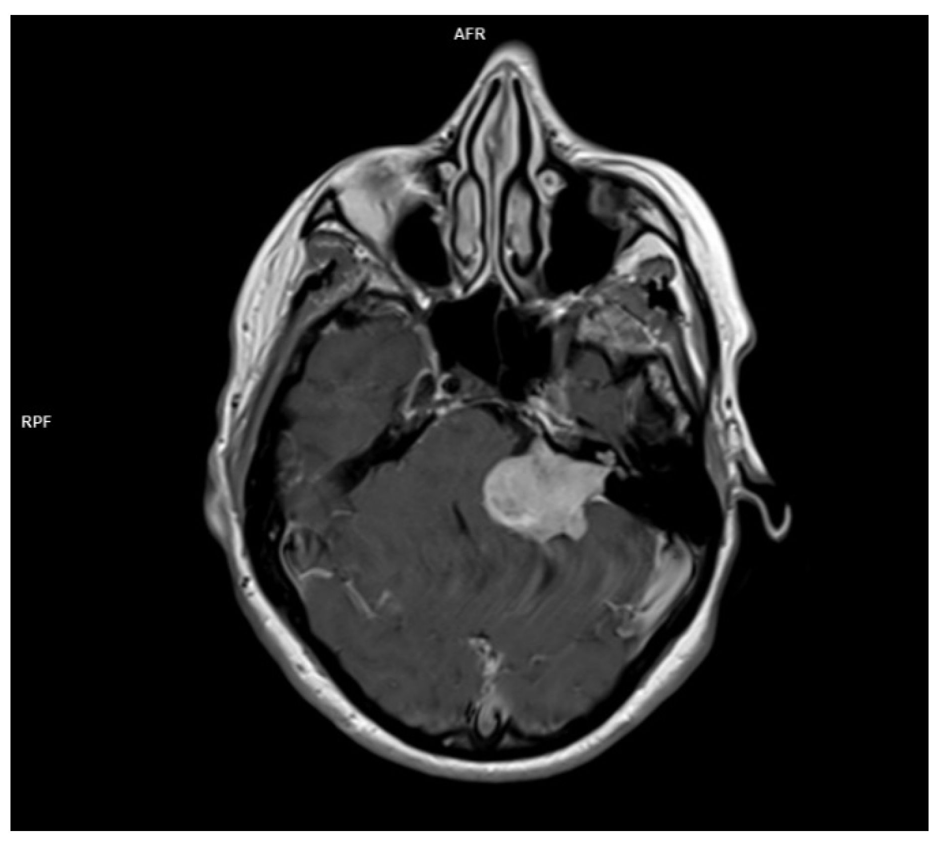
3.1.4. Acoustic Neuroma
3.1.5. Superior Canal Dehiscence
3.1.6. Differential Diagnosis with Central Vertigo
4.1. Conclusions
Authors contributions
Funding
Institutional Review Board Statement
Informed consent statement
Acknowledgments
Conflicts of Interest
References
- Neuhauser H K, “Handbook of clinical neurology.”.
- R. Teggi et al., “Prevalenza dei sintomi vertigine e instabilità in un campione di 2672 soggetti e correlazione con il sintomo cefalea,” Acta Otorhinolaryngologica Italica, vol. 36, no. 3, pp. 215–219, May 2016. [CrossRef]
- H. W. Lin and N. Bhattacharyya, “Balance disorders in the elderly: Epidemiology and functional impact,” Laryngoscope, vol. 122, no. 8, pp. 1858–1861, Aug. 2012. [CrossRef]
- M. Strupp and T. Brandt, “Peripheral vestibular disorders,” Curr Opin Neurol, vol. 26, no. 1, pp. 81–89, Feb. 2013. [CrossRef]
- T. Brandt and M. Dieterich, “The dizzy patient: don’t forget disorders of the central vestibular system,” Nat Rev Neurol, vol. 13, no. 6, pp. 352–362, Jun. 2017. [CrossRef]
- V. Renga, “Clinical Evaluation of Patients with Vestibular Dysfunction,” Neurol Res Int, vol. 2019, pp. 1–8, Feb. 2019. [CrossRef]
- M. Strupp and V. Arbusow, “Acute vestibulopathy,” Curr Opin Neurol, vol. 14, no. 1, pp. 11–20, Feb. 2001. [CrossRef]
- J.-Y. Choi and J.-S. Kim, “Nystagmus and central vestibular disorders,” Curr Opin Neurol, vol. 30, no. 1, pp. 98–106, Feb. 2017. [CrossRef]
- B. M. Seemungal, “Neuro-otological emergencies,” Curr Opin Neurol, vol. 20, no. 1, pp. 32–39, Feb. 2007. [CrossRef]
- A. G. Feldman and L. Zhang, “Eye and head movements and vestibulo-ocular reflex in the context of indirect, referent control of motor actions,” J Neurophysiol, vol. 124, no. 1, pp. 115–133, Jul. 2020. [CrossRef]
- M. Bronstein, M. Patel, and Q. Arshad, “A brief review of the clinical anatomy of the vestibular-ocular connections—how much do we know?,” Eye, vol. 29, no. 2, pp. 163–170, Feb. 2015. [CrossRef]
- J. A. Edlow and D. Newman-Toker, “Using the Physical Examination to Diagnose Patients with Acute Dizziness and Vertigo,” J Emerg Med, vol. 50, no. 4, pp. 617–628, Apr. 2016. [CrossRef]
- A. Serra, “Diagnostic value of nystagmus: spontaneous and induced ocular oscillations,” J Neurol Neurosurg Psychiatry, vol. 73, no. 6, pp. 615–618, Dec. 2002. [CrossRef]
- S. D. Z. Eggers et al., “Classification of vestibular signs and examination techniques: Nystagmus and nystagmus-like movements,” Journal of Vestibular Research, vol. 29, no. 2–3, pp. 57–87, Jul. 2019. [CrossRef]
- M. M. Ganança, H. H. Caovilla, and F. F. Ganança, “Eletronistagmografia versus videonistagmografia,” Braz J Otorhinolaryngol, vol. 76, no. 3, pp. 399–403, Jun. 2010. [CrossRef]
- S. A. Zuniga and M. E. Adams, “Efficient Use of Vestibular Testing,” Otolaryngol Clin North Am, vol. 54, no. 5, pp. 875–891, Oct. 2021. [CrossRef]
- V. M. Rao and D. C. Levin, “The Overuse of Diagnostic Imaging and the Choosing Wisely Initiative,” Ann Intern Med, vol. 157, no. 8, p. 574, Oct. 2012. [CrossRef]
- S. E. J. Connor and N. Sriskandan, “Imaging of dizziness,” Clin Radiol, vol. 69, no. 2, pp. 111–122, Feb. 2014. [CrossRef]
- Bakous and Douglas, Vertigo and Disequilibrium: A Practical Guide to Diagnosis and Management, Second edition. 2017.
- D. Patkar, G. Yevankar, and R. Parikh, “Radiology in Vertigo and Dizziness,” An International Journal of Otorhinolaryngology Clinics, vol. 4, no. 2, pp. 86–92, Aug. 2012. [CrossRef]
- A. Guarnizo, K. Farah, D. A. Lelli, D. Tse, and N. Zakhari, “Limited usefulness of routine head and neck CT angiogram in the imaging assessment of dizziness in the emergency department,” Neuroradiol J, vol. 34, no. 4, pp. 335–340, Aug. 2021. [CrossRef]
- R. Farhat et al., “The ‘Vestibular Eye Sign’—‘VES’: a new radiological sign of vestibular neuronitis can help to determine the affected vestibule and support the diagnosis,” J Neurol, vol. 270, no. 9, pp. 4360–4367, Sep. 2023. [CrossRef]
- H.-M. Shi, H.-C. Sun, and F.-H. Ju, “Recommendations for reducing exposure to medical X-ray irradiation (Review),” Medicine International, vol. 2, no. 4, p. 22, Jul. 2022. [CrossRef]
- A. E. CHANG et al., “Magnetic Resonance Imaging Versus Computed Tomography in the Evaluation of Soft Tissue Tumors of the Extremities,” Ann Surg, vol. 205, no. 4, pp. 340–348, Apr. 1987. [CrossRef]
- S. Vadera and D. Smith, “MRI brain (summary),” in Radiopaedia.org, Radiopaedia.org, 2015. [CrossRef]
- M. Strupp et al., “Acute unilateral vestibulopathy/vestibular neuritis: Diagnostic criteria,” Journal of Vestibular Research, vol. 32, no. 5, pp. 389–406, Oct. 2022. [CrossRef]
- S. Himmelein et al., “Differential Involvement during Latent Herpes Simplex Virus 1 Infection of the Superior and Inferior Divisions of the Vestibular Ganglia: Implications for Vestibular Neuritis,” J Virol, vol. 91, no. 14, Jul. 2017. [CrossRef]
- Adamec, M. Krbot Skorić, J. Handžić, and M. Habek, “Incidence, seasonality and comorbidity in vestibular neuritis,” Neurological Sciences, vol. 36, no. 1, pp. 91–95, Jan. 2015. [CrossRef]
- M. Eliezer et al., “Detection of intralabyrinthine abnormalities using post-contrast delayed 3D-FLAIR MRI sequences in patients with acute vestibular syndrome,” Eur Radiol, vol. 29, no. 6, pp. 2760–2769, Jun. 2019. [CrossRef]
- H. Byun, J. H. Chung, S. H. Lee, C. W. Park, D. W. Park, and T. Y. Kim, “Clinical value of 4-hour delayed gadolinium-Enhanced 3D FLAIR MR Images in Acute Vestibular Neuritis,” Laryngoscope, vol. 128, no. 8, pp. 1946–1951, Aug. 2018. [CrossRef]
- B. Navi et al., “Rate and Predictors of Serious Neurologic Causes of Dizziness in the Emergency Department,” Mayo Clin Proc, vol. 87, no. 11, pp. 1080–1088, Nov. 2012. [CrossRef]
- R. W. Baloh, “Prosper Ménière and His Disease,” Arch Neurol, vol. 58, no. 7, p. 1151, Jul. 2001. [CrossRef]
- H. Thai-Van, M.-J. Bounaix, and B. Fraysse, “Meni??re???s Disease,” Drugs, vol. 61, no. 8, pp. 1089–1102, 2001. [CrossRef]
- Y. Watanabe, K. Mizukoshi, H. Shojaku, I. Watanabe, M. Hinoki, and M. Kitahara, “Epidemiological and Clinical Characteristics of Meniere’s Disease in Japan,” Acta Otolaryngol, vol. 115, no. sup519, pp. 206–210, Jan. 1995. [CrossRef]
- G. J. Basura et al., “Clinical Practice Guideline: Ménière’s Disease,” Otolaryngology–Head and Neck Surgery, vol. 162, no. S2, Apr. 2020. [CrossRef]
- K. Sharma, “Audiological Assessment in Meniere’s Disease,” in Up to Date on Meniere’s Disease, InTech, 2017. [CrossRef]
- A. Ciorba, P. H. Skarżyński, V. Corazzi, C. Bianchini, C. Aimoni, and S. Hatzopoulos, “Assessment Tools for Use in Patients with Ménière Disease: An Update,” Medical Science Monitor, vol. 23, pp. 6144–6149, Dec. 2017. [CrossRef]
- Kay-Rivest, D. R. Friedmann, and J. T. Roland, “Imaging for Menière Disease,” American Journal of Neuroradiology, vol. 41, no. 11, pp. 1964–1965, Nov. 2020. [CrossRef]
- H. Yamane et al., “Practical 3DCT imaging of the vestibular aqueduct for Meniere’s disease,” Acta Otolaryngol, vol. 135, no. 8, pp. 799–806, Aug. 2015. [CrossRef]
- T. Miyashita, Y. Toyama, R. Inamoto, and N. Mori, “Evaluation of the vestibular aqueduct in Ménière’s disease using multiplanar reconstruction images of CT,” Auris Nasus Larynx, vol. 39, no. 6, pp. 567–571, Dec. 2012. [CrossRef]
- A. Venkatasamy et al., “Imaging of the saccule for the diagnosis of endolymphatic hydrops in Meniere disease, using a three-dimensional T2-weighted steady state free precession sequence: accurate, fast, and without contrast material intravenous injection,” Eur Radiol Exp, vol. 1, no. 1, p. 14, Dec. 2017. [CrossRef]
- J.-H. Park, A. Shen, S. Keil, N. Kraemer, and M. Westhofen, “Radiological findings of the cochlear aqueduct in patients with Meniere’s disease using high-resolution CT and high-resolution MRI,” European Archives of Oto-Rhino-Laryngology, vol. 271, no. 12, pp. 3325–3331, Dec. 2014. [CrossRef]
- T. Nakashima et al., “Endolymphatic hydrops revealed by intravenous gadolinium injection in patients with Meniere’s disease,” Acta Otolaryngol, pp. 1–6, 2009. [CrossRef]
- A. Bernaerts, “MRI in Menière’s Disease,” J Belg Soc Radiol, vol. 102, no. S1, Nov. 2018. [CrossRef]
- G. Conte et al., “MR imaging of endolymphatic hydrops in Ménière’s disease: not all that glitters is gold,” Acta Otorhinolaryngologica Italica, vol. 38, no. 4, pp. 369–376, Aug. 2018. [CrossRef]
- T. Nakashima et al., “Grading of endolymphatic hydrops using magnetic resonance imaging,” Acta Otolaryngol, vol. 129, no. sup560, pp. 5–8, Jan. 2009. [CrossRef]
- Baráth, B. Schuknecht, A. M. Naldi, T. Schrepfer, C. J. Bockisch, and S. C. A. Hegemann, “Detection and Grading of Endolymphatic Hydrops in Menière Disease Using MR Imaging,” American Journal of Neuroradiology, vol. 35, no. 7, pp. 1387–1392, Jul. 2014. [CrossRef]
- Eliezer et al., “Clinical and radiological characteristics of patients with collapse or fistula of the saccule as evaluated by inner ear MRI,” Acta Otolaryngol, vol. 140, no. 4, pp. 262–269, Apr. 2020. [CrossRef]
- Dubrulle, V. Chaton, M. Risoud, H. Farah, Q. Charley, and C. Vincent, “The round window sign: a sensitive sign to detect perilymphatic fistulae on delayed postcontrast 3D-FLAIR sequence,” Eur Radiol, vol. 30, no. 11, pp. 6303–6310, Nov. 2020. [CrossRef]
- D. A. FROEHLING, M. D. SILVERSTEIN, D. N. MOHR, C. W. BEATTY, K. P. OFFORD, and D. J. BALLARD, “Benign Positional Vertigo: Incidence and Prognosis in a Population-Based Study in Olmsted County, Minnesota,” Mayo Clin Proc, vol. 66, no. 6, pp. 596–601, Jun. 1991. [CrossRef]
- P. Hilton and D. K. Pinder, “The Epley (canalith repositioning) manoeuvre for benign paroxysmal positional vertigo,” Cochrane Database of Systematic Reviews, Dec. 2014. [CrossRef]
- R. B. Halker, D. M. Barrs, K. E. Wellik, D. M. Wingerchuk, and B. M. Demaerschalk, “Establishing a Diagnosis of Benign Paroxysmal Positional Vertigo Through the Dix-Hallpike and Side-Lying Maneuvers,” Neurologist, vol. 14, no. 3, pp. 201–204, May 2008. [CrossRef]
- D. Bell, “Benign paroxysmal positional vertigo,” in Radiopaedia.org, Radiopaedia.org, 2017. [CrossRef]
- Bhattacharyya et al., “Clinical Practice Guideline: Benign Paroxysmal Positional Vertigo (Update),” Otolaryngology–Head and Neck Surgery, vol. 156, no. S3, Mar. 2017. [CrossRef]
- L. Kluwe, “Molecular study of frequency of mosaicism in neurofibromatosis 2 patients with bilateral vestibular schwannomas,” J Med Genet, vol. 40, no. 2, pp. 109–114, Feb. 2003. [CrossRef]
- R. Babu, R. Sharma, J. H. Bagley, J. Hatef, A. H. Friedman, and C. Adamson, “Vestibular schwannomas in the modern era: epidemiology, treatment trends, and disparities in management,” J Neurosurg, vol. 119, no. 1, pp. 121–130, Jul. 2013. [CrossRef]
- C. Matthies and M. Samii, “Management of 1000 Vestibular Schwannomas (Acoustic Neuromas): Clinical Presentation,” Neurosurgery, vol. 40, no. 1, pp. 1–10, Jan. 1997. [CrossRef]
- X. HUANG et al., “Clinical features of intracranial vestibular schwannomas,” Oncol Lett, vol. 5, no. 1, pp. 57–62, Jan. 2013. [CrossRef]
- A. Venkatasamy, C. Nicolas-Ong, H. Vuong, A. Charpiot, and F. Veillon, “Extension patterns of vestibular schwannomas towards the middle ear: three new cases and review of the literature,” European Archives of Oto-Rhino-Laryngology, vol. 276, no. 4, pp. 969–976, Apr. 2019. [CrossRef]
- W. T. Koos, J. D. Day, C. Matula, and D. I. Levy, “Neurotopographic considerations in the microsurgical treatment of small acoustic neurinomas,” J Neurosurg, vol. 88, no. 3, pp. 506–512, Mar. 1998. [CrossRef]
- L. R. Gentry, C. G. Jacoby, P. A. Turski, L. W. Houston, C. M. Strother, and J. F. Sackett, “Cerebellopontine angle-petromastoid mass lesions: comparative study of diagnosis with MR imaging and CT.,” Radiology, vol. 162, no. 2, pp. 513–520, Feb. 1987. [CrossRef]
- Deng and F. Gaillard, “Vestibular schwannoma,” in Radiopaedia.org, Radiopaedia.org, 2008. [CrossRef]
- E. Hofmann and L. Choné, “Neuroradiologische Bildgebung des Akustikusneurinoms (Vestibularisschwannoms),” HNO, vol. 59, no. 1, pp. 9–15, Jan. 2011. [CrossRef]
- T. H. Mulkens et al., “Acoustic schwannoma: MR findings in 84 tumors.,” American Journal of Roentgenology, vol. 160, no. 2, pp. 395–398, Feb. 1993. [CrossRef]
- V. Dallari et al., “Cochlear Implantation Following Transcanal Infrapromontorial Approach for Vestibular Schwannoma: A Case Series,” Audiol Res, vol. 13, no. 1, pp. 1–11, Dec. 2022. [CrossRef]
- M. Song et al., “Sudden sensorineural hearing loss as the initial symptom in patients with acoustic neuroma,” Front Neurol, vol. 13, Aug. 2022. [CrossRef]
- J. Y. Park and C.-H. Kim, “Vestibular Schwannoma Presenting as Acute Vertigo Mimicking Vestibular Neuritis,” Case Rep Neurol, vol. 14, no. 3, pp. 464–468, Nov. 2022. [CrossRef]
- C. J. Belden, N. Weg, L. B. Minor, and S. J. Zinreich, “CT Evaluation of Bone Dehiscence of the Superior Semicircular Canal as a Cause of Sound- and/or Pressure-induced Vertigo,” Radiology, vol. 226, no. 2, pp. 337–343, Feb. 2003. [CrossRef]
- M. Palma Diaz, J. Cisneros Lesser, and A. Vega Alarcón, “Superior Semicircular Canal Dehiscence Syndrome – Diagnosis and Surgical Management,” Int Arch Otorhinolaryngol, vol. 21, no. 02, pp. 195–198, Apr. 2017. [CrossRef]
- J. Addams-Williams, K. Wu, and J. Ray, “The experiments behind the Tullio phenomenon,” J Laryngol Otol, vol. 128, no. 3, pp. 223–227, Mar. 2014. [CrossRef]
- A. G. Shuman, S. S. Rizvi, C. W. Pirouet, and K. D. Heidenreich, “Hennebert’s sign in superior semicircular canal dehiscence syndrome: A Video Case Report,” Laryngoscope, vol. 122, no. 2, pp. 412–414, Feb. 2012. [CrossRef]
- Minor L B, “Superior canal dehiscence syndrome,” Am J Otol . vol. 1, no. 21, pp. 9–19, 2000.
- W. W. Chien, J. P. Carey, and L. B. Minor, “Canal dehiscence,” Curr Opin Neurol, vol. 24, no. 1, pp. 25–31, Feb. 2011. [CrossRef]
- B. K. Ward et al., “Superior semicircular canal dehiscence syndrome: Diagnostic criteria consensus document of the committee for the classification of vestibular disorders of the Bárány Society,” Journal of Vestibular Research, vol. 31, no. 3, pp. 131–141, May 2021. [CrossRef]
- Y. S. Cheng, S. Raufer, X. Guan, C. F. Halpin, D. J. Lee, and H. H. Nakajima, “Superior Canal Dehiscence Similarly Affects Cochlear Pressures in Temporal Bones and Audiograms in Patients,” Ear Hear, vol. 41, no. 4, pp. 804–810, Jul. 2020. [CrossRef]
- K. S. Noij and S. D. Rauch, “Vestibular Evoked Myogenic Potential (VEMP) Testing for Diagnosis of Superior Semicircular Canal Dehiscence,” Front Neurol, vol. 11, Jul. 2020. [CrossRef]
- Browaeys, T. L. Larson, M. L. Wong, and U. Patel, “Can MRI Replace CT in Evaluating Semicircular Canal Dehiscence?,” American Journal of Neuroradiology, vol. 34, no. 7, pp. 1421–1427, Jul. 2013. [CrossRef]
- J. S. Kim and H. Lee, “Posterior Circulation Stroke and Vestibular Syndromes,” in Oxford Textbook of Vertigo and Imbalance, Oxford University Press, 2013, pp. 251–266. [CrossRef]
- A. Zwergal and M. Dieterich, “Vertigo and dizziness in the emergency room,” Curr Opin Neurol, vol. 33, no. 1, pp. 117–125, Feb. 2020. [CrossRef]
- M. Gottlieb, G. D. Peksa, and J. N. Carlson, “Head impulse, nystagmus, and test of skew examination for diagnosing central causes of acute vestibular syndrome,” Cochrane Database of Systematic Reviews, vol. 2023, no. 11, Nov. 2023. [CrossRef]
- S. Datar and A. A. Rabinstein, “Cerebellar Hemorrhage,” Neurol Clin, vol. 32, no. 4, pp. 993–1007, Nov. 2014. [CrossRef]
- T. Bulut, A. Yildirim, B. Ekmekci, N. Eskut, and H. P. Gunbey, “False-Negative Diffusion-Weighted Imaging in Acute Stroke and its Frequency in Anterior and Posterior Circulation Ischemia,” J Comput Assist Tomogr, vol. 38, no. 5, pp. 627–633, 2014. [CrossRef]
- D. Sankalia, S. Kothari, and D. Phalgune, “Diagnosing Stroke in Acute Vertigo: Sensitivity and Specificity of HINTS Battery in Indian Population,” Neurol India, vol. 69, no. 1, p. 97, 2021. [CrossRef]
- T. Qiu, X. Dai, X. Xu, G. Zhang, L. Huang, and Q. Gong, “A prospective study on the application of HINTS in distinguishing the localization of acute vestibular syndrome,” BMC Neurol, vol. 22, no. 1, p. 378, Oct. 2022. [CrossRef]
- C. L. Warner, L. Bunn, N. Koohi, G. Schmidtmann, J. Freeman, and D. Kaski, “Clinician’s perspectives in using head impulse-nystagmus-test of skew (HINTS) for acute vestibular syndrome: UK experience,” Stroke Vasc Neurol, vol. 7, no. 2, pp. 172–175, Apr. 2022. [CrossRef]
- I.-A. Taciuc et al., “Applications and challenges of neural networks in otolaryngology (Review),” Biomed Rep, vol. 20, no. 6, p. 92, Apr. 2024. [CrossRef]

| Vestibular Disorder | Primary Symptoms | Diagnostic Methods | Imaging Role |
|---|---|---|---|
| Vestibular Neuritis | Vertigo, imbalance, nausea | Clinical examination, VNG | MRI (for differential diagnosis) |
| Meniere’s Disease | Vertigo, hearing loss, tinnitus | Clinical examination, audiometry | MRI (for hydrops) |
| BPPV | Positional vertigo | Dix-Hallpike maneuver | Not typically required |
| Acoustic neuroma (vestibular schwanoma) |
Unilateral hearing loss, tinnitus, imbalance | Audiometry, VNG, MRI | MRI (gold standard), essential for diagnosis |
| Superior canal dehiscence |
Autophony, sound intolerance, vertigo | Audiometry, VEMP, CT | CT for confirmation, MRI (optional) |
Disclaimer/Publisher’s Note: The statements, opinions and data contained in all publications are solely those of the individual author(s) and contributor(s) and not of MDPI and/or the editor(s). MDPI and/or the editor(s) disclaim responsibility for any injury to people or property resulting from any ideas, methods, instructions or products referred to in the content. |
© 2024 by the authors. Licensee MDPI, Basel, Switzerland. This article is an open access article distributed under the terms and conditions of the Creative Commons Attribution (CC BY) license (http://creativecommons.org/licenses/by/4.0/).


