Submitted:
22 October 2024
Posted:
25 October 2024
You are already at the latest version
Abstract
The skin, being the largest organ of the human body, serves as the primary barrier against external insults, including UV radiation, pollutants, and microbial pathogens. However, prolonged exposure to these environmental stressors can lead to the generation of reactive oxygen species (ROS), causing oxidative stress, inflammation, and ultimately, skin aging and diseases. Antioxidants play a crucial role in neutralizing ROS and preserving skin health by preventing oxidative damage.
In recent years, nanotechnology has emerged as a powerful tool for enhancing the delivery of an-tioxidants onto the skin. In particular, liposomal formulations have offered unique advantages such as improved stability, controlled release, and enhanced penetration through the skin barrier. This has led to a surge in research focused on developing liposomal-based antioxidant delivery systems tailored for skin health applications.
Through a comprehensive analysis of the literature from the 2019-2024 period, this review provides an overview of emerging trends in the use of liposomal delivery systems developed for antioxidants aimed at improving skin health. It explores the latest advancements in liposomal formulation strategies, vesicles characterization and their applications in delivering antioxidants to combat oxidative stress-induced skin damage and other associated skin pathologies.
Keywords:
antioxidants
; delivery systems
; liposomes
; ethosomes
; niosomes
1. Introduction
Skin cells play a crucial role as the body's first line of defense against external stressors. These stressors, including UV rays, pollution, and various infection, can increase reactive oxygen species (ROS) (O2., HOO., H2O2, HO., etc.) which are naturally produced as byproducts in cell metabolism and used in redox signaling [1].
Cells are able to regulate ROS concentration with antioxidants (vitamin E, and C, glutathione, coenzyme Q10, carotenoids) and enzymes (superoxide dismutase, catalases, glutathione peroxidase, etc.) that maintain redox homeostasis [2,3,4]. However, this regulatory capacity declines with age [5] leading to an imbalance in redox equilibrium, which results in oxidative stress and inflammation.
In the skin, chronic oxidative stress can contribute to the development of wrinkles, loss of elasticity, and hyperpigmentation as well as various pathological conditions like psoriasis, dermatitis, and even cancer [6,7].
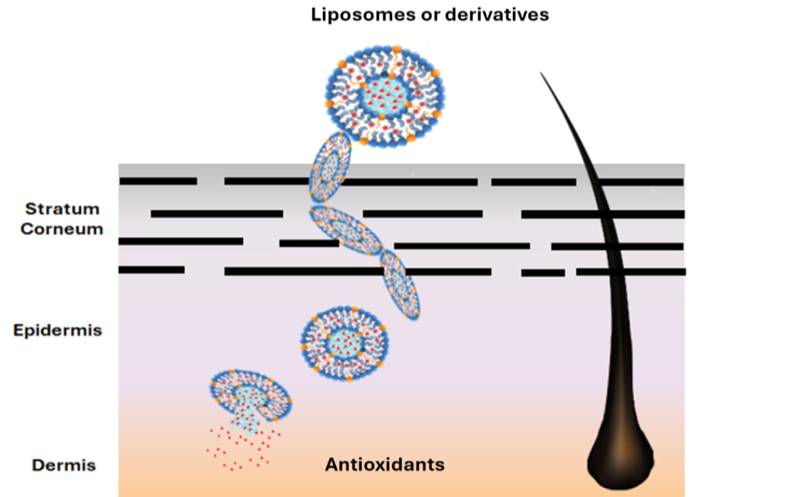
It is well known that the use of exogenous antioxidants supplied by diet enhances the body's response against ROS [2,8]. However, many natural antioxidants are not as effective in vivo, due to their short lifetime, low absorption through cell membranes, rapid metabolic degradation and excretion [9]. Topical application on the skin suffers from similar disadvantages, because many natural antioxidants are unable to penetrate the stratum corneum (SC). Structure and composition of a skin layer is shown in Figure 1.
In recent decades, several nanomaterials have been developed, gaining significant attention for their applications across various fields. Notably, extensive research has also been conducted in delivering molecules with poor solubility [10].
Some examples of nano-delivery systems include vesicles [11,12] liposomes [13], polymer nanoparticles [14], solid lipid nanoparticles [15] and liquid crystals [16]. These systems are designed to encapsulate and deliver drugs or other active compounds to specific target sites in the body, offering improved efficacy, reduced side effects, and enhanced therapeutic outcomes. Each system has its own unique properties, advantages, and drawbacks, making it suitable for different applications. In the case of skincare, specialized liposomal carriers have been tailored to target the unique characteristics of the skin's external surface and to pass through the specific structure of the stratum corneum.
This review aims to provide a comprehensive synopsis of various liposomal systems developed for delivering antioxidants into skin cells. It offers a general description of the potential benefits and limitations of each type of vesicle, along with recent applications for delivering specific antioxidants to the skin.
2. Natural and Synthetic Vesicles
A natural vesicle is a small, membrane-bound cavity. Liposome vesicles are formed by cell's membrane phospholipids and can vary in size from nanometers to micrometers. These sacs play crucial roles in transporting, storing, and processing various substances within the cells. Since Bangham's discovery of the possibility to produce synthetic liposomes in laboratory [14], their use as drug delivery systems has expanded significantly. Initially formed only by natural phospholipids (PL), such as dipalmitoyl phosphatidylcholine (lecithin or DPPC), phosphatidylethanolamine (cephalin) and phosphatidylserine derived by soybean, egg, and milk, they have also been produced later with synthetic or semisynthetic molecules. More recent formulations envisaged the use of organic solvents (ethosomes) or surfactants (transethosomes) to enhance the vesicle deformability. On the other hand, the use of polyethylene glycol bound to the membrane to produce stealth liposomes permitted to avoid immunity system recognition. Specific proteins can also be added to the outer layer to enhance the cell specificity and facilitate the phagocytosis (actively targeted liposomes) [17]. Their biocompatibility and biodegradability have made them a popular choice in nanomedicine [18,19]. This has proven to be extremely useful in anticancer therapies with common drugs [20] and is paving the way for the pharmacological application of natural bioactive molecules that were previously overlooked due to their low solubility and poor pharmacokinetics [21].
Many natural antioxidants suffer the same problems and are seldom used, despite being highly effective against inflammation and diseases associated with ROS imbalance. Liposomes and derivative vesicles specifically designed for antioxidant delivery to skin cells allow for direct application on the epidermis, enhancing efficacy and absorption. The liposomal systems that were analyzed and the antioxidants that were presented are listed in Table 1.
2.1.1. Liposomes
Synthetic liposomes (LPS) can be produced with various methodologies and specific reviews are available dealing with this subject [39,40,41]. LPS are composed of one or multiple bilayer membranes with variable diameters, made up of natural or synthetic phospholipids [42]. The phospholipids used to prepare these vesicles influence their physico-chemical properties. Liposomes formed by saturated phospholipids have higher gel-liquid crystalline phase transition temperature, are more rigid and better retain drugs in their bilayer. One potential limitation of these vesicles is their excessive stability, which can lead to a reduced release of the active principle trapped inside. On the contrary, those LPS mainly formed by unsaturated phospholipids, have opposed properties. So, the proper design of a vesicle nanocarrier should be based on a suitable choice of a proper mixture of phospholipids to obtain a balance between stability and drug release [43]. LPS can carry hydrophilic or hydrophobic molecules within their core or bilayer, making them versatile carriers for improving drug efficacy and reducing side effects [44]. The efficiency of the active principle's absorption can be measured by calculating the encapsulation efficiency (EE), which is the percentage of the active principle that remains in the vesicles compared to the amount initially present in the solution. A schematic view of several kinds of liposomes is shown in Figure 2.
Since the initial use of LPS as drug delivery system, some drawbacks have been envisaged and many efforts have been pursued to solve them. To enhance the bilayer stability versus mechanical and chemical stress, additives as cholesterol or vitamin E were added. Cholesterol can modulate membrane rigidity by reducing the interactions between saturated chains and enhancing those between unsaturated ones [45]. More recently surfactants were also used to balance membrane stability and deformability [28]. Moreover, as vesicles can reassemble together, charged phospholipids were preferred in order to keep vesicles away from each other by electrostatic repulsion [46]. The surface potential of vesicles is influenced by the choice of phospholipid head groups in the bilayer and the presence of a drug with a specific charge. The ζ potential (ZP), typically determined by microelectrophoresis, provides insight into this parameter. In particular, anionic LPS are easily phagocytized by macrophages and can penetrate cells by endocytosis, making this two aspects particularly useful in drug delivery through the skin [44].
The vesicle size is another important parameter to control during LPS preparation. See for instance Figure 3. Vesicles with a mean diameter (MD) in the range of 20-40 nm, known as small unilamellar vesicles (SUV), can evade immune system attacks, resulting in longer clearance times. In contrast, only vesicles smaller than 100 nm, referred to as medium unilamellar vesicles (MUV), can prolong their circulation in the blood and be easily absorbed by cells [18].
In the case of dermal drug delivery, even larger vesicles (LUV) can be used, provided they can penetrate the SC. It is also important to recall that in loaded LPS the chemo-physical properties of the entrapped organic compounds can alter the dimensions of the vesicles, especially if the molecules interact with the bilayer. In some case vesicles dimensions were enlarged by the loosening of the interaction between phospholipids due to the intercalation of the drug molecule [23,28], while the opposite effect is detected with other drugs [13,30]. A parameter that represents the distribution of the vesicle size is the polydispersity index (PDI). It characterizes the distribution of molecular mass among the vesicles in the sample and provides insight into the heterogeneity of the vesicles' volume. Determined by Dynamic Light Scattering (DLS), a PDI lower than 0.1-0.2 indicates a narrow size distribution and a more uniform population of liposomes. The uniformity is often desirable for consistent drug delivery and predictable behavior in biological systems. Techniques generally used to enhance the liposomal size homogeneity are sonication during mixing operation [23] or filtration of liposomes through filters smaller than their previous average diameter [25].
Many liposomal formulations have been used to deliver antioxidants, either pure or derived by complex natural extracts. Bavarsad et al. [22] developed liposomal formulations of pure quercetin for treating skin pressure ulcers. They employed a fusion method of phospholipid bilayers in the presence of 0.01-0.04 g/ml quercetin buffer solutions, without the use of organic solvents. The small unilamellar vesicles (SUV) produced had a homogeneous particle size with a MD of 10-16 nm, demonstrated good encapsulation efficiency (EE) of 65-78%, and were sufficiently viscous to be directly applied to the affected areas. This delivery system showed a high efficiency of retention (RE) into the skin (RE 46-51%) and a good level of penetration ratio (retention/penetration ratio 7.1-9.0), indicating that for the most part quercetin LPS were trapped in the skin. The LPS-loaded active ingredient regulated vascular circulation and necrosis, reducing inflammation and shortening recovery time for pressure ulcers. With minimal systemic absorption, the liposomal formulation had fewer side effects compared to phenytoin cream, which was used as the reference drug.
An example of plant extracts delivery has been brought up using Moringa Oleifera leaves [13]. Rich in carotenoids, glucosinolates, vitamins, amino acids, and many phenolic antioxidants, Moringa Oleifera is well known for its anti-microbial, anticholesterolemic and anti-inflammatory properties. In the skin, this plant extract helps to maintain collagen and hydration level, thus enhancing defense against ageing and photo-damage. The hydroethanolic extracts of the Moringa Oleifera leaves were encapsulated in LPS formed by DPPC and cholesterol (4:1 ratio). The presence of the extracts lowered the diameter of the LPS from the size of a giant unilamellar vesicle (GUV, 736±100 nm MD) to a large one (LUV, 154±1 nm MD). The loaded vesicles possessed a low polydispersity index (PDI 0.17±0.01), a weak positive charge (ZP +6.6±0.1), and a good retention rate (RR >80% at 35°C for 6 h). The antioxidant activity (AA), determined by the DPPH assay, of the pure extract (AA 85.8 ± 1.5%) proved to be similar to that of the reference, gallic acid (AA 87 ± 2%), while the liposomal formulation was less active (AA 70.0 ± 1.0%). Additionally, the antioxidant effect of liposomes, determined by ORAC experiments, diminished faster (60 minutes instead of 120 minutes for the pure extract). A gel formulation with propylene glycol, hydroxyethyl cellulose and glycerin was used to analyze effects on skin. Results demonstrated that even if the free hydroethanolic extracts had greater antioxidant properties and could better enhance hydration, the liposomal formulation was more effective at preventing transepidermal water loss.
Another interesting application of liposomal delivery was the preparation of vesicles loaded with berry extract from Myrtus Communis [23]. This mediterranean bush is well known for the presence of antioxidants in its berries. HPLC-DAD analyses detected high concentration of anthocyanins, galloyl derivatives and flavonoids in the macerated ethanolic extract (1:1 w/v). The dry extracts were used to prepare the liposomes using fat-free soybean phospholipids (70% phosphatidylcholine) in PBS solution under sonication cycles. This very simple method produced liposomes of 102±5.6 nm MD, slightly bigger than the corresponding empty vesicles, but similar in PDI (0,22 ±0.02) and ZP (-10.00 ±0.08 mV). The research group determined the entrapment efficiency (EE) of each main antioxidant of the myrtle extract: the EE ranges from 71.4% of Myricetin-3-O-galactoside to over 95% of most of the anthocyanins. The antioxidant activity of the loaded liposomes, measured by DPPH analysis, was only slightly lower (326±17 μg Trolox Equivalent/ml) than that of the free myrtle extract (344±22 μg TE/ml). The reducing power remained unchanged, as confirmed by FRAP analysis, with values of 1831±70 and 1867±32 μg Fe²⁺ equivalents/mL for the loaded liposomes and myrtle solution, respectively. The 3-(4,5-Dimethylthiazol-2-yl)-2,5-diphenyltetrazolium bromide (MTT) assay on cell viability demonstrated the low cell cytotoxicity of myrtle extracts and the even higher vitality of cells treated with loaded liposomes vs control cells. Furthermore, anti ROS-activity experiments made on fibroblasts exposed to AAPH radical generator showed that liposomes, loaded with increasing quantity of myrtle extracts (from 0.1 to 10 μg), significatively reduced the oxidative stress, even better than the pure myrtle extract after 24 hours. Unfortunately, no direct test on skin were performed by authors even with this encouraging results.
Permana, Rahman et al. [24] investigated the use of liposomes to enhance the antioxidant activity, permeability and skin retention of propolis extracts (PEs). The more efficient extraction (75% v/v of ethanol/water, PE75) in term of total polyphenolic content (TPC 179.3±9.3 mg/ g PE) and total flavonoid concentration (TFC 277.2± 3.4 mg/ gPE) turned to be the more antioxidant either as radical scavenger capacity (DPPH IC50 43.2± 3.0) or as lipid peroxidation inhibition (LP, 65.3±3.4 μg/ml). These PEs are rich in caffeic acid (maximum 4.15±0.01 mg/g in PE75), quercetin (0.72±0.01 mg/g in PE75) and kaempferol (2.62±0.01 in PE75), as confirmed by HPLC analyses. They were encapsulated into liposomal vesicles using different amount of phosphatidylcholine (PC) applying the thin-layer hydration technique with subsequent solvent evaporation. Higher concentrations of phospholipids, increasing the viscosity of the solution, generally produced larger vesicles due to the higher surface tension. On the other hand, the use of a lower weight ratio of PE/phosphatidylcholine combined with a longer duration of homogenization or bath sonication allowed for a reduction in liposomal size from 620±54 to 248±22 nm of diameter, with PDI and ZP remained constant. As the major interaction of the three main antioxidants depend on hydrogen bonds with the bilayer either as acceptor or donor, the EE seemed to depend on their hydrogen bond capacity. Quercetin exhibited better encapsulation (maximum 77±6%) than kaempferol or caffeic acid (maximum 45±4% and 55±5% respectively), the extent of EE being also dependent on the size of the LPS and the time of homogenization and sonication. The obtained vesicles enhanced the water solubility of caffeic acid, kaempferol, and quercetin by 5-fold, 11-fold, and 12-fold, respectively. LPS were dissolved in a hydrogel (Carbomer 940 and glycerol in water), in order to analyze their potentiality as skin formulation. Antioxidant analysis confirmed that their antioxidant activity either as radical scavenger (DPPH IC50 48± 5) or as lipid peroxidation inhibitor (LP, 68 ± 5 μg/ml) remained similar to that of hydrogel containing simple propolis extract. On the other hand, tests in vitro on mouse skin, showed that the use of LPS-hydrogel enhanced the skin permeation and retention of all antioxidants by 4- to 8-fold compared to the hydrogel containing PEs. This was ascribed to the poor absorption of the free antioxidants into the skin, and the opposite effect of phospholipidic bilayer that facilitate the penetration into the stratum corneum. This hydrogel formulation containing propolis extract also acted as photoprotector against UVA and UVB aggression, as stated by the measured sun protection factor of 17± 2.
The combined use of liposomes and other carriers, such as cyclodextrin (CD), has been designed in pharmaceutical applications to enhance stability of loaded drugs and modulate their release (Modulatory Liposomal Controlled Release, MCLR) [47].
Gardikis et al. [25] analyzed the use of liposomes and CD to enhance the adsorption of propolis antioxidants and study their biochemical activity in skin. In their research they used micronized propolis extracted in presence of CD. The LPS mixed with the CD/propolis extract, produced large micelles that were made smaller through membrane filtration (0.22 mm membrane pore size). Using 10% w/w propolis concentration, the obtained liposomes showed an initial MD of 255±36 nm, decent PDI (0.366±0.051) and good ZP (-21±5 mV). This preparation gave higher EE (84±4%) with respect to previous works, thanks to the presence of CD. The TPC and the antioxidant activity determined by Folin-Ciocalteu's method and DPPH assay, respectively, evidenced a high polyphenols concentration of 100.0± 0.7 mg gallic acid eq/ml and a significant antioxidant activity of 33.7±0.2mM Trolox equivalent. However, both parameters decreased over time or when vesicles were stored at temperature higher than 6°C. The liposomal formulation was also studied in vitro on human skin cell (HaCaT) by biochemical and genetic tests. LPS enhanced skin cells metabolism enhancing ATP levels, and protected cells against UVB lowering photoaging. The upregulation of various genes connected with cell proliferation (AQP3), antiaging (TNFα), cell defense against pathogens (IL4) and immune protection (ITGB2) confirmed the beneficial effect of the propolis and the efficacy of the liposomal carrier in enhancing the absorbance of the active principles into the skin. Finally, a cosmetic formulation (1% LPS) proved that the loaded liposomes were not irritating for human skin cells (MTT on reconstructed skin model), opening to possible marketing application.
Oskoueiana, Karimi and coworkers produced nanoliposomes of Owhadi pistachio hull extract (PHE). The aim was to enhance the stability and applicability of the antioxidant mixture containing gallic acid, rutin, quercetin, myricetin, catechin and pyrogallol [26]. The extract containing the higher quantity of polyphenols (TPC 52±g GAE/100g dry extract) was used in the liposomal encapsulation. The LPS were formed by mixing hot water solution of lecithin and ethanolic organic extract and then sonicating. Despite being sufficiently small (MD 96.3 ± 7.2 nm) and exhibiting a negative charge (ZP -48.0 ± 0.8 mV), the vesicles demonstrated a low entrapment efficiency (EE of 38.4 ± 2.7%). As indicated by the MTT assay, the liposomal formulations maintained high cell viability in murine hepatocytes, at extract concentrations up to 50 μg/ml. The antioxidant activity was confirmed by DPPH assay, even if LPS radical scavenging capacity was lower (IC50 12.6±2.9 μg/L) in comparison with gallic acid reference (IC50 8.8±0.6 μg/L). The LPS were also able to up-regulate the genes of endogenous antioxidant enzymes in murine hepatocytes: SOD (+3.2 fold change), CAT (+2.1), GPX (+1.9), and Nrf2 (+2.7). However, this effect was not as large as that observed with gallic acid, which showed fold changes of +3.8, +2.2, +2.8, and +3.3, respectively, for the same enzymes. Furthermore, the PHE liposomal formulation demonstrated activity against melanogenesis: it inhibited tyrosinase activity in mushroom cells (IC50 21.8 ± 4.3 μg/ml), although to a lesser extent than the kojic acid reference (16.2 ± 4.3 μg/ml). Additionally, it reduced melanin production and partially suppressed the expression of tyrosinase, MTIF, TRP-1 and TRP-2 genes in melanoma cells. These data confirmed the potential use of this extract in skin whitening processes.
Recently, the problem of scalability and industrial production, with all the challenges that this involves, has been raised from various groups of research. Caddeo, Pucci and coworkers [27] analyzed the preparation of vesicle loaded with resveratrol from a commercial liposomal preparation to check the possibility of an easy-to-handle formulation for skin application. The Pronanosome LIPO-N (PLIPO-N) by Nanovex Biotechnologies SL (Llanera, Spain) is a commercial powder of dry LPS. The easy preparation of the liposomal carrier, through an overnight mixing of resveratrol, Tween 80 surfactant, and PLIPO-N in water, followed by sonication, produced highly homogeneous nanovesicles (PDI 0.19) having MD (80 nm) and ZP (-25 mV) statistically consistent with the same regenerated empty liposomes. The loading of the resveratrol was high (83%) and long-term tests confirmed the stability of the physico-chemical properties of the vesicles for more than three months. DPPH (321.0 ± 7.0 μg of Trolox equivalents/ml) and FRAP (8.1 ± 0.7 mg of Fe2+ equivalents/ml) assay proved that resveratrol antioxidant activity was maintained even in the liposomal formulation. More surprisingly, while resveratrol ethanolic solution lowered 3T3-L1 cell viability up to 20 % at 10 μg maximum concentration, the loaded LPS proved to be less toxic with a 55% viability in the same condition. The ability of resveratrol to protect fibroblasts from ROS injuries was analyzed by dichlorofluorescein (DCF) assay in which the fluorescent radical DCF was produced by a ROS activator (AAPH 250 μM). In the DCF experiments, the fluorescence lowered as resveratrol concentration increased much faster than with the liposomal formulation. On the other hand, microscope images confirmed that, using free resveratrol, the cell mortality increased so much to invalidate the fluorescence data. Instead, the ROS scavenging capacity of liposomal resveratrol did not produce cell death at the higher concentration thus supporting the usefulness of the liposomal formulation.
Surfactants, which are amphiphilic, generally are used to stabilize the vesicles, reduce their diameter and increase their deformability by lowering the energy required to modify membrane curvature. For this reason, adding surfactant to liposomal formulation could be useful for a dermal formulation. In the case of naringenin, another antioxidant that is poorly soluble in water but shows high potential as anti-inflammatory agent and regulator of fibroblast and melanoma cell growth, Lowry et al. [28] analyzed its incorporation into liposomal formulations containing different percentage of Tween 20 surfactant. The positioning of naringenin in the bilayer increased the diameter of the vesicles. The so formed loaded liposomes had a MD of 392 ± 32 nm that decrease to 235 ± 9 nm in presence of 10% Tween 20. The vesicles resulted neutral or slightly negative (ZP from 4.12 to -022 mV) but with a PDI of 0.3%. The EE of the active principle was also decreased by Tween 20, from 90.8±4.6% to 64.3±5.6% at 10% Tween 20 concentration. It was supposed that the surfactant competed with lipophilic naringenin for a position within the membrane. As the authors expected, the elasticity of the different liposomal formulations increased in presence of a higher percentage of surfactant as evidenced by a decrease in the deformability index (DI) from 80.7 ± 2.0% (without Tween 20) to 59.2 ± 4.4% (with 10% Tween 20). Gel derived by hydroxyethyl cellulose (HEC) and hydroxypropyl methylcellulose (HPMC) were added to naringenin and to the loaded liposomal formulations to compare the release of the active principle using side-by-side diffusion cell experiments. The results showed that the liposomes released naringenin faster than the solution, and the release rate increased as concentration of Tween 20 reached 10%. On the other hand, the comparison of the formulations containing either loaded liposomes, with 2% Tween 20 or without, and one cellulose gel evidenced that HMPC gel slowed the release of the naringenin better than HEC gel even in presence of the surfactant that enhanced the process in both formulations. The study of the uptake of naringenin by HaCat and HDFa cells, using sodium 3'–(1–phenylamino)–carbonyl–3,4–tetrazolium (XTT) assay with a fluorescent dye, confirmed the ability of gel liposomes to penetrate the cells, probably, by ionic interaction with cell membrane. These data illustrate the importance of both the structure of the vesicles and the formulation used for the delivery system in achieving a synergistic effect on the transport and adsorption of the active principle.
2.1.2. Ethosomes and Transethosomes
In the last years the use of vesicles has been further increased by the development of liposomal derivatives with higher membrane mobility and deformability. This kind of vesicles are particularly useful in dermal treatments. Ethosomes (ETH) are soft, malleable structure composed of phospholipids, ethanol (20-45%), and water, classified as ultra deformable vesicles [48]. They penetrate deeper layers and increase drug permeation compared to traditional LPS. Their flexible structure and ability to enhance skin penetration, is due to ethanol ability to perturb the organization of SC lipids. The deformability index (DI) can parametrize the deformability of these vesicles, with a lower DI corresponding to a high degree of deformability, and it can be determined by analyzing the ethosomes’ diameter and weight before and after extrusion through membrane filters with pores smaller than the average diameter of the vesicles under examination. The deformability of ETH make them a valuable tool for transdermal drug delivery in pharmaceutical and cosmetic formulations. Furthermore, ethosomes are particularly advantageous for delivering drugs with poor water solubility or high molecular weight.
Sguizzato and coworker [29] prepared ETH containing Coenzyme Q10 (CoQ10) to enhance the bioavailability of this important antioxidant in the skin. CoQ10 is insoluble in the most common solvents and poorly in ethanol (0.3 mg/ml). The preparation of ETH from a solution of soybeans PC and CoQ10 in ethanolic solution was performed at room temperature and enhanced the solubility of the antioxidant to 1 mg/ml, permitting a 98% adsorption into the vesicles. This procedure resulted in better performance if compared to that obtained with solid lipid nanoparticles or simple LPS which permitted an adsorption of only 73% and 89% respectively [49]. Considering EE parameter, the more stable vesicles obtained were those derived by a 0.9% w/w concentration of PC in ethanol, with a 254-271 nm MD, and a mixed unilamellar and multilamellar structure. The stability of CoQ10 inside the ETH in three months was higher that the free molecule or other kind of supramolecular preparations. ETH alone or with CoQ10 was found to be safe for cells in MTT and LDH experiments. The research group demonstrated the ability of loaded vesicles to penetrate fibroblast membranes, probably by endocytosis, and protect human fibroblast cells from H2O2 insults in ex-vivo experiments, as demonstrated by analysis of the production of 4-hydroxynonenale (4-HNE) from lipids. The same fluorescence experiments on reconstructed human epidermis, and TEM images confirmed ETH adsorption within the stratum corneum (SC) and even into the stratum basale, as well as their protective effect against hydrogen peroxide-induced lipid degradation, resulting in a 35% reduction in the production of 4-HNE.
To fully utilize the strong antioxidant properties of rutin, given its difficulty to go beyond the SC, the set-up of an effective delivery system is almost essential. To reach this goal, Rolim Baby et al. [30] prepared ethosomes with PC from egg yolk (2% w/v), rutin (0.03%w/v) and an hydroalcoholic solution (20% ethanol). The vesicles produced were bigger (369.5±5.2 nm) than the empty one, with a lower ZP (-19.6±0.4 mV) and a higher PDI (0.43±0.01). In particular a PDI higher than 0.3 could be due either to the formation of vesicles agglomerates during the addition of the rutin ethanolic solution or to a more disperse geometry of the vesicles. In vivo tape-stripping tests on human skin demonstrated that rutin-loaded ETH were not irritating and that they penetrated the lower layers of the stratum corneum (SC) three times more than the rutin solution. Even if the antioxidant activity in vitro was confirmed for the ETH formulation, the ex vivo DPPH assay either on the upper or deeper layer of the skin, did not evidenced relevant differences between the loaded ethosomal formulation, the free rutin solution and the control. The authors ascribed this negative result to the low concentration of rutin in the formulation.
Two years later the group of Paolino et al. [31] succeeded in enhancing the rutin antioxidant efficacy respect to the simple rutin solution by studying a different ethosomal composition. The ETH were prepared with Phospholipon 90G©, water and rutin ethanolic solutions, in the dark to avoid rutin photodegradation. The concentration of rutin solutions, from 0.5 to 4 mg/ml, into the ETH was inversely correlated with the size (MD from 110 ± 8 nm to 575 ± 6 nm) and also with the elasticity of the vesicles. However, DI values (from 3.5 to 10%) permitted a reasonable degree of membrane elasticity. Also, the ZP of the different ETH, varied as a function of the rutin adsorbed into the bilayer. In all cases, ranging from -22.4 to -29.5 mV, ZP values were consistently negative and low enough to ensure effective repulsion of the vesicles. Interestingly, in comparison with the other formulations, those ETH prepared with higher concentration of ethanol (40%w/v instead 30%w/v) produced smaller ZP (from -5.8 to -12.8 mV) and were discarded. The EE was dependent on different parameters: a higher concentration of rutin solution significantly enhanced the EE, while a higher ethanol content reduced it. On the other hand, rutin diminished the release of the active principle by enhancing bilayer rigidity. Considering the higher EE (67.5 ±5.2 %) and DI (3.5±0.5%) obtained, the best ethosome composition was found to be 4 mg of rutin dissolved in 30 ml of EtOH, 69 ml water and 1%w/v PL90G©. In comparison with rutin solution, the experiments conducted with this ETH showed that the delivery system stabilized the antioxidant and strongly reduced its photodegradation in presence of UV light. MTT tests on cell lines subjected to H2O2 oxidative stress, showed that the ETH not only maintained cell viability, but also exerted antioxidant protection on the cells, unlike the pure compound. This protection depended on the concentration of rutin, and increased as the concentration of the active ingredient rose up to 100 μM. Finally, the anti-inflammatory effect of rutin ETH, conducted on conveniently irritated epidermis of human volunteers, confirmed the higher effect of the loaded vesicles in comparison with simple solution of the active principle. It should be noted that rutin properties counterbalanced the initial irritating effect of ethanol on the skin due to the hydroalcoholic preparation, while the erythema recovery, in presence of rutin ETHs, was more rapid and with better results after 4-5h.
In case of Euphorbia Characia latex extract, ETH were conveniently used as skin penetration enhancer of the quercetin derivatives [32]. This extract inhibited collagenase, hyaluronidase and elastase enzymes responsible of skin ageing. Suitable ETH were prepared to analyze its inhibitory capacity against tyrosinase, responsible of skin melanogenesis. The obtained vesicles were nanostructures (MD 101.1 ± 9.7nm, PDI 0.29 ± 0.07%) with high ZP (−70.6 ± 7.3 mV) and a good EE of 85-86% depending on the glycoside bound to quercetin. MTT experiments pointed out the safety of the vesicles, with cell viability >82% for empty and even higher for loaded ETH, while the DPPH assay confirmed the maintenance of high antioxidant activity of 94.8± 0.4%, in the liposomal formulation. The anti-melanogenic effect of tyrosinase, determined by zymography on cells treated with the L-DOPA enzyme activator, confirmed a high level of inhibition from the plant extract, which was further enhanced by the loaded ETH. All of these properties, along with the long-term stability of the prepared ETH, confirm their potential for delivering plant extract bioactive compounds through the skin, even though no specific experiments were conducted on skin cells in this case.
Another recent application of nano delivery systems is based on the use of two or more components together for a synergistic effect. This is the case of the work of Ferrara et al. [33] that used either ETH of curcumin (Cur) and piperine (Pip) to check their capacity to reduce the skin damages derived by environmental pollution. While Cur is well known as an powerful antioxidant, antibacterial and antiviral with effects against inflammation, and various pathologies [50], Pip shares this properties but it also increases the bioavailability of other molecules [51] as well, by slowing down the cell efflux systems and enhancing skin cells permeation [52]. In their research, Ferrara and coworkers compared the bulk production method with microfluidic technique for creating ETH. In the microfluidic approach, two syringes containing an ethanolic solution of phospholipids and distilled water were mixed at a cross-junction. The flow ratio of the two solutions was maintained at 2:1 (v/v), while varying total flow rates were applied. During flows mixing the LPS were formed with homogeneous size as a function of the total flow rates. Nevertheless, the bulk technique turned out to be more rapid (5 ml vs 0.36 ml volume of 200 nm diameter ETH), also considering a possible scaling up of the production. The ETH so prepared were multilamellar and showed an EE of 97 and 79% for Cur and Pip respectively. In a second step, the researchers compared the antioxidant effect of Cur and Pip solutions and their ETH either alone or together. The results evidence that the mixed ETH solutions enhance the drug efficacy of Cur in the FRAP assay. However, some drawbacks can be envisaged in the release profile of the two drugs. In comparison with the drug loaded in the vesicles, the percentage of drug released from ETH was 33% in case of Pip and 15% for Cur, lower than the values obtained by the corresponding drug solutions that were 57% and 24% respectively. In the permeability tests, Cur and Pip behaved differently. Pip was faster in diffusing through the synthetic membrane (STRAT-M®) that simulate the SC, either in solution or in loaded ETH, the second being slower. Conversely, Cur in ETH showed a partition coefficient higher than the Cur solution even if both had a longer lag time than the Pip systems. If compared to those of Pip, Cur either in solution and in ETH also showed low permeability coefficient. Nevertheless, the Cur ETH system seemed to be more efficient than the corresponding solution in providing a prolonged protective effect. Additionally, the combined use of Cur ETH and Pip ETH, further enhanced this behavior against oxidative stress in skin submitted to diesel engine exhausts.
Transethosomes (TETH) are a further variation of ethosomes containing, together with ethanol, a surfactant that works as SC permeation enhancer, particularly useful for dermal delivery [53].
To study the antioxidant activity of mangiferin, a glucitol xanthone present in many plants and fruits (papaya, mango), Valacchi, Esposito et al., considering its higher solubility in ethanol (0.20 mg/ml) respect to water (0.11 mg/ml), prepared ETH and TETH as nano delivery systems [34]. They analyzed the different loaded vesicles in their physico-chemical characteristics, antioxidant activity, and anti-inflammatory properties on skin keratinocytes. The ETH were formed mixing PC and ethanol with mangiferin, enhancing its solubility to 3.3 mg/ml thanks to PC presence. TETH were obtained adding, to the same ingredients, different quantity of surfactants (0.1-0.6%) such as hydrophilic polysorbate (Twin 20, Twin 80), lipophilic sorbitane monoleate (SP20, SP 80), or ionic dimethyldidodecylammonium bromide (DDAB). Depending on the surfactant used, the vesicle size and zeta potential (ZP) varied, while the PDI was always less than 0.2, indicating good homogeneity in vesicle volumes. In general, a higher concentration of surfactant produced a lowering of the diameter and zeta potential. Except in case of DDAB that gave highly positively charged vesicles, the ZP were always negative. As already stated, stability tests confirmed that the presence of a low ZPs was detrimental to the stability of the vesicles, which could fuse over time due to the low repulsion. For these reasons the research group chose to continue the study with mangiferin (0.1%w/w), only on the vesicles containing either Twin 80 (MD 169.3 ± 0.46, ZP−28.29 ± 0.4) or DDAB (MD 86.41 ± 1.09, ZP 84.08 ± 0.6) as surfactant. However, in MTT tests, while ETH+TW80 produced low cytotoxicity that increased with mangiferin concentration, either empty or loaded DDAB ethosomes showed high toxicity and were discarded from further experiments. This is probably because the positive charged vesicles tented to destabilize the keratinocytes, leading to their lysis. The diffusion test, conducted with Franz cell using a nylon membrane, confirmed a lower diffusion capacity for both formulations compared to a simple Mangiferin solution. However, TETH showed better performance compared to ETH, because the surfactant most likely facilitated the release of the active principle from the vesicles. Finally, exposure to cigarette smoke was used to test the antioxidant and anti-inflammatory effect of the vesicles on HaCaT cells. The results demonstrated that both ETH and TETH permitted mangiferin to penetrate cells (TEM images) and to lower the inflammation of exposed keratinocytes as further demonstrated by the reduction of OH-1 and IL-6 inflammatory enzyme expression.
Subsequently, the same research group studied the anti-melanoma activity of different concentrations of pure quercetin loaded onto either ETH or TETH [35]. Building on previous results, only Twin 80 was used as surfactant. Both kinds of the vesicles were multilamellar with similar diameters dependent on the PC and surfactant concentration but with generally uniform dispersity index. Quercetin, standing in the bilayer, produced a loosening of the membrane and an enlargement of vesicles diameter while Twin 80 seemed to enhance vesicles stability (ETH MD 258 ± 21nm, TETH MD 240 ± 21nm).
The entrapment capacity of TETH was determined to be slightly higher than that of ETH, with a percentage of 59.2% and 56.4% respectively, while antioxidant capacity on lipid proved to be almost equal for the two delivery systems (3.16 μmol TE/g vs 3.26 μmol TE/g) but lower than free quercetin solution (3.82 ± 0.05 μmol TE/g). In in vitro release test (IVRT) on lipophilic polytetrafluoroethylene (PTFE) membrane, which is more similar to human skin, the release rate remained constant over time (zero-order kinetics), confirming the ability of the vesicles to control and prolong the release of the active principle. In vitro permeation test (IVPT) confirmed that transethosomes possessed a better capacity in letting quercetin penetrate into the SC respect to ETH (4-fold lower), and quercetin solution (10-fold lower). The MTT experiments, conducted on both HT144 melanoma and HaCaT cells, demonstrated low cytotoxicity at quercetin concentrations below 5 µg/ml. The subsequent wound healing assay, performed at 2.5μg/ml quercetin concentration, evidenced a lower capacity of TETH and ETH to favor wound resolution in both the cell lines. This has been ascribed to the capacity of active principle to inhibit ROS production so lowering the natural response of cell to repair the tissue damage, partially activated by ROS as second messenger.
2.1.3. Niosomes
A different improvement in the use of synthetic vesicles has been the development of nonionic surfactant bilayer (alkyl ethers, alkylated sugars, polyethylene fatty acid esters, etc.) called niosomes (NIS). Niosomes exhibit higher chemical stability compared to LPS, and allowed for better control of shape and size, prolonged drug release and improved bioavailability, making them a preferred option in many delivery systems. Additionally, the cost-effectiveness and scalability of niosome production further contribute to their advantages over conventional LPS [54,55].
Al Saqr and coworkers [36] prepared NIS to study the skin adsorption of hydroxytyrosol, an antioxidant present in olive oil with various benefits, including anti-inflammatory, anti-platelet aggregation, and anti-UV-B DNA damage properties, among others [56]. Vesicles containing different percentages of Span 60 surfactant (27.6-45%), lecithin (0-15%), ethanol (40-45%), and buffered water (0-9%), along with cholesterol (5% w/w) and hydroxytyrosol (1% w/w), were prepared. Span 60 was chosen as surfactant for its higher transition temperature (53-57°C) and its high EE. Different percentage of lecithin and surfactant produced vesicles with similar small diameters (MD around 1 nm), high negative charge (ZP between -41.2 and -56.4 mV) and high entrapment efficiency (all EE > 92%). In the TEM images, the niosomes used in gel formulation, were found to consist of non-aggregated, spherical-shaped vesicles. Niosomes release capacity was determined and compared with either ethanolic or PBS aqueous solutions of hydroxytyrosol. In vitro results confirmed the slower releasing properties of niosomes related to Span 60/water concentration rate and to the surfactant capacity to load hydroxytyrosol into the bilayer. In vivo permeation and retention experiments were performed using human cadaver skin. Results confirmed the higher ability of niosomes to transport hydroxytyrosol through the SC and let it penetrate into skin cells in comparison with antioxidant solutions. Therefore, the authors attributed this result to the ability of Span 60 to deconstruct the SC. NIS water concentration was also important as it could be seen by the lower hydroxytyrosol transdermal permeation and retention from niosomes prepared without water. Analogously, higher concentration of lecithin lowered the delivery of the active principle.
The group led by Jingyuan Wen investigated the optimal niosome formulation for delivering epigallocatechin gallate (EGCG) into skin cells using mathematical methodologies, together with statistical optimization [37]. They analyzed the effects of the concentration of EGCG, surfactant (Twin 40 or Span 60) and dihexadecyl phosphate (DCP), cholesterol/surfactant ratio, buffer amount (PBS solution + 10% EtOH, 7.4pH) and hydration time on the encapsulation efficiency. Different batches of niosomes were prepared, and their encapsulation efficiency (EE) was measured. A multivariable equation in which EE is the dependent variable was developed. The ANOVA statistics on the effects of these variables on the EE confirmed the importance of both the amount of antioxidant and the cholesterol/surfactant ratio in achieving higher EE. The subsequent experimental data confirmed that the niosomes containing 1.4 mg of EGCG and with a cholesterol/surfactant ratio of 0.9 had the best EE, which was 53.05 ± 4.46%. These niosomes were spherical, as shown in the TEM images, and had a MD of 235.4 ± 15.6 nm, a negative ZP of −45.20 ± 0.03 mV and a good PDI of 0.267 ± 0.053. Studies on the release of EGCG from loaded NIS, using Franz cell, evidenced a longer release time (35% after 3 h, 73% after 21 h) in comparison with the of EGCG solution (2h). Also, the deposition of EGCG from NIS onto the skin, after both 12 (69.0 ± 13.87 µmg/cm2) and 24 hours (54.38 ± 8.86 µmg/cm2), was proven twice as effective compared to the EGCG solution. Experiments with Fluorescein 5(6)-isothiocyanate (FITC) solution free or loaded in the same NIS demonstrated that the molecule was absorbed deeper in the human SC when released from the vesicles compared to the solution. The antioxidant activity of EGCG-loaded NIS was evaluated by measuring malondialdehyde (MDA) production in UV-stressed fetal bovine cells. Cells treated with EGCG-loaded NIS exhibited significantly lower MDA levels (0.80 ± 0.33 μmol/L/mg protein) compared to those treated with EGCG solution (2.08 ± 0.33 μmol/L/mg protein), confirming the enhanced protective effect of the loaded antioxidant. This was attributed to the activation of glutathione peroxidase (GPX) production by EGCG release in the treated cells. Notably, the cellular uptake of NIS requires energy, suggesting a time- and concentration-dependent mechanism mediated by endocytosis.
2.1.4. Unsaturated Fatty Acid Liposomes
Vesicles with a bilayer membrane can also be prepared using simple fatty acids. In particular, vesicles formed by unsaturated fatty acids are called ufasomes (UFS, unsaturated fatty acid liposomes). The use of ufasomes has followed a parallel but slower, development in the last decades [57,58]. UFS are composed of salified and non-ionized acids. Their formation is easy and can be controlled by the pH of the solution. pH should generally range from 7 to 9, depending on the acids used, to ensure the presence of both forms and achieve stable vesicles rather than micelles (at higher pH) or oily emulsions (at lower pH). As UFS are vesicles formed by a single lipophilic chain they resulted more deformable. Furthermore, there is a dynamic equilibrium between the free acid molecules in the solution and those present in the membrane bilayer. Oleic acid (C18, Δ9) and linoleic acid (C18, Δ9-12) are generally used because their biocompatibility and intrinsic biological importance as functional food. Moreover, in skin treatment oleic and linoleic acid showed whitening properties by inhibiting melanin production [59]. Paolino et al. [38] produced UFS containing equal molar quantities of oleic and linoleic acids, with (WL) or without lecithin as an aggregating molecule. These vesicles were loaded with the antioxidant and anti-inflammatory ammonium glycyrrhizinate (3 mg/ml) to study their capacity as topical nanodelivery system. The obtained vesicles empty or loaded had all good PDI and negative ZP from -42 to -50 mV. The MD, higher for empty vesicle or without the presence of lecithin, ranges from 146 ± 1 nm to 153 ± 3 nm in the loaded samples, due to an aggregating effect of the active principle. Both compositions showed a high EE (UFS WL 80.92 ± 1.03%, UFS 84.98 ± 1.2%). As expected, the deformability index of UFS was good in all formulations and the presence of the antioxidant enhanced this property (UFS WL 9.55 ± 0.59, UFS 10.02 ± 1.00). However, in the Franz diffusion cell test, an unexpectedly low release capacity was observed in vesicles containing only unsaturated acids (6.0 ± 0.8%) and in those supplemented with lecithin (15.5 ± 1.0%). The comparison of percutaneous permeation on SC cells between the two UFS formulations and an ethanolic solution of ammonium glycyrrhizinate demonstrated more clearly the superior permeation capacity of the vesicles. In fact, a permeation rate of 15.0 ± 4.9 μg/cm² h for UFS WL and 16.6 ± 5.2 μg/cm² h for UFS was obtained. In comparison, the permeation rate for the ethanolic solution was only 8.1 ± 1.1 μg/cm² h, as ammonium glycyrrhizinate had limited ability to penetrate the SC due to its large amphiphilic molecular structure. Experiments on NCTC2544 cells with UFS loaded with 0.03 mM ammonium glycyrrhizinate confirmed the complete safety of both the vesicles formulations. The UFS demonstrated superior protection for cells stressed by H2O2 compared to the ammonium glycyrrhizinate solution at the same concentration, resulting in cell viability exceeding 80% for all formulations. Furthermore, these vesicles succeeded in decreasing the release of lactic dehydrogenase (LH) to 21 % and 25%, for UFS WL and UFS respectively, in comparison with 40% of glycyrrhizinate solution. As the unloaded UFS did not exert any antioxidant activity, the enhancement of glycyrrhizinate's effect should be attributed to the higher internalization of the active ingredient into the cells, facilitated by the delivery system. The skin tolerability test of the UFS confirmed that the vesicles did perturb the structure of skin lipids, so facilitating the antioxidant penetration, but without causing any damage to the tissue.
3. Conclusions
Poorly soluble or unstable antioxidants represent an unexploited resource that could be extremely useful if an appropriate delivery system is developed to facilitate their broader application. In recent decades, research has developed various nanodelivery systems. Liposomes, along with their modifications (ethosomes, transethosomes, niosomes, and UFA), have responded to the demand for a reliable, stable, and effective delivery system. It is evident that, at this stage of research, it is insufficient to solely focus on the properties of the materials to be used in order to prepare suitable liposomes. Each antioxidant can interact with and alter the characteristics of the bilayer. However, research conducted in the last years showed several correlations between the composition of the liposomes and the delivery system properties, which can facilitate future development. This overview has highlighted the main questions that a researcher must address when designing a liposomal delivery system, as well as the parameters (MD, ZP, PI, EE) that should be considered before and during the research. In particular the antioxidant drug, if lipophilic, dissolves in the bilayer and can modify the stability of the membrane inducing changes in tension or curvature that can either increase or decrease the size of the liposome, varying the MD values. On the other hand, the drug loading has generally less effect on the polidispersity and Z potential of the liposomal structure. The first factor is more related to the components used (phospholipids, surfactant, cholesterol, ethanol), the preparation procedure, and eventual further manipulation (extrusion, sonication, etc.) which lead to a more defined distribution of vesicles diameters (smaller PDI). The membrane charge, measured by ZP, depends on the charge of the phospholipids used and eventually to the other charged molecules present in the preparation. In the context of skin applications, negative zeta potentials have been shown to be more advantageous. Focusing in transdermal delivery, liposomes have been overtaken by the use of more malleable vesicles such as ethosomes, transethosomes, niosomes and ufasomes. Thanks to the use of ethanol a higher drug loading capacity and more deformable structure can be pursued, while the use of surfactants permits to enhance membrane stability along with a high deformability suitable for reaching the SC in transdermal applications.
Author Contributions
Conceptualization, D.T.; investigation, D.T.; A.R.; L.S; T.G.; data curation, D.T.; L.S.; A.R.; writing—original draft preparation, D.T.; writing—review and editing, T.G.; L.S; A.R.; F.B.; D.T.; All authors have read and agreed to the published version of the manuscript.
Funding
This research was also funded by The Grant Excellence Departments 2023-2027 (Department of Science, Roma Tre University, MIUR (articolo 1, commi 314-337 legge 232/2016), Rome Technopole Project (Flagship 7, research Lines 3 and 6, CUP: F83B22000040006).
Data Availability Statement
No new data were created.
Conflicts of Interest
The authors declare no conflicts of interest.
References
- Sies, H.; Jones, D.P. Reactive Oxygen Species (ROS) as Pleiotropic Physiological Signalling Agents. Nat. Rev. Mol. Cell Biol. 2020, 21, 363–383. [Google Scholar] [CrossRef] [PubMed]
- Snezhkina, A.V.; Kudryavtseva, A.V.; Kardymon, O.L.; Savvateeva, M.V.; Melnikova, N.V.; Krasnov, G.S.; Dmitriev, A.A. ROS Generation and Antioxidant Defense Systems in Normal and Malignant Cells. Oxid. Med. Cell. Longev. 2019, 2019, e6175804. [Google Scholar] [CrossRef] [PubMed]
- Juan, C.A.; Pérez de la Lastra, J.M.; Plou, F.J.; Pérez-Lebeña, E. The Chemistry of Reactive Oxygen Species (ROS) Revisited: Outlining Their Role in Biological Macromolecules (DNA, Lipids and Proteins) and Induced Pathologies. Int. J. Mol. Sci. 2021, 22, 4642. [Google Scholar] [CrossRef] [PubMed]
- Halliwell, B. Understanding Mechanisms of Antioxidant Action in Health and Disease. Nat. Rev. Mol. Cell Biol. 2024, 25, 13–33. [Google Scholar] [CrossRef] [PubMed]
- Anik, M.I.; Mahmud, N.; Masud, A.A.; Khan, M.I.; Islam, M.N.; Uddin, S.; Hossain, M.K. Role of Reactive Oxygen Species in Aging and Age-Related Diseases: A Review. ACS Appl. Bio Mater. 2022, 5, 4028–4054. [Google Scholar] [CrossRef]
- Rinnerthaler, M.; Bischof, J.; Streubel, M.K.; Trost, A.; Richter, K. Oxidative Stress in Aging Human Skin. Biomolecules 2015, 5, 545–589. [Google Scholar] [CrossRef] [PubMed]
- Liu, H.-M.; Cheng, M.-Y.; Xun, M.-H.; Zhao, Z.-W.; Zhang, Y.; Tang, W.; Cheng, J.; Ni, J.; Wang, W. Possible Mechanisms of Oxidative Stress-Induced Skin Cellular Senescence, Inflammation, and Cancer and the Therapeutic Potential of Plant Polyphenols. Int. J. Mol. Sci. 2023, 24, 3755. [Google Scholar] [CrossRef] [PubMed]
- Poljšak, B.; Fink, R. The Protective Role of Antioxidants in the Defence against ROS/RNS-Mediated Environmental Pollution. Oxid. Med. Cell. Longev. 2014, 2014, 671539. [Google Scholar] [CrossRef] [PubMed]
- Ndhlala, A.R.; Moyo, M.; Van Staden, J. Natural Antioxidants: Fascinating or Mythical Biomolecules? Molecules 2010, 15, 6905–6930. [Google Scholar] [CrossRef] [PubMed]
- Afzal, O.; Altamimi, A.S.A.; Nadeem, M.S.; Alzarea, S.I.; Almalki, W.H.; Tariq, A.; Mubeen, B.; Murtaza, B.N.; Iftikhar, S.; Riaz, N.; et al. Nanoparticles in Drug Delivery: From History to Therapeutic Applications. Nanomaterials 2022, 12, 4494. [Google Scholar] [CrossRef]
- Li, D.; Martini, N.; Liu, M.; Falconer, J.R.; Locke, M.; Wu, Z.; Wen, J. Non-Ionic Surfactant Vesicles as a Carrier System for Dermal Delivery of (+)-Catechin and Their Antioxidant Effects. J. Drug Target. 2021, 29, 310–322. [Google Scholar] [CrossRef] [PubMed]
- Sguizzato, M.; Ferrara, F.; Mariani, P.; Pepe, A.; Cortesi, R.; Huang, N.; Simelière, F.; Boldrini, P.; Baldisserotto, A.; Valacchi, G.; et al. “Plurethosome” as Vesicular System for Cutaneous Administration of Mangiferin: Formulative Study and 3D Skin Tissue Evaluation. Pharmaceutics 2021, 13, 1124. [Google Scholar] [CrossRef]
- Gimenis, J.M.; Figueiredo, C.C.M.; Gomes, A. da C.; Granero, F.O.; Ximenes, V.F.; Silva, L.P.; Bronzel Junior, J.L.; Leite e Silva, V.R.; Silva, R.M.G. da Microencapsulation of Moringa Oleifera L. Extract in Liposomes: Evaluation of Antioxidant and Antiglycation Activities, Cytotoxicity and Skin Biometric Parameters. J. Drug Deliv. Sci. Technol. 2023, 88, 104972. [Google Scholar] [CrossRef]
- Elmowafy, M.; Shalaby, K.; Elkomy, M.H.; Alsaidan, O.A.; Gomaa, H.A.M.; Abdelgawad, M.A.; Mostafa, E.M. Polymeric Nanoparticles for Delivery of Natural Bioactive Agents: Recent Advances and Challenges. Polymers 2023, 15, 1123. [Google Scholar] [CrossRef] [PubMed]
- Sguizzato, M.; Mariani, P.; Ferrara, F.; Drechsler, M.; Hallan, S.S.; Huang, N.; Simelière, F.; Khunti, N.; Cortesi, R.; Marchetti, N.; et al. Nanoparticulate Gels for Cutaneous Administration of Caffeic Acid. Nanomaterials 2020, 10, 961. [Google Scholar] [CrossRef]
- Mancuso, A.; Tarsitano, M.; Cavaliere, R.; Fresta, M.; Cristiano, M.C.; Paolino, D. Gelled Liquid Crystal Nanocarriers for Improved Antioxidant Activity of Resveratrol. Gels 2023, 9, 872. [Google Scholar] [CrossRef] [PubMed]
- Nsairat, H.; Khater, D.; Sayed, U.; Odeh, F.; Al Bawab, A.; Alshaer, W. Liposomes: Structure, Composition, Types, and Clinical Applications. Heliyon 2022, 8, e09394. [Google Scholar] [CrossRef] [PubMed]
- Kraft, J.C.; Freeling, J.P.; Wang, Ziyao. ; Ho, R.J.Y. Emerging Research and Clinical Development Trends of Liposome and Lipid Nanoparticle Drug Delivery Systems. J. Pharm. Sci. 2014, 103, 29–52. [Google Scholar] [CrossRef] [PubMed]
- Akombaetwa, N.; Ilangala, A.B.; Thom, L.; Memvanga, P.B.; Witika, B.A.; Buya, A.B. Current Advances in Lipid Nanosystems Intended for Topical and Transdermal Drug Delivery Applications. Pharmaceutics 2023, 15, 656. [Google Scholar] [CrossRef]
- Paolino, D.; d’Avanzo, N.; Canato, E.; Ciriolo, L.; Grigoletto, A.; Chiara Cristiano, M.; Mancuso, A.; Celia, C.; Pasut, G.; Fresta, M. Improved Anti-Breast Cancer Activity by Doxorubicin-Loaded Super Stealth Liposomes. Biomater. Sci. 2024, 12, 3933–3946. [Google Scholar] [CrossRef] [PubMed]
- Zarrabi, A.; Zarepour, A.; Khosravi, A.; Alimohammadi, Z.; Thakur, V.K. Synthesis of Curcumin Loaded Smart pH-Responsive Stealth Liposome as a Novel Nanocarrier for Cancer Treatment. Fibers 2021, 9, 19. [Google Scholar] [CrossRef]
- Bavarsad, N.; Karampour, N.S.; Hemmati, G.; Rezaie, A. The Effect of Topical Quercetin Loaded Liposome on Pressure Ulcer Healing in Rats. Nanomedicine J. 2021, 8, 187–199. [Google Scholar] [CrossRef]
- De Luca, M.; Lucchesi, D.; Tuberoso, C.I.G.; Fernàndez-Busquets, X.; Vassallo, A.; Martelli, G.; Fadda, A.M.; Pucci, L.; Caddeo, C. Liposomal Formulations to Improve Antioxidant Power of Myrtle Berry Extract for Potential Skin Application. Pharmaceutics 2022, 14, 910. [Google Scholar] [CrossRef] [PubMed]
- Permana, A.D.; Utami, R.N.; Courtenay, A.J.; Manggau, M.A.; Donnelly, R.F.; Rahman, L. Phytosomal Nanocarriers as Platforms for Improved Delivery of Natural Antioxidant and Photoprotective Compounds in Propolis: An Approach for Enhanced Both Dissolution Behaviour in Biorelevant Media and Skin Retention Profiles. J. Photochem. Photobiol. B 2020, 205, 111846. [Google Scholar] [CrossRef]
- Spanidi, E.; Karapetsas, A.; Voulgaridou, G.-P.; Letsiou, S.; Aligiannis, N.; Tsochantaridis, I.; Kynigopoulos, S.; Lambropoulou, M.; Mourtzinos, I.; Pappa, A.; et al. A New Controlled Release System for Propolis Polyphenols and Its Biochemical Activity for Skin Applications. Plants 2021, 10, 420. [Google Scholar] [CrossRef]
- Oskoueian, E.; Karimi, E.; Noura, R.; Ebrahimi, M.; Shafaei, N.; Karimi, E. Nanoliposomes Encapsulation of Enriched Phenolic Fraction from Pistachio Hulls and Its Antioxidant, Anti-Inflammatory, and Anti-Melanogenic Activities. J. Microencapsul. 2020, 37, 1–13. [Google Scholar] [CrossRef] [PubMed]
- Caddeo, C.; Lucchesi, D.; Fernàndez-Busquets, X.; Valenti, D.; Penno, G.; Fadda, A.M.; Pucci, L. Efficacy of a Resveratrol Nanoformulation Based on a Commercially Available Liposomal Platform. Int. J. Pharm. 2021, 608, 121086. [Google Scholar] [CrossRef]
- Marwah, M.; Badhan, R.K.S.; Lowry, D. Development of a Novel Polymer-Based Carrier for Deformable Liposomes for the Controlled Dermal Delivery of Naringenin. J. Liposome Res. 2022, 32, 181–194. [Google Scholar] [CrossRef]
- Sguizzato, M.; Mariani, P.; Spinozzi, F.; Benedusi, M.; Cervellati, F.; Cortesi, R.; Drechsler, M.; Prieux, R.; Valacchi, G.; Esposito, E. Ethosomes for Coenzyme Q10 Cutaneous Administration: From Design to 3D Skin Tissue Evaluation. Antioxidants 2020, 9, 485. [Google Scholar] [CrossRef]
- Cândido, T.M.; De Oliveira, C.A.; Ariede, M.B.; Velasco, M.V.R.; Rosado, C.; Baby, A.R. Safety and Antioxidant Efficacy Profiles of Rutin-Loaded Ethosomes for Topical Application. AAPS PharmSciTech 2018, 19, 1773–1780. [Google Scholar] [CrossRef]
- Cristiano, M.C.; Barone, A.; Mancuso, A.; Torella, D.; Paolino, D. Rutin-Loaded Nanovesicles for Improved Stability and Enhanced Topical Efficacy of Natural Compound. J. Funct. Biomater. 2021, 12, 74. [Google Scholar] [CrossRef] [PubMed]
- Pintus, F.; Floris, S.; Fais, A.; Era, B.; Porcedda, C.; Tuberoso, C.I.G.; Caddeo, C. Euphorbia Characias Extract: Inhibition of Skin Aging-Related Enzymes and Nanoformulation. Plants 2022, 11, 1849. [Google Scholar] [CrossRef] [PubMed]
- Ferrara, F.; Bondi, A.; Pula, W.; Contado, C.; Baldisserotto, A.; Manfredini, S.; Boldrini, P.; Sguizzato, M.; Montesi, L.; Benedusi, M.; et al. Ethosomes for Curcumin and Piperine Cutaneous Delivery to Prevent Environmental-Stressor-Induced Skin Damage. Antioxidants 2024, 13, 91. [Google Scholar] [CrossRef]
- Sguizzato, M.; Ferrara, F.; Hallan, S.S.; Baldisserotto, A.; Drechsler, M.; Malatesta, M.; Costanzo, M.; Cortesi, R.; Puglia, C.; Valacchi, G.; et al. Ethosomes and Transethosomes for Mangiferin Transdermal Delivery. Antioxidants 2021, 10, 768. [Google Scholar] [CrossRef] [PubMed]
- Ferrara, F.; Benedusi, M.; Sguizzato, M.; Cortesi, R.; Baldisserotto, A.; Buzzi, R.; Valacchi, G.; Esposito, E. Ethosomes and Transethosomes as Cutaneous Delivery Systems for Quercetin: A Preliminary Study on Melanoma Cells. Pharmaceutics 2022, 14, 1038. [Google Scholar] [CrossRef] [PubMed]
- Al Saqr, A.; Annaji, M.; Poudel, I.; Rangari, S.; Boddu, S.H.S.; Tiwari, A.K.; Babu, R.J. Niosomal Formulation of Hydroxytyrosol, a Polyphenolic Antioxidant, for Enhancing Transdermal Delivery across Human Cadaver Skin. Pharm. Dev. Technol. 2022, 27, 155–163. [Google Scholar] [CrossRef] [PubMed]
- Li, D.; Martini, N.; Wu, Z.; Chen, S.; Falconer, J.R.; Locke, M.; Zhang, Z.; Wen, J. Niosomal Nanocarriers for Enhanced Dermal Delivery of Epigallocatechin Gallate for Protection against Oxidative Stress of the Skin. Pharmaceutics 2022, 14, 726. [Google Scholar] [CrossRef]
- Cristiano, M.C.; Mancuso, A.; Fresta, M.; Torella, D.; De Gaetano, F.; Ventura, C.A.; Paolino, D. Topical Unsaturated Fatty Acid Vesicles Improve Antioxidant Activity of Ammonium Glycyrrhizinate. Pharmaceutics 2021, 13, 548. [Google Scholar] [CrossRef] [PubMed]
- Patil, Y.P.; Jadhav, S. Novel Methods for Liposome Preparation. Chem. Phys. Lipids 2014, 177, 8–18. [Google Scholar] [CrossRef] [PubMed]
- Has, C.; Sunthar, P. A Comprehensive Review on Recent Preparation Techniques of Liposomes. J. Liposome Res. 2020, 30, 336–365. [Google Scholar] [CrossRef]
- Jiang, Y.; Li, W.; Wang, Z.; Lu, J. Lipid-Based Nanotechnology: Liposome. Pharmaceutics 2024, 16, 34. [Google Scholar] [CrossRef] [PubMed]
- Trucillo, P.; Campardelli, R.; Reverchon, E. Liposomes: From Bangham to Supercritical Fluids. Processes 2020, 8, 1022. [Google Scholar] [CrossRef]
- Mouritsen, O.G. Lipids, Curvature, and Nano-Medicine. Eur. J. Lipid Sci. Technol. 2011, 113, 1174–1187. [Google Scholar] [CrossRef] [PubMed]
- Lombardo, D.; Kiselev, M.A. Methods of Liposomes Preparation: Formation and Control Factors of Versatile Nanocarriers for Biomedical and Nanomedicine Application. Pharmaceutics 2022, 14, 543. [Google Scholar] [CrossRef]
- Andra, V.V.S.N.L.; Pammi, S.V.N.; Bhatraju, L.V.K.P.; Ruddaraju, L.K. A Comprehensive Review on Novel Liposomal Methodologies, Commercial Formulations, Clinical Trials and Patents. Bionanoscience 2022, 12, 274–291. [Google Scholar] [CrossRef]
- Miller, C.R.; Bondurant, B.; McLean, S.D.; McGovern, K.A.; O’Brien, D.F. Liposome−Cell Interactions in Vitro: Effect of Liposome Surface Charge on the Binding and Endocytosis of Conventional and Sterically Stabilized Liposomes. Biochemistry 1998, 37, 12875–12883. [Google Scholar] [CrossRef]
- Papagiannaros, A.; Dimas, K.; Papaioannou, G.T.; Demetzos, C. Doxorubicin-PAMAM Dendrimer Complex Attached to Liposomes: Cytotoxic Studies against Human Cancer Cell Lines. Int. J. Pharm. 2005, 302, 29–38. [Google Scholar] [CrossRef] [PubMed]
- Ascenso, A.; Raposo, S.; Batista, C.; Cardoso, P.; Mendes, T.; Praça, F.G.; Bentley, M.V.L.B.; Simões, S. Development, Characterization, and Skin Delivery Studies of Related Ultradeformable Vesicles: Transfersomes, Ethosomes, and Transethosomes. Int. J. Nanomedicine 2015, 10, 5837–5851. [Google Scholar] [CrossRef]
- Gokce, E.H.; Korkmaz, E.; Tuncay-Tanrıverdi, S.; Dellera, E.; Sandri, G.; Bonferoni, M.C.; Ozer, O. A Comparative Evaluation of Coenzyme Q10-Loaded Liposomes and Solid Lipid Nanoparticles as Dermal Antioxidant Carriers. Int. J. Nanomedicine 2012, 7, 5109–5117. [Google Scholar] [CrossRef]
- Sohn, S.-I.; Priya, A.; Balasubramaniam, B.; Muthuramalingam, P.; Sivasankar, C.; Selvaraj, A.; Valliammai, A.; Jothi, R.; Pandian, S. Biomedical Applications and Bioavailability of Curcumin—An Updated Overview. Pharmaceutics 2021, 13, 2102. [Google Scholar] [CrossRef]
- Shi, L.; Xun, W.; Peng, W.; Hu, H.; Cao, T.; Hou, G. Effect of the Single and Combined Use of Curcumin and Piperine on Growth Performance, Intestinal Barrier Function, and Antioxidant Capacity of Weaned Wuzhishan Piglets. Front. Vet. Sci. 2020, 7, 418. [Google Scholar] [CrossRef]
- Jantarat, C.; Sirathanarun, P.; Boonmee, S.; Meechoosin, W.; Wangpittaya, H. Effect of Piperine on Skin Permeation of Curcumin from a Bacterially Derived Cellulose-Composite Double-Layer Membrane for Transdermal Curcumin Delivery. Sci. Pharm. 2018, 86, 39. [Google Scholar] [CrossRef] [PubMed]
- Ma, M.; Wang, J.; Guo, F.; Lei, M.; Tan, F.; Li, N. Development of Nanovesicular Systems for Dermal Imiquimod Delivery: Physicochemical Characterization and in Vitro/in Vivo Evaluation. J. Mater. Sci. Mater. Med. 2015, 26, 191. [Google Scholar] [CrossRef] [PubMed]
- Bhardwaj, P.; Tripathi, P.; Gupta, R.; Pandey, S. Niosomes: A Review on Niosomal Research in the Last Decade. J. Drug Deliv. Sci. Technol. 2020, 56, 101581. [Google Scholar] [CrossRef]
- Liga, S.; Paul, C.; Moacă, E.-A.; Péter, F. Niosomes: Composition, Formulation Techniques, and Recent Progress as Delivery Systems in Cancer Therapy. Pharmaceutics 2024, 16, 223. [Google Scholar] [CrossRef] [PubMed]
- Wang, E.; Jiang, Y.; Zhao, C. Hydroxytyrosol Isolation, Comparison of Synthetic Routes and Potential Biological Activities. Food Sci. Nutr. n/a. [CrossRef] [PubMed]
- Morigaki, K.; Walde, P. Fatty Acid Vesicles. Curr. Opin. Colloid Interface Sci. 2007, 12, 75–80. [Google Scholar] [CrossRef]
- Kaur, M.; Kaur, L.; Singh, G.; Singh, L.; Kaur, A.; Dhawan, R.K. Recent Advancements in Biomimetic Drug Delivery System of Single- Chain Fatty Acids as Ufasomes and Ufosomes: A Comprehensive Review. Curr. Nanosci. 2023, 19, 362–371. [Google Scholar] [CrossRef]
- Ando, H.; Ryu, A.; Hashimoto, A.; Oka, M.; Ichihashi, M. Linoleic Acid and α-Linolenic Acid Lightens Ultraviolet-Induced Hyperpigmentation of the Skin. Arch. Dermatol. Res. 1998, 290, 375–381. [Google Scholar] [CrossRef]
Figure 1.
Structure of layers of epidermis (https://commons.wikimedia.org/wiki/File:502_Layers_of_epidermis.jpg).
Figure 1.
Structure of layers of epidermis (https://commons.wikimedia.org/wiki/File:502_Layers_of_epidermis.jpg).
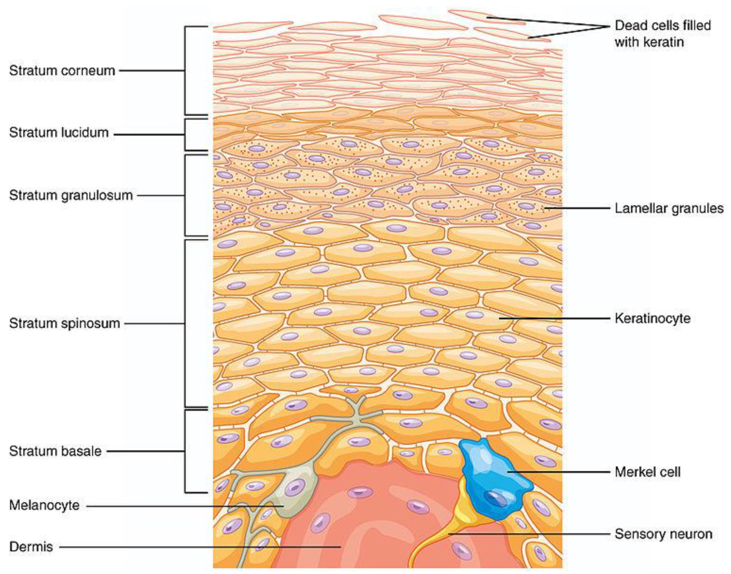
Figure 2.
Schematic representation of the different types of liposomal drug delivery systems (https://commons.wikimedia.org/wiki/File:Liposomas_articulo_liberaci%C3%B3n_activos.jpg).
Figure 2.
Schematic representation of the different types of liposomal drug delivery systems (https://commons.wikimedia.org/wiki/File:Liposomas_articulo_liberaci%C3%B3n_activos.jpg).
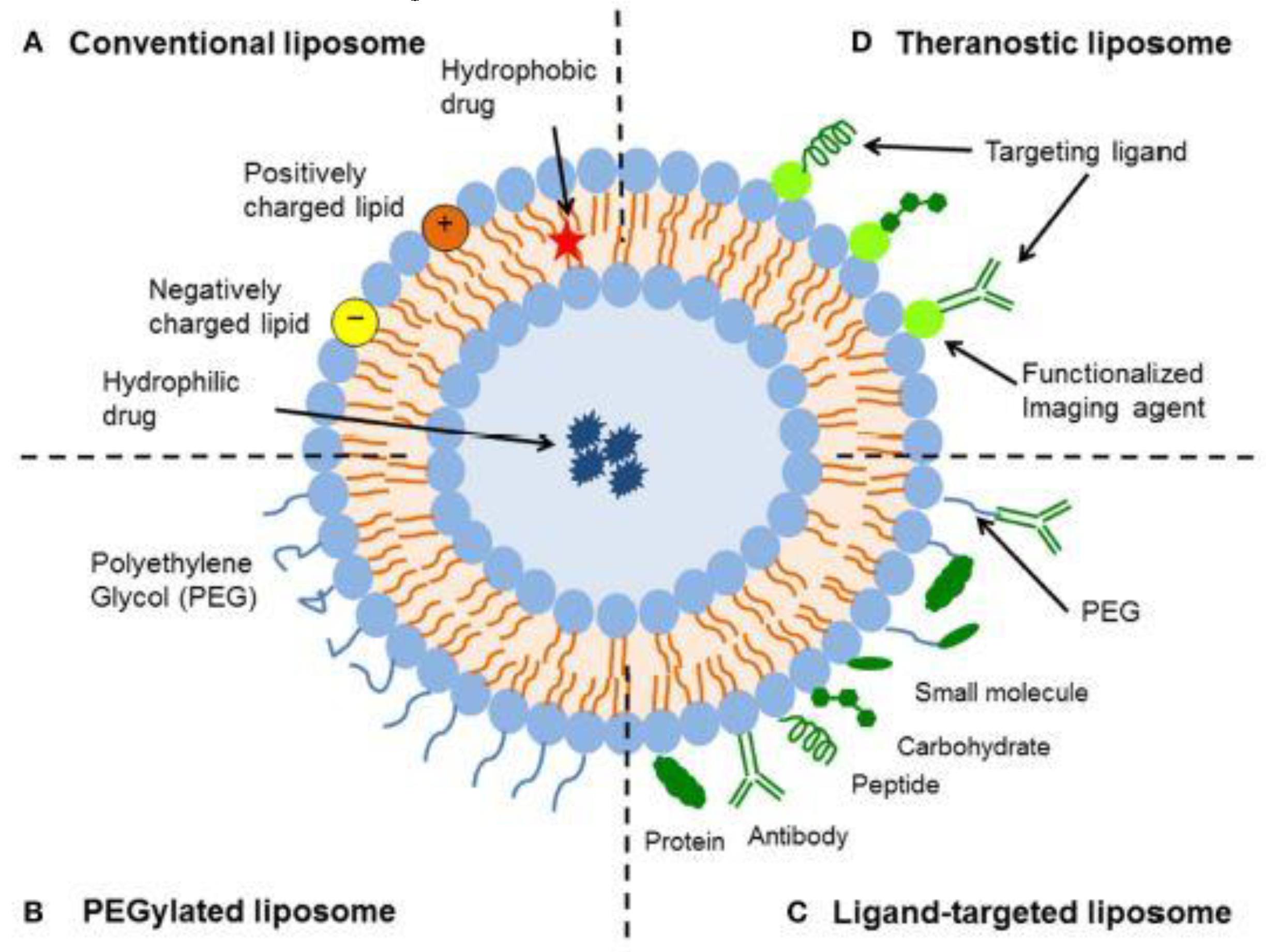
Figure 3.
Different size and lamellarity of liposomes. V: vesicles, U: unilamellar; G giant, L: Large; M: medium; S: small; OLV: oligolamellar vesicles; MLV: multilamellar vesicles; (https://commons.wikimedia.org/wiki/File:1-s2.0-S0168365921005034-gr6_lrg.jpg#filelinks).
Figure 3.
Different size and lamellarity of liposomes. V: vesicles, U: unilamellar; G giant, L: Large; M: medium; S: small; OLV: oligolamellar vesicles; MLV: multilamellar vesicles; (https://commons.wikimedia.org/wiki/File:1-s2.0-S0168365921005034-gr6_lrg.jpg#filelinks).
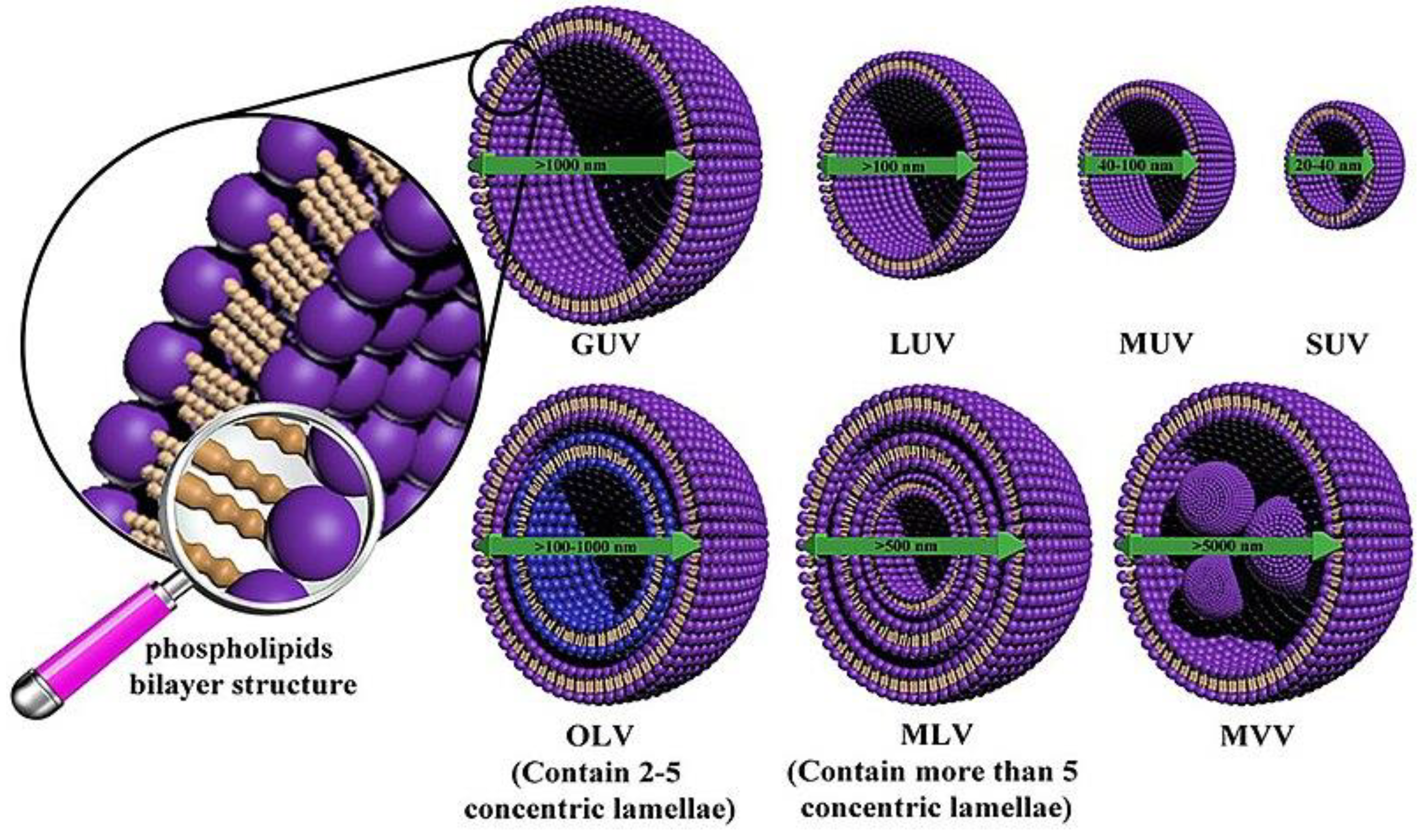

Table 1.
List of antioxidants analyzed with the formulation and the liposomal delivery system used.
| entry | antioxidant | formulation | delivery system1 | properties analyzed2 | ref. |
|---|---|---|---|---|---|
| 1 | quercetin | pure comp. | LPS | A-Inf; VCir; | [22] |
| 2 | Moringa Oleifera | extract, cholesterol | LPS | A-ox | [13] |
| 3 | Myrtus communis | extract | LPS | A-ox | [23] |
| 4 | propolis | extract | LPS | A-ox; SAbs; PhP | [24] |
| 5 | propolis | extract, cyclodextrins |
LPS | A-ox; GR; PhP | [25] |
| 6 | onhadi pistachio | extract | LPS | A-ox; GR; A-mel | [26] |
| 7 | resveratrol | pure comp. | LPS | A-ox | [27] |
| 8 | naringenin | pure comp., Tween 20 |
TLPS | A-ox | [28] |
| 9 | coenzime Q10 | pure comp., EtOH | ETH | A-ox; SAbs | [29] |
| 10 | rutin | pure comp., EtOH | ETH | A-ox; SAbs | [30] |
| 11 | rutin | pure comp., EtOH | ETH | A-ox; A-Inf; PhP | [31] |
| 12 | euphorbia characias | pure comp., EtOH | ETH | A-ox; A-mel; PhP | [32] |
| 13 | curcumin, piperine | pure comp., EtOH | ETH | A-ox | [33] |
| 14 | mangiferin | pure comp., EtOH, surfactants | ETH, TETH | A-ox; A-Inf; SAbs; | [34] |
| 15 | quercetin | pure comp., EtOH, surfactants |
ETH, TETH | A-ox; SAbs; A-mel | [35] |
| 16 | hydroxytirosol | pure comp., EtOH, Span 60 |
NIS | SAbs | [36] |
| 17 | epigallocatechin gallate | pure comp., EtOH, surfactants |
NIS | A-ox; SAbs | [37] |
| 18 | glicirrate | pure comp., Fatty acids |
UFS | A-ox; SAbs; EM | [38] |
1.LPS liposomes; ETH ethosomes; TETH transethosomes; NIS niosomes; UFS unsaturated fatty acids liposomes. 2.A-ox antioxidant; A-inf ant-inflammation; VCir vascular circulation; SAbs skin absorption; PhP photo-protection; GR gene regulation; A-mel anti-melanogenesis; E enzyme modulation.
Disclaimer/Publisher’s Note: The statements, opinions and data contained in all publications are solely those of the individual author(s) and contributor(s) and not of MDPI and/or the editor(s). MDPI and/or the editor(s) disclaim responsibility for any injury to people or property resulting from any ideas, methods, instructions or products referred to in the content. |
© 2024 by the authors. Licensee MDPI, Basel, Switzerland. This article is an open access article distributed under the terms and conditions of the Creative Commons Attribution (CC BY) license (http://creativecommons.org/licenses/by/4.0/).
Copyright: This open access article is published under a Creative Commons CC BY 4.0 license, which permit the free download, distribution, and reuse, provided that the author and preprint are cited in any reuse.


