Preprint
Review

This version is not peer-reviewed.

DNA as a Double-Coding Device for Information Conversion and Organization of Self-Referential Unity

A peer-reviewed article of this preprint also exists.

Submitted:

14 October 2024

Posted:

16 October 2024

You are already at the latest version

Abstract

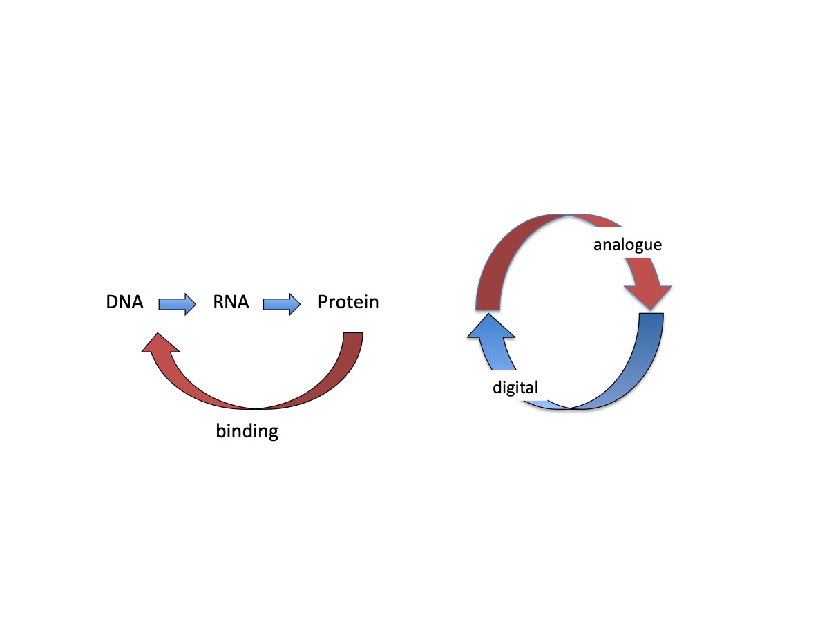
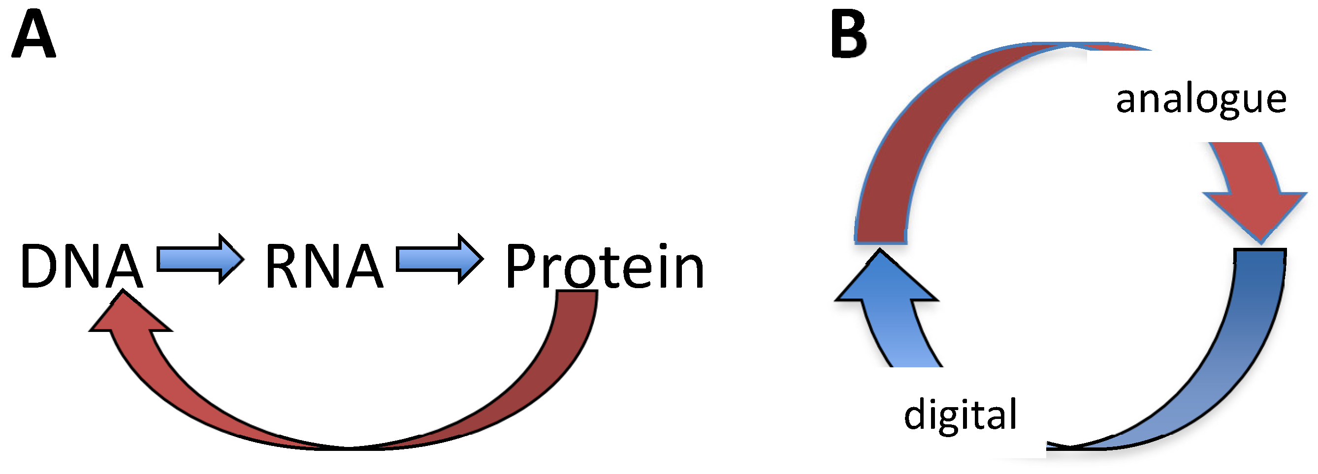
Living systems are capable on the one hand, of eliciting a coordinated response to changing environment (aka adaptation) and on the other hand, they are capable of reproducing themselves. Notably, adaptation to environmental change requires the monitoring of the surroundings, while reproduction requires monitoring oneself. These two tasks appear separate and making use of different sources of information. Yet both the process of adaptation as well as that of reproduction are inextricably coupled to alterations of genomic DNA expression, while a cell behaves as an indivisible unity in which apparently independent processes and mechanisms are both integrated and coordinated. We argue that at the most basic level, this integration is enabled by the unique property of the DNA to act as a double coding device harboring two logically distinct types of information. We review biological systems of different complexity and infer that inter-conversion of these two distinct types of DNA information represents a fundamental self-referential device underlying both systemic integration and coordinated adaptive response.

Keywords: 
;  ;  ;  ;  ;  

1. Introduction

The distinctive organizational hallmark of living systems is their ability to self-reproduce. For that matter, living systems are regarded as self-referential systems implying the capacity to monitor oneself, that is, to perpetually assess their status quo [1,2]. At the same time, living systems are capable of monitoring their surroundings and eliciting a functionally coordinated adaptive response to environmental change (Figure 1). The capacity of ‘monitoring oneself’ assumes that the system divides itself, as it were, into two parts, that which monitors and that which is monitored. Furthermore, monitoring oneself and monitoring environment appear as separate tasks utilizing different sources of information. In multicellular eukaryotes relative independence of the information used to control cellular reproduction and functional specialization is apparent in separation of the processes of proliferation and differentiation [3] observed in most cells, especially during development.
In unicellular organisms such as bacteria, alteration of e.g. the cell motility in response to environmental signals (chemotaxis) and the process of cell division are also regulated independently. For example, control of bacterial cell density (quorum sensing) is executed by autocrine/paracrine signaling pathways involving autoinducer molecules [4], whereas bacterial chemotaxis is induced by environmental factors including e.g., those produced by plants [5,6,7]. In Caulobacter system the cell differentiation can be decoupled from DNA replication [8] and the processes of cell division and differentiation are distributed between the two morphologically and developmentally distinct daughter cells [9]. However, both in eukaryotes and prokaryotes the information underpinning the apparently independent processes of reproduction and adaptation is encoded in the very same DNA genome being reflected in, and largely governed by, the genetic control mechanisms. Furthermore, even if the cellular reproduction and the adaptive environmental response may utilize different information sources and independent regulation mechanisms, it is obvious that the intrinsic organization of a cell endows it with the capacity to behave as a whole, indivisible unity in which apparently independent processes and mechanisms are both coordinated and integrated [3,7,10]. Importantly, a coordinated switch in gene expression during transition between bacterial motile and biofilm lifestyles appears to involve a change in chromosome structure [11]. Also the switching between alternative gene expression programs both during the growth cycle and in response to various stress impacts involves coordinated alterations of DNA topology, coherently modulating the gene expression in extended chromosomal domains [12,13]. So then, assuming that both the genetic expression and the structural dynamics of the genomic DNA polymer are intimately involved in this integration process, the central question to address is the nature of the coordinating device.

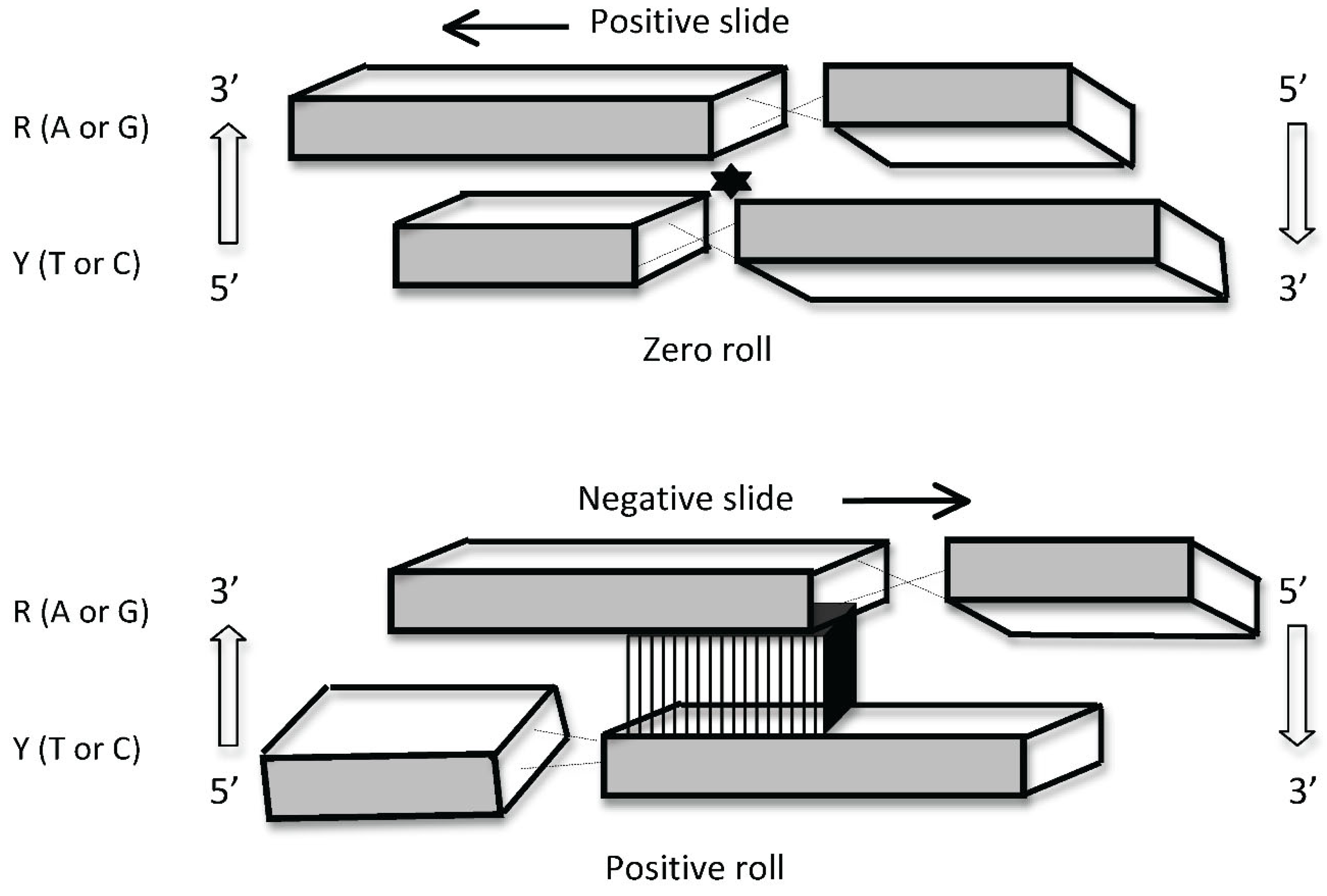
2. DNA Is a Source of Two Distinct Types of Information

The still largely underappreciated characteristic of the double helical DNA polymer is that it is a source of two logically distinct types of information. One is the well-known linear genetic code, which is discontinuous (digital) being embodied in discrete triplets of base pairs – the codons (Figure 2). The other source of information stored in the DNA is of continuous (analogue) nature, being embodied in juxtaposition of distinct base steps, which partly overlap [14,15,16].
Since in contrast to the codons, the base steps are overlapping (that is, each first base of any base step is the second base of a previous step and each second base of any base step is the first base of a following step), it is exactly this latter feature, which confers the character of continuity to the DNA analogue ‘code’. Importantly, various base steps are characterized by distinct stacking/melting energy levels (Table 1) and also, can adopt different preferential conformations [14,15,16,17,18] (Figure 3).
The contiguous base steps favoring various local conformations can determine the 3D configuration and average trajectory of the DNA [15,16,18,19]. Importantly, the base stacking and accordingly, the conformation of DNA base steps can be modulated by environmental conditions inducing alterations of DNA twist eventually affecting the DNA helical repeat and the torque accommodated by the double helix. The configuration of DNA depends both on sequence organization and average superhelical density [20,21,22] as well as on the size of the affected topological domain [23].
In the bacterium E. coli the DNA superhelical density varies as a function of cellular energy charge, which depends on, and changes with, the environmental conditions. The level of negative DNA superhelicity varies as a function of ATP/ADP ratio, primarily because ATP is utilized by DNA gyrase, an enzyme introducing negative supercoils into the DNA [24,25,26,27]. Both the ATP/ADP ratio as well as gyrase activity increase on nutritional shift-up, e.g., when the starved bacterial cells are inoculated in fresh growth medium. However, at this stage there is also a more direct effect on DNA topology (namely on DNA twist) mediated by changing ionic composition [28,29,30,31,32,33]. In other words, changing environmental conditions altering the DNA superhelicity eventually stabilize distinct DNA structures depending on sequence organisaton and size of the affected domain. Ultimately, the sensing of available metabolic energy and ionic composition by DNA would select the configuration and topology optimally adapted to given environmental impact. In turn, alterations of DNA configuration are relevant to gene expression, as the DNA binding ligands, architectural proteins and enzymes (e.g. the transcription and replication machinery) show preferences for particular DNA topology [34,35,36,37,38,39,40,41,42,43,44]. The DNA binding proteins can in turn stabilize different DNA deformations such as bending, over- or under-twisting, wrapping, looping, bridging as well as can constrain DNA supercoils [45,46,47,48,49]. All these effects are pertinent, as the various assembled nucleoprotein complexes modulate the genetic expression. Furthermore, on translocation along the DNA template, the transcription and replication machineries directionally modulate the DNA superhelicity, inducing positive supercoils ahead and negative supercoils in their wake [50]. Diffusion of these induced free supercoils can distinctly affect the activity of neighboring genes in genome [51,52,53]. In addition, this topological differentiation of DNA on opposite sides of the moving DNA translocases has a potential to spatially organize the binding of regulatory proteins recognizing distinct DNA supercoil structures [23].
Thus in principle, coordination of different genetic programs (e.g. those governing self-reproduction and those for adaptive response) could be achieved simply by arranging the genomic DNA analogue information in such a way as to couple the emergence of distinct 3D DNA structures and particular DNA topology to different internal and external impacts on the one hand, and on the other hand, to employ these distinct structures for selective and coordinated readout of the digital (genetic) code optimizing the expression of traits apt for coping with given demands. The expression of different genetic programs in response to both environmental and internal signals would be then integrated and coordinated by variation of a single, continuous tunable parameter sensitive to both internal alterations and environmental change. Conceivably, the superhelicity of DNA serving as an interface between the external and internal milieu, is the most plausible contender for the role of pivotal variable adjusting the dynamics of DNA analogue information (i.e. the genomic DNA configuration) and the pattern of gene expression in response to both internal and external signals. Indeed, in bacteria the induction of distinctly different patterns of gene transcription coupled to activation of disparate genetic functions have been observed in response to directional modulation of DNA superhelical density by environmental stress or topoisomerase poisons and inhibitors as well as in response to topoisomerase gene mutations [12,13,54,55,56,57,58,59].
In bacteria, the role of DNA topology in coordinating the genetic adaptive response with various environmental cues is well documented [13,60,61]. Also in experimental evolution studies, modulation of global regulatory networks and DNA topology were identified as main internal factors subject to the process of selection [62,63]. During the bacterial growth cycle, the successive stages of cell reproduction (aka exponential growth phase) and maintenance (aka stationary phase) are long known to be associated with distinct – respectively high and low – negative superhelical densities of the DNA [64]. Notably, the spatial separation of relatively G/C-rich and relatively A/T-rich sequences, organized respectively around the oriC and ter poles of the E. coli chromosome, allows for temporal separation of gene expression at the two chromosomal poles due to growth phase-dependent changes of superhelicity. Indeed, the chromosomal oriC pole is not only G/C-rich relatively to the ter pole but is also enriched for gyrase binding sites [54,65,66]. On nutritional shift-up, the increase of negative superhelicity at this chromosomal pole is reinforced by production of negative supercoils trailing both the translocating replisomes and the trains of RNA polymerase 70 holoenzyme molecules transcribing the numerous strong ribosomal RNA operons, all of which are directionally oriented from oriC towards the terminus of chromosomal replication [65]. The vegetative 70 RNA polymerase and the stationary phase S holoenzymes respectively prefer highly supercoiled and relaxed DNA templates and accordingly, are activated in succession during the growth cycle [34,36,67,68]. The frequency distributions of 70 and S binding sites form correspondingly decreasing and increasing spatial gradients along the chromosomal oriC-ter axis [66]. Thus the oriC pole (the Ori macrodomain and the flanking left and right non-structured domains) of the E. coli chromosome is enriched for 70 binding sites and transcribed by 70 RNA polymerase both earlier and more actively than the ter pole enriched for S binding sites, giving rise to early gene products underpinning fast growth and replication [12,66,69]. Furthermore, the anabolic and catabolic genes are respectively enriched at the oriC and ter poles of the E. coli chromosome [70]. As a result, anabolic pathways are activated early during the reproduction stage under conditions of high negative superhelicity and catabolic pathways are activated later under conditions of low negative superhelicity characteristic of the maintenance stage [12,56]. Thus during the bacterial growth cycle, the temporal separation of anabolic (reproductive) and catabolic (maintenance) gene expression ‘subprograms’ is achieved by strategic spatial organization of the DNA analogue information (such as the oriC-ter gradients of the DNA thermodynamic stability and relative frequencies of the gyrase, 70 and S binding sites) coordinated with asymmetric enrichment of anabolic and catabolic genes around the chromosomal poles (Figure 4).
In addition to the enrichment of anabolic and catabolic genes around opposite chromosomal poles, also the order of cognate regulatory genes along the oriC-ter axis is correlated with their successive expression during the growth cycle [66]. The genes of reproduction stage regulators are located in the vicinity of the oriC pole, whereas the genes of maintenance function regulators are positioned closer to the ter pole of the chromosome (Figure 5).
Therefore, also these regulators are expressed sequentially, first because as already mentioned, on the commencement of growth the oriC pole is activated earlier than the ter pole and second, because the iterative rounds of chromosomal replication initiation at oriC increase the copy numbers of early regulatory genes located in its vicinity relative to that of the genes of maintenance regulators located closer to the chromosomal replication terminus [12,66,73,74,75]. Most important among these sequentially expressed regulators, besides the DNA topoisomerases and RNA polymerase sigma factors, are the highly abundant nucleoid-associated proteins (NAPs), which potentially form spatiotemporal gradients (Figure 5) interacting with cognate binding sites spatially organized in the genome [66,76,77,78,79,80]. Importantly, the abundant NAPs bind DNA with different affinities depending on its 3D structure, are capable of constraining supercoils and stabilizing topological domains and thus of partitioning and storing the superhelical energy [81,82,83] which can be used to do work, e.g. to separate the DNA strands and facilitate transcription initiation.
Similar organizational logic applies to the operation of the aerobic/anaerobic switch during the bacterial growth. The atp operon responsible for ATP production under aerobic growth conditions is located in close vicinity of oriC while the fnr gene, encoding the major DNA binding regulator of anaerobic growth, is located in the vicinity of ter. Accordingly, the arcA and arcB genes encoding the two-component system responsible for gene regulation under conditions of microaerobiosis are located in-between atp and fnr (Figure 6A). Thus again, these genes are spatially ordered in the genome according to their sequential requirement during growth while the expression of aerobic and anaerobic gene groups appears to be correlated with the gradual alteration of oxygen partial pressure (Figure 6B). As with nutritional shift-up, the topoisomerase activities are involved in the regulation of DNA supercoiling during aerobic-anaerobic transitions in E. coli [84], whereby the growth under high oxygen conditions is correlated with high negative superhelicity of plasmid DNA [85].

3. Coupling of Logically Distinct Types of Information

The pivotal question is as to how the coupling of DNA analogue information (i.e. the spatial distribution of DNA torsional energy, which is a continuous variable) with the digital information (i.e. selective expression of unique genes manifesting a discontinuous pattern) is accomplished in the genome? More compellingly, how do the DNA analogue information and digital code communicate with each other? In bacteria, it has been shown that variation in G/C content of the promoter sequence context [55,89] as well as the peculiar sequence organization of the different promoter elements such as e.g. the deviation of the -35 hexamer from the consensus sequence, as well as the G/C-richness and/or extension of the discriminator sequence and the length of the spacer between the -10 and -35 hexamers, confer the ability to distinctly respond to alterations of DNA superhelical density [90,91,92,93,94,95]. Also the sequences located upstream of the core promoter and characterized by anisotropic bending modulate the response to DNA superhelicity [91,96,97,98]. Thus a simple way to produce a coordinated transcriptional response to changes of supercoiling would be to put all the functionally relevant genes under the control of promoters with similar sequence organisation and indeed, that is the case for many stringently regulated genes (that is, the genes down-regulated by the alarmone ppGpp; see below) including the stable RNA (transfer and ribosomal RNA) operons [99]. However, several studies identified supercoiling-dependent, spatially extending coherent gene expression patterns in the bacterial genome, which cannot be readily expounded by the promoter sequence similarity scenario [12,13,54,80,82,100]. Various explanations have been proposed for the organization of such extended topological domains including coherent domains of gene expression (aka CODOs) [12,13,71,101] but the issue remains controversial [71,102,103,104]. Importantly, the CODOs were found to harbor distinct genetic functions [12,13,71] consistent with spatial coupling of the DNA analog information and the digital code in genome.

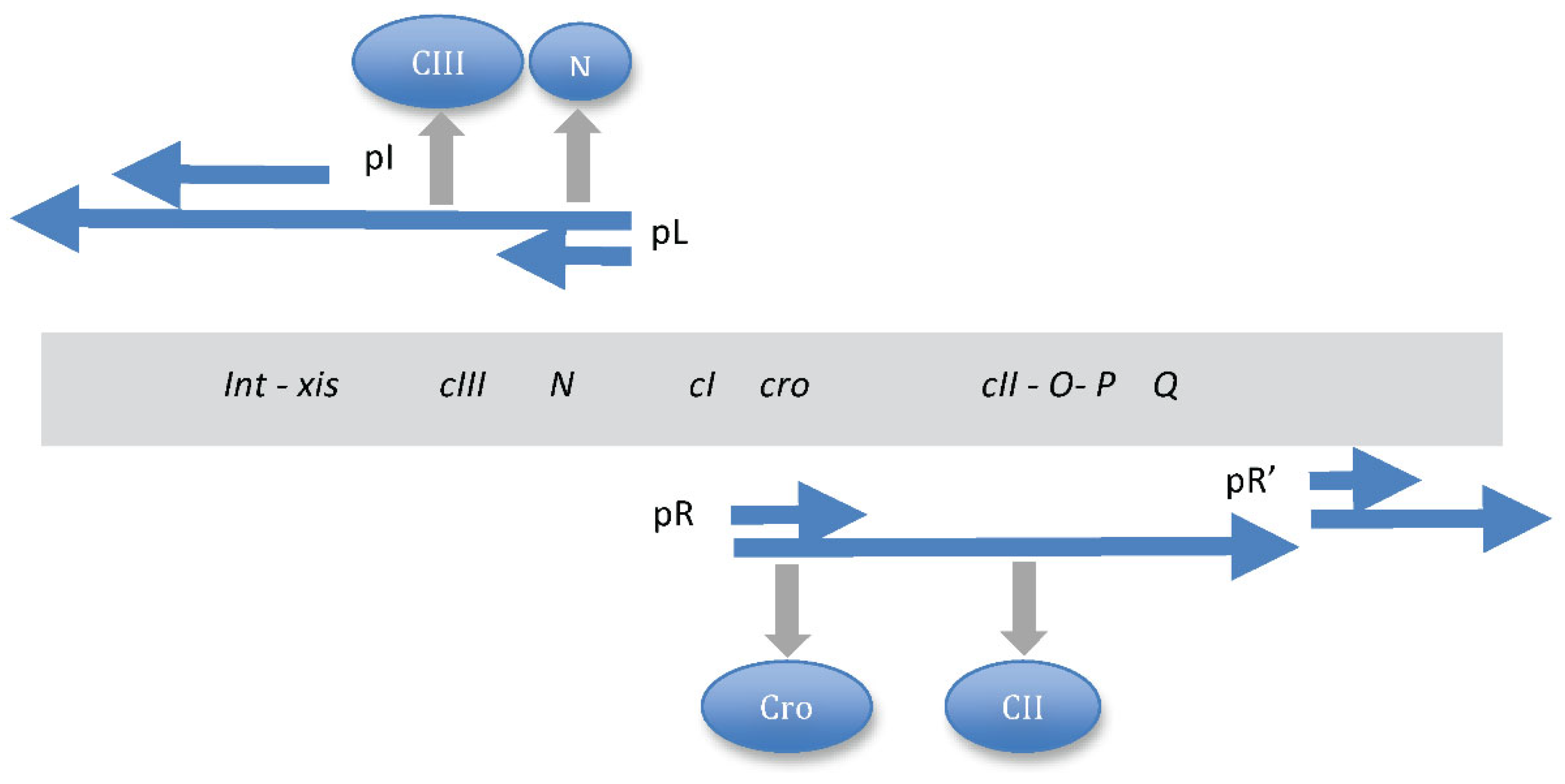
4. Switching between Alternative Gene Expression Programs

In bacteria, global alterations of gene expression can be induced not only by alterations of DNA superhelicity but also by small intracellular effectors, such as the nucleotide guanosine tetraphosphate (ppGpp). In metazoan cells the role of ppGpp is less clear [105] while in bacteria it serves as an alarmone, reprograming the cell physiology by interacting directly with the transcription and translation machinery [106,107]. However, ppGpp also interferes with replication initiation by modulating the DNA topology at oriC [108]. For that matter, ppGpp, the production of which is sharply induced on shortage of nutritional resources, appears to act as a switch curtailing cell reproduction and promoting the establishment of maintenance program.
This ppGpp-dependent switch in gene expression occurs on exhaustion of nutritional resources at later stage of growth. At this stage ribosome production and gyrase activity subside, whereas sharply increased ppGpp concentration facilitates the compositional change of the transcription machinery, e.g. partial substitution of 70 by stationary phase S factor in the RNAP holoenzyme and for that matter, ppGpp also switches the supercoiling preferences of polymerase. While RNA polymerase is a direct target of ppGpp, the ppGpp sensitivity of the 70 holoenzyme in vitro can be attenuated by increased DNA superhelicity [109]. In addition, ppGpp appears to stabilise the so-called ‘tight’ conformer of the 70 holoenzyme at the expense of the ‘ratcheted’ conformer favoring supercoiled DNA (Malcolm Buckle, GM and AT, manuscript in preparation). Also the composition of the abundant NAPs changes at this stage such that overall, the intracellular milieu and the bacterial chromatin composition facilitate the transcription of more relaxed and relatively A/T rich DNA around the terminus of replication (the Ter macrodomain), which is enriched for genes involved in maintenance functions. The ppGpp-facilitated growth phase-dependent substitution of 70 by the stationary phase S factor in the RNAP holoenzyme is associated with switching between the reproduction and maintenance programs and thus, resembles the genetic switch between the alternative growth pathways of temperate bacterial phages such as phage . The  switch is also sensitive to both the cell density and metabolic state [110,111] as well as to supercoiling level of the DNA [112,113].
In these two systems, despite the huge difference in complexity, there is a notable organizational similarity manifest in the conversion of distinct information types occurring during the establishment of both the bacterial switch between the reproduction and maintenance programs and the phage switch between the lytic and lysogenic pathways. In the latter case the system is much simpler and ultimately, the switch boils down to competition between two DNA binding transcriptional regulators (the Cro and CI repressors) for binding specific operator sites in the regulatory region. However, in both systems first the information of continuous (analogue) type is produced and then converted into information of discontinuous (digital) type.
In E. coli system as mentioned above, analogue information is manifest in the oriC-ter skew of DNA binding site frequencies for DNA gyrase, 70 and S, interacting with the changing ratio of the RNAP 70 and S holoenzymes, the spatiotemporal gradient of chromosomal superhelical density and the temporal concentration gradients evident in various growth phase-dependent combinations of the NAPs (Table 2). These DNA architectural proteins form distinct spatiotemporal patterns of regulatory nucleoprotein complexes in genome [69,114,115,116]. The NAPs compete for stabilization of alternative supercoil structures (Figure 7) but can also cooperate depending on DNA sequence organisation [47,117]. The mutations of NAP genes alter both the gene expression patterns and DNA topology, consistent with the notion that the NAPs coordinate the growth phase-dependent chromosome structure and function [11,13,56,65,118,119,120].
In the case of bacteriophage  the analogue information is manifest as continual bidirectional extension of transcription initiated from the divergent pR and pL promoters located in the  control region, producing on extension distinct sets of regulatory proteins, including those involved in sensing physiological conditions (e.g. CII) and eventually, by modulating the CI/Cro repressor ratio, favoring either the lytic or lysogenic pathway (Figure 8). Continual transcription is both contingent on, and also results in, formation of a spatiotemporal pattern of regulatory nucleoprotein complexes.
In E. coli the switch between the reproduction and maintenance stages is primarily dictated by the energy status, which in turn depends on environmental conditions. Notwithstanding the difficulty of considering phage an organism, it not only can reproduce itself (albeit hijacking the cellular components and machinery) but also responds to environmental conditions. For example, the  phage may prefer lysogenic to lytic growth under conditions of starvation, perhaps since starving cells cannot provide components supporting efficient lytic growth [110]. Starving bacterial cells produce low amounts of proteases, which at high concentrations observed in rich medium destroy the phage CII protein required for activation of the phage cI and int genes essential for establishing lysogeny. The CI repressor produced under conditions of high CII activity inhibits transcription from the divergent pR and pL promoters in the  regulatory region and thus turns off the expression of all the phage genes except that of its own. However, if CII is rapidly degraded no CI repressor is synthesized, the Cro repressor occupies the  regulatory region instead of CI and lytic growth ensues. So while the phage senses the energy status of the cell, this latter is ultimately translated into specific nucleoprotein complexes competing for binding at the  regulatory region and acting as a switch between alternative developmental pathways. Furthermore, regulation of the lysogenic/lytic switch by CI repressor appears sensitive to DNA supercoiling [112,113], as is also the RNAP 70/S holoenzyme switch in E. coli [36]. A similar relationship of DNA topology-dependent competitive binding at the overlapping DNA sites has been observed between the early and late NAPs, respectively FIS and Lrp, involved in the control of type 1 fimbrial genetic switch in E. coli [122].
Ptashne [110] suggested that the regulatory sequences initiated at the  control region essentially generate a ‘cascade’ along each pathway, sequentially turning on and off groups of genes. In this cascade, one regulatory protein turns on or off a block of genes, which includes another regulatory gene the product of which in turn regulates another block of genes and so on. Here, the regulatory cascade is established by protein binding only at a few DNA sites on the phage genome. This type of regulation is made possible due to peculiar spatial organization of functionally related genes in genome, as they are grouped together and also transcribed in the same direction. Thus while emphasizing the role of cascades, this mode of regulation also implicates the spatial gene organization and the directional extension of transcription – properties, considered here as belonging to analogue (continuous) information type – by contrast to the ‘cascade control’ which essentially turns the genes on or off and therefore, provides purely digital information.
Despite the difference in complexity and details, in both the bacterial and phage systems there is a discernible common organisational design: initial utilization of analogue information (spatial oriC-ter gradients of DNA binding sites interacting with temporal gradients of regulatory proteins in the former, and gradually extending transcription starting from the divergent pR and pL promoters in the latter) and its subsequent conversion into digital information (the differing nucleoprotein complexes producing specific gene expression patterns sustaining either reproduction or maintenance in the former and, the distinct sets of regulatory proteins underpinning either the lytic or lysogenic pathway in the latter).

5. Analogue/Digital Information Conversion Operates as a Regulatory Device in Living Systems of Diverse Structural Complexity

Given the similarity of underlying regulatory design in the bacterial and phage systems the pertinent question is, whether this mode of information conversion obtains also in more complex multicellular organisms. Indeed, over three decades ago Ptashne [110] drew parallels between the processes of gene regulation in phage and higher organisms, in particular the process of Drosophila embryogenesis, where formation of the pattern of stripes expressing the segmentation gene even-skipped (eve) depends on sequential turning on and off of transcriptional regulators, a form of cascade control similar to that of the life cycle. Actually, during Drosophila embryogenesis, notwithstanding the role of digital on or off type ‘cascade control’, the importance of analogue information in the pattern formation is most conspicuous.
During Drosophila embryonic development the maternal gene messages are strategically deposited at opposite – anterior and posterior - poles of the embryo, such that the translated proteins diffuse from the poles forming spatial concentration gradients along the anterior-posterior axis (Figure 9). These overlapping concentration gradients lead to spatially determined, locally fixed ratios of transcriptional regulators and thus establish boundaries of target gene expression. Spatially determined threshold concentrations of transcriptional regulators lead to sequential activation of the various segmentation genes eventually producing a distinct pattern of seven stripes expressing the even-skipped pair-rule gene, which is essential for segmentation of the embryo [123,124]. So here we have a clear case of the conversion of analogue information (continuous protein concentration gradients) into digital information (specific pattern of seven discrete eve stripes). This conversion of protein concentration gradients into particular pattern of stripes is enabled by existence of seven distinct enhancers of the eve gene (one for each stripe), each of which binds different combinations of regulatory proteins depending on their spatially determined threshold concentrations established along the anterior-posterior axis. Thus seven distinct enhancers binding different combinations of regulatory proteins generate alternative nucleoprotein complexes independently activating eve expression - albeit in a spatially defined manner – and producing the specific pattern of seven stripes.
At this stage of development (syncytial blastoderm) the Drosophila embryo is not yet cellularized and contains about 1500 nuclei evenly distributed underneath the membrane, while each stripe extends over 6 nuclei on average [110]. The Drosophila genome is about 180Mb in size, so a single row of nuclei expressing even-skipped gene would contain about 1Gb of DNA. In contrast, the E. coli genome is 4.6Mb in size and that of phage  is about 48.5Kb. The Drosophila embryo is (longitudinally) about 500 times the size of E. coli cell. Thus, concerning the spatial extension of implicated gradients, there is a difference in orders of magnitude. Furthermore, in the case of phage and E. coli, the gradients (directionally elongating transcripts in the former and the putative sigma factor and NAP gradients in the latter) extend over single genomes (albeit differing in size by two orders of magnitude), whereas in the case of Drosophila the protein concentration gradients extend over more than a thousand of spatially arranged genomes (nuclei) in the embryo. Finally, in the phage and bacterial genomes the regulatory proteins have a relatively easy access to DNA binding sites, whereas the binding of cognate regulatory sites is obstructed in Drosophila nuclei by tight packaging of the DNA in chromatin. Furthermore, in phagethe spatial extension of genomic transcription produces distinct sets of proteins that are put to work in temporal succession. Also the temporal gradients of NAPs and sigma factors do not coexist in a single bacterial cell but are successively established in the progeny. In contrast, the opposite concentration gradients of Bicoid and Caudal extending from the poles and responsible for the formation of the anterior and posterior structures coexist in a single Drosophila embryo.
Notwithstanding the abovementioned differences, there are also remarkable similarities between these systems. During Drosophila embryonic development as well as during the E. coli growth cycle the transcriptional and metabolic programs appear tightly correlated [56,125,126]. Regulation of the activity of DNA topoisomerases is associated with both the E. coli growth cycle and the Drosophila embryogenesis [27,127]. Also, the Drosophila development time, the phase transition during E. coli growth cycle and the  phage lytic/lysogeny decision, all respond to nutritional supply [110,128,129]. However, the most important similarity between the systems is the phenomenon of conversion of continuous data (analogue information) into discrete data (digital information). First, in all three systems there is a directional dispersion of analogue information (gradients of proteins in Drosophila and E. coli and continually elongating transcripts in phage ) from spatially localized sources (anterior and posterior poles in Drosophila, proximities of the chromosomal oriC and ter poles in E. coli and divergent pR and pL promoters in regulatory region of phage ). Second, in all three systems the conversion of analogue into digital information is manifest in formation of distinct nucleoprotein complexes involving DNA binding proteins interacting with DNA sites spatially organized in corresponding genomes, or in the embryo. In the latter case, this interaction is facilitated and fine-tuned by ATP-driven chromatin remodelers [130]. Finally and more generally, assuming that the initial gradients possess higher entropy than the ensuing discrete patterns of DNA-protein interactions, we may also assume an energy-driven decrease of entropy associated with pattern making in all three systems.
Thus, despite the substantial differences in size, complexity and structural detail between these three living systems (although the phage system can barely qualify as such), in all cases we have a spatiotemporally organized gene regulation program. The temporally organized regulatory cascades alone cannot provide for the unity of living system - a cascade has a beginning and an end – yet it does not necessarily close onto itself while as mentioned above, from systems-theoretical perspective, the living system constitutes a self-referential circuit. What is assumed here is the closure of the system onto itself and this organisation of unity implicates spatial coordinates [131,132,133]. A relevant example of integration of cell division and differentiation by coordinating the temporal gene expression and spatial organization of gene products and protein gradients has been provided in studies of the Caulobacter crescentus system [9,134,135]. All the three systems discussed here have similar organization embodied in the conversion of two distinct information types manifesting a coordinated unity (Figure 10). On this view, a phage acquires the properties of a living system primarily by tapping into its intrinsic organization, namely, appropriating the device of analogue/digital information conversion.

6. Conclusions

The central dogma of molecular biology is stating that genetic information flows only in one direction, from DNA, to RNA, to protein, or from RNA directly to protein. This theory highlighting the unidirectional flow of genetic information does not consider the DNA analogue information and the crosstalk between the DNA and DNA binding proteins – essentially a feedback (Figure 10A, brown curved arrow). It thus cannot account for the main organizational hallmark of the living system manifesting a self-referential circuit. We argue here that this latter organizational feature is inherent in the structure of the DNA double helix, representing a basic device for interconverting information. This conversion of information is made possible by existence of two logically different – digital and analogue – information types stored in the DNA. While the digital genetic information encodes all the DNA binding proteins and enzymes, the DNA appears to ‘read itself’ via DNA-protein interactions. These interactions are informative as they lead to modulation of gene expression according to nascent external and/or internal signals. The coordinated DNA ‘self-readout’ mediated by DNA binding proteins is based on a strategic spatial organization of regulatory DNA binding sites and regulated genes in genome (or spatial organization of nuclei in case of Drosophila embryo). This spatial organization in turn is determinative for successive formation of distinct regulatory nucleoprotein complexes and the emergence of temporal regulatory ‘cascades.’ Thus the DNA genome can generate spatiotemporally coordinated patterns of activated and repressed genes in response to both internal and external signals. And so then, the genomic DNA acting as a double-coding device, represents the integrative interface where ‘that which monitors and that which is monitored’, meet to generate a coordinated unity (Figure 10B) responding to both the internal and external signals as an indivisible whole.

Author Contributions

Conceptualization, G.M., W.N., S.R. and A.T.; methodology, G.M., W.H., S.R. and A.T.; formal analysis, G.M., W.N., S.R. and A.T.; writing—original draft preparation, G.M., W.N., S.R. and A.T.; writing—review and editing, G.M., W.N., S.R. and A.T.; visualization, G.M., W.N., S.R. and A.T.; supervision, G.M., W.H., S.R. and A.T.; All authors have read and agreed to the published version of the manuscript.

Data Availability Statement

No new data were created in this study.

Conflicts of Interest

The authors declare no conflicts of interest.

References

  1. Kaufman L.H. Self reference and recursive forms. J. Social Biol. Struct. (1987)10, 53-72. 1987 Academic Press Inc. (London) Limited.
  2. Muskhelishvili G, Sobetzko P, Travers A. Spatiotemporal Coupling of DNA Supercoiling and Genomic Sequence Organization-A Timing Chain for the Bacterial Growth Cycle? Biomolecules. 2022 Jun 15;12(6):831. [CrossRef] [PubMed]
  3. Ruijtenberg S, van den Heuvel S. Coordinating cell proliferation and differentiation: Antagonism between cell cycle regulators and cell type-specific gene expression. Cell Cycle. 2016;15(2):196-212. [CrossRef] [PubMed]
  4. Papenfort K, Bassler BL. Quorum sensing signal-response systems in Gram-negative bacteria. Nat Rev Microbiol. 2016 Aug 11;14(9):576-88. [CrossRef] [PubMed]
  5. Antunez-Lamas M, Cabrera E, Lopez-Solanilla E, Solano R, González-Melendi P, Chico JM, Toth I, Birch P, Pritchard L, Liu H, Rodriguez-Palenzuela P. Bacterial chemoattraction towards jasmonate plays a role in the entry of Dickeya dadantii through wounded tissues. Mol Microbiol. 2009 Nov;74(3):662-71. Epub 2009 Oct 8. Erratum in: Mol Microbiol. 2009 Dec;74(6):1543. Prichard, Leighton [corrected to Pritchard, Leighton]. [CrossRef] [PubMed]
  6. Río-Álvarez I, Muñoz-Gómez C, Navas-Vásquez M, Martínez-García PM, Antúnez-Lamas M, Rodríguez-Palenzuela P, López-Solanilla E. Role of Dickeya dadantii 3937 chemoreceptors in the entry to Arabidopsis leaves through wounds. Mol Plant Pathol. 2015 Sep;16(7):685-98. [CrossRef] [PubMed]
  7. Jiang X, Zghidi-Abouzid O, Oger-Desfeux C, Hommais F, Greliche N, Muskhelishvili G, Nasser W, Reverchon S. Global transcriptional response of Dickeya dadantii to environmental stimuli relevant to the plant infection. Environ Microbiol. 2016 Nov;18(11):3651-3672. [CrossRef] [PubMed]
  8. Hallgren J, Koonce K, Felletti M, Mortier J, Turco E, Jonas K. Phosphate starvation decouples cell differentiation from DNA replication control in the dimorphic bacterium Caulobacter crescentus. PLoS Genet. 2023 Nov 27;19(11):e1010882. [CrossRef] [PubMed]
  9. Barrows JM, Goley ED. Synchronized Swarmers and Sticky Stalks: Caulobacter crescentus as a Model for Bacterial Cell Biology. J Bacteriol. 2023 Feb 22;205(2):e0038422. [CrossRef] [PubMed]
  10. Dudin O, Geiselmann J, Ogasawara H, Ishihama A, Lacour S. Repression of flagellar genes in exponential phase by CsgD and CpxR, two crucial modulators of Escherichia coli biofilm formation. J Bacteriol. 2014 Feb;196(3):707-15. [CrossRef] [PubMed]
  11. Scolari VF, Bassetti B, Sclavi B, Lagomarsino MC. Gene clusters reflecting macrodomain structure respond to nucleoid perturbations. Mol Biosyst. 2011 Mar;7(3):878-88. [CrossRef] [PubMed]
  12. Sobetzko P, Glinkowska M, Travers A, Muskhelishvili G. DNA thermodynamic stability and supercoil dynamics determine the gene expression program during the bacterial growth cycle. Mol Biosyst. 2013 Jul;9(7):1643-51. [CrossRef] [PubMed]
  13. Jiang X, Sobetzko P, Nasser W, Reverchon S, Muskhelishvili G. Chromosomal "stress-response" domains govern the spatiotemporal expression of the bacterial virulence program. mBio. 2015 Apr 28;6(3):e00353-15. [CrossRef] [PubMed]
  14. SantaLucia J, Jr. A unified view of polymer, dumbbell, and oligonucleotide DNA nearest-neighbor thermodynamics. Proc Natl Acad Sci U S A. 1998 Feb 17;95(4):1460-5. [CrossRef] [PubMed]
  15. Packer MJ, Dauncey MP, Hunter CA. Sequence-dependent DNA structure: dinucleotide conformational maps. J Mol Biol. 2000 Jan 7;295(1):71-83. [CrossRef] [PubMed]
  16. Travers AA, Muskhelishvili G, Thompson JM. DNA information: from digital code to analogue structure. Philos Trans A Math Phys Eng Sci. 2012 Jun 28;370(1969):2960-86. [CrossRef] [PubMed]
  17. El Hassan MA, Calladine CR. The assessment of the geometry of dinucleotide steps in double-helical DNA; a new local calculation scheme. J Mol Biol. 1995 Sep 1;251(5):648-64. [CrossRef] [PubMed]
  18. Calladine CR, Drew HR, Luisi BF, Travers A.A. Understanding DNA. The Molecule & How It Works. 2004, Third ed. ELSEVIER Academic Press.
  19. Palecek, E. Local supercoil-stabilized DNA structures. Crit Rev Biochem Mol Biol. 1991;26(2):151-226. [CrossRef] [PubMed]
  20. Irobalieva RN, Fogg JM, Catanese DJ Jr, Sutthibutpong T, Chen M, Barker AK, Ludtke SJ, Harris SA, Schmid MF, Chiu W, Zechiedrich L. Structural diversity of supercoiled DNA. Nat Commun. 2015 Oct 12;6:8440. Erratum in: Nat Commun. 2015 Oct 29;6:8851. 10.1038/ncomms9851. Catanese, Daniel J [Corrected to Catanese, Daniel J Jr]. PMCID: PMC4608029. [CrossRef] [PubMed]
  21. Wang Q, Irobalieva RN, Chiu W, Schmid MF, Fogg JM, Zechiedrich L, Pettitt BM. Influence of DNA sequence on the structure of minicircles under torsional stress. Nucleic Acids Res. 2017 Jul 27;45(13):7633-7642. [CrossRef] [PubMed]
  22. Pyne ALB, Noy A, Main KHS, Velasco-Berrelleza V, Piperakis MM, Mitchenall LA, Cugliandolo FM, Beton JG, Stevenson CEM, Hoogenboom BW, Bates AD, Maxwell A, Harris SA. Base-pair resolution analysis of the effect of supercoiling on DNA flexibility and major groove recognition by triplex-forming oligonucleotides. Nat Commun. 2021 Feb 16;12(1):1053. [CrossRef] [PubMed]
  23. Muskhelishvili G, Travers A. The regulatory role of DNA supercoiling in nucleoprotein complex assembly and genetic activity. Biophys Rev. 2016 Nov;8(Suppl 1):5-22. [CrossRef] [PubMed]
  24. Hsieh LS, Burger RM, Drlica K. Bacterial DNA supercoiling and [ATP]/[ADP]. Changes associated with a transition to anaerobic growth. J Mol Biol. 1991 Jun 5;219(3):443-50. [CrossRef] [PubMed]
  25. Drlica, K. Control of bacterial DNA supercoiling. Mol Microbiol. 1992 Feb;6(4):425-33. [CrossRef] [PubMed]
  26. Van Workum M, van Dooren SJ, Oldenburg N, Molenaar D, Jensen PR, Snoep JL, Westerhoff HV. DNA supercoiling depends on the phosphorylation potential in Escherichia coli. Mol Microbiol. 1996 Apr;20(2):351-60. [CrossRef] [PubMed]
  27. Snoep JL, van der Weijden CC, Andersen HW, Westerhoff HV, Jensen PR. DNA supercoiling in Escherichia coli is under tight and subtle homeostatic control, involving gene-expression and metabolic regulation of both topoisomerase I and DNA gyrase. Eur J Biochem. 2002 Mar;269(6):1662-9. [CrossRef] [PubMed]
  28. Schultz SG, Solomon AK. Cation transport in Escherichia coli. I. Intracellular Na and K concentrations and net cation movement. J Gen Physiol. 1961 Nov;45(2):355-69. [CrossRef] [PubMed]
  29. Hempfling WP, Höfer M, Harris EJ, Pressman BC. Correlation between changes in metabolite concentrations and rate of ion transport following glucose addition to Escherichia coli B. Biochim Biophys Acta. 1967 Jul 25;141(2):391-400. [CrossRef] [PubMed]
  30. Anderson P, Bauer W. Supercoiling in closed circular DNA: dependence upon ion type and concentration. Biochemistry. 1978 Feb 21;17(4):594-601. [CrossRef] [PubMed]
  31. Vologodskii AV, Cozzarelli NR. Conformational and thermodynamic properties of supercoiled DNA. Annu Rev Biophys Biomol Struct. 1994;23:609-43. [CrossRef] [PubMed]
  32. Rybenkov VV, Vologodskii AV, Cozzarelli NR. The effect of ionic conditions on DNA helical repeat, effective diameter and free energy of supercoiling. Nucleic Acids Res. 1997 Apr 1;25(7):1412-8. [CrossRef] [PubMed]
  33. Xu YC, Bremer H. Winding of the DNA helix by divalent metal ions. Nucleic Acids Res. 1997 Oct 15;25(20):4067-71. [CrossRef] [PubMed]
  34. Kusano S, Ding Q, Fujita N, Ishihama A. Promoter selectivity of Escherichia coli RNA polymerase E sigma 70 and E sigma 38 holoenzymes. Effect of DNA supercoiling. J Biol Chem. 1996 Jan 26;271(4):1998-2004. [CrossRef] [PubMed]
  35. Schneider R, Travers A, Muskhelishvili G. FIS modulates growth phase-dependent topological transitions of DNA in Escherichia coli. Mol Microbiol. 1997 Nov;26(3):519-30. [CrossRef] [PubMed]
  36. Bordes P, Conter A, Morales V, Bouvier J, Kolb A, Gutierrez C. DNA supercoiling contributes to disconnect sigmaS accumulation from sigmaS-dependent transcription in Escherichia coli. Mol Microbiol. 2003 Apr;48(2):561-71. [CrossRef] [PubMed]
  37. Fulcrand, G. , Dages, S., Zhi, X. et al. DNA supercoiling, a critical signal regulating the basal expression of the lac operon in Escherichia coli. Sci Rep 6, 19243 (2016). [CrossRef]
  38. Gerganova V, Maurer S, Stoliar L, Japaridze A, Dietler G, Nasser W, Kutateladze T, Travers A, Muskhelishvili G. Upstream binding of idling RNA polymerase modulates transcription initiation from a nearby promoter. J Biol Chem. 2015 Mar 27;290(13):8095-109. [CrossRef] [PubMed]
  39. Japaridze A, Muskhelishvili G, Benedetti F, Gavriilidou AF, Zenobi R, De Los Rios P, Longo G, Dietler G. Hyperplectonemes: A Higher Order Compact and Dynamic DNA Self-Organization. Nano Lett. 2017 Mar 8;17(3):1938-1948. [CrossRef] [PubMed]
  40. Guo MS, Haakonsen DL, Zeng W, Schumacher MA, Laub MT. A Bacterial Chromosome Structuring Protein Binds Overtwisted DNA to Stimulate Type II Topoisomerases and Enable DNA Replication. Cell. 2018 Oct 4;175(2):583-597.e23. [CrossRef] [PubMed]
  41. Tarry MJ, Harmel C, Taylor JA, Marczynski GT, Schmeing TM. Structures of GapR reveal a central channel which could accommodate B-DNA. Sci Rep. 2019 Nov 13;9(1):16679. [CrossRef] [PubMed]
  42. Huang Q, Duan B, Qu Z, Fan S, Xia B. The DNA Recognition Motif of GapR Has an Intrinsic DNA Binding Preference towards AT-rich DNA. Molecules. 2021 Sep 24;26(19):5776. [CrossRef] [PubMed]
  43. Xu W, Yan Y, Artsimovitch I, Dunlap D, Finzi L. Positive supercoiling favors transcription elongation through lac repressor-mediated DNA loops. Nucleic Acids Res. 2022 Mar 21;50(5):2826-2835. [CrossRef] [PubMed]
  44. Visser BJ, Sharma S, Chen PJ, McMullin AB, Bates ML, Bates D. Psoralen mapping reveals a bacterial genome supercoiling landscape dominated by transcription. Nucleic Acids Res. 2022 May 6;50(8):4436-4449. [CrossRef] [PubMed]
  45. Schneider R, Lurz R, Lüder G, Tolksdorf C, Travers A, Muskhelishvili G. An architectural role of the Escherichia coli chromatin protein FIS in organising DNA. Nucleic Acids Res. 2001 Dec 15;29(24):5107-14. [CrossRef] [PubMed]
  46. Guo F, Adhya S. Spiral structure of Escherichia coli HUalphabeta provides foundation for DNA supercoiling. Proc Natl Acad Sci U S A. 2007 Mar 13;104(11):4309-14. [CrossRef] [PubMed]
  47. Maurer S, Fritz J, Muskhelishvili G. A systematic in vitro study of nucleoprotein complexes formed by bacterial nucleoid-associated proteins revealing novel types of DNA organization. J Mol Biol. 2009 Apr 17;387(5):1261-76. [CrossRef] [PubMed]
  48. Dame RT, Kalmykowa OJ, Grainger DC. Chromosomal macrodomains and associated proteins: implications for DNA organization and replication in gram negative bacteria. PLoS Genet. 2011 Jun;7(6):e1002123. [CrossRef] [PubMed]
  49. Verma SC, Qian Z, Adhya SL. Architecture of the Escherichia coli nucleoid. PLoS Genet. 2019 Dec 12;15(12):e1008456. Erratum in: PLoS Genet. 2020 Oct 21;16(10):e1009148. [CrossRef] [PubMed]
  50. Liu LF, Wang JC. Supercoiling of the DNA template during transcription. Proc Natl Acad Sci U S A. 1987 Oct;84(20):7024-7. [CrossRef] [PubMed]
  51. Sobetzko, P. Transcription-coupled DNA supercoiling dictates the chromosomal arrangement of bacterial genes. Nucleic Acids Res. 2016 Feb 29;44(4):1514-24.
  52. doi: 10.1093/nar/gkw007.
  53. Dages S, Dages K, Zhi X, Leng F. Inhibition of the gyrA promoter by transcription-coupled DNA supercoiling in Escherichia coli. Sci Rep. 2018 Oct 3;8(1):14759. [CrossRef] [PubMed]
  54. El Houdaigui B, Forquet R, Hindré T, Schneider D, Nasser W, Reverchon S, Meyer S. Bacterial genome architecture shapes global transcriptional regulation by DNA supercoiling. Nucleic Acids Res. 2019 Jun 20;47(11):5648-5657. [CrossRef] [PubMed]
  55. Jeong KS, Ahn J, Khodursky AB. Spatial patterns of transcriptional activity in the chromosome of Escherichia coli. Genome Biol. 2004;5(11):R86. [CrossRef] [PubMed]
  56. Peter BJ, Arsuaga J, Breier AM, Khodursky AB, Brown PO, Cozzarelli NR. Genomic transcriptional response to loss of chromosomal supercoiling in Escherichia coli. Genome Biol. 2004;5(11):R87. [CrossRef] [PubMed]
  57. Blot N, Mavathur R, Geertz M, Travers A, Muskhelishvili G. Homeostatic regulation of supercoiling sensitivity coordinates transcription of the bacterial genome. EMBO Rep. 2006 Jul;7(7):710-5. [CrossRef] [PubMed]
  58. Ferrándiz MJ, Martín-Galiano AJ, Arnanz C, Camacho-Soguero I, Tirado-Vélez JM, de la Campa AG. An increase in negative supercoiling in bacteria reveals topology-reacting gene clusters and a homeostatic response mediated by the DNA topoisomerase I gene. Nucleic Acids Res. 2016 Sep 6;44(15):7292-303. [CrossRef] [PubMed]
  59. Behle A, Dietsch M, Goldschmidt L, Murugathas W, Berwanger LC, Burmester J, Yao L, Brandt D, Busche T, Kalinowski J, Hudson EP, Ebenhöh O, Axmann IM, Machné R. Manipulation of topoisomerase expression inhibits cell division but not growth and reveals a distinctive promoter structure in Synechocystis. Nucleic Acids Res. 2022 Dec 9;50(22):12790-12808. [CrossRef] [PubMed]
  60. Pineau M, Martis B S, Forquet R, Baude J, Villard C, Grand L, Popowycz F, Soulère L, Hommais F, Nasser W, Reverchon S, Meyer S. What is a supercoiling-sensitive gene? Insights from topoisomerase I inhibition in the Gram-negative bacterium Dickeya dadantii. Nucleic Acids Res. 2022 Sep 9;50(16):9149-9161. [CrossRef] [PubMed]
  61. Dorman CJ, Ni Bhriain N, Higgins CF. DNA supercoiling and environmental regulation of virulence gene expression in Shigella flexneri. Nature. 1990 Apr 19;344(6268):789-92. [CrossRef] [PubMed]
  62. Hsieh LS, Rouviere-Yaniv J, Drlica K. Bacterial DNA supercoiling and [ATP]/[ADP] ratio: changes associated with salt shock. J Bacteriol. 1991 Jun;173(12):3914-7. [CrossRef] [PubMed]
  63. Crozat E, Winkworth C, Gaffé J, Hallin PF, Riley MA, Lenski RE, Schneider D. Parallel genetic and phenotypic evolution of DNA superhelicity in experimental populations of Escherichia coli. Mol Biol Evol. 2010 Sep;27(9):2113-28. [CrossRef] [PubMed]
  64. Hindré T, Knibbe C, Beslon G, Schneider D. New insights into bacterial adaptation through in vivo and in silico experimental evolution. Nat Rev Microbiol. 2012 Mar 27;10(5):352-65. [CrossRef] [PubMed]
  65. Balke VL, Gralla JD. Changes in the linking number of supercoiled DNA accompany growth transitions in Escherichia coli. J Bacteriol. 1987 Oct;169(10):4499-506. [CrossRef] [PubMed]
  66. Berger M, Farcas A, Geertz M, Zhelyazkova P, Brix K, Travers A, Muskhelishvili G. Coordination of genomic structure and transcription by the main bacterial nucleoid-associated protein HU. EMBO Rep. 2010 Jan;11(1):59-64. [CrossRef] [PubMed]
  67. Sobetzko P, Travers A, Muskhelishvili G. Gene order and chromosome dynamics coordinate spatiotemporal gene expression during the bacterial growth cycle. Proc Natl Acad Sci U S A. 2012 Jan 10;109(2):E42-50. [CrossRef] [PubMed]
  68. Hengge-Aronis, R. Stationary phase gene regulation: what makes an Escherichia coli promoter sigmaS-selective? Curr Opin Microbiol. 2002 Dec;5(6):591-5. [CrossRef] [PubMed]
  69. Klauck E, Typas A, Hengge R. The sigmaS subunit of RNA polymerase as a signal integrator and network master regulator in the general stress response in Escherichia coli. Sci Prog. 2007;90(Pt 2-3):103-27. [CrossRef] [PubMed]
  70. Cameron ADS, Dillon SC, Kröger C, Beran L, Dorman CJ. Broad-scale redistribution of mRNA abundance and transcriptional machinery in response to growth rate in Salmonella enterica serovar Typhimurium. Microb Genom. 2017 Aug 4;3(10):e000127. [CrossRef] [PubMed]
  71. Nigatu D, Henkel W, Sobetzko P, Muskhelishvili G. Relationship between digital information and thermodynamic stability in bacterial genomes. EURASIP J Bioinform Syst Biol. 2016 Feb 2;2016(1):4. [CrossRef] [PubMed]
  72. Meyer S, Reverchon S, Nasser W, Muskhelishvili G. Chromosomal organization of transcription: in a nutshell. Curr Genet. 2018 Jun;64(3):555-565. [CrossRef] [PubMed]
  73. Muskhelishvili G, Sobetzko P, Geertz M, Berger M. General organisational principles of the transcriptional regulation system: a tree or a circle? Mol Biosyst. 2010 Apr;6(4):662-76. [CrossRef] [PubMed]
  74. Sousa C, de Lorenzo V, Cebolla A. Modulation of gene expression through chromosomal positioning in Escherichia coli. Microbiology (Reading). 1997 Jun;143 ( Pt 6):2071-2078. [CrossRef] [PubMed]
  75. Teufel M, Henkel W, Sobetzko P. The role of replication-induced chromosomal copy numbers in spatio-temporal gene regulation and evolutionary chromosome plasticity. Front Microbiol. 2023 Apr 20;14:1119878. [CrossRef] [PubMed]
  76. Pountain AW, Jiang P, Yao T, Homaee E, Guan Y, McDonald KJC, Podkowik M, Shopsin B, Torres VJ, Golding I, Yanai I. Transcription-replication interactions reveal bacterial genome regulation. Nature. 2024 Feb;626(7999):661-669. [CrossRef] [PubMed]
  77. Lang B, Blot N, Bouffartigues E, Buckle M, Geertz M, Gualerzi CO, Mavathur R, Muskhelishvili G, Pon CL, Rimsky S, Stella S, Babu MM, Travers A. High-affinity DNA binding sites for H-NS provide a molecular basis for selective silencing within proteobacterial genomes. Nucleic Acids Res. 2007;35(18):6330-7. [CrossRef] [PubMed]
  78. Montero Llopis P, Jackson AF, Sliusarenko O, Surovtsev I, Heinritz J, Emonet T, Jacobs-Wagner C. Spatial organization of the flow of genetic information in bacteria. Nature. 2010 Jul 1;466(7302):77-81. [CrossRef] [PubMed]
  79. Kuhlman TE, Cox EC. Gene location and DNA density determine transcription factor distributions in Escherichia coli. Mol Syst Biol. 2012;8:610. [CrossRef] [PubMed]
  80. Antipov SS, Tutukina MN, Preobrazhenskaya EV, Kondrashov FA, Patrushev MV, Toshchakov SV, Dominova I, Shvyreva US, Vrublevskaya VV, Morenkov OS, Sukharicheva NA, Panyukov VV, Ozoline ON. The nucleoid protein Dps binds genomic DNA of Escherichia coli in a non-random manner. PLoS One. 2017 Aug 11;12(8):e0182800. [CrossRef] [PubMed]
  81. Reverchon S, Meyer S, Forquet R, Hommais F, Muskhelishvili G, Nasser W. The nucleoid-associated protein IHF acts as a 'transcriptional domainin' protein coordinating the bacterial virulence traits with global transcription. Nucleic Acids Res. 2021 Jan 25;49(2):776-790. [CrossRef] [PubMed]
  82. Hardy CD, Cozzarelli NR. A genetic selection for supercoiling mutants of Escherichia coli reveals proteins implicated in chromosome structure. Mol Microbiol. 2005 Sep;57(6):1636-52. [CrossRef] [PubMed]
  83. Martín-Galiano AJ, Ferrándiz MJ, de la Campa AG. Bridging Chromosomal Architecture and Pathophysiology of Streptococcus pneumoniae. Genome Biol Evol. 2017 Feb 1;9(2):350-361. [CrossRef] [PubMed]
  84. Yan Y, Xu W, Kumar S, Zhang A, Leng F, Dunlap D, Finzi L. Negative DNA supercoiling makes protein-mediated looping deterministic and ergodic within the bacterial doubling time. Nucleic Acids Res. 2021 Nov 18;49(20):11550-11559. [CrossRef] [PubMed]
  85. Cortassa S, Aon MA. Altered topoisomerase activities may be involved in the regulation of DNA supercoiling in aerobic-anaerobic transitions in Escherichia coli. Mol Cell Biochem. 1993 Sep 22;126(2):115-24. [CrossRef] [PubMed]
  86. Jaén KE, Sigala JC, Olivares-Hernández R, Niehaus K, Lara AR. Heterogeneous oxygen availability affects the titer and topology but not the fidelity of plasmid DNA produced by Escherichia coli. BMC Biotechnol. 2017 Jul 4;17(1):60. [CrossRef] [PubMed]
  87. Alexeeva S, Hellingwerf KJ, Teixeira de Mattos MJ (2003) Requirement of ArcA for redox regulation in Escherichia coli under microaerobic but not anaerobic or aerobic conditions. J Bacteriol 185:204–209.
  88. Levanon SS, San KY, Bennett GN (2005) Effect of oxygen on the Escherichia coli ArcA and FNR regulation systems and metabolic responses. Biotechnol Bioeng 89:556–564.
  89. Mika F, Hengge R (2005) A two-component phosphotransfer network involving ArcB, ArcA, and RssB coordinates synthesis and proteolysis of sigmaS (RpoS) in E. coli. Genes Dev 19:2770–2781.
  90. Muskhelishvili G, Sobetzko P, Mehandziska S, Travers A. Composition of Transcription Machinery and Its Crosstalk with Nucleoid-Associated Proteins and Global Transcription Factors. Biomolecules. 2021 Jun 22;11(7):924. [CrossRef] [PubMed]
  91. Borowiec JA, Gralla JD. All three elements of the lac ps promoter mediate its transcriptional response to DNA supercoiling. J Mol Biol. 1987 May 5;195(1):89-97. [CrossRef] [PubMed]
  92. Auner H, Buckle M, Deufel A, Kutateladze T, Lazarus L, Mavathur R, Muskhelishvili G, Pemberton I, Schneider R, Travers A. Mechanism of transcriptional activation by FIS: role of core promoter structure and DNA topology. J Mol Biol. 2003 Aug 8;331(2):331-44. [CrossRef] [PubMed]
  93. Travers A, Muskhelishvili G. DNA supercoiling - a global transcriptional regulator for enterobacterial growth? Nat Rev Microbiol. 2005 Feb;3(2):157-69. [CrossRef] [PubMed]
  94. Forquet R, Pineau M, Nasser W, Reverchon S, Meyer S. Role of the Discriminator Sequence in the Supercoiling Sensitivity of Bacterial Promoters. mSystems. 2021 Aug 31;6(4):e0097821. [CrossRef] [PubMed]
  95. Forquet R, Nasser W, Reverchon S, Meyer S. Quantitative contribution of the spacer length in the supercoiling-sensitivity of bacterial promoters. Nucleic Acids Res. 2022 Jul 22;50(13):7287-7297. [CrossRef] [PubMed]
  96. Klein CA, Teufel M, Weile CJ, Sobetzko P. The bacterial promoter spacer modulates promoter strength and timing by length, TG-motifs and DNA supercoiling sensitivity. Sci Rep. 2021 Dec 22;11(1):24399. [CrossRef] [PubMed]
  97. Rochman M, Aviv M, Glaser G, Muskhelishvili G. Promoter protection by a transcription factor acting as a local topological homeostat. EMBO Rep. 2002 Apr;3(4):355-60. [CrossRef] [PubMed]
  98. Olivares-Zavaleta N, Jáuregui R, Merino E. Genome analysis of Escherichia coli promoter sequences evidences that DNA static curvature plays a more important role in gene transcription than has previously been anticipated. Genomics. 2006 Mar;87(3):329-37. [CrossRef] [PubMed]
  99. Balas D, Fernández-Moreira E, De La Campa AG. Molecular characterization of the gene encoding the DNA gyrase A subunit of Streptococcus pneumoniae. J Bacteriol. 1998 Jun;180(11):2854-61. [CrossRef] [PubMed]
  100. Hirvonen CA, Ross W, Wozniak CE, Marasco E, Anthony JR, Aiyar SE, Newburn VH, Gourse RL. Contributions of UP elements and the transcription factor FIS to expression from the seven rrn P1 promoters in Escherichia coli. J Bacteriol. 2001 Nov;183(21):6305-14. [CrossRef] [PubMed]
  101. Vijayan V, Zuzow R, O'Shea EK. Oscillations in supercoiling drive circadian gene expression in cyanobacteria. Proc Natl Acad Sci U S A. 2009 Dec 29;106(52):22564-8. [CrossRef] [PubMed]
  102. Muskhelishvili G, Forquet R, Reverchon S, Meyer S, Nasser W. Coherent Domains of Transcription Coordinate Gene Expression During Bacterial Growth and Adaptation. Microorganisms. 2019 Dec 13;7(12):694. [CrossRef] [PubMed]
  103. Le TB, Imakaev MV, Mirny LA, Laub MT. High-resolution mapping of the spatial organization of a bacterial chromosome. Science. 2013 Nov 8;342(6159):731-4. [CrossRef] [PubMed]
  104. Le TB, Laub MT. Transcription rate and transcript length drive formation of chromosomal interaction domain boundaries. EMBO J. 2016 Jul 15;35(14):1582-95. [CrossRef] [PubMed]
  105. Booker BM, Deng S, Higgins NP. DNA topology of highly transcribed operons in Salmonella enterica serovar Typhimurium. Mol Microbiol. 2010 Dec;78(6):1348-64. [CrossRef] [PubMed]
  106. Ito D, Kawamura H, Oikawa A, Ihara Y, Shibata T, Nakamura N, Asano T, Kawabata SI, Suzuki T, Masuda S. ppGpp functions as an alarmone in metazoa. Commun Biol. 2020 Nov 13;3(1):671. [CrossRef] [PubMed]
  107. Potrykus K, Cashel M. (p)ppGpp: still magical? Annu Rev Microbiol. 2008;62:35-51. [CrossRef] [PubMed]
  108. Gonzalez D, Collier J. Effects of (p)ppGpp on the progression of the cell cycle of Caulobacter crescentus. J Bacteriol. 2014 Jul;196(14):2514-25. [CrossRef] [PubMed]
  109. Kraemer JA, Sanderlin AG, Laub MT. The Stringent Response Inhibits DNA Replication Initiation in E. coli by Modulating Supercoiling of oriC. mBio. 2019 Jul 2;10(4):e01330-19. [CrossRef] [PubMed]
  110. Cashel, M. Inhibition of RNA polymerase by ppGpp, a nucleotide accumulated during the stringent response to aminoacid starvation in E. coli. (1970) Cold Spring Harb. Symp. Quant. Biol. 35, 407–413.
  111. Ptashne M (1992) A Genetic Switch - 2nd ed. Blackwell Scientific Publications & Cell Press.
  112. Laganenka L, Sander T, Lagonenko A, Chen Y, Link H, Sourjik V.2019.Quorum Sensing and Metabolic State of the Host Control Lysogeny-Lysis Switch of Bacteriophage T1. mBio10:10.1128/mbio.01884-19. [CrossRef]
  113. Norregaard K, Andersson M, Sneppen K, Nielsen PE, Brown S, Oddershede LB. Effect of supercoiling on the λ switch. Bacteriophage. 2014 Jan 1;4(1):e27517. [CrossRef] [PubMed]
  114. Ding Y, Manzo C, Fulcrand G, Leng F, Dunlap D, Finzi L. DNA supercoiling: a regulatory signal for the λ repressor. Proc Natl Acad Sci U S A. 2014 Oct 28;111(43):15402-7. [CrossRef] [PubMed]
  115. Kahramanoglou C, Prieto AI, Khedkar S, Haase B, Gupta A, Benes V, Fraser GM, Luscombe NM, Seshasayee AS. Genomics of DNA cytosine methylation in Escherichia coli reveals its role in stationary phase transcription. Nat Commun. 2012 Jun 6;3:886. [CrossRef] [PubMed]
  116. Kahramanoglou C, Seshasayee AS, Prieto AI, Ibberson D, Schmidt S, ZimmermannJ, Benes V, Fraser GM, Luscombe NM. Direct and indirect effects of H-NS and Fison global gene expression control in Escherichia coli. Nucleic Acids Res. 2011Mar;39(6):2073-91. [CrossRef] [PubMed]
  117. Prieto AI, Kahramanoglou C, Ali RM, Fraser GM, Seshasayee AS, Luscombe NM. Genomic analysis of DNA binding and gene regulation by homologous nucleoid-associated proteins IHF and HU in Escherichia coli K12. Nucleic Acids Res. 2012 Apr;40(8):3524-37. [CrossRef] [PubMed]
  118. Japaridze A, Yang W, Dekker C, Nasser W, Muskhelishvili G. DNA sequence-directed cooperation between nucleoid-associated proteins. iScience. 2021 Apr 20;24(5):102408. [CrossRef] [PubMed]
  119. Hommais F, Krin E, Laurent-Winter C, Soutourina O, Malpertuy A, Le Caer JP, Danchin A, Bertin P. Large-scale monitoring of pleiotropic regulation of gene expression by the prokaryotic nucleoid-associated protein, H-NS. Mol Microbiol. 2001 Apr;40(1):20-36. [CrossRef] [PubMed]
  120. Berger M, Gerganova V, Berger P, Rapiteanu R, Lisicovas V, Dobrindt U. Genes on a Wire: The Nucleoid-Associated Protein HU Insulates Transcription Units in Escherichia coli. Sci Rep. 2016 Aug 22;6:31512. [CrossRef] [PubMed]
  121. Rashid FM, Dame RT. 2024: A "nucleoid space" odyssey featuring H-NS. Bioessays. 2024 Sep 26:e2400098. [CrossRef] [PubMed]
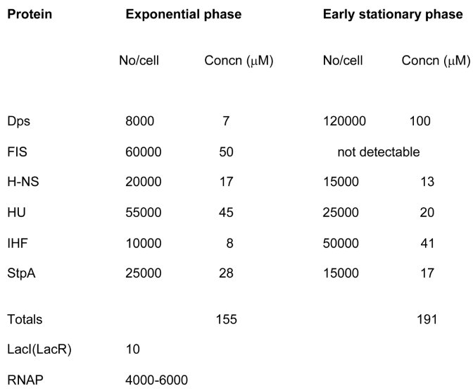
  122. Ali Azam T, Iwata A, Nishimura A, Ueda S, Ishihama A. Growth phase-dependent variation in protein composition of the Escherichia coli nucleoid. J Bacteriol. 1999 Oct;181(20):6361-70. [CrossRef] [PubMed]
  123. Conway C, Beckett MC, Dorman CJ. The DNA relaxation-dependent OFF-to-ON biasing of the type 1 fimbrial genetic switch requires the Fis nucleoid-associated protein. Microbiology (Reading). 2023 Jan;169(1):001283. [CrossRef] [PubMed]
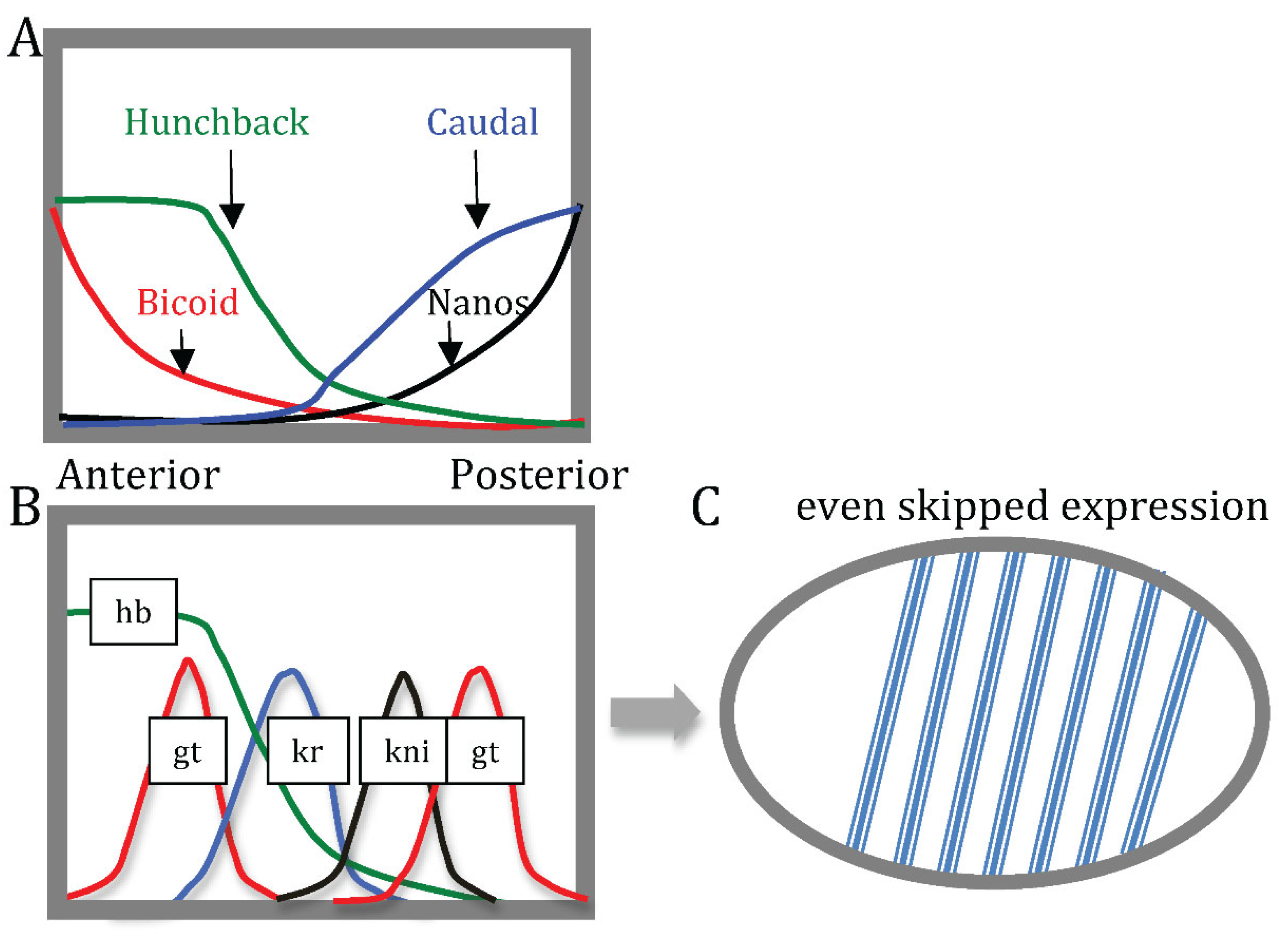
  124. Fujioka M, Jaynes JB, Goto T. Early even-skipped stripes act as morphogenetic gradients at the single cell level to establish engrailed expression. Development. 1995 Dec;121(12):4371-82. [CrossRef] [PubMed]
  125. Nakamura Y, Tomonari S, Kawamoto K, Yamashita T, Watanabe T, Ishimaru Y, Noji S, Mito T. Evolutionarily conserved function of the even-skipped ortholog in insects revealed by gene knock-out analyses in Gryllus bimaculatus. Dev Biol. 2022 May;485:1-8. [CrossRef] [PubMed]
  126. Sonnenschein N, Geertz M, Muskhelishvili G, Hütt MT. Analog regulation of metabolic demand. BMC Syst Biol. 2011 Mar 15;5:40. [CrossRef] [PubMed]
  127. Pérez-Mojica JE, Enders L, Walsh J, Lau KH, Lempradl A. Continuous transcriptome analysis reveals novel patterns of early gene expression in Drosophila embryos. Cell Genom. 2023 Feb 15;3(3):100265. [CrossRef] [PubMed]
  128. Gemkow MJ, Dichter J, Arndt-Jovin DJ. Developmental regulation of DNA-topoisomerases during Drosophila embryogenesis. Exp Cell Res. 2001 Jan 15;262(2):114-21. [CrossRef] [PubMed]
  129. Marr, AG. Growth rate of Escherichia coli. Microbiol Rev. 1991 Jun;55(2):316-33. [CrossRef] [PubMed]
  130. Koyama T, Mirth CK. Unravelling the diversity of mechanisms through which nutrition regulates body size in insects. Curr Opin Insect Sci. 2018 Feb;25:1-8. [CrossRef] [PubMed]
  131. Moshkin YM, Chalkley GE, Kan TW, Reddy BA, Ozgur Z, van Ijcken WF, Dekkers DH, Demmers JA, Travers AA, Verrijzer CP. Remodelers organize cellular chromatin by counteracting intrinsic histone-DNA sequence preferences in a class-specific manner. Mol Cell Biol. 2012 Feb;32(3):675-88. [CrossRef] [PubMed]
  132. Kosmidis K, Hütt MT. The E. coli transcriptional regulatory network and its spatial embedding. Eur Phys J E Soft Matter. 2019 Mar 20;42(3):30. [CrossRef] [PubMed]
  133. Kosmidis K, Jablonski KP, Muskhelishvili G, Hütt MT. Chromosomal origin of replication coordinates logically distinct types of bacterial genetic regulation. NPJ Syst Biol Appl. 2020 Feb 17;6(1):5. [CrossRef] [PubMed]
  134. Ricci DP, Melfi MD, Lasker K, Dill DL, McAdams HH, Shapiro L. Cell cycle progression in Caulobacter requires a nucleoid-associated protein with high AT sequence recognition. Proc Natl Acad Sci U S A. 2016 Oct 4;113(40):E5952-E5961. [CrossRef] [PubMed]
  135. Biondi EG, Reisinger SJ, Skerker JM, Arif M, Perchuk BS, Ryan KR, Laub MT. 2006. Regulation of the bacterial cell cycle by an integrated genetic circuit. Nature 444:899–904. [CrossRef]
  136. Laub MT, Shapiro L, McAdams HH. 2007. Systems biology of Caulobacter. Annu Rev Genet 41:429–441. [CrossRef]
  137. Sobetzko, P.; Glinkowska, M.; Muskhelishvili, G.; GSE65244: Temporal Gene Expression in Escherichia coli. Gene Expression Omnibus. 2017. Available online: https://www.ncbi.nlm.nih.gov/geo/query/acc.cgi?acc=GSE65244 (accessed on 20 June 2021).
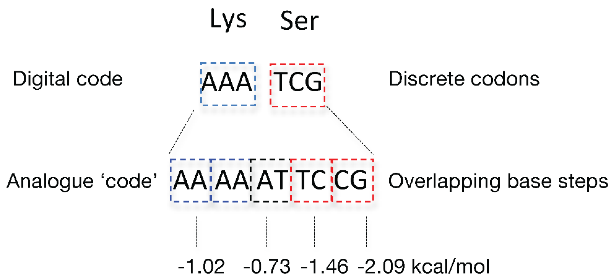
Figure 1. The self-referential organisation of living system is represented by an arrow, which closes on itself (left panel). The self-pointing arrow is a symbol for the condition in which the system divides itself into that which monitors and that which is monitored [1]. The system thus constitutes an isolated, operationally closed circuit. The environment (the space outside of the circle circumference) is marked as ‘E’ (right panel). The environmental impact on the system is indicated by black arrow crossing the circle circumference from outside to inside. Unless this environmental impact is deteriorating, the operational closure of the system is retained.
Preprints 121183 g001
Figure 2. Relation between the digital genetic code and the analog information stored in overlapping base steps of the DNA. Two deliberately chosen triplets coding e.g. for lysine and serine are indicated as ‘digital code’. Indicated below as ‘analogue code’ are the five consecutive overlapping base steps harbored in these two codons. The free stacking/melting energies of base steps [14] are indicated underneath (see also Table 1).
Preprints 121183 g002
Figure 3. Pyrimidine-purine (YR) base steps are flexible and can adopt various configurations. Purine and pyrimidine bases respectively are indicated by large and small rectangles. The asterisk in upper panel indicates the potential steric clash between large purine bases, which is avoided by positive slide of the base pairs. In lower panel this base step has a negative slide and positive roll, and this alternative configuration is stabilized by increased cross-chain stacking interaction between the large purine bases (vertically striated region between the large rectangles). The minor groove side of the bases is shaded (after [18]).
Preprints 121183 g003
Figure 4. Organisation of the gene expression program during the E. coli growth cycle. The circular bacterial chromosome is indicated on the top folded, with two arms aligned along the oriC-ter axis. Indicated below are the spatial gradients of DNA average negative stacking/melting free energy (approx. the G/C content), relative frequencies of gyrase binding sites, of the 70 and S binding sites and the spatial organisation of anabolic and catabolic genes along the oriC- ter axis. Shown underneath is the spatiotemporal gradient of negative superhelical density (-), which changes both temporally with growth phase (from ~ -0.068 on shift-up to ~ -0.043 in stationary phase) as well as forms a spatial gradient along the oriC-ter axis of the chromosome [44,66]. This spatiotemporal gradient of superhelical density is proposed to coordinate the sequential expression of the anabolic and catabolic genes during the bacterial growth cycle [66,71].
Preprints 121183 g004
Figure 5. Switch between the reproduction and maintenance programs in E. coli. (A). Growth phase-dependent expression of the NAP and sigma factor genes (data from [136]). The different expression curves were normalized to [0;1] to compare them in one plot. Minimum and maximum values are indicated in brackets in the legend. Abscissa—time in minutes after inoculation of cells in fresh growth medium. The Escherichia coli CSH50 overnight (16 h) cultures were inoculated at an initial OD600 of 0.1 in rich double yeast-tryptone (dYT) medium and grown in a fermenter under constant pH 7.4 and high aeration (5 L air per min) at 37 ◦C for 7 h (420min). Samples for RNA-seq were taken at 1, 2, 3, 5 and 7 h after inoculation. The different curves were normalized to [0;1] to compare them in one plot. B. The circular chromosomes are depicted with Ori and Ter poles indicated. The reproduction regulatory genes are indicated in blue (left panel), the maintenance program regulatory genes are indicated in red (right panel). The original position of these genes on the circular chromosome is approximated. Note that the reproduction and the maintenance regulators are located around the opposite poles of the chromosome. Colored areas indicate the putative spatiotemporal concentration gradients of regulators. Connecting lines indicate the crosstalk between regulatory genes [72]. The ‘alarmone’ ppGpp produced on shortage of nutritional resources acts as a switch from reproduction to maintenance program. Conversely, high ATP/ADP ratio (established e.g. on nutritional shift-up) favors the commencement of reproduction program.
Preprints 121183 g005
Figure 6. Chromosomal order of regulators and temporal pattern of regulated gene expression. A. Spatial ordering of aerobic/anaerobic growth regulatory genes on the E. coli chromosome along the oriC–ter axis. Genes on the clockwise (right) replichore are indicated on the upper bar, and genes on anti-clockwise (left) replichore are indicated on the lower bar. The atp operon encodes ATP synthase. arcA/arcB encode a two-component system active under microaerobic conditions [86,87]. ArcA also represses rpoS [88]. fnr has a dominant role under more strictly anaerobic conditions [86]. B. Temporal dynamics of expression of various gene classes (data from [136]). The Escherichia coli CSH50 overnight (16 h) cultures were inoculated at an initial OD600 of 0.1 in rich double yeast-tryptone (dYT) medium and grown in a fermenter under constant pH 7.4 and high aeration (5L air per min) at 37 ◦C for 7 h. Samples for RNA-seq were taken at 1, 2, 3, 5 and 7 h after inoculation. The different curves were normalized to [0;1] to compare them in one plot. The envelopes of the curves indicate the standard deviation at 10% random remapping of the expression patterns to genes. Minimum and maximum values are indicated in brackets in the legend. Expression values (anabolic, catabolic, aerobic, anaerobic) in brackets are normalized to the expression of all genes. The optical density and partial oxygen pressure are indicated respectively, by the dashed, light blue and green lines. Note the correlation between the maximal expression of anaerobic genes and minimal partial oxygen pressure.
Preprints 121183 g006
Figure 7. Competition between the bacterial NAPs, HU and H-NS, which stabilise alternative supercoil structures on binding DNA. Distinct supercoil structures stabilized by HU and H-NS are indicated by white arrows. HU stabilizes more open toroidal coils, whereas H-NS stabilizes tightly interwound, stiff plectonemic DNA structures (AFM image, courtesy Sebastian Maurer).
Preprints 121183 g007
Figure 8. Regulation of lysis-lysogeny decision in λ phage development. Gene and early transcription map of λ is shown (simplified). Genes are indicated in the shaded rectangle. The early transcripts produced from pL and pR promoters are shown as blue arrows. The immediate early gene (short arrows) products are N and Cro. On extension of the transcripts known as ‘delayed early transcription’ (long arrows underneath) the CIII and CII proteins are produced. The N and Cro proteins support lytic development, whereas CIII and CII proteins support lysogeny. Note that synthesis of transcripts of different lengths (i.e. generation of analogue information) results in production of distinct sets of specific proteins (digital information). O and P are DNA replication genes involved in lytic growth. Q protein turns on the late genes for production of phage tails and heads. cI, cII, cIII and int genes are involved in the establishment of lysogeny. The xis gene (together with int) is involved in excision of the integrated prophage. Cro and CI are repressor proteins competing for binding at the operator sites in the regulatory region. Critical for the active production of CI repressor is the CII protein, the stability of which in turn is sensitive to physiological conditions.
Preprints 121183 g008
Figure 9. Generation of anterior-posterior pattern of even skipped (eve pair-rule gene) expression initiated by gradients of the Drosophila maternal effect genes. A. The Bicoid protein gradient extends from anterior to posterior while the Nanos protein gradient extends from posterior to anterior. Nanos inhibits the translation of the hunchback message (in the posterior), while Bicoid prevents the translation of the caudal message (in the anterior). This inhibition results in opposing Caudal and Hunchback gradients. (B) Spatial distribution of Bicoid-responsive segmentation (gap) gene expression such as giant (gt), krueppel (kr) and knirps (kni). The gap gene products mutually repress each other’s expression, resulting in spatially defined domains of gap gene expression along the anterior-posterior axis of the embryo, which ultimately define the pattern of even skipped (a pair-rule gene) expression in seven stripes (C). Each stripe has an independent transcriptional control system consisting of different constellations of transcription factors (specific combinations of activators) interacting with seven different enhancer sequences all of which can activate the even skipped gene expression.
Preprints 121183 g009
Figure 10. Organization of information flow and inter-conversion in the living system. A. Depicted is the central dogma of molecular biology: digital genetic information flows only in one direction, from DNA, to RNA, to protein (blue arrows). However the DNA analogue information is recognized directly by the DNA binding proteins stabilizing various 3D conformations of the DNA (curved brown arrow) and thus modulating the content of digital information. B. Systems-theoretical model for inter-conversion of DNA information. The terms ‘analogue’ and ‘digital’ refer to two distinct information types stored in the genomic DNA polymer. DNA analogue information implies both static (sequence organization viz. arrangement of base steps) and dynamic parameters (DNA 3D configuration and superhelicity). Digital information implies differential gene expression patterns and the gene interaction networks (including regulatory cascades) emerging thereof. Analogue information provides an integrative sensory interface for both internal and external signals as well as a regulatory context for digital code expression, whereas the latter provides information for reproduction and maintenance of the former. Together, the inter-converting DNA ‘codes’ form a coordinated self-referential circuit responding to both the internal and external signals as an indivisible unity.
Preprints 121183 g010
Table 1. Free stacking/melting energy.
Base steps kcal/mol
AA/TT -1.02
AT/AT -0.73
TA/TA -0.6
CA/TG -1.38
GT/AC -1.43
CT/AG -1.16
GA/CT -1.46
CG/CG -2.09
GC/GC -2.28
GG/CC -1.77
Table 2. Estimated number and concentrations of the most abundant nucleoid-associated proteins in E. coli [121]. Approximate numbers for RNA polymerase and lac repressor are shown for comparison.
Preprints 121183 i001
Disclaimer/Publisher’s Note: The statements, opinions and data contained in all publications are solely those of the individual author(s) and contributor(s) and not of MDPI and/or the editor(s). MDPI and/or the editor(s) disclaim responsibility for any injury to people or property resulting from any ideas, methods, instructions or products referred to in the content.
Copyright: This open access article is published under a Creative Commons CC BY 4.0 license, which permit the free download, distribution, and reuse, provided that the author and preprint are cited in any reuse.
Prerpints.org logo

Preprints.org is a free preprint server supported by MDPI in Basel, Switzerland.

Subscribe

Disclaimer

Terms of Use

Privacy Policy

Privacy Settings

© 2025 MDPI (Basel, Switzerland) unless otherwise stated