Submitted:
25 July 2024
Posted:
26 July 2024
You are already at the latest version
Abstract
Photodynamic therapy has emerged as a non-invasive treatment modality for various diseases, leveraging photosensitizing compounds to induce localized cytotoxicity upon light activation. This review explores the isolation and characterization of photosensitizing compounds sourced from microalgae, focusing on their potential applications in photodynamic therapy. Microalgae represent a rich source of bioactive molecules, including chlorophylls, phycobiliproteins, and carotenoids, which exhibit inherent photosensitizing properties. Methodologies for the extraction and purification of these compounds from different microalgal species are discussed, emphasizing the importance of optimizing extraction techniques to enhance yield and purity. Characterization techniques such as UV-Vis absorption spectroscopy, fluorescence spectroscopy, and mass spectrometry are highlighted for elucidating the chemical structures and photophysical properties of isolated compounds. Furthermore, the review examines the photodynamic efficacy of microalgae-derived photosensitizers through in vitro and in vivo studies, assessing their cytotoxic effects on cancer cells and antimicrobial activity against pathogens. The integration of microalgae-derived photosensitizers into PDT protocols represents a promising avenue for developing sustainable and effective therapeutic strategies, underscoring the potential of these natural compounds in advancing biomedical applications.
Keywords:
Chlorella
; photodynamic therapy
; photosensitizers
; microalgae
1. Introduction
Photodynamic therapy has emerged as a promising modality for the treatment of various types of cancers and other medical conditions [1]. This non-invasive approach involves the administration of a photosensitizing agent that preferentially accumulates within the target tissue, followed by exposure to a specific wavelength of light, resulting in the generation of cytotoxic reactive oxygen species and subsequent cell death [2,1,3]. In recent years, there has been a growing interest in exploring microalgae as a source of novel photosensitizing compounds for photodynamic therapy (PDT) applications.
Microalgae are a diverse group of photosynthetic microorganisms that have the potential to produce a wide range of bioactive compounds, including pigments, lipids, and other secondary metabolites. These organisms have garnered attention due to their ability to produce a variety of photosensitizing compounds, such as chlorophyll derivatives, carotenoids, and anthraquinones, which possess the necessary photophysical and photochemical properties for effective photodynamic therapy [4,1,5].
Several studies have evaluated the potential of semi-purified extracts from various microalgae strains as photosensitizers for photodynamic therapy. These extracts have been shown to exhibit significant effects on the viability of different tumor cell lines, decreasing it by as much as 95% for certain microalgae-derived constituents. [1,5]The fluorescence measurements and spectral intensities of these extracts have revealed characteristic features of their photosensitizing compounds, which can be activated by blue light, red light, or a combination of both. Additionally, the molecular, cellular, and tumor responses associated with photodynamic therapy, such as the subcellular and tumor localization of photosensitizing agents, have been extensively studied and discussed in the literature. [7,8,9,10,11]
The use of engineered algae has also been explored as a potential oxygen-generating system for the effective treatment of hypoxic cancer [12].
Photodynamic therapy has been approved for the treatment of various cancers, including early and advanced-stage lung, digestive tract, and genitourinary tract cancers, in several countries since the 1990s [13]. The potential of microalgae-derived photosensitizers to enhance the efficacy and selectivity of photodynamic therapy is an area of active research, with promising results reported in preclinical studies.[6,7]
Continued efforts to isolate, characterize, and optimize the use of microalgae-derived photosensitizing compounds may lead to the development of improved photodynamic therapy approaches for the treatment of various types of cancer and other medical conditions [6,1,12].
Microalgae have been recognized as a promising source of novel photosensitizing compounds for photodynamic therapy applications. Several studies have demonstrated the potential of semi-purified extracts from various microalgae strains to exhibit significant effects on the viability of different tumor cell lines, with certain microalgae-derived constituents capable of decreasing cell viability by as much as 95% [1,12]. The photosensitizing properties of these extracts have been attributed to the presence of characteristic photosensitizing compounds, which can be activated by blue light, red light, or a combination of both.
The use of photodynamic therapy for the treatment of various cancers has been approved in several countries since the 1990s, and the potential of microalgae-derived photosensitizers to enhance the efficacy and selectivity of this approach is an area of active research. Continued efforts to isolate, characterize, and optimize the use of these photosensitizing compounds may lead to the development of improved photodynamic therapy approaches for the treatment of a wide range of medical conditions [6,1,13].
Recent advances in the field of photodynamic therapy have sparked a renewed interest in the isolation and characterization of photosensitizing compounds from microalgae. The potential of these compounds to revolutionize the treatment of various cancers and medical conditions is a driving force behind ongoing research efforts.[5,14]One of the key areas of focus in this research is the identification and isolation of specific photosensitizing compounds from microalgae that exhibit potent cytotoxicity towards cancer cells while sparing healthy tissues. Scientists are employing advanced techniques such as liquid chromatography, mass spectrometry, and nuclear magnetic resonance spectroscopy to isolate and characterize these compounds at a molecular level.[6,16,17]Understanding the chemical structure and properties of these compounds is crucial in elucidating their mechanism of action and optimizing their efficacy for photodynamic therapy.[18,19,20].
Furthermore, the cellular and subcellular localization of these photosensitizing agents within tumor tissues is a subject of intense investigation. By utilizing fluorescence microscopy and other imaging technologies, researchers aim to gain insights into the distribution and uptake of these compounds in cancer cells. This knowledge is vital for enhancing the targeting and specificity of photodynamic therapy, thereby minimizing off-target effects and maximizing the destruction of malignant cells.[21,4,22,23]
The application of photodynamic therapy in the treatment of various cancers, including lung, digestive tract, and genitourinary tract cancers, has been approved in several countries since the 1990s. This approval underscores the clinical efficacy and safety of this therapeutic approach.[13,11,24]However, the search for more potent and selective photosensitizing agents continues, with microalgae-derived compounds emerging as promising candidates.[13,25,4,1]
In addition to the molecular and cellular aspects, studies are also delving into the tumor responses associated with photodynamic therapy using microalgae-derived photosensitizers. This includes analyzing the interaction of these compounds with the tumor microenvironment and understanding the cascading effects that lead to cell death upon light activation. Through in-depth characterization and understanding of these processes, researchers aspire to refine photodynamic therapy protocols and optimize treatment outcomes.[26,11,19,4]
Overall, the isolation and characterization of photosensitizing compounds from microalgae for photodynamic therapy is a rapidly evolving field of research with significant implications for the treatment of various cancers and other medical conditions.[1,5,4,14]The continued efforts in this area hold the promise of developing more effective and targeted photodynamic therapy approaches that can improve patient outcomes and quality of life.[27,28,29]
As the quest for novel photosensitizing compounds from microalgae continues, the potential for these natural sources to elevate the efficacy and selectivity of photodynamic therapy in the clinical setting becomes increasingly promising. With each discovery and breakthrough in the isolation and characterization of these compounds, the horizon of photodynamic therapy widens, offering new hope for improved treatment approaches for cancer and other medical conditions.[29,24,30]
2. Investigating the Mechanism of Action for Enhanced Efficacy.
In order to fully harness the potential of microalgae-derived photosensitizing compounds for photodynamic therapy, it is imperative to delve into the intricate mechanisms underlying their enhanced efficacy and selectivity. Researchers are delving into the molecular pathways through which these compounds exert their cytotoxic effects on cancer cells and the factors that contribute to their preferential accumulation within tumor tissues (Figure 1).[31]
One of the key focus areas is the investigation of the subcellular localization of microalgae-derived photosensitizers and their interactions with specific cellular organelles and biomolecules. By utilizing advanced imaging techniques, scientists are gaining insights into the distribution and uptake of these compounds within cancer cells, which is crucial for enhancing the targeting and specificity of photodynamic therapy [24,11,21].
Additionally, researchers are exploring the impact of these photosensitizers on the tumor microenvironment and the cascading cellular responses that lead to the selective destruction of malignant cells upon light activation. Understanding these complex interactions is essential for optimizing the treatment protocols and maximizing the therapeutic benefits of photodynamic therapy.[32,33,19]
Ultimately, the continued efforts to isolate, characterize, and elucidate the mechanisms of action of microalgae-derived photosensitizing compounds will pave the way for the development of improved photodynamic therapy approaches that offer enhanced efficacy, selectivity, and clinical outcomes for patients.[1,5,34]
Understanding the molecular pathways involved in the enhanced efficacy and selectivity of microalgae-derived photosensitizing compounds for photodynamic therapy is a multifaceted endeavor that requires a comprehensive approach. Researchers are delving into the intricate interactions between these compounds and the cellular components of tumor tissues to unravel the underlying mechanisms that drive their therapeutic potential.[35,36,37,38]
One crucial aspect is the investigation of the subcellular localization of these photosensitizers and their interactions with specific organelles and biomolecules within cancer cells.[39,40,41]
By utilizing advanced imaging techniques, such as fluorescence microscopy and confocal microscopy, scientists are gaining valuable insights into the distribution and uptake of these compounds within the tumor cells.[42,43]
This information is crucial for enhancing the targeting and specificity of photodynamic therapy, as it enables the optimization of treatment protocols to ensure the selective accumulation of the photosensitizers in malignant cells while minimizing off-target effects.[44]
A critical aspect of this investigation is the subcellular localization of microalgae-derived photosensitizers within cancer cells. Advanced imaging techniques, including confocal microscopy and super-resolution microscopy, are being employed to visualize the precise localization of these compounds within specific cellular organelles such as mitochondria, endoplasmic reticulum, and lysosomes. By elucidating the subcellular distribution patterns, researchers aim to gain a deeper understanding of the cellular interactions that dictate the cytotoxic effects of the photosensitizers upon light activation.[45,46,47,48]
Furthermore, the interactions of these compounds with specific biomolecules within the cellular milieu are being scrutinized. Proteomic and metabolomic analyses are shedding light on the molecular targets and signaling pathways modulated by the photosensitizers, providing invaluable insights into the complex network of cellular responses elicited upon their activation.[49,50]
Figure 1.
The effect of photosensitizers on cancer cells.

Beyond the direct interactions within cancer cells, researchers are investigating the broader impact of microalgae-derived photosensitizers on the tumor microenvironment. By examining the interplay between the photosensitizers and stromal cells, immune cells, and extracellular matrix components, scientists seek to discern the dynamic changes that occur within the tumor milieu following photodynamic therapy. This includes unraveling the immunomodulatory effects triggered by the treatment and delineating the mechanisms that contribute to the localized destruction of malignant cells while preserving healthy tissue integrity.[51,52]
Moreover, the cascading cellular responses initiated upon light activation of the photosensitizers are under intense scrutiny. From the generation of cytotoxic reactive oxygen species to the induction of apoptotic and necrotic pathways, researchers are aiming to unravel the sequence of events that culminate in the selective eradication of cancer cells while mitigating systemic adverse effects.[53,54,55,56,57]
The in-depth exploration of the molecular pathways underlying the enhanced efficacy and selectivity of microalgae-derived photosensitizing compounds holds profound implications for the clinical translation of photodynamic therapy. By delineating the precise molecular targets and intracellular dynamics, researchers are poised to refine treatment protocols, optimize dosing regimens, and tailor therapeutic strategies to individual patient profiles.[58,59,60]
Furthermore, this mechanistic understanding sets the stage for the design of next-generation photosensitizers with enhanced targeting capabilities, improved photochemical properties, and augmented biocompatibility. These advancements hold the promise of broadening the applicability of photodynamic therapy beyond cancer, encompassing diverse medical conditions where localized, precise therapeutic interventions are warranted.[61,62,63]
In essence, the relentless pursuit of unraveling the molecular pathways for the enhanced efficacy of microalgae-derived photosensitizing compounds in photodynamic therapy stands to revolutionize the landscape of cancer treatment and beyond. Each revelation brings us closer to realizing the full potential of this promising therapeutic modality and improving the quality of life for patients across the globe. [61,64]
Studies on microalgae have shown promising results in the field of photodynamic therapy. The use of chlorella sp. in particular has garnered significant attention due to its potential in enhancing the efficacy and selectivity of photosensitizing compounds. With an increased focus on subcellular localization and molecular interactions, researchers are uncovering a wealth of information that could propel the development of improved photodynamic therapy approach [65]
3. Chlorella sp. and Subcellular Localization
The exploration of chlorella sp. and its role in subcellular localization within cancer cells has opened up new avenues for understanding the precise mechanisms at play. Cutting-edge imaging techniques, such as super-resolution microscopy, are providing insights into the specific cellular organelles where chlorella-derived photosensitizers accumulate. The visualization of these localization patterns is instrumental in enhancing the targeting and specificity of photodynamic therapy, thereby maximizing the therapeutic benefits while minimizing off-target effects. Figure 2 shows which sensitizing substances are released from microalgae [66,67,68].
Compounds (1, 2, 3) are usually present in different fractions or amounts depending on the chlorella strain and growing conditions. Each of these chemicals plays an important role, for example, in photosynthesis (chlorophylls), antioxidant protection (carotenoids, astaxanthin) and possibly in biotechnological applications due to their functional properties. [69,70]
Various authors consider the main released substances from microalgae used in photodynamic therapy and their potential in modern clinical practice.
Microalgae, as a source of biologically active substances, attract the attention of scientists and doctors due to their unique ability to photosynthesis and accumulation of useful compounds. In recent years, special attention has been paid to substances isolated from microalgae, which have shown potential in photodynamic therapy. This approach to treatment, based on the activation of photosensitive compounds with light for the targeted destruction of tumor cells, opens up new prospects in the fight against oncological diseases and other pathologies. The photosensitizing substances that are released from Chlorella sp are listed below.:
Chlorophyll a (C₅₅H₇₂O₅N₄Mg) (1) and Chlorophyll b (C₅₅H₇₀O₆N₄Mg) (2):
Usually found in chloroplasts of chlorella cells. It is necessary for photosynthesis, the conversion of light energy into chemical energy. Chlorophylls are also being studied for their antioxidant properties and potential health benefits. [71,72]
Chlorophylls are the most important pigments in photosynthesis, absorbing light energy to convert carbon dioxide and water into glucose and oxygen. Chlorophyll a is the main pigment in photosystems I and II, while chlorophyll b helps capture light energy and transfer it to chlorophyll A.
The extraction of chlorophyll from chlorella usually involves solvent extraction (e.g., acetone or ethanol) followed by purification methods such as column chromatography or filtration.
Carotenoids (β-carotene, C₄₀H₅₆) (3) :
Found in fat-soluble chlorella fractions. Carotenoids act as antioxidants, protecting cells from oxidative stress. They also give chlorella a green color and are of interest as natural pigments and compounds that promote health. [73,74]
Carotenoids are auxiliary pigments that play an important role in light absorption and photoprotection. β-carotene, in particular, acts as a precursor for the synthesis of vitamin A in animals and has powerful antioxidant properties.
Carotenoids can be extracted from chlorella biomass using organic solvents (e.g., hexane, acetone) or supercritical liquid extraction methods. Purification includes methods such as chromatography or crystallization.
Astaxanthin (C₄₀H₅₂O₄) (4):
Usually found in fat-soluble extracts. Astaxanthin is a powerful antioxidant and anti-inflammatory agent. It is known for its bright red color and is used in aquaculture to improve the coloration of fish and crustaceans. It can also find potential applications in nutraceuticals and cosmetics due to its health benefits. [75,76]
Astaxanthin is a xanthophyll carotenoid with powerful antioxidant properties, about 10 times stronger than β-carotene and 100 times stronger than vitamin E. It protects cells from oxidative damage and plays an important role in photoprotection.
Methods such as extraction with organic solvents (e.g., acetone, methanol) or supercritical CO2 extraction are used to obtain astaxanthin from chlorella. Chromatography or crystallization is usually used after purification.
Astaxanthin is used in aquaculture to improve pigmentation of fish and crustaceans. It is also popular as a dietary supplement due to its potential benefits for the cardiovascular system, eye health and skin protection from ultraviolet radiation. Due to its antioxidant and anti-inflammatory properties, phycocyanin is used in cosmetology as an anti-aging agent. [85,86,87]
Phycocyanin (C₃₇H₆₄N₈O₁₆S₄) (5):
A water-soluble pigment found in cyanobacteria, including some chlorella species. Phycocyanin is a blue pigment involved in the absorption of light during photosynthesis. It has antioxidant and anti-inflammatory properties and is used as a natural dye in food and cosmetics. It is also promising in medical research as a therapeutic agent.
These chemicals are not only an integral part of the biological processes in chlorella cells, but also have important commercial and research applications. They are studied due to their antioxidant, anti-inflammatory and coloring properties, thanks to which Chlorella sp. a valuable source of natural compounds for various industries, including food, pharmaceutical and cosmetics. [77,78]
Phycocyanin is a water-soluble pigment belonging to the phycobiliprotein family. It absorbs orange and red light and transfers energy to chlorophyll for photosynthesis. It also has antioxidant and anti-inflammatory properties.
Phycocyanin extraction consists in the destruction of chlorella cells, followed by centrifugation or filtration to separate the pigment from the biomass. Further purification may include precipitation and chromatography.
Phycocyanin is used as a natural blue food coloring (E163) in beverages and confectionery. Its antioxidant properties make it a valuable ingredient in dietary supplements to promote health and wellness. Research is underway on its potential therapeutic applications, including anti-cancer and anti-inflammatory properties [88,89,90].
The structural formulas of astaxanthin and phycocyanin are shown in Figure 3.
In addition to subcellular localization, the impact of chlorella sp. on molecular interactions within the cellular environment is a key area of investigation. By scrutinizing the interactions between chlorella-derived photosensitizers and specific biomolecules, researchers are gaining a deeper understanding of the complex network of cellular responses triggered upon light activation. This comprehensive analysis extends to the cascading cellular responses, including the generation of reactive oxygen species and the modulation of apoptotic pathways, ultimately leading to the selective eradication of cancer cells.[91,92,93]
The profound implications of these findings extend to the clinical translation of photodynamic therapy. Armed with a detailed understanding of the molecular pathways and intracellular dynamics facilitated by chlorella-derived photosensitizers, researchers are poised to revolutionize treatment protocols and individualize therapeutic strategies. Furthermore, this knowledge serves as a catalyst for the design of next-generation photosensitizers that offer enhanced targeting capabilities and improved biocompatibility, promising advancements that could expand the scope of photodynamic therapy beyond cancer treatment.[94,95]
In summary, the continued exploration of chlorella sp. and its implications for photodynamic therapy holds immense potential to reshape the landscape of cancer treatment and beyond. With each revelation, we move closer to unlocking the full therapeutic potential of this groundbreaking approach, ultimately enhancing the quality of life for patients worldwide.[96,97]
4. Isolation of Sensitizing Substances from Microalgae Using Column Chromatography.
The exploration of microalgae for the isolation of sensitizing substances has gained traction in the field of photodynamic therapy. In particular, the utilization of column chromatography has emerged as a vital technique for the isolation and purification of these compounds from microalgae. This method allows for the separation of sensitizing substances based on their chemical properties, paving the way for a more comprehensive understanding of their structure and function.[98,99,100]
Column chromatography enables the enhanced purification of sensitizing substances derived from microalgae by isolating individual components from complex mixtures. This process not only facilitates the identification of specific compounds but also aids in the characterization of their chemical properties, such as polarity, molecular weight, and composition. As a result, researchers can gain a deeper insight into the diverse array of sensitizing substances present in microalgae, laying the groundwork for their potential application in photodynamic therapy and other biomedical fields.[101,102,103]
The isolated sensitizing substances obtained through column chromatography provide a unique opportunity to investigate their subcellular localization within cancer cells. By employing advanced imaging techniques and fluorescent labeling, researchers can elucidate the precise distribution of these compounds within cellular organelles, shedding light on their functional significance in the context of photodynamic therapy. This knowledge is instrumental in optimizing the targeting efficiency and specificity of these substances, ultimately enhancing their therapeutic efficacy while minimizing off-target effects.[104,105]
Furthermore, the purification and isolation of sensitizing substances from microalgae lay the foundation for conducting comprehensive mechanistic studies to unravel their interactions with biomolecules and their impact on cellular responses upon light activation.[106,107,108]
In conclusion, the employment of column chromatography for the isolation of sensitizing substances from microalgae represents a pivotal stride in advancing our understanding of these compounds and their potential applications in photodynamic therapy. Through meticulous purification and characterization, researchers are poised to uncover the intricate details that underpin the therapeutic capabilities of these substances, setting the stage for transformative developments in the field of cancer treatment and beyond.[109]
In order to further advance the study of photosensitizing compounds derived from Chlorella sp., researchers have turned to column chromatography for the isolation of sensitizing substances. This technique allows for the separation of complex mixtures, enabling the purification of individual compounds based on their differential affinity for the stationary phase.[110,111,112]
The isolation of sensitizing substances from Chlorella sp. through column chromatography presents a significant step towards characterizing the specific molecules responsible for the observed therapeutic effects. By obtaining purified compounds, researchers can conduct more targeted studies to elucidate the mechanisms of action, subcellular localization, and molecular interactions of these substances within the cellular environment.[113,114,115]
The application of column chromatography not only aids in the identification and isolation of sensitizing compounds but also serves as a crucial preparative step for subsequent analytical techniques, paving the way for deeper insights into the potential of Chlorella-derived photosensitizers in photodynamic therapy.[116,117]
The authors A. G. Fasya, N. Millati, L. M. Rahmawati, R. Iyani, A. Hanapi, R. Ningsih, D. Yuliani and D. S. Megawati in their articles determined the toxicity and antioxidant activity in steroid compounds in petroleum ether in microalgae Chlorella sp.
Chlorella sp. It was grown in a 4% medium with a sprout extract using a solvent - methanol.
The fraction containing steroids was separated by thin-layer and column chromatography. And the level of toxicity and antioxidant analysis were determined by DPPH (2,2-diphenyl-1-picrylhydrazyl).
For column chromatography, the authors used a mixture of n-hexane and ethyl acetate in a ratio (4:1) as the mobile phase (eluent), while silica gel 60 (0.063-0.200 mm) was used as the stationary phase. The column diameter was 1.5 cm and the flow rate for elution was 1.5 ml/min.
For the preparation of the stationary phase: 10 g of silica gel was activated at a temperature of 110 ° C for 2 hours, then cooled for 15 minutes in a desiccator. An eluent was added to the silica gel and homogenized to form a suspension and sent to the column for 24 hours. The ethyl acetate fraction (0.1 g) was diluted in 1 ml of the eluent. The sample was placed in a treated column for elution. Every 2 ml of eluate was taken into a vial in the form of a fraction. [118,119,120]
As a result, the steroid compounds A2 - β-sitosterol, A8 - stigmasterin, A10 - campesterin and A12 - erythrodiol were found.
β-sitosterol is used in various cosmetic and pharmaceutical products in the treatment of inflammatory diseases such as arthritis.
Stigmasterine and campesterine are used to prevent cardiovascular diseases and other conditions associated with inflammation.
Erythrodiol may have antitumor activity, which makes it potentially useful for cancer treatment. [121,122]
A group of Yousef Sultan researchers in their articles obtained antifungal drugs from extracts, fractions, subfractions and pure compounds of Chlorella sp. to protect against various strains of mycotoxigenic fungi. [123]
Chlorella sp. it was obtained in the Laboratory of Marine Toxins of the National Research Center of Egypt. [124]
They used thin-layer and column chromatography to separate these compounds. And the level of toxicity and antioxidant analysis were determined by the DPPH method. [125,126,127,128]
The diethyl ether extract was fractionated by column chromatography. To do this, a glass column (30 × 500 mm) was first filled with 5 g of anhydrous sodium sulfate, then 30 g of silica gel, using chloroform as a solvent carrier to obtain a suspension. Finally, 5 g of anhydrous sodium sulfate was added on top of the silica gel to prevent the column from drying out. A portion of diethyl ether (500 mg) diluted in 10 ml of chloroform was loaded into a column and passed through it at a rate of 1 drop per second.
The silica gel column was eluted with a solution of various mixtures (by volume) of chloroform: methanol (90:10, F1), (80:20, F2), (50:50, F3), (25:75, F4) and, finally, methanol 100% (F5) to obtain 5 fractions. Fractions of 50 ml each were collected, evaporated under vacuum and stored for analysis of antifungal activity. Fractions F3 and F5 were dissolved in a mixture of chloroform:methanol (50:50) and methanol (100%), respectively, were passed through newly prepared columns. Each fraction was divided into 10 sub-fractions (5 ml each). [123]
As a result, antifungal compounds were obtained: A. flavus, A. Parasiticus, A. Carbonarius, A. Ochraceus, F. Verticilioides, P. Verrucosum.
They are used in medical research, for example, to study its genetics or to create infection models in the study of antimicrobial agents. They are known for their ability to produce mycotoxins such as aflatoxins, which can be harmful to human and animal health. Aflatoxins can lead to various diseases, including cancer. [129,130,131,132,133,134,135,136]
Shinya Shibata and co-authors in their research obtained highly purified lutein using column chromatography on silica gel. [137]
For the experiment, a spray-dried Chlorella sp. powder was used, which was obtained from Nihon Chlorella Co. Ltd. (Tokyo, Japan).
Column chromatography was performed using PUMP 540 and PREP UV-10V equipment (Yamazen Corp., Osaka, Japan).
The crude carotenoid solution was concentrated to a yellow-orange residue using a rotary evaporator. This residue was dissolved in 100 ml of ether and an equal volume of hexane was added. After filtration, this solution was chromatographed on an instantaneous column (30 mm in diameter × 260 mm) on silica gel. The eluent flowed through the column at a pressure of 1-2 kg/cm2. First, the column was eluted with hexane to obtain a mixture of α and β carotene.When the elution of the carotene strip was completed, the eluent was replaced with hexane-acetone-chloroform (7:2:1 by volume). Fractions 1-3 were selected based on the absorption coefficient of ultraviolet radiation (fraction 1) or the color scale (yellow-orange, fractions 2 and 3). [138,139]
As a result, lutein, α-carotene and β-carotene were obtained. They are used as dietary supplements to support vision, skin health and the immune system. Research shows that carotenoids can help prevent various diseases, including eye diseases and cancer. [140]
Yen-Ju Lee and co-authors in their research received aminolevulinic acid for photodynamic therapy against pathogens and cancer cells. [141]
To do this, they used a highly acidic cation exchange resin (Amberlite® IR 120) placed in a column (7.07 cm2, height 20 cm). First, the resin was immersed in 50 ml of 1.5 M HCl for 1.5 hours, then 50 ml of 1.5 M NaOH was added. 50 ml of 1.5 M HCl was passed through a column to obtain the H+ form. The resin was washed in distilled water once between each stage. Before adsorption, the microalgae solution was adjusted to a pH of 4.2-4.8 using acetate acid. 600 ml of the solution was applied to the column, and then 100 ml of distilled water was passed through it to wash out the residual medium. In this study, HCl, sodium acetate buffer and ammonia were used to study the effectiveness of microalgae desorption at various concentrations and pH. At the end, 85% phosphate acid was added to the desorbed solution and the pH was adjusted to 3.0.
As a result, purified 5-ALA (5-aminolevulinic acid) was used to destroy cancer cells and pathogenic microorganisms, achieving an efficiency of 83% and 100%, respectively.
5-aminolevulinic acid is used in photodynamic therapy to treat various dermatological diseases such as acne, rosacea and pigmented disorders. Patients are applied a thin layer of 5-ALA to the skin, which is then absorbed into the cells. After that, the area is irradiated with light of a certain wavelength, which leads to the activation of 5-ALA and the destruction of the affected cells.
5-aminolevulinic acid (1) is used in some cancer diagnostic methods, such as urological cytology. It can help identify altered cells, including cancerous ones, in urine, blood and other biological samples. [142,143,144,145,146,147]
The authors Feng Liang and other researchers in their articles isolated photosensitizing substances from microalgae Chlorella vulgaris for use in photodynamic therapy. [148]
The Chlorella vulgaris strain was obtained at Guangyu Biological Technology (Shanghai, China) and cultured in BG11 medium.
A group of Bing-Chung Liaua researchers in their research obtained extracts of carotenoids from microalgae with supercritical liquids and purified them from solvents. [149]
The method of column chromatography was used to isolate the extracts. Freeze-dried microalgae (10.0 g) were thoroughly boiled in CH2Cl2 using a Soxlet apparatus. The extracts were concentrated under vacuum to obtain CH2Cl2 extract (0.85 g), and all extracts were dissolved in ethyl acetate and n-hexane (5:1). The solution was additionally chromatographed on a silica gel column (3 cm in diameter × 25 cm in diameter) by elution of hexane, ethyl acetate.
Each fraction of the eluent had a volume of 100-150 ml; a total of 12 fractions were collected. The fractionation of chemical components was monitored using thin-layer chromatography (TLC). The compounds collected from fractions 8-11 were additionally purified using a programmed gradient reverse-phase HPLC.
As a result, zeaxanthin compounds were obtained. Zeaxanthin is a natural carotenoid that belongs to the xanthine class. This yellow-orange pigment belongs to a group of carotenoids called macular carotenoids, which play an important role in protecting the eyes and maintaining visual health. Zeaxanthin is a powerful antioxidant that helps protect cells from damage caused by free radicals and oxidative stress.
It is used to support visual health, protect against oxidative stress, improve skin condition, maintain heart and vascular health and bone health. [150,151,152,153,154,155,156,157]
Victor Abrahamsson and co-authors in their research determined the content of carotenoids in microalgae Chlorella sp. using supercritical liquid extraction and column chromatography. [158]
The separation of carotenoids using a sequentially connected column C18 and a column with 2-ethylpyridine (2-EP) containing silicon dioxide was optimized by testing both methanol and ethanol as co-solvents, various profiles of the gradient of co-solvents, pressure, temperature and flow. The initial optimization was carried out using a mixture of 8 standard carotenoids, which are often found in microalgae and include astaxanthin, β-carotene, canthaxanthin, echinenone, lutein, neoxanthin, violaxanthin and zeaxanthin.
As a result, carotenoids were isolated from microalgae using SFC. When combined in series A C18 and column 2-EP, a standard mixture of astaxanthin, β-carotene, canthaxanthin, echinenone, lutein, neoxanthin, violaxanthin and zeaxanthin was isolated in less than 10 minutes.
Astaxanthin, β-carotene, lutein, neoxanthin, violaxanthin and zeaxanthin – all these substances belong to the class of carotenoids, which are pigments that provide the red, orange and yellow color of many plants, animals and microorganisms.
Some studies show that carotenoids can have positive health effects, including protecting the eyes from age-related changes and reducing the risk of developing chronic diseases such as cancer and cardiovascular diseases. However, additional research is required to confirm these effects. [159,160,161,162,163,164,165,166]
The authors of Sonja Srdanovic in the article The photodynamic activity of 13-[2-(2-pyridyl)ethylamine] Chlorin e6 photosensitizer in human esophageal cancer synthesized a pyridine-substituted derivative of Chlorin e6 (Chlorin A) [167].
It has a characteristic long-wave absorption at 664 nm and a wavelength of radiation at 667 nm. The rate of formation of singlet oxygen in this compound is higher than that of Temoporphine. In vitro, chlorine has demonstrated higher phototoxicity to human esophageal cancer cells than Temoporphine, with lower toxicity in the dark. Its accumulation effect in mitochondria, lysosomes and endoplasmic reticulum has been traced in subcellular localization tests. With flow cytometry, obvious cell apoptosis was observed after 2 hours of irradiation.
Chlorin A from dimethylpheophorbide was synthesized by nucleophilic addition of 2-pyridylethylamine to the exocyclic ring. As a result of the reaction at room temperature in dry tetrahydrofuran, a separate product was obtained, which was purified using column chromatography. The synthesis of chloride a is shown in Figure 4. [168,169].
Significant antitumor efficacy of photodynamic drugs in vivo has also been demonstrated in mice with esophageal cancer. Thus, Chlorin A can be proposed as a promising antitumor drug for photodynamic therapy.
As a result of PDT, necrotic lesions were found, which suggested that Chlorin A has optimal properties in PDT [167].
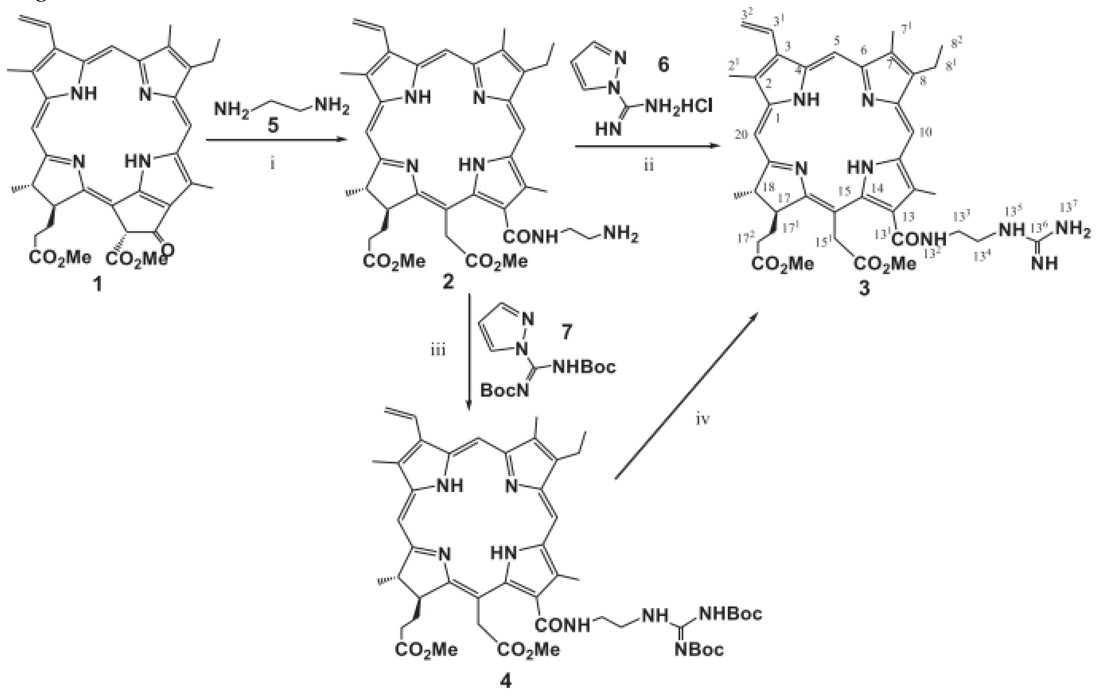
A group of Ying-Hua Gao researchers in the work “The photodynamic activities of dimethyl 13-[2-(guanidinyl)ethylamino] Chlorin e6 photosensitizers in A549 tumor” synthesized two new dimethyl 2-(guanidinyl)ethylamino chlorin e6 photosensitizers and investigated their effectiveness during PDT in A549 tumors. It has been shown that compounds 3 and 4 have a long absorption wavelength in the near infrared region and strong fluorescent radiation with a low photobleaching rate [170].
Chlorin e6 diaminoethylcarboxamide was obtained by the interaction of dimethylpheophorbide a with ethylenediamine in accordance with the Smith technique. The reaction mechanism is shown in Figure 5 [171]:
When amine 2 was guanidinylated with 1H-pyrazole-1-carboxomidine reagent 6, a guanidine conjugate of chloride e6 was obtained with a yield of 67%. Guanidine conjugate 3 can also be obtained in two stages of synthesis using the guanidinylation/de-protection sequence. In this synthesis method, guanidinylation of amine 2 using N,N0-di-Boc-1H-pyrazole-1-carboxomidine reagent 7 [172]
They showed lower cytotoxicity and higher photocytotoxicity in vitro compared to the well-known antitumor drug m-THPC in the in vitro MTT assay.
In the DCFDA analysis, it was found that intracellular generation is responsible for apoptotic cell death. Subcellular localization confirmed the site of damage to compounds 3 and 4 in PDT. These data suggest that two new photosensitizers may serve as potential photosensitizers to enhance the therapeutic effectiveness of PDT.
Compound 3 showed better tumor inhibition than compound 4. This may be related to the metabolism of compounds in mice. Subcellular localization confirmed that compounds 3 and 4 cause severe intracellular oxidative processes, photodynamic damage in lung cancer cells is manifested in the destruction of cellular organelles such as mitochondria, lysosomal and endoplasmic reticulum. Thus, compounds 3 and 4, which are shown in Figure 5, can be potential photosensitizers for photodiagnostics and photodynamic therapy of cancer [173].
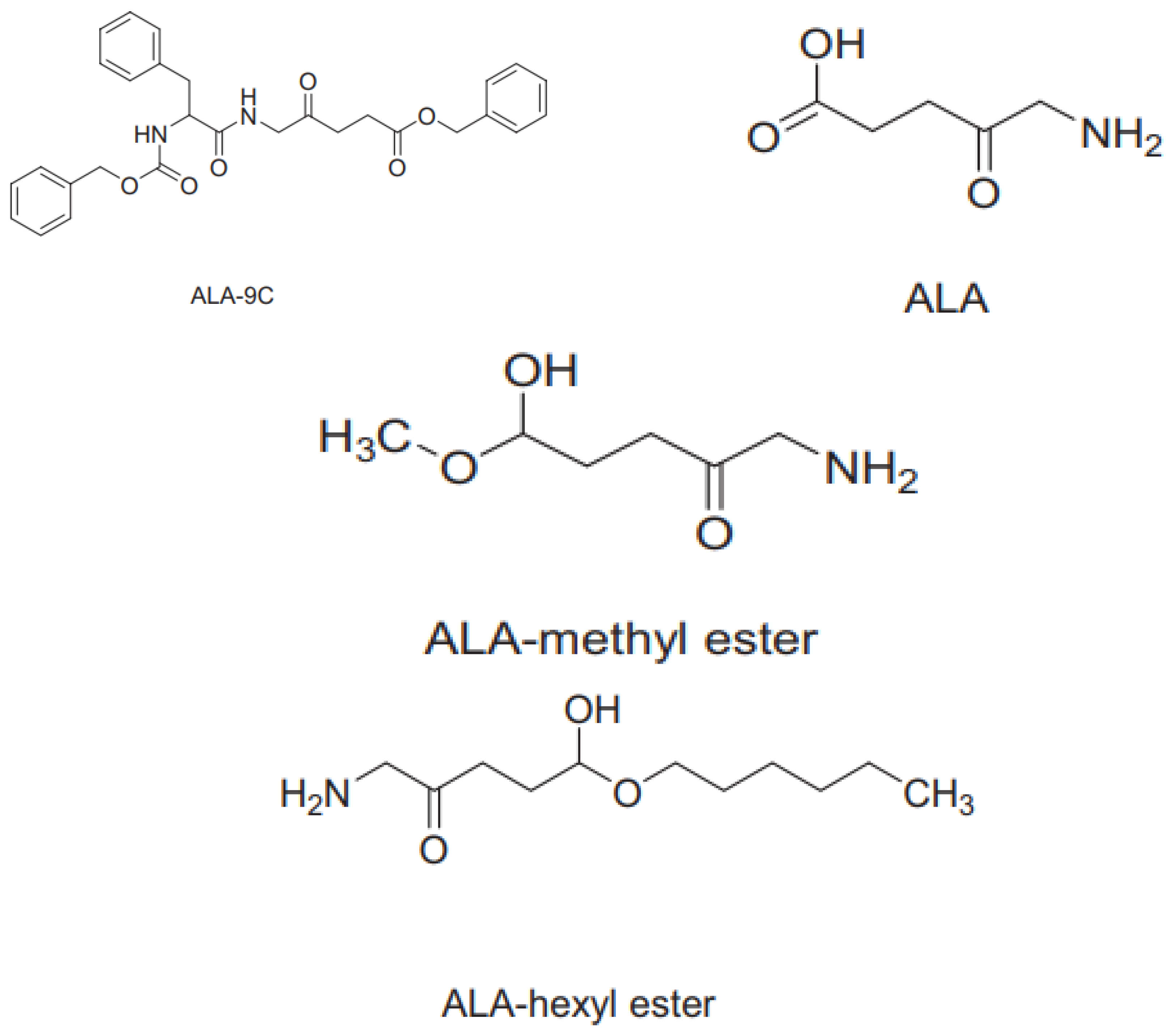
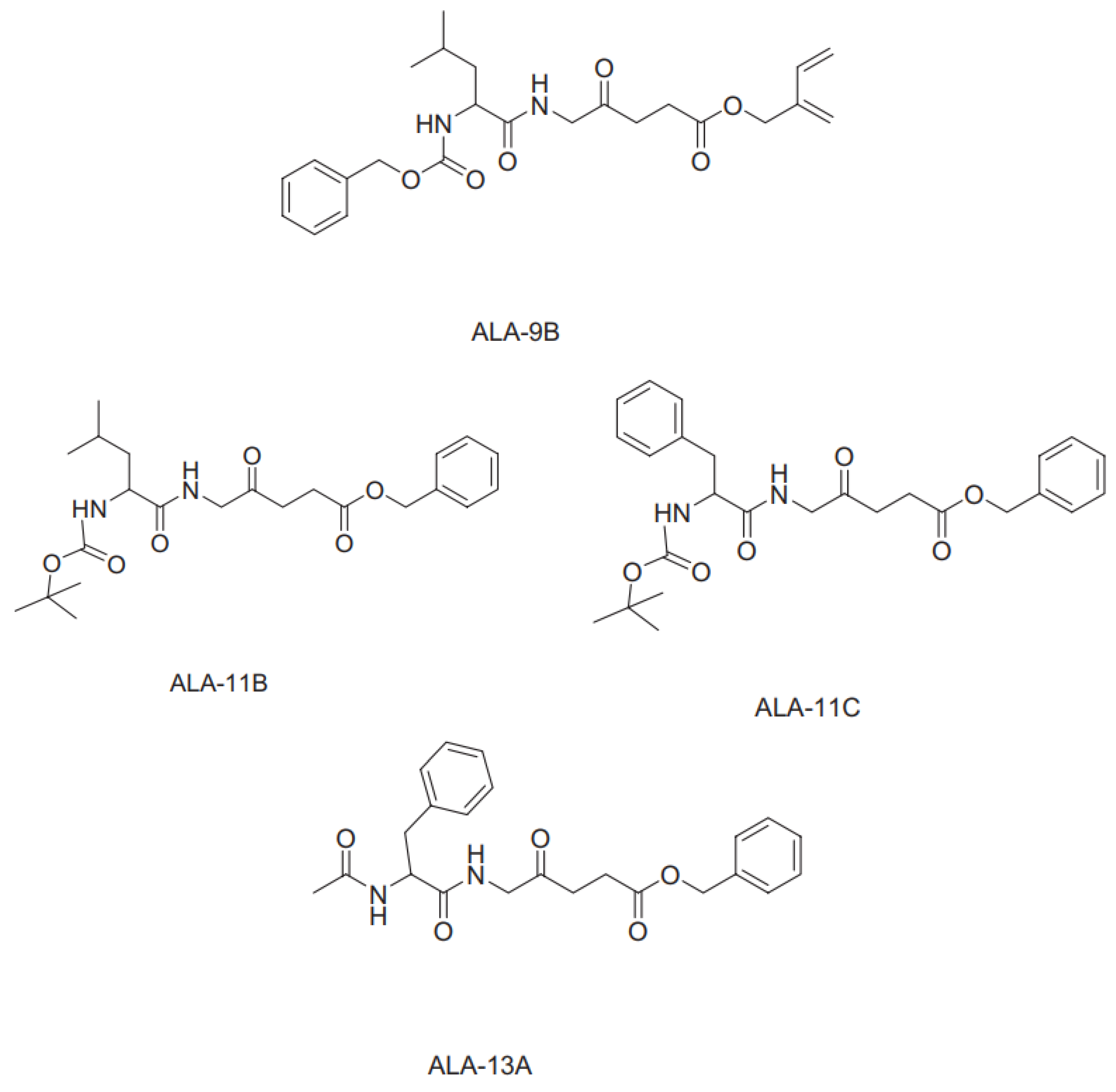
Faiza Sajjad and co-authors in their article “Evaluation of antimicrobial photodynamic activities of 5-aminolevulinic acid derivatives” evaluated the antimicrobial effect of ALA derivatives using photodynamic therapy. In this study, the authors evaluated the aPDT effect of various 5-ALA derivatives. In vivo and in vitro studies were conducted to determine antimicrobial activity. To test the antibacterial effect of drugs, as well as to detect any physiological changes in animal models after treatment, various small doses and different concentrations of drugs were used.
In vivo studies have shown that ALA-methyl ether, ALA-hexyl ether and ALA13A are powerful photosensitizers. In vitro studies evaluated wound healing rate, body weight and diet, and the results showed that ALA, ALA-methyl ether, ALA-hexyl ether and ALA-13A have good antibacterial properties, heal quickly and do not affect other physical parameters.
All compounds were synthesized and their structures can be seen in Figure 6. Ps was dissolved in DMSO, and the excitation wavelength of PS was selected in accordance with the absorption spectrum. The PS irradiation was carried out using a light system (Nd: YAG) consisting of laser radiation, the lamps of which are evenly distributed in the device to ensure uniform irradiation of the plate.
Both isolates were contained in Luria-Bertani-LB medium (trypton, yeast extract, NaCl) and agar in a solid medium. For experiments, these bacteria were individually seeded in 5 ml LB and grown aerobically overnight at a temperature of 37 °C. Each culture was collected after centrifugation at 671 g for 10 minutes, washed twice with sterile distilled water and suspended in PBS. Cell suspensions were standardized using a spectrophotometer calibrated at a wavelength of 600 nm [174].
Figure 6.
Structural formulas of synthesized substances.
5. Conclusion
In conclusion, the isolation and characterization of photosensitizing compounds from microalgae holds great promise for advancing the field of photodynamic therapy. These natural sources have been found to harbor a wealth of potent photosensitizers capable of selectively targeting and destroying cancer cells upon light activation, with some extracts demonstrating up to 95% reduction in cell viability.
The ongoing research efforts to identify, isolate, and elucidate the mechanisms of action of these microalgae-derived photosensitizers are crucial for unlocking their full potential in the clinical setting. By leveraging advanced analytical techniques and delving into the subcellular interactions and tumor responses, researchers are striving to enhance the efficacy and selectivity of photodynamic therapy, ultimately leading to improved treatment outcomes for patients.
As the field continues to evolve, the integration of microalgae-derived photosensitizers into clinical practice holds the promise of revolutionizing the management of a wide range of cancers and other medical conditions.
In addition, an important aspect is the ongoing deepening into the study of the mechanisms of action of these photosensitizers at the level of subcellular processes and interactions with the tumor. This line of research is aimed at improving the effectiveness and selectivity of photodynamic therapy. It is expected that such efforts will lead to improved patient outcomes, which is especially important in the context of a wide range of cancers and other pathologies, where the prospect of a revolutionary change in therapy approaches is already outlined.
Moreover, increasing attention is being paid not only to the identification and isolation of photosensitizers from microalgae, but also to understanding their effects at the molecular level. The use of advanced analytical methods allows researchers to delve deeper into the mechanisms of interaction of these compounds with cancer cells, which contributes to the development of more accurate and effective treatment strategies. Such efforts have the potential to revolutionize clinical practice and significantly improve the prognosis for patients suffering from various types of cancer and other serious diseases.
In addition, active research is aimed at optimizing the processes of extracting photosensitizers from microalgae in order to increase their purity and concentration. This is important to ensure the stability and predictability of the results of photodynamic therapy in clinical practice. The development of new methods of analysis and diagnosis also plays a key role in the further progress of this field, contributing to a deeper understanding of the mechanisms of action and optimization of therapeutic protocols.
Funding
This research is funded by the Committee of Science of the Ministry of Science and Education of the Republic of Kazakhstan (Grant No AP19677547 “Isolation of sensitizing substances used in photodynamic therapy from algae in reservoirs of Kazakhstan and study of their properties”).
List of Abbreviations
| PDT | photodynamic therapy |
| eg | exampleli gratia |
| 2-EP | 2-ethylpyridine |
| DPPH | 2,2-diphenyl-1-picrylhydrazyl radical. |
| 5-ALA | (5-aminolevulinic acid) |
| PS | photosensitizers |
| g | gramm |
| kg | kilogramm |
| etc | et cetera |
| ml | milliliter |
| i.e., | id est |
| ALA | alanine |
| DMSO | Dimethyl Sulfoxide |
| UV | ultraviolet |
| UV-Vis | ultraviolet-visible |
| PBS | Phosphate Buffered Saline |
| TLC | Thin Layer Chromatography |
| HPLC | High Performance Liquid Chromatography |
| DCFDA | Dichlorodihydrofluorescein diacetate |
References
- Jabeen et al., “Effect of the Photodynamic Therapy Applications with Potent Microalgae Constituents on Several Types of Tumor”.
- W. Pang et al., “Nucleolus-Targeted Photodynamic Anticancer Therapy Using Renal-Clearable Carbon Dots”.
- J. A. Miller et al., “Photodynamic therapy with the phthalocyanine photosensitizer Pc 4: The case experience with preclinical mechanistic and early clinical–translational studies”.
- Yoon, J. Li and Y. K. Shim, “Advance in Photosensitizers and Light Delivery for Photodynamic Therapy”.
- Saide, C. Lauritano and A. Ianora, “Pheophorbide a: State of the Art”.
- K. C. D. Andrade, C. Lauritano, G. Romano and A. Ianora, “Marine Microalgae with Anti-Cancer Properties”.
- “Photodynamic Therapy to Treat Cancer - NCI”.
- Y. Tian, L. L. Wang and W. Wang, “Progress in photodynamic therapy on tumors”.
- 9. T J Dougherty, C J Gomer, B W Henderson, G Jori, D Kessel, M Korbelik, J Moan, Q Peng Photodynamic therapy.
- Paul Harrod-Kim, “Tumor ablation with photodynamic therapy: introduction to mechanism and clinical applications.”.
- Conte, F. Ungaro, A. Mazzaglia and F. Quaglia, “Photodynamic Therapy for Cancer: Principles, Clinical Applications, and Nanotechnological Approaches”.
- Y. Qiao et al., “Engineered algae: A novel oxygen-generating system for effective treatment of hypoxic cancer”.
- T. J. Dougherty et al., “Photodynamic Therapy”.
- S. Cogno, P. Gilardi, L. R. Comini, S. C. Núñez-Montoya, J. L. Cabrera and V. Rivarola, “Natural photosensitizers in photodynamic therapy: In vitro activity against monolayers and spheroids of human colorectal adenocarcinoma SW480 cells”.
- K. L. M. Santos et al., “Prospective application of phthalocyanines in the photodynamic therapy against microorganisms and tumor cells: a mini-review.”.
- Y. Karakaş, H. T. Şahin, B. İnan, D. Özçimen and Y. Erginer, “In vitro cytotoxic activity of microalgal extracts loaded nano–micro particles produced via electrospraying and microemulsion methods”.
- S. Braune, A. Krüger-Genge, S. Kammerer, F. Jung and J. Küpper, “Phycocyanin from Arthrospira platensis as Potential Anti-Cancer Drug: Review of In Vitro and In Vivo Studies”.
- S. S. Kulthe, Y. Choudhari, N. Inamdar and V. Mourya, “Polymeric micelles: authoritative aspects for drug delivery”.
- R. R. Allison and K. Moghissi, “Photodynamic Therapy (PDT): PDT Mechanisms”.
- Mazzaglia, “Photodynamic Tumor Therapy with Cyclodextrin Nanoassemblies”.
- M. Olszowy, M. Nowak-Perlak and M. Woźniak, “Current Strategies in Photodynamic Therapy (PDT) and Photodynamic Diagnostics (PDD) and the Future Potential of Nanotechnology in Cancer Treatment”.
- L. E. Ibarra et al., “Selective Photo-Assisted Eradication of Triple-Negative Breast Cancer Cells through Aptamer Decoration of Doped Conjugated Polymer Nanoparticles”.
- Lima and L. V. Reis, “Photodynamic Therapy: From the Basics to the Current Progress of N-Heterocyclic-Bearing Dyes as Effective Photosensitizers”.
- P. Agostinis et al., “Photodynamic therapy of cancer: An update”.
- X. Wang, D. Luo and J. P. Basilion, “Photodynamic Therapy: Targeting Cancer Biomarkers for the Treatment of Cancers”.
- T. A. Mishchenko, I. V. Balalaeva, A. A. Gorokhova, M. V. Vedunova and D. V. Krysko, “Which cell death modality wins the contest for photodynamic therapy of cancer?”.
- V. Straten, V. Mashayekhi, H. S. D. Bruijn, S. Oliveira and D. J. Robinson, “Oncologic Photodynamic Therapy: Basic Principles, Current Clinical Status and Future Directions”.
- T. Nelius, W. D. Riese and S. Filleur, “Photodynamic therapy: a promising alternative in oncology”.
- U. Chilakamarthi and L. Giribabu, “Photodynamic Therapy: Past, Present and Future”.
- C. Serra et al., “A look at clinical applications and developments of photodynamic therapy”.
- T. Tran et al., “Identification of Small Molecule Modulators of Gene Transcription with Anticancer Activity”.
- Paul Harrod-Kim “Tumor ablation with photodynamic therapy”.
- Thomas J. Dougherty, Charles J. Gomer, Barbara W. Henderson, Giulio Jori, David Kessel, Mladen Korbelik, Johan Moan, and Qian Peng* “Photodynamic Therapy.
- Chun-Yan Wang “Photosensitization of phycocyanin extracted from Microcystis in human hepatocellular carcinoma cells: implication of mitochondria-dependent apoptosis.”.
- D C Shackley, “Photodynamic therapy”.
- C. J. Gomer, A. Ferrario, M. Luna, N. Rucker and S. Wong, “Photodynamic therapy: Combined modality approaches targeting the tumor microenvironment”.
- S. Kwiatkowski et al., “Photodynamic therapy – mechanisms, photosensitizers and combinations”.
- H. Barr, C. Kendall, J. Reyes-Goddard and N. Stone, “Clinical Aspects of Photodynamic Therapy”.
- J. Zhang, C. Jiang, J. P. F. Longo, R. B. Azevedo, H. Zhang and L. A. Muehlmann, “An updated overview on the development of new photosensitizers for anticancer photodynamic therapy”.
- Qian Peng, Johan Moan, Jahn M Nesland, Peng n.d. “Correlation of subcellular and intratumoral photosensitizer localization with ultrastructural features after photodynamic therapy.”.
- Magdalena Cañete, Universidad Autónoma de Madrid, Cantoblanco, “Preclinical photodynamic therapy research in Spain. 3. Localization of photosensitizers and mechanisms of cell death in vitro”.
- D. Chen, C. A. Dougherty, K. Zhu and H. Hong, “Theranostic applications of carbon nanomaterials in cancer: Focus on imaging and cargo delivery”.
- T. Schluep et al., “Pharmacokinetics and tumor dynamics of the nanoparticle IT-101 from PET imaging and tumor histological measurements”.
- W. Park et al., “Advanced smart-photosensitizers for more effective cancer treatment”.
- D. Kessel, “Correlation between subcellular localization and photodynamic efficacy”.
- P. Mróz, A. Yaroslavsky, G. B. Kharkwal and M. R. Hamblin, “Cell Death Pathways in Photodynamic Therapy of Cancer”.
- Xiao Yan He, “Effectiveness of photosensitive dye during uptake and redistribution”.
- L. Yang et al., “Boosting the photodynamic therapy efficiency with a mitochondria-targeted nanophotosensitizer”.
- S. Sansaloni-Pastor, J. Bouilloux and N. Lange, “The Dark Side: Photosensitizer Prodrugs”.
- C. Constantin and M. Neagu, “Photosensitizers Imprinting Intracellular Signaling Pathways in Dermato-Oncology Therapy”.
- T. M. Busch, “Local physiological changes during photodynamic therapy”.
- “Photodynamic Therapy to Treat Cancer”.
- S. Kumari, A. K. Badana, M. M. G, G. Shailender and R. Malla, “Reactive Oxygen Species: A Key Constituent in Cancer Survival”.
- P. Jia, C. Dai, P. Cao, D. I. Sun, R. Ouyang and Y. Miao, “The role of reactive oxygen species in tumor treatment”.
- W. H. Ahsan, “Reactive oxygen species: role in the development of cancer and various chronic conditions”.
- Yu, J. Yan, Z. Li, L. Yang, F. Ju and Y. Sun, “Recent trends in emerging strategies for ferroptosis-based cancer therapy”.
- J. P. Fruehauf and F. L. Meyskens, “Reactive Oxygen Species: A Breath of Life or Death?”.
- T. Hu, Z. Wang, W. Shen, R. Liang, D. Yan and M. Wei, “Recent advances in innovative strategies for enhanced cancer photodynamic therapy”.
- S. Mallidi, S. Anbil, A. Bulin, G. Obaid, M. Ichikawa and T. Hasan, “Beyond the Barriers of Light Penetration: Strategies, Perspectives and Possibilities for Photodynamic Therapy”.
- Shannon M Gallagher-colombo, Amanda L Maas, “Photodynamic therapy-induced angiogenic signaling: consequences and solutions to improve therapeutic response.”.
- R. L. Yanovsky, D. W. Bartenstein, G. S. Rogers, S. J. Isakoff and S. T. Chen, “Photodynamic therapy for solid tumors: A review of the literature”.
- C P Lowdell, “Photodynamic therapy: an update.”.
- N. Shah et al., “Deep-Tissue Activation of Photonanomedicines: An Update and Clinical Perspectives”.
- “Advances in Photodynamic Therapy of Cancer Bentham Science”.
- D. L. Sai, J. Lee, D. L. Nguyen and Y. Kim, “Tailoring photosensitive ROS for advanced photodynamic therapy”.
- R. K. Pandey et al., “Nature: A rich source for developing multifunctional agents. tumor-imaging and photodynamic therapy”.
- Agnieszka Szurko “Spectroscopic and biological studies of a novel synthetic chlorin derivative with prospects for use in PDT.”.
- Z. Zhuo, Z. Song, Z. Ma, Y. Zhang, G. Xu and G. Chen, “Chlorophylline6-mediated photodynamic therapy inhibits proliferation and induces apoptosis in human bladder cancer cells”.
- 69. Shaikh Abdur Razzak Comprehensive overview of microalgae-derived carotenoids and their applications in diverse industries.
- 70. Po-Fung Ip, Feng Chen Production of astaxanthin by the green microalga Chlorella zofingiensis in the dark.
- Mafalda Trovão, Lucas Cardoso, Lisa Schüler, Adriana Machado, Gonçalo Espírito Santo, Humberto Pedroso, Ana Reis, Ana Barros, Nádia Correia, Monya Costa, Sara Ferreira, Helena Cardoso, Marília Mateus, Joanam Silva, Hugo Pereira, Filomena Freitas, João Varela. Oxyfluorfen: a novel metabolic inhibitor to select microalgal chlorophyll-deficient mutant strains for nutritional applications.
- T. Lafarga Effect of microalgal biomass incorporation into foods: nutritional and sensorial attributes of the end products.
- Andrêssa S. Fernandes, Patrícia A. Caetano, Eduardo Jacob-Lopes, Leila Queiroz Zepka, Veridiana Vera de Rosso. Alternative green solvents associated with ultrasound-assisted extraction: A green chemistry approach for the extraction of carotenoids and chlorophylls from microalgae.
- Arti Sharma, Meenu Chhabra, Shashi Kumar. Performance evaluation of genetically modified microalgae in photosynthetic microbial fuel cells for carotenoids and power generation.
- Yamini Sumathi, Prashant Kumar, Reeta Rani Singhania, Chiu-Wen Chen, Baskar Gurunathan, Cheng-Di Dong, Anil Kumar Patel. Harnessing Fe3O4 nanoparticles for sustainable harvesting of astaxanthin-producing microalgae: Advancing industrial-scale biorefinery.
- Yanlong Gu, Michelle Yee Mun Teo, Lionel Lian Aun In, Kazuya Shimizu, Kyu-Jung Chae, Tran Thi Ngoc Thu, Kuan Shiong Khoo. Genetic engineering of Haematococcus pluvialis microalgae for the enhancement of astaxanthin production.
- Zengyu Yu, Weiyang Zhao, Han Sun, Haijin Mou, Jin Liu, Hui Yu, Lei Dai, Qing Kong, Shufang Yang. Phycocyanin from microalgae: A comprehensive review covering microalgal culture, phycocyanin sources and stability.
- Shuyu Liu, Zitong Wu, Xin Min, Hong Liu, Nijuan Nian, Pei Zhang, Xiaoyu Li. Synergism Variation between intracellular Glutathione, phycocyanin and SOD in microalgae by carbon quantum dot fluorescence.
- 79. Israel Emiezi Agarry, Desheng Ding, Yunchang Li, Zihan Jin, Huiling Deng, Jiang Hu, Tian Cai, Jianquan Kan, Kewei Chen In vitro bioaccessibility evaluation of chlorophyll pigments in single and binary carriers.
- Eric Biehler, Anouk Kaulmann, Lucien Hoffmann, Elmar Krause, Torsten Bohn. Dietary and host-related factors influencing carotenoid bioaccessibility from spinach (Spinacia oleracea).
- Kewei Chen, María Roca. In vitro digestion of chlorophyll pigments from edible seaweeds.
- Zhuo Chen, Jiu-Qiang Xiong. Recovery mechanism of a microalgal species, Chlorella sp. from toxicity of doxylamine: Physiological and biochemical changes, and transcriptomics.
- Nathanan Preechaphonkul, Sukrit Sirikwanpong, Cherdsak Maneeruttanarungroj. Freshwater green alga Chlorella sp. KLSc59 produced all forms of omega-3 oil: ALA, EPA, and DHA.
- Gökhun Çağatay Erbil, Mahmut Elp, Yaşar Durmaz. Myo-inositol as a carbon source in Chlorella sp. production.
- Vaibhav Sunil Tambat, Anil Kumar Patel, Reeta Rani Singhania, Akash Pralhad Vadrale, Archana Tiwari, Chiu-Wen Chen, Cheng-Di Dong. Sustainable mixotrophic microalgae refinery of astaxanthin and lipid from Chlorella zofingiensis.
- Yamini Sumathi, Prashant Kumar, Reeta Rani Singhania, Chiu-Wen Chen, Baskar Gurunathan, Cheng-Di Dong, Anil Kumar Patel. Harnessing Fe3O4 nanoparticles for sustainable harvesting of astaxanthin-producing microalgae: Advancing industrial-scale biorefinery.
- Dong-Yeon Kim, Durairaj Vijayan, Ramasamy Praveenkumar, Jong-In Han, Kyubock Lee, Ji-Yeon Park, Won-Seok Chang, Jin-Suk Lee, You-Kwan Oh. Cell-wall disruption and lipid/astaxanthin extraction from microalgae: Chlorella and Haematococcus.
- Birgitta Narindri Rara Winayu, Kuan-Ya Chiu, Hsin-Ta Hsueh, Hsin Chu. Thermosynechococcus sp. CL-1 (TCL-1) as an efficient cyanobacterium in CO2 fixation, C-phycocyanin production, and removal of Cd and Pb.
- Birgitta Narindri Rara Winayu, Yu-Ting Lin, Hsin-Ta Hsueh, Hsin Chu. Importance of lighting color and period for CO2 fixation and C-phycocyanin production during Thermosynechococcus sp. CL-1 growth.
- Birgitta Narindri Rara Winayu, Hsin-Ta Hsueh, Hsin Chu. CO2 fixation and cultivation of Thermosynechococcus sp. CL-1 for the production of phycocyanin.
- F. D. Santos et al., “Distinct photo-oxidation-induced cell death pathways lead to selective killing of human breast cancer cells”.
- Moserová and J. Králová, “Role of ER Stress Response in Photodynamic Therapy: ROS Generated in Different Subcellular Compartments Trigger Diverse Cell Death Pathways”.
- Y. Adar et al., “Imidazoacridinone-dependent lysosomal photodestruction: a pharmacological Trojan horse approach to eradicate multidrug-resistant cancers”.
- W. Jiang, M. Liang, Q. Lei, G. Li and S. Wu, “The Current Status of Photodynamic Therapy in Cancer Treatment”.
- S. Sharma, P. Mróz, T. Dai, Y. Huang, T. G. S. Denis and M. R. Hamblin, “Photodynamic Therapy for Cancer and for Infections: What Is the Difference?”.
- M. Zahra, A. Chota, H. Abrahamse and B. P. George, “Efficacy of Green Synthesized Nanoparticles in Photodynamic Therapy: A Therapeutic Approach”.
- J. Nyst, I. B. Tan, F. Stewart and A. J. M. Balm, “Is photodynamic therapy a good alternative to surgery and radiotherapy in the treatment of head and neck cancer?”.
- L. Onofrejová et al., “Bioactive phenols in algae: The application of pressurized-liquid and solid-phase extraction techniques”.
- M. Plaza, M. Herrero, A. Cifuentes and E. Ibáñez, “Innovative natural functional ingredients from microalgae.”.
- Jesus, M. Correia-da-Silva, C. Afonso, M. Pinto and H. Cidade, “Isolation and Potential Biological Applications of Haloaryl Secondary Metabolites from Macroalgae”.
- Saide, K. A. Martínez, A. Ianora and C. Lauritano, “Unlocking the Health Potential of Microalgae as Sustainable Sources of Bioactive Compounds”.
- P. Abreu, R. Martins and J. Nunes, “Emerging Applications of Chlorella sp. and Spirulina (Arthrospira) sp.”.
- G. A. Colusse, J. Carneiro, M. E. R. Duarte, J. C. D. Carvalho and M. D. Noseda, “Advances in microalgal cell wall polysaccharides: a review focused on structure, production, and biological application”.
- M. Cañete, J. C. Stockert and A. Villanueva, “Preclinical photodynamic therapy research in Spain. 3. Localization of photosensitizers and mechanisms of cell death in vitro”.
- R. Wang, X. Li and J. Yoon, “Organelle-Targeted Photosensitizers for Precision Photodynamic Therapy”.
- S. P. M. Ventura et al., “Extraction of value-added compounds from microalgae”.
- G. Perin, E. Cimetta, F. Monetti, T. Morosinotto and F. Bezzo, “Novel micro-photobioreactor design and monitoring method for assessing microalgae response to light intensity”.
- Pulz and P. Kretschmer, “Perspectives of phototrophic microorganisms in environment protection and ecology”.
- R. Calori, B. Hong and A. C. Tedesco, “Expanding the Limits of Photodynamic Therapy: The Design of Organelles and Hypoxia-Targeting Nanomaterials for Enhanced Photokilling of Cancer”.
- “Chromatography Adsorbents - Silica Gel and Aluminium Oxide”.
- Saitoh, I. Awaka and N. Suzuki, “Determination of chlorophylls by reversed-phase high-performance liquid chromatography with isocratic elution and the column-switching technique”.
- H. Inoue, K. Furuya, K. Watanabe, K. Tanaka, T. Shirai and E. Miyoshi, “Separation and Determination of Copper(II) Chlorophylls by Reversed-Phase High Performance Liquid Chromatography”.
- W. Zheng, N. Thorne and J. C. McKew, “Phenotypic screens as a renewed approach for drug discovery”.
- S. Kraljević, P. J. Stambrook and K. Pavelić, “Accelerating drug discovery”.
- “Discovery and Development”.
- N. Suvorov, V. Pogorilyy, E. Diachkova, Y. Vasil’ev, А. Ф. Мирoнoв and M. A. Grin, “Derivatives of Natural Chlorophylls as Agents for Antimicrobial Photodynamic Therapy”.
- A. H. A. Akhras, “Introducing the Effect of Chinese Chlorella as a Photosensitizing Drug at Different Temperatures”.
- G. Fasya, N. Millati, L. M. Rahmawati, R. Iyani, A. Hanapi, R. Ningsih, D. Yuliani и D. S. Megawati Isolation and bioactivity of steroids isolates from petroleum ether fraction of Chlorella sp.
- N. Meyer, N. R. Ferrigni, J. E. Putnarn, L. B. Jacobsen, D. E. Nichols, and J. L. McLaughlin, Planta Medica 45 (5), 31–34 (1982).
- G. Fasya, A. R. Dinasti, S. M. Syofiyah, L. M. Rahmawati, N. Millati, D. A. Safitri, S. Handoko, A. Hanapi, and R. Ningsih, ALCHEMY: Journal of Chemistry 5 (1), 5–9 (2016).
- F. Aprelia and Suyatno, UNESA Journal of Chemistry 2 (3), 94–99 (2013).
- D. Astuti, A. Maulana, and E. M. Kuntowati, Prosiding Seminar Nasional Kimia Universitas Negeri Surabaya, (Universitas Negeri Surabaya, 2014).
- Yousef Y. Sultan 1 , Diaa A. Marrez Isolation and Purification of Antifungal Compounds from the Green Microalga Chlorella vulgaris.
- Marrez DA, Naguib MM, Sultan YY, Daw ZY, Zaher SS, Higazy AM. Phytoplankton profile and toxicity assessment of dominant algal species from different Egyptian aquatic ecosystems. Res J Pharm Biol Chem Sci. 2016;7(2):1453-61.
- Yang J, Guo J, Yuan J. In vitro antioxidant properties of rutin. LWT Food Sci Technol. 2008;41(6):1060-6. [CrossRef]
- Prabhadevi V, Sahaya SS, Johnson M, Venkatramani B, Janakiraman N. Phytochemical studies on Allamanda cathartica L. using GC–MS. Asian Pac J Trop Biomed. 2012;2(2):S550-4. [CrossRef]
- Mujeeb F, Bajpai P, Pathak N. Phytochemical evaluation, antimicrobial activity, and determination of bioactive components from leaves of Aegle marmelos. Biomed Res Int. 2014;2014:497606. [CrossRef]
- Senthilkumar P, Sambath R, Vasantharaj S. Antimicrobial potential and screening of antimicrobial compounds of Ruellia tuberose using GC-MS. Int J Pharm Sci Rev Res. 2013;20(1):184-9.
- Marrez DA, Sultan YY. Antifungal activity of the cyanobacterium Microcystis aeruginosa against mycotoxigenic fungi. J Appl Pharm Sci. 2016;6(11):191- 8. [CrossRef]
- Sultan YY, Ali MA, Darwesh OM, Embaby MA, Marrez DA. Influence of nitrogen source in culture media on antimicrobial activity of Microcoleus lacustris and Oscillatoria rubescens. Res J Pharm Biol Chem Sci. 2016;7(2):1444-52.
- Marrez, DA, Sultan YY, Embaby MA. Biological activity of the cyanobacterium Oscillatoria brevis extracts as a source of nutraceutical and bio-preservative agents. Int J Pharmacol. 2017;13(8):1010-19. [CrossRef]
- Marrez DA, Naguib MM, Sultan YY, Higazy AM. Antimicrobial and anticancer activities of Scenedesmus obliquus metabolites. Heliyon. 2019;5(3):e01404. doi:10 .1016/j.heliyon.2019.e01404.
- Marrez DA, Sultan YY, Naguib MM, Higazy AM. Antimicrobial Activity, Cytotoxicity and Chemical Constituents of the Freshwater Microalga Oscillatoria princeps. Biointerface Res Appl Chem. 2022, 12(1):961- 77. [CrossRef]
- Ordog V, Stirk WA, Lenobel R, Bancířová M, Strnad M, Van Staden J, et al. Screening microalgae for some potentially useful agricultural and pharmaceutical secondary metabolites. J Appl Phycol. 2004;16(4):309- 14. [CrossRef]
- Khosravi K. The potential Health Benefits of Atgae and Micro Algae in Medicine: A Review on Spirulina Platensis. Curr Nutr Food Sci. 2011;7(4):279-85. [CrossRef]
- Hosseini S, Shahbazizadeh S, Khosravi-Darani K, Mozafari M. Spirulina paltensis: Food and Function. Curr Nutr Food Sci. 2013;9(3):189-93. [CrossRef]
- Shinya Shibata, Chiyoko Ishihara, Keisuke Matsumoto. Improved separation method for highly purified lutein from Chlorella powder using jet mill and flash column chromatography on silica gel.
- Matsuno, T. Structure and characterization of carotenoids from various habitats and natural sources. Methods Enzymol. 1992, 213, 22-31.
- Goodwin, T. W.; Britton, G. Distribution and analysis of carotenoids. In Plant Pigments; Goodwin, T. W., Ed.; Academic Press: London, 1988; pp 61-127.
- Ichioka, M.; Endo, H. Effect of light on cellular carotenoids formation of Chlorella regularis S-50 grown on glucose (in Japanese). Annu. Rep. Yakult Central Inst. Microbiol. Res. 1974, 5, 91-99.
- Yen-Ju Lee, Ying-Chen Yi, Yu-Chieh Lin, Chao-Chung Chen, Jia-Horung Hung, Jia-Yi Lin,I-Son Ng. Purification and biofabrication of 5-aminolevulinic acid for photodynamic therapy against pathogens and cancer cells.
- Armbruster CE, Mobley HL. Merging mythology and morphology: the multifaceted lifestyle of Proteus mirabilis. Nat Rev Microbiol. 2012;10:743–754. [CrossRef]
- Bunke A, Zerbe O, Schmid H, Burmeister G, Merkle HP, Gander B. Degradation mechanism and stability of 5-aminolevulinic acid. J Pharm Sci. 2000;89:1335–1341. [CrossRef]
- Cai J, Zheng Q, Huang H, Li B. 5-aminolevulinic acid mediated photodynamic therapy inhibits survival activity and promotes apoptosis of A375 and A431 cells. Photodiagnosis Photodyn Ther. 2018;21:257–262. [CrossRef]
- Chen H, Jiang JG. Toxic effects of chemical pesticides (trichlorfon and dimehypo) on Dunaliella salina. Chemosphere. 2011;84:664–670. [CrossRef]
- Di Venosa G, Fukuda H, Perotti C, Batlle A, Casas A. A method for separating ALA from ALA derivatives using ionic exchange extraction. J Photochem Photobiol B. 2004;75:7–163. [CrossRef]
- Din, Lim SJ, Maskat MY, Abd Mutalib S, Zaini NAM. Lactic acid separation and recovery from fermentation broth by ion-exchange resin: a review. Bioresour Bioprocess. 2021;8:1–23. [CrossRef]
- Feng Lianga, Xueying Anc, Ruoxi Wang, Wenshu Wu, Lin Yang , Yixin Zheng, Qing Jiang, Xingquan Xu, Danni Zhong, Min Zhou. Microalgae-based drug delivery system for tumor microenvironment photo-modulating and synergistic chemo-photodynamic therapy of osteosarcoma.
- Bing-Chung Liau, Chun-Ting Shen, Fong-Ping Liang, Siang-En Hong, Shih-Lan Hsu, Ting-Ting Jong, Chieh-Ming J. Chang. Supercritical fluids extraction and anti-solvent purification of carotenoids from microalgae and associated bioactivity.
- M.M. Rebolloso-Fuentes, A. Navarro-Perez, F. Garcia-Camacho, J.J. RamosMiras, J.L. Guil-Guerrero, Biomass nutrient profiles of the microalga Nannochloropsis, J. Agricultural Food Chemistry 49 (2001) 2966–2972.
- T.L. Walker, S. Purton, D.K. Becker, C. Collet, Microalgae as bioreactors, Plant Cell Reports 24 (2005) 629–641.
- P.Z. Margalith, Production of ketocarotenoids by microalgae, Appl. Microbiol. Biotechnol. 51 (1999) 431–438.
- Herrero, A. Cifuentes, E. Ibanez, Sub- and supercritical fluid extraction ˜ of functional ingredients from different natural sources: plants, foodby-products, algae and microalgae: a review, Food Chem. 98 (2006) 136–148.
- L. Rodolfi, G. Chini Zittelli, N. Bassi, G. Padovani, N. Biondi, G. Bonini,M.R. Tredici, Microalgae for oil: strain selection, induction of lipid synthesis and outdoor mass cultivation in a low-cost photobioreactor, Biotechnol. Bioeng. 102 (2009) 100–112.
- Bhosale, Environmental and cultural stimulants in the production of carotenoids from microorganisms, Appl. Microbiol. Biotechnol. 63 (2004) 351–361.
- M.D. Macias-Sanchez, C. Mantell Serrano, M. Rodriguez Rodriguez, E. Martinez de la Ossa, L.M. Lubian, O. Montero, Extraction of carotenoids and chlorophyll from microalgae with supercritical carbon dioxide and ethanol as cosolvent, J. Sep. Sci. 31 (2008) 1352–1362.
- C.J. Chang, A.D. Randolph, Precipitation of microsize organic particles from supercritical fluids, AIChE J. 35 (1989) 1876–1882.
- 158. Victor Abrahamsson, Irene Rodriguez-Meizoso, Charlotta Turner Determination of carotenoids in microalgae using supercritical fluid extraction and chromatography.
- A.V. Rao, L.G. Rao, Pharmacological Research 55 (2007) 207.
- B.D. Ribeiro, D.W. Barreto, M.A.Z. Coelho, Food and Bioprocess Technology 4 (2011) 693.
- Rodríguez-Bernaldo de Quirós, H.S. Costa, Journal of Food Composition and Analysis 19 (2006) 97.
- L.C. Sander, K.E. Sharpless, M. Pursch, Journal of Chromatography A 880 (2000) 189.
- Su, K.G. Rowley, N.D.H. Balazs, Journal of Chromatography B: Analytical Technologies in the Biomedical and Life Sciences 781 (2002) 393.
- J. Oliver, A. Palou, Journal of Chromatography A 881 (2000) 543.
- C.J. Welch, N. Wu, M. Biba, R. Hartman, T. Brkovic, X. Gong, R. Helmy, W. Schafer, J. Cuff, Z. Pirzada, L. Zhou, TrAC - Trends in Analytical Chemistry 29 (2010) 667.
- M.D. Macías-Sánchez, C. Mantell, M. Rodríguez, E. Martínez de la Ossa, L.M. Lubián, O. Montero, Journal of Supercritical Fluids 39 (2007) 323.
- Sonja Srdanovic , Ying-Hua Gao, Dan-Ye Chen, Yi-Jia Yan, Davor Margetic, Zhi-Long Chen. The photodynamic activity of 13-[2-(2-pyridyl)ethylamine] Сhlorin e6 photosensitizer in human esophageal cancer.
- Briš A, Marinic. Zˇ , Chen ZL, Margetic. D. Synthesis of chlorins by Diels-Alder cycloadditions of pheophorbide a and its derivatives. Synlett. 2015;26:991–994.
- Belykh DV, Kopylov EA, Gruzdev IV, Kuchin AV. Opening of the extra ring in pheophorbide a methyl ester by the action of amines as a one-step method for introduction of additional fragments at the periphery of chlorin macroring. Russ J Org Chem. 2010;46:577–585.
- Ying-Hua Gao, Vanda Lovrekovic, Akmaral Kussayeva, Dan-Ye Chen, Davor Margetic, Zhi-Long Chen. The photodynamic activities of dimethyl 13-[2-(guanidinyl)ethylamino] Сhlorin e6 photosensitizers in A549 tumor.
- R.G.W. Jinadasa, X. Hu, M.G.H. Vicente, K.M. Smith, Synthesis and cellular investigations of 173-, 152- and 131- amino acid derivatives of Chlorin e6, J. Med. Chem. 54 (2011) 7464-7476.
- M. Dud, Z. Glasovac, D. Margetic. The utilization of ball-milling in synthesis of aryl guanidines through guanidinylation and N-Boc-deprotection sequence, Tetrahedron 75 (2019) 109-115.
- 173. Ying-Hua Gao, Vanda Lovrekovi, Akmaral Kussayeva, Dan-Ye Chen, Davor Margetic, Zhi-Long Chen, The photodynamic activities of dimethyl 131 -[2-(guanidinyl) ethylamino] chlorin e6 photosensitizers in A549 tumor.
- 174. Автoры Faiza Sajjad, Ning-Ning Sun, Ting Chen, Yi-Jia Yan, Davor Margetić, Zhi-Long Chen «Evaluation of antimicrobial photodynamic activities of 5-aminolevulinic acid derivatives».
Figure 2.
Sensitizing substances released from microalgae.

Figure 3.
Structural formulas of the main sensitizers isolated from microalgae.
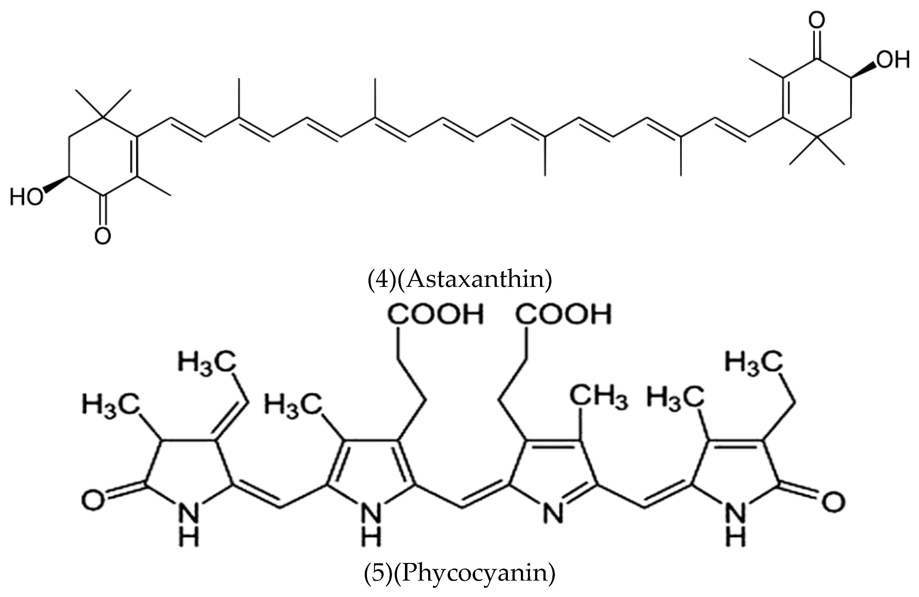
Figure 4.
Synthesis of Chlorin A.
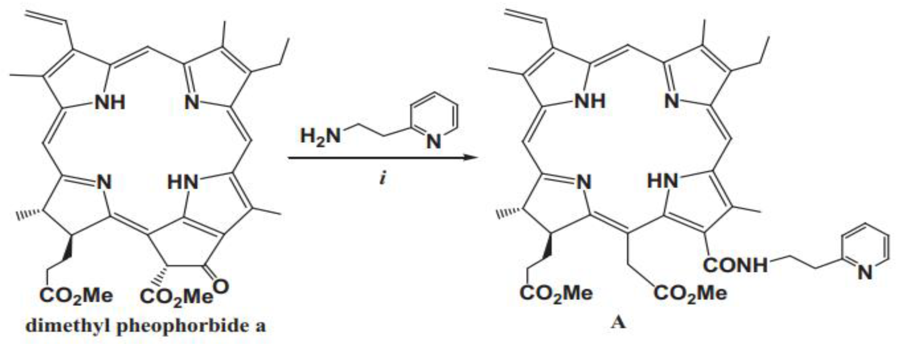
Figure 5.
Obtaining Chlorin e6 by the Smith method.
Disclaimer/Publisher’s Note: The statements, opinions and data contained in all publications are solely those of the individual author(s) and contributor(s) and not of MDPI and/or the editor(s). MDPI and/or the editor(s) disclaim responsibility for any injury to people or property resulting from any ideas, methods, instructions or products referred to in the content. |
© 2024 by the authors. Licensee MDPI, Basel, Switzerland. This article is an open access article distributed under the terms and conditions of the Creative Commons Attribution (CC BY) license (http://creativecommons.org/licenses/by/4.0/).
Copyright: This open access article is published under a Creative Commons CC BY 4.0 license, which permit the free download, distribution, and reuse, provided that the author and preprint are cited in any reuse.


