Submitted:
19 July 2024
Posted:
22 July 2024
You are already at the latest version
Abstract
Despite effective vaccines, yellow fever outbreaks persist, increasing the urgency to find antivirals for patient treatment. This study aimed to discover compounds with potential antiviral properties against the wild-type yellow fever virus (wt-YFV) in Hippeastrum puniceum, of the Amaryllidaceae family, through bioassay-guided fractionation of the crude bulb extract using ultra-high-performance liquid chromatography (UHPLC) coupled with high-resolution ESI-QTOF mass spectrometry and subsequent analysis of the pharmacokinetic and toxicological properties (ADMET) of the annotated compounds. In the active fractions against wt-YFV, six alkaloids were proposed such as bulbisine, cathinone, trigonelline, tetrahydroharman-3-carboxylic acid, and 2,7-dimethoxyhomolycorine (or 3-O-acetylnarcissidine). Co-occurring in these fractions were the amino acids arginine, asparagine, tryptophan, and glutamic acid. In silico ADMET analysis of the alkaloids predicted good absorption, distribution, excretion, favorable metabolization profiles, and non-mutagenic toxicity, indicating their potential for drug development. These findings justify further in vitro assessment of the compounds against YFV and in vivo testing in animal models. This strategy seems to be an efficient approach for discovering antiviral compounds against YFV. Such discoveries hold promise not only in combating YFV but also in addressing a spectrum of other flaviviruses.
Keywords:
antivirals
; wild-type yellow fever virus
; Hippeastrum puniceum
; natural compound
; bioassay-guided fractionation
; UHPLC-HRMS/MS
; In silico ADMET prediction
1. Introduction
Yellow fever (YF) is a non-contagious acute febrile illness of short duration and variable severity caused by the yellow fever virus (YFV), one of the most dangerous arboviruses still circulating in several countries [1]. The YFV is transmitted to humans through the bite of infected Aedes aegypti mosquitoes, responsible for its urban transmission cycle. Moreover, Haemagogus and Sabethes mosquitoes are accountable for its sylvatic cycle [2].
It is worth mentioning that the YFV was renamed by the International Committee on Taxonomy of Viruses (ICTV) in 2023 as Orthoflavivirus flavi [3,4]. Currently, 47 countries, including Brazil, are considered endemic for the disease [5]. It is estimated that the actual number of yellow fever cases is 10 to 250 times greater than the number of reported cases, due to underreporting. This underreporting may be due to asymptomatic infections or mild infections with nonspecific symptoms [5,6].
Despite the availability of an effective and cost-free yellow fever vaccine in Brazil since 1939, some areas still have low distribution and vaccination coverage, which favors yellow fever outbreaks [7]. In addition, a considerable percentage of individuals cannot be vaccinated against yellow fever, including those allergic to egg proteins present in the 17DD vaccine formulation used in Brazil, and immunosuppressed patients. [8,9,10].
Approximately 15% of individuals with symptomatic yellow fever develop the toxic form of the disease, known as "hepatorenal hemorrhagic syndrome," which is associated with a high mortality rate ranging from 40% to 70%. Given the absence of specific treatment for yellow fever, there is an urgent need to develop antiviral drugs to reduce the viral load in patients [8,11].
Medicinal plants are promising sources for discovering and developing sustainable, efficient, and cost-effective alternatives against viral infections. Crude plant extracts may contain a variety of secondary metabolites with bioactivity. Therefore, bioassay-guided fractionation of these extracts has proven to be an advantageous strategy for identifying bioactive compounds, aiming at developing new drugs, including antivirals [12].
The Amaryllidaceae family has garnered significant attention for its alkaloids, which are notable for their valuable pharmacological properties [13]. Bulbs of some species belonging to this family are used in traditional medicine as emetics, purgatives, wound healers, and remedies for stomach aches [14,15]. Plants of the genus Hippeastrum occur in Brazil in areas of sandbanks and Atlantic Forest, and they have been widely used in Brazilian folk medicine. Bulb extracts of Hippeastrum puniceum were recently chemically characterized by gas chromatography-mass spectrometry (GC-MS), and fifteen alkaloids were identified from an ethyl acetate bulb extract. Lycorine was the major compound, and the alkaloids pseudolycorine, 9-O-dimethyllicoramine, and pancratinin C were detected in significant proportions [16].
In addition, ultra-high-performance liquid chromatography coupled with high-resolution mass spectrometry (UHPLC-HRMS) was employed by our group to analyze ethanolic bulb extracts of H. puniceum due to their inhibitory properties against Dengue virus (DENV) and Zika virus (ZIKV). The MS/MS spectra of the active fractions against those viruses were consistent with compounds such as narcissidine acetate, narciclasine, lycorine, kalbreclasine, lycoramine E, lycoramine C, acetylnerbowdine, incartine, crisarnine, pseudolycorine, N-norlycoramine, and narciclasine-4-O-β-D-xylopyranoside (NXP) [17,18].
The aim of this study was to explore the antiviral activities of H. puniceum bulb extract against a wild-type yellow fever virus (wt-YFV) utilizing bioassay-guided fractionation to identify new potential antiviral compounds using UHPLC-HRMS. Additionally, in silico predictions of pharmacokinetic and toxicity (ADMET) properties of the annotated compounds were evaluated to assess their potential for further in vitro and in vivo assessment. Indeed, computational analyses play a crucial role in predicting the pharmacology of new drugs, enabling a detailed understanding of essential properties such as absorption, distribution, metabolism, excretion, and toxicity (ADMET). So, in addition to identifying the most promising candidates, these analyses also suggest areas for potential improvement in pharmaceutical development.
2. Materials and Methods
2.1. Reagents and Solvents
All reagents used in this study were of analytical grade. The absolute ethanol used for extract production was purchased from LS Chemicals, Brazil. Dimethyl sulfoxide (DMSO) and dimethyl-2-thiazolyl tetrazolium bromide (MTT) were purchased from Sigma-Aldrich, France, and formic acid was purchased from Fluka, USA. Acetonitrile and 2-propanol were purchased from Merck, Supelco, Germany. The water was purified using the Milli-Q IQ7000 ultrapure water system, Merck Millipore, USA.
2.2. Cells, Wild Type YFV, and Interferon alfa-2a
C6/36 cells (ATCC #CRL-1660), derived from Aedes albopictus mosquito larvae, obtained from the Rio de Janeiro Cell Bank (BCRJ #0343) were used for viral multiplication. These cells were cultured in Leibovitz's L-15 medium (cat# 41300039, Thermo Fisher Sci, USA), supplemented with 2% fetal bovine serum (FBS) (Gibco) and 100 U/mL of penicillin/streptomycin (cat# 1514012, Thermo Fisher Sci, USA). Vero CCL-81 cells, derived from the kidney of a normal adult African green monkey (Cercopithecus aethiops), were purchased from BCRJ (#0245).
Vero cells were cultured in Dulbecco's Modified Eagle Medium with low glucose content (DMEM) (Gibco, #31600034), supplemented with 2% FBS and 100 U/mL of penicillin/streptomycin, at 37°C in a 5% CO2 atmosphere incubator (Thermo Scientific). These cells were used for viral titration and for cytotoxicity and antiviral activity assays. The wild-type yellow fever virus (wt-YFV) used in this study was isolated from a hospitalized patient during the yellow fever outbreak in Minas Gerais State, Brazil, in 2017. A working stock of low passage of wt-YFV in C6/36 cells was stored at − 80°C and titrated by plaque formation assay in Vero cells as described [19].
Human recombinant interferon alfa-2a (Bergamo®, Brazil) was used as a positive control in all antiviral assays.
2.3. Plant Material
Bulbs of Hippeastrum puniceum (Lam.) Kuntze were collected by Dr. Carlos Alberto Ferreira Junior at Fundação Zoobotânica, located in the municipality of Belo Horizonte, Minas Gerais State, Brazil, in April 2022. Carlos Alberto Ferreira Junior was also responsible for the identification of plant specimens under the voucher code BHZB 12069. The access to the genetic heritage of the plant has been registered in the National System of Genetic Heritage Management (SISGEN) under the number AF030EA.
2.4. Ethanolic Extract Preparation
Briefly, 300 g of bulbs of H. puniceum were cut into small pieces, transferred to a 1000 mL flask which was then filled with absolute ethanol. The plant material was then left in contact at room temperature in the dark for one week. The macerate was filtered, and the solvent removed at 40°C under vacuum. A stock solution of the crude extract at 20 mg/mL was prepared using 90% DMSO (Merck) and stored at -20°C until being used in the experiments.
2.5. Bioassay-Guided Chromatographic Fractionation
A Nexera ultra-high-performance liquid chromatograph equipped with a Shim-Pack XR-ODS-III column (C18, 2.2 μm, 2.0 × 150 mm, Shimadzu, Japan) held at 40°C was used for the fractionation of the H. puniceum extracts. The column effluent was directed to a diode-array detector followed by a MaXis ETD high-resolution ESI-QTOF mass spectrometer (Bruker, Germany). This setup was controlled by the Compass 1.7 software package (Bruker, Germany). The mobile phase, composed of a mixture in different proportions of A (0.1% formic acid) and B (acetonitrile with 0.1% formic acid) was pumped at a flow rate of 400 μL/min. The chromatography started with 5% B during 5 min, then reaching 100% B in 45 min and hold at this proportion for 5 min. The mass spectra were acquired in positive mode at a spectra rate of 5 Hz. Ion-source parameters were set to 500 V endplate offset, 4500 V capillary voltage, 3.0 bar nebulizer pressure, 8 L/min, and 200°C dry gas flow and temperature, respectively. Data-dependent MS/MS was run using a collision energy range between 15 and 60 eV. Ion cooler settings were optimized for an m/z 100–1500 range using a calibrant solution of 1 mM sodium formate in 50% 2-propanol. Mass calibration was achieved by initial ion-source infusion of 20 μL of sodium formate calibrant solution and post-acquisition recalibration of the raw data. Compound detection was performed by chromatographic peak dissection with subsequent formula prediction using Bruker software. Putative identification (annotation) was based on the comparison of compound fragment spectra (MS2) with in-house and public spectral libraries, and with standard compounds [20].
The sample was diluted in DMSO/Acetonitrile to a final concentration of 5 mg/mL. For spectral analysis, 3 μL (15 μg) were fractionated using the above conditions and the UV and HRMS and MS/MS data recorded. To collect fractions for the bioassays while keeping identical retention times with the previous run, 10 μL (50 μg) of the extract were used and the column effluent directed to an automatic fraction collector (Advantec, USA) without passing through the mass detector. Eighty 200 μL fractions were collected in 96-well polypropylene microtiter plates. These plates were put in a vacuum centrifuge at 40°C (Thermo Savant, USA) to eliminate all solvents before being used in the bioassays.
2.6. Cytotoxicity Assay and wt-YFV Replication Inhibition Assay
The crude extract of H. puniceum and all chromatographic fractions were analyzed for their cytotoxic and anti-wt-YFV activities in Vero-CCL81 cells.
For cytotoxicity assays, cells were seeded in 96-well plates at a density of 1x10⁴ cells per well in complete DMEM medium and incubated at 37°C in a 5% CO2 atmosphere for 24 hours until reaching approximately 80% confluence. After, cells were treated with the crude extract at a final concentration of 20 μg/mL in DMEM medium supplemented with 2% fetal bovine serum (FBS), in a final volume of 200 μL per well. The plates were then incubated at 37°C in a 5% CO2 atmosphere for five days.
For the wt-YFV replication inhibition assay, a 100 µL volume of viral suspension with a multiplicity of infection (m.o.i.) of 0.1 was added to the cell monolayers at 80% confluence. Viral adsorption was performed for 1 h followed by treatment the samples at same concentration used in the cytotoxicity assays. The final volume in each well was adjusted to 200 µL with DMEM, and the plates were incubated at 37°C and 5% CO2 atmosphere. Cytotoxic and cytopathic effects were observed daily by optical microscopy [21]. Each sample was evaluated in duplicate. Control sets of untreated infected and untreated non-infected cells with and without DMSO addition were included. Interferon alfa-2a, at a final concentration of 600 U/mL, was used as a positive antiviral control in all antiviral assays. Cytotoxicity and antiviral assays were revealed by the MTT colorimetric method [22]. Briefly, the supernatant was removed and replaced with 30 µL per well of an MTT solution (2 mg/mL) dissolved in phosphate-buffered saline. The MTT-treated cells were then incubated for 90 minutes at 37 °C. Subsequently, 130 µL of DMSO was added to each well, and the plates were homogenized for 5 minutes at 500 rpm. Absorbance values of each reaction were measured using an ELISA reader (Spectra Max, Molecular Devices, USA) at λ 540 nm. After five days the reduction of the viral cytopathic effect (CPE) and the cytotoxic effect were accessed. The extract or fractions were considered active if they showed at least 40% protection.
2.7. In Silico ADMET Prediction
The pharmacokinetic parameters, including absorption, distribution, metabolism, excretion, and toxicity (ADMET), of the compounds detected in the active fractions against wt-YFV were predicted using the pkCSM platform (https://biosig.lab.uq.edu.au/pkcsm/prediction) [23]. pkCSM is a free-to-use, user-friendly machine-learning platform that leverages graph-based signatures to build predictive models via supervised learning for over 30 ADMET endpoints [24]. The chemical structures of the annotated compounds were provided to the web-based platform in their canonical representation using the Simplified Molecular Input Line Entry System (SMILES).
2.8. Statistical Analyses
Statistical analyses were performed using the GraphPad Prism 8 software. The Shapiro-Wilk normality test was previously applied to all data. Two-way ANOVA test followed by Sidak multiple comparisons were used to analyze the parametric data. For non-parametric data, the Kruskal-Wallis test was applied followed by Dunn's multiple comparisons test. Differences were considered statistically significant when the P-value was less than or equal to 0.05.
3. Results
3.1. Crude Extract Screening against wt-YFV
To evaluate the antiviral effect of the crude extract of H. puniceum against wt-YFV, Vero cells were infected with an m.o.i. of 0.1 and subsequently treated with the crude extract at 20 µg/mL. After 120 hours, the MTT assay was performed and showed 58% protection of the treated cells compared to the untreated infected cells.
3.2. Bioassay-Guided Fractionation of H. puniceum Bulb Extract against wt-YFV
To detect antiviral compounds, the ethanolic bulb extract of H. puniceum was subjected to bioassay-guided fractionation using UHPLC-DAD as described in M&M. This approach yielded three fractions that exhibited activity against wt-YFV: fraction 02 from well B2, fraction 16 from well A3, and fraction 23 from well G4. These fractions demonstrated varying degrees of protection of cells infected with wild-type YFV, with fraction 02 showing 44% protection, fraction 16 showing 41% protection, and fraction 23 showing 51% protection. Importantly, none of these fractions displayed cytotoxic effects on Vero-CCL81 cells. These fractions are represented as gray bars in the base peak chromatogram of the Figure 1, which was obtained in the first UHPLC run, as described before.
The parent ion ([M+H]+) with m/z 175.1190 showed a fragmentation pattern compatible with arginine, asparagine at m/z 133.0606, glutamic acid at m/z 148.0603, trigonelline at m/z 138.0547, cathinone at m/z 150.0911, tryptophan at m/z 205.0972, bulbisine at m/z 320.1490, tetrahydroharman-3-carboxylic acid at m/z 231.1126, 2,7-dimethoxyhomolycorine or 3-O-acetylnarcissidine at m/z 376.1755.
3.3. In Silico ADMET Properties
In the present work, ADMET properties of the annotated alkaloids were computed using the pkCSM platform aiming at predicting physicochemical attributes encompassing over 30 different ADMET parameters [37,38,39] (shown in Table 2) and taking into account the Lipinski’s Rule of Five (RO5) [37]. The ADMET properties of the annotated amino acids were not shown since they are constituents of the primary metabolism.
The parameters established by the RO5 indicate that the molecular weight of a compound should not exceed 500 Da [37]. The calculated data in Table 2 shows that the alkaloids detected in this study fit within this parameter. When analyzing lipophilicity, the RO5 establishes a LogP value of ≤ 5, ideally between 1 and 4 [38,40]. The predicted values of this parameter for cathinone (LogP = 1.2165), tetrahydroharman-3-carboxylic acid (LogP = 1.8278), 3-O-acetylnarcissidine (LogP = 1.2328) and 2,7-dimethoxyhomolycorine (LogP = 1.9941) are within the described reference values.
According to the RO5, hydrogen bond acceptors and donors are also analyzed (the number of hydrogen bond acceptors should not exceed 10, and the number of donors should not exceed 5) [40]. Our ten compounds aligned with these parameters: trigonelline (acceptors: 2, donors: 0), cathinone (acceptors: 2, donors: 1), bulbisine (acceptors: 6, donors: 2), tetrahydroharman-3-carboxylic acid (acceptors: 2, donors: 3), 3-O-acetylnarcissidine (acceptors: 7, donors: 1) and 2,7-dimethoxyhomolycorine (acceptors: 7, donors: 0).
It is accepted that if an investigated compound does not meet two or more parameters analyzed in RO5, it is likely to have lower oral bioavailability, indicating the necessity to reassess its relevance in the context of drug development [41,42]. All the alkaloids analyzed in this study overcome this constraint, making them potential candidates for drug development against yellow fever.
Concerning the drug permeability in Caco2 cells, the values predicted for cathinone (log Papp = 1.237) and 2,7-dimethoxyhomolycorine (log Papp= 1.472) points to a potentially good oral absorption. The compounds tetrahydroharman-3-carboxylic acid and 3-O-acetylnarcissidine had predicted values close to the reference value (log P app > 0.90), with LogP app = 0.779; 0.832; and 0.705, respectively.
Considering the prediction of intestinal absorption, three compounds stand out for their high absorption rate: trigonelline, estimated to have 96.4% absorption rate, tetrahydroharman-3-carboxylic acid with 94.5%, and 2,7-dimethoxyhomolycorine with 97.7%. The compounds predicted to have intermediate intestinal absorption were cathinone (76,9%) and bulbisine (70.9%).
The calculated volume of distribution steady state (VDss), for which higher values indicates a broader distribution of the compound in tissues compared to plasma, shows that cathinone (log VDss = 0.465) has the highest predicted value, followed by 2,7-dimethoxyhomolycorine (VDss = 0.402) and bulbisine (VDss = 0.401). This volume represents the theoretical volume required for the total dose of a drug to be evenly distributed throughout the body, reaching a concentration equivalent to that found in plasma [23].
The capacity of molecules to cross the blood-brain barrier (BBB) and enter the central nervous system (CNS) is crucial as, depending on the therapeutic objective, the drug´s ability to reach the brain might not be desirable, thus a negative result for this parameter is preferred [40]. Among the alkaloids investigated, tetrahydroharman-3-carboxylic acid was predicted to have the highest ability to cross the blood-brain barrier (log BB = 0.225).
The cytochrome P450 (CYP45) family involves enzymes responsible for drug metabolism, performing biotransformation of a myriad of compounds. Predominantly located in the liver, CYP450 enzymes play a crucial role in detoxifying the body by oxidizing xenobiotics to facilitate their excretion [23,43]. The CYP450 system consists of isoenzymes grouped classified into subfamilies based on their amino acid sequences similarities. The CYP1, CYP2, and CYP3 families have been described as the most significant in drug biotransformation. Among these families, the isoenzymes 1A2, 2C9, 2C19, 2D6, and 3A3/4 are the most significantly involved in drug metabolism [44].
Tetrahydroharman-3-carboxylic acid was identified as potential substrate for the CYP2D6 isoenzyme, which is involved in the metabolism of fatty acids, steroids, and retinoids [40]. CYP3A4 is involved in the metabolism of sterols, steroid hormones, retinoids, and fatty acids [40]. Among the alkaloids bulbisine, 3-O-acetylnarcissidine, and 2,7-dimethoxyhomolycorine emerged as putative substrates and may be metabolized by this pathway. Concerning CYP1A2, which is involved in the metabolization of endogenous substrates such as fatty acids, steroid hormones, and vitamins [40], cathinone was predicted to act as an inhibitor. Finally, none of the analyzed molecules showed potential as inhibitors for CYP2C9, CYP2D6 and CYP3A4 enzymes.
The predicted excretion profiles showed that bulbisine would exhibit higher clearance capacity (logCL = 1.137), consistent with its predicted metabolization, which is foreseen to be a substrate of CYP3A4 and excreted after P450-mediation.
The decreasing order of clearance of the other alkaloids was bulbisine (logCL = 1.137), 3-O-acetylnarcissidine (logCL = 0.902), cathinone (logCL = 0.811), tetrahydroharman-3-carboxylic acid (logCL = 0.694), 2,7-dimethoxyhomolycorine (logCL = 0.636), trigonelline (logCL = 0.378).
None of the proposed alkaloids were predicted to induce genetic mutations (AMES toxicity) or cause skin sensitization. However, 3-O-acetylnarcissidine and bulbisine were identified as potentially hepatotoxic. Regarding the values predicted for the maximum recommended therapeutic dose (MRTD), 3-O-acetylnarcissidine showed the highest [-0.558log (mg/kg/day)], followed by the compound 2,7-dimethoxyhomolycorine [-0.099log (mg/kg/day)].
4. Discussion
Aiming at disclosing new potential antivirals against wild-type yellow fever virus (wt-YFV), a bioassay based on the replication of this virus on Vero cells was used to guide the fractionation of the crude extract of H. puniceum bulb using ultra-high performance liquid chromatography coupled to high-resolution mass spectrometry. With the help of computational analysis using the pkCSM platform, predictions of the pharmacological potential of the bioactive compounds were generated.
Previous work with this plant species in our group showed the activity of H. puniceum bulb extract against Zika virus and Dengue virus [17]. Here, we used the bioassay with the wt-YFV to reveal fractions able to control the viral replication. Among the eighty fractions collected, three were considered active. Based on the analysis of the high-resolution mass spectrometric data of these three fractions, we found that trigonelline and cathinone were present in the most polar fraction (RT 0.5-1 min), together with arginine, asparagine, and glutamic acid. In the second active fraction (RT 7.5-8 min), we were able to propose the presence of bulbisine and tryptophan. In the last fraction, with RT 11-11.5 min, the presence of the alkaloids 3-O-acetylnarcissidine, tetrahydroharman-3-carboxylic acid and 2,7-dimethoxyhomolychorin was proposed.
The presence of free amino acids, such as glutamine, arginine, threonine, asparagine, and alanine, was previously described in protoplasts of Hippeastrum [45]. Amino acids play a crucial role as gene expression regulators. Physiological concentrations of amino acids and their metabolites such as nitric oxide, polyamines, glutathione, taurine, thyroid hormones, and serotonin, are necessary for their proper functions [46]. Indeed, antiviral properties of some amino acids have been reported.
Glutamic acid, a precursor of glutamine, plays crucial metabolic and signaling roles in the human body, including neurotransmission, energy metabolism, and regulation of acid-base balance [47,48]. A polymer poly-γ-glutamic acid (γ-PGA), induced innate immune responses through the TLR4-MD2 complex, resulting in an antiviral state effective against SARS coronavirus and hepatitis C virus [49]. Further studies are needed to investigate the antiviral activity of this amino acid against YFV.
Asparagine and aspartate metabolism has been suggested to be important to fine-tune macrophage-mediated inflammation, however there are no reports of their antiviral properties [50]. Their function might be particularly significant in the context of YF pathogenesis since YFV infects lymphoid cells and macrophages, where it carries out its replicative cycle [51]. Hence, targeting cellular signaling pathways potentially influenced by asparagine may hold promise as an important therapeutic strategy for YF management.
Tryptophan, an essential amino acid, was detected in extracts of Narcissus tazetta (Amaryllidaceae) using an ultra-performance liquid chromatography-diode array detection method [52]. Tryptophan and its metabolites, play key roles in various physiological processes, however its antiviral action alone or in combination with other compounds still requires investigation.
A number of alkaloids have significant antiviral properties against various infectious viruses inhibiting important steps of viral replication indicating that they could serve as effective and safe antiviral medications if further pursued in medicinal and pharmacological investigations [53].
Trigonelline is an alkaloid found in plants of the Amaryllidaceae family such as Narcissus pseudonarcissus L. [54] and in Areca vestiaria (Arecaceae) [55]. More and colleagues (2022) also identified this alkaloid in plant extracts with antiviral properties against Rift Valley fever virus (RVFV) obtained from Adansonia digitata (Malvacea), Elephantorrhiza elephantina and Sutherlandia frutescens (Fabacea) using proton nuclear magnetic resonance spectroscopy (1H NMR) coupled with multivariate data analysis (MVDA) [29]. Trigonelline significantly inhibited the survival of herpes simplex virus (type 1) [56]. Our in silico calculations predicted that trigonelline could have low mutagenic activity, high intestinal absorption rate and intermediate ability to penetrate the CNS. Together, these data encourage further in vitro and in vivo investigations of this alkaloid against YFV.
Cathinone is described here for the first time in the Amaryllidaceae family. This compound is found in Catha edulis leaves and possesses psychoactive effects like amphetamines, earning it the nickname “natural amphetamine” due to its structural and pharmacological resemblance [57]. However, current studies have not reported the potential use of cathinones as antiviral agents. There is a scarcity of studies exploring the potential use of cathinones as antiviral agents, however lead screening for HIV-1 integrase inhibition by simulating molecular docking and molecular dynamics selected cathinone as putative antiviral against this virus [58]. Cathinone was predicted to have good oral absorption, moderate intestinal and cutaneous absorption. Additionally, it showed the highest prediction for a steady-state volume of distribution, indicating widespread distribution in body tissues and compartments, including the central nervous system.
Bulbisine, also named bowdensine, is found in bulbs of Hippeastrum littoralis [32] and Nerine sarniensis, another Amaryllidaceae [59]. So far, no biological and antiviral properties of this alkaloid are reported. ADMET prediction indicates that bulbisine is unlikely to penetrate the central nervous system, have intermediate intestinal absorption and is potentially hepatotoxic.
The presence of tetrahydroharman-3-carboxylic acid, a derivative of tetrahydro-β-carboline, is reported here for the first time in an Amaryllidaceae species. Although there is no information on the antimicrobial activity of tetrahydroharman-3-carboxylic acid. Its precursor, tetrahydro-β-carboline, exhibits well-documented antioxidant activities [60]. The computational analysis for tetrahydroharman-3-carboxylic acid predicted high likelihood of being absorbed in the intestine, easily crossing the blood-brain barrier, nor capable of inducing genetic mutations.
The presence of 3-O-acetylnarcissidine in H. puniceum was previously described by Santana and colleagues (2008) [35]. However, we could not find any report related to its biological activity, making it a good candidate for additional investigations to assess its efficacy against YFV. In silico prediction can give us a glimpse of the physicochemical properties of 3-O-acetylnarcissidine. ADMET prediction indicated to a good oral absorption and moderate intestinal and cutaneous absorption, little likelihood of penetrating the CNS. However, it was predicted to have the lowest MRTD among the alkaloids analyzed, besides its potential hepatotoxicity.
2,7-Dimethoxyhomolycorine is a derivative of the alkaloid lycorine. Previous studies conducted by our group indicated that lycorine exhibits antiviral activity against dengue virus type 2 (DENV-2) and Zika virus (ZIKV) [18]. These findings are consistent with the research of Chen et al. (2020), which also observed in vivo antiviral activity of lycorine against ZIKV in AG6 mice, resulting in reduced viral load in the blood and consequent decrease in mortality [61]. Lycorine has also demonstrated broad-spectrum antiviral activity against various other viruses, including poliovirus [62], herpes simplex virus type 1 [63], SARS-CoV [35], and West Nile virus [36]. These data encourage us to investigate the antiviral activity of 2,7-dimethoxyhomolycorine against wt-YFV.
5. Conclusions
The search for an effective compound against YFV is both urgent and necessary. Our data suggest that bio-guided fractionation of Hippeastrum puniceum using ultra-high-performance liquid chromatography coupled with high-resolution mass spectrometry is an efficient strategy to unveil new antiviral compounds since enabled annotation of putative compounds with potential antiviral properties against wild-type yellow fever virus (wt-YFV). In addition, in silico prediction of their ADMET properties may indicate favorable profiles for further in vivo testing of some compounds. Our work reinforces the significant potential of Hippeastrum puniceum bulb extract as a source of natural compounds with promising antiviral activity for development of antivirals to treat yellow fever patients.
Author Contributions
Conceived and designed the experiments: J.G.O; T.M.A.A; E.F.S; A.S.P.C; E.C.B. Performed the experiments: E.F.S, T.V.M.S; A.S.P.C; T.M.A. Analyzed the data: E.F.S; A.S.P.C; C.C.P.M; J.G.O; T.M.A.A; C.L.Z; D.E.V.P; C.E.C.S. Contributed reagents/materials/analysis, tools: J.G.O; T.M.A.A; C.L.Z; C.E.C.S; E.F.S; D.E.V.P. Wrote the paper: E.F.S; A.S.P.C; C.C.P.M; N.C.C.T.P; C.L.Z.; T.M.A.A; J.G.O.
Funding
Conselho Nacional de Desenvolvimento Científico e Tecnológico (CNPq), Coordenação de Aperfeiçoamento de Pessoal de Nível Superior (CAPES), Fundação de Amparo à Pesquisa do Estado de Minas Gerais (FAPEMIG) and Inova Fiocruz Program.
Institutional Review Board Statement
Not applicable.
Informed Consent Statement
Not applicable.
Data Availability Statement
Not applicable.
Acknowledgments
We would like to express our gratitude to Dr. Pedro Augusto Alves, Dr. Andreza Parreiras Gonçalves, and Dr. Letícia Trindade Almeida for providing us with the wt-YFV sample; to Carlos Alberto Ferreira Junior from the Fundação Zoobotânica of Belo Horizonte-MG for the collection and identification of Hippeastrum; to Daniela Nabak Bueno Maia and Dr. Mariana Costa Ferreira for their support in using the Natural Product Bioprospection Platform - MG (RPT10A) at Fiocruz Minas. We are also thankful for the financial support provided by Conselho Nacional de Desenvolvimento Científico e Tecnológico (CNPq), Coordenação de Aperfeiçoamento de Pessoal de Nível Superior (CAPES), Fundação de Amparo à Pesquisa do Estado de Minas Gerais (FAPEMIG) and Inova Fiocruz Program.
Conflicts of Interest
The authors declare no conflict of interest.
References
- Thomas, C.; Michaud, C.; Gaillet, M.; Carrión-Nessi, F.S.; Forero-Peña, D.A.; Lacerda, M.V.G.; Duchemin, J.-B.; Rodovalho, S.; Vreden, S.; Ramos, R. Yellow Fever Reemergence Risk in the Guiana Shield: A Comprehensive Review of Cases Between 1990 and 2022. Current Tropical Medicine Reports 2023, 10, 138–145. [Google Scholar] [CrossRef]
- Stanzani, L.M. de A.; Motta, M. de A.; Erbisti, R.S.; Abreu, F.V.S. de; Nascimento-Pereira, A.C.; Ferreira-de-Brito, A.; Neves, M.S.A.S.; Pereira, G.R.; Pereira, G.R.; Santos, C.B. dos; et al. Back to Where It Was First Described: Vectors of Sylvatic Yellow Fever Transmission in the 2017 Outbreak in Espírito Santo, Brazil. Viruses 2022, 14, 2805. [Google Scholar] [CrossRef]
- Taxon Details | ICTV. Available online: https://ictv.global/taxonomy/taxondetails?taxnode_id=202303121&taxon_name=Orthoflavivirus%20flavi (accessed on 25 June 2024).
- Postler, T.S.; Beer, M.; Blitvich, B.J.; Bukh, J.; de Lamballerie, X.; Drexler, J.F.; Imrie, A.; Kapoor, A.; Karganova, G.G.; Lemey, P.; et al. Renaming of the Genus Flavivirus to Orthoflavivirus and Extension of Binomial Species Names within the Family Flaviviridae. Arch Virol 2023, 168, 224. [Google Scholar] [CrossRef] [PubMed]
- Yellow Fever - Number of Reported Cases. Available online: https://www.who.int/data/gho/data/indicators/indicator-details/GHO/yellow-fever---number-of-reported-cases (accessed on 14 December 2023).
- Klitting, R.; Roth, L.; Rey, F.A.; de Lamballerie, X. Molecular Determinants of Yellow Fever Virus Pathogenicity in Syrian Golden Hamsters: One Mutation Away from Virulence. Emerg Microbes Infect 2018, 7, 51. [Google Scholar] [CrossRef] [PubMed]
- Phan, M.V.T.; Mendonca Melo, M.; van Nood, E.; Aron, G.; Kreeft-Voermans, J.J.C.; Koopmans, M.P.G.; Reusken, C.; GeurtsvanKessel, C.H.; Cotten, M. Shedding of Yellow Fever Virus from an Imported Case in the Netherlands After Travel to Brazil. Open Forum Infectious Diseases 2020, 7, ofaa020. [Google Scholar] [CrossRef] [PubMed]
- Cancado, B.; Aranda, C.; Mallozi, M.; Weckx, L.; Sole, D. Yellow Fever Vaccine and Egg Allergy. The Lancet Infectious Diseases 2019, 19, 812. [Google Scholar] [CrossRef] [PubMed]
- Vacina Febre Amarela (Atenuada) 5 e 10 Doses. [Bula] Rio de Janeiro: Instituto de Tecnologia Em Imunobiológicos (BIO-MANGUINHOS) – Fundação Oswaldo Cruz (FIOCRUZ). Available online: https://www.bio.fiocruz.br/en/images/stories/pdfs/bulas/fa/BM_BUL_045_00_V_190702_FA10Nacional.pdf (accessed on 7 August 2023).
- Pileggi, G.S.; Da Mota, L.M.H.; Kakehasi, A.M.; De Souza, A.W.; Rocha, A.; de Melo, A.K.G.; da Fonte, C.A.M.; Bortoletto, C.; Brenol, C.V.; Marques, C.D.L.; et al. Brazilian Recommendations on the Safety and Effectiveness of the Yellow Fever Vaccination in Patients with Chronic Immune-Mediated Inflammatory Diseases. Advances in Rheumatology 2019, 59, 17. [Google Scholar] [CrossRef] [PubMed]
- Simon, L.V.; Hashmi, M.F.; Torp, K.D. Yellow Fever. In StatPearls; StatPearls Publishing: Treasure Island (FL), 2023. [Google Scholar]
- Leal, C.M.; Leitão, S.G.; de Mello, L.L.O.; Rangel, I. de C.; da Silva, C.V.A.; Miranda, M.D.; Tucci, A.R.; de Assis, C.B.; Sacramento, C. de Q.; Fintelman-Rodrigues, N.; et al. Bioassay-Guided Fractionation of Siparuna Glycycarpa n-Butanol Extract with Inhibitory Activity against Influenza A(H1N1) Pdm09 Virus by Centrifugal Partition Chromatography (CPC). Molecules 2022, 27, 399. [Google Scholar] [CrossRef]
- Hippeastrum Puniceum (Lam.) Voss — Herbário. Available online: https://www.unirio.br/ccbs/ibio/herbariohuni/hippeastrum-puniceum-lam-voss (accessed on 13 December 2023).
- Hippeastrum Puniceum - Useful Tropical Plants. Available online: https://tropical.theferns.info/viewtropical.php?id=Hippeastrum+puniceum (accessed on 13 December 2023).
- Mitchell, S.A.; Ahmad, M.H. A Review of Medicinal Plant Research at the University of the West Indies, Jamaica, 1948-2001. West Indian Med J 2006, 55, 243–269. [Google Scholar] [CrossRef]
- Soprani, L.C.; Andrade, J.P. de; Santos, V.D. dos; Alves-Araújo, A.; Bastida, J.; Silva, C.A.G.; Silveira, D.; Borges, W. de S.; Jamal, C.M. Chemical Evaluation and Anticholinesterase Activity of Hippeastrum Puniceum (Lam.) Kuntz Bulbs (Amaryllidaceae). Braz. J. Pharm. Sci. 2021, 57, e19154. [Google Scholar] [CrossRef]
- Barbosa, E. de C. Avaliação da atividade antiviral de extratos brutos e substâncias, obtidos de plantas e de fungos, contra os vírus Dengue, Zika e Chikungunya. Thesis, 2019.
- de Castro Barbosa, E.; Alves, T.M.A.; Kohlhoff, M.; Jangola, S.T.G.; Pires, D.E.V.; Figueiredo, A.C.C.; Alves, É.A.R.; Calzavara-Silva, C.E.; Sobral, M.; Kroon, E.G.; et al. Searching for Plant-Derived Antivirals against Dengue Virus and Zika Virus. Virology Journal 2022, 19, 31. [Google Scholar] [CrossRef] [PubMed]
- Dulbecco, R.; Vogt, M. Some Problems of Animal Virology as Studied by the Plaque Technique. Cold Spring Harb Symp Quant Biol 1953, 18, 273–279. [Google Scholar] [CrossRef]
- Horai, H.; Arita, M.; Kanaya, S.; Nihei, Y.; Ikeda, T.; Suwa, K.; Ojima, Y.; Tanaka, K.; Tanaka, S.; Aoshima, K.; et al. MassBank: A Public Repository for Sharing Mass Spectral Data for Life Sciences. J Mass Spectrom 2010, 45, 703–714. [Google Scholar] [CrossRef]
- Kudi, A.C.; Myint, S.H. Antiviral Activity of Some Nigerian Medicinal Plant Extracts. J Ethnopharmacol 1999, 68, 289–294. [Google Scholar] [CrossRef] [PubMed]
- Mosmann, T. Rapid Colorimetric Assay for Cellular Growth and Survival: Application to Proliferation and Cytotoxicity Assays. J Immunol Methods 1983, 65, 55–63. [Google Scholar] [CrossRef] [PubMed]
- Pires, D.E.V.; Blundell, T.L.; Ascher, D.B. PkCSM: Predicting Small-Molecule Pharmacokinetic and Toxicity Properties Using Graph-Based Signatures. J Med Chem 2015, 58, 4066–4072. [Google Scholar] [CrossRef]
- Pires, D.E.V.; Kaminskas, L.M.; Ascher, D.B. Prediction and Optimization of Pharmacokinetic and Toxicity Properties of the Ligand. Methods Mol Biol 2018, 1762, 271–284. [Google Scholar] [CrossRef]
- Wagner, G.J. Content and Vacuole/Extravacuole Distribution of Neutral Sugars, Free Amino Acids, and Anthocyanin in Protoplasts 1. Plant Physiol 1979, 64, 88–93. [Google Scholar] [CrossRef] [PubMed]
- Hu, L.-J.; Li, X.-F.; Hu, J.-Q.; Ni, X.-J.; Lu, H.-Y.; Wang, J.-J.; Huang, X.-N.; Lin, C.-X.; Shang, D.-W.; Wen, Y.-G. A Simple HPLC–MS/MS Method for Determination of Tryptophan, Kynurenine and Kynurenic Acid in Human Serum and Its Potential for Monitoring Antidepressant Therapy. Journal of Analytical Toxicology 2017, 41, 37–44. [Google Scholar] [CrossRef]
- Guo, N.; Yang, D.; Yang, X.; Yan, H.; Fan, B.; Dai, J.; Lei, Y.; Yan, D. A Rapid, Sensitive, and Widely Applicable Method for Quantitative Analysis of Underivatized Amino Acids in Different Biological Matrices by UHPLC-MS/MS. Journal of Separation Science 2019, 42, 3173–3181. [Google Scholar] [CrossRef]
- Tömösi, F.; Kecskeméti, G.; Cseh, E.K.; Szabó, E.; Rajda, C.; Kormány, R.; Szabó, Z.; Vécsei, L.; Janáky, T. A Validated UHPLC-MS Method for Tryptophan Metabolites: Application in the Diagnosis of Multiple Sclerosis. Journal of Pharmaceutical and Biomedical Analysis 2020, 185, 113246. [Google Scholar] [CrossRef]
- More, G.K.; Vervoort, J.; Steenkamp, P.A.; Prinsloo, G. Metabolomic Profile of Medicinal Plants with Anti-RVFV Activity. Heliyon 2022, 8, e08936. [Google Scholar] [CrossRef] [PubMed]
- Giordani, R.B.; de Andrade, J.P.; Verli, H.; Dutilh, J.H.; Henriques, A.T.; Berkov, S.; Bastida, J.; Zuanazzi, J.A.S. Alkaloids from Hippeastrum Morelianum Lem.(Amaryllidaceae). Magnetic Resonance in Chemistry 2011, 49, 668–672. [Google Scholar] [CrossRef]
- Catherine, S. Lane Rapid LC-MS-MS Analysis of Free Amino Acids in Extracellular Matrix.
- Lopez, M.H.M. Potencial de Inibição de Enzimas de Interesse Farmacêutico Por Espécies de Amaryllidaceae. 2018.
- Klupczynska, A.; Misiura, M.; Miltyk, W.; Oscilowska, I.; Palka, J.; Kokot, Z.J.; Matysiak, J. Development of an LC-MS Targeted Metabolomics Methodology to Study Proline Metabolism in Mammalian Cell Cultures. Molecules 2020, 25, 4639. [Google Scholar] [CrossRef] [PubMed]
- Bowerbank, S.L.; Gallidabino, M.D.; Dean, J.R. Plant Poisons in the Garden: A Human Risk Assessment. Separations 2022, 9, 308. [Google Scholar] [CrossRef]
- Santana, O.; Reinab, M.; Anaya, A.L.; Hernández, F.; Izquierdo, M.E.; González-Coloma, A. 3-O-Acetyl-Narcissidine, a Bioactive Alkaloid from Hippeastrum Puniceum Lam. (Amaryllidaceae). Z Naturforsch C J Biosci 2008, 63, 639–643. [Google Scholar] [CrossRef] [PubMed]
- Dührkop, K.; Fleischauer, M.; Ludwig, M.; Aksenov, A.A.; Melnik, A.V.; Meusel, M.; Dorrestein, P.C.; Rousu, J.; Böcker, S. SIRIUS 4: A Rapid Tool for Turning Tandem Mass Spectra into Metabolite Structure Information. Nat Methods 2019, 16, 299–302. [Google Scholar] [CrossRef] [PubMed]
- Lipinski, C.A. Lead- and Drug-like Compounds: The Rule-of-Five Revolution. Drug Discov Today Technol 2004, 1, 337–341. [Google Scholar] [CrossRef]
- Lipinski, C.A.; Lombardo, F.; Dominy, B.W.; Feeney, P.J. Experimental and Computational Approaches to Estimate Solubility and Permeability in Drug Discovery and Development Settings. Advanced Drug Delivery Reviews 1997, 23, 3–25. [Google Scholar] [CrossRef]
- Santos, V.L. dos A.; Gonsalves, A. de A.; Araújo, C.R.M. ABORDAGEM DIDÁTICA PARA O DESENVOLVIMENTO DE MOLÉCULAS BIOATIVAS: REGRA DOS CINCO DE LIPINSKI E PREPARAÇÃO DE HETEROCICLO 1,3,4-OXADIAZOL EM FORNO DE MICRO-ONDAS DOMÉSTICO. Quím. Nova 2018, 41, 110–115. [Google Scholar] [CrossRef]
- Barros, A.G. Avaliação ADMET de substâncias. BIOINFO 2023, 3, 25. [Google Scholar] [CrossRef]
- Mandal, S.K.; Rehman, M.M.-U.; Katyal, A.; Rajvanshi, K.; Kannan, M.; Garg, M.; Murugesan, S.; Deepa, P.R. In Silico Anti-Viral Assessment of Phytoconstituents in a Traditional (Siddha Medicine) Polyherbal Formulation - Targeting Mpro and Pan-Coronavirus Post-Fusion Spike Protein. J Tradit Complement Med 2024, 14, 55–69. [Google Scholar] [CrossRef]
- Roskoski, R. Rule of Five Violations among the FDA-Approved Small Molecule Protein Kinase Inhibitors. Pharmacol Res 2023, 191, 106774. [Google Scholar] [CrossRef]
- Volpato, D.C.; Oliveira, E.A.; Okawa, R.T.; Teixeira, J.J.V. Idade e polifarmácia como fatores de risco para potenciais interações de drogas psicotrópicos via CYP450. Revista Contexto & Saúde 2022, 22, e9543–e9543. [Google Scholar] [CrossRef]
- Audi, E.A.; Pussi, F.D. Isoenzimas Do CYP450 e Biotransformação de Drogas. 2000, 22, 599–604.
- Wagner, G.J. Content and Vacuole/Extravacuole Distribution of Neutral Sugars, Free Amino Acids, and Anthocyanin in Protoplasts 1. Plant Physiol 1979, 64, 88–93. [Google Scholar] [CrossRef]
- Wu, G. Amino Acids: Metabolism, Functions, and Nutrition. Amino Acids 2009, 37, 1–17. [Google Scholar] [CrossRef]
- Shah, R.; Chen, S. Metabolic Signaling Cascades Prompted by Glutaminolysis in Cancer. Cancers 2020, 12, 2624. [Google Scholar] [CrossRef]
- Yoo, H.C.; Yu, Y.C.; Sung, Y.; Han, J.M. Glutamine Reliance in Cell Metabolism. Exp Mol Med 2020, 52, 1496–1516. [Google Scholar] [CrossRef]
- Lee, W.; Lee, S.-H.; Ahn, D.-G.; Cho, H.; Sung, M.-H.; Han, S.H.; Oh, J.-W. The Antiviral Activity of Poly-γ-Glutamic Acid, a Polypeptide Secreted by Bacillus Sp., through Induction of CD14-Dependent Type I Interferon Responses. Biomaterials 2013, 34, 9700–9708. [Google Scholar] [CrossRef]
- Wang, H.; Zheng, X.; Liu, B.; Xia, Y.; Xin, Z.; Deng, B.; He, L.; Deng, J.; Ren, W. Aspartate Metabolism Facilitates IL-1β Production in Inflammatory Macrophages. Front Immunol 2021, 12, 753092. [Google Scholar] [CrossRef]
- Vasconcelos, P.F. da C. Febre amarela. Rev. Soc. Bras. Med. Trop. 2003, 36, 275–293. [Google Scholar] [CrossRef]
- Katoch, D.; Sharma, U. Simultaneous Quantification and Identification of Amaryllidaceae Alkaloids in Narcissus Tazetta by Ultra Performance Liquid Chromatography-Diode Array Detector-Electrospray Ionisation Tandem Mass Spectrometry. Journal of Pharmaceutical and Biomedical Analysis 2019, 175, 112750. [Google Scholar] [CrossRef]
- Faisal, S.; Badshah, S.L.; Kubra, B.; Emwas, A.-H.; Jaremko, M. Alkaloids as Potential Antivirals. A Comprehensive Review. Nat Prod Bioprospect 2023, 13, 4. [Google Scholar] [CrossRef]
- Boshra, Y.R.; Fahim, J.R.; Hamed, A.N.E.; Desoukey, S.Y. Phytochemical and Biological Attributes of Narcissus Pseudonarcissus L. (Amaryllidaceae): A Review. South African Journal of Botany 2022, 146, 437–458. [Google Scholar] [CrossRef]
- Simbala, H.E.I.; Nurkolis, F.; Mayulu, N.; Rotty, L.W.A. Metabolites of Pinang Yaki (Areca Vestiaria) Fruit Extract: A Metabolite Profiling Study. F1000Res 2022, 10, 1021. [Google Scholar] [CrossRef]
- Özçelik, B.; Kartal, M.; Orhan, I. Cytotoxicity, Antiviral and Antimicrobial Activities of Alkaloids, Flavonoids, and Phenolic Acids. Pharmaceutical Biology 2011, 49, 396–402. [Google Scholar] [CrossRef]
- Bedada, W.; de Andrés, F.; Engidawork, E.; Hussein, J.; LLerena, A.; Aklillu, E. Effects of Khat (Catha Edulis) Use on Catalytic Activities of Major Drug-Metabolizing Cytochrome P450 Enzymes and Implication of Pharmacogenetic Variations. Sci Rep 2018, 8, 12726. [Google Scholar] [CrossRef]
- Hung, T.-C.; Lee, W.-Y.; Chen, K.-B.; Chan, Y.-C.; Chen, C.Y.-C. Lead Screening for HIV-1 Integrase (IN) Inhibited by Traditional Chinese Medicine. Biomed Res Int 2014, 2014, 479367. [Google Scholar] [CrossRef]
- Masi, M.; Cala, A.; Tabanca, N.; Cimmino, A.; Green, I.R.; Bloomquist, J.R.; Van Otterlo, W.A.L.; Macias, F.A.; Evidente, A. Alkaloids with Activity against the Zika Virus Vector Aedes Aegypti (L.)—Crinsarnine and Sarniensinol, Two New Crinine and Mesembrine Type Alkaloids Isolated from the South African Plant Nerine Sarniensis. Molecules 2016, 21, 1432. [Google Scholar] [CrossRef]
- Herraiz, T.; Galisteo, J. Tetrahydro-β-Carboline Alkaloids Occur in Fruits and Fruit Juices. Activity as Antioxidants and Radical Scavengers. J. Agric. Food Chem. 2003, 51, 7156–7161. [Google Scholar] [CrossRef]
- Chen, H.; Lao, Z.; Xu, J.; Li, Z.; Long, H.; Li, D.; Lin, L.; Liu, X.; Yu, L.; Liu, W.; et al. Antiviral Activity of Lycorine against Zika Virus in Vivo and in Vitro. Virology 2020, 546, 88–97. [Google Scholar] [CrossRef] [PubMed]
- Hwang, Y.-C.; Chu, J.J.-H.; Yang, P.L.; Chen, W.; Yates, M.V. Rapid Identification of Inhibitors That Interfere with Poliovirus Replication Using a Cell-Based Assay. Antiviral Res 2008, 77, 232–236. [Google Scholar] [CrossRef] [PubMed]
- Renard-Nozaki, J.; Kim, T.; Imakura, Y.; Kihara, M.; Kobayashi, S. Effect of Alkaloids Isolated from Amaryllidaceae on Herpes Simplex Virus. Research in Virology 1989, 140, 115–128. [Google Scholar] [CrossRef] [PubMed]
Figure 1.
UHPLC-ESI-QTOF base peak chromatogram profiles of the ethanolic crude extract (A). Deconvoluted peaks for B2 fraction (B), A3 fraction (C) and G4 fraction (D) from Hippeastrum puniceum. Annotated compounds: see Table 1 for annotation and Figure 2 for chemical structures.
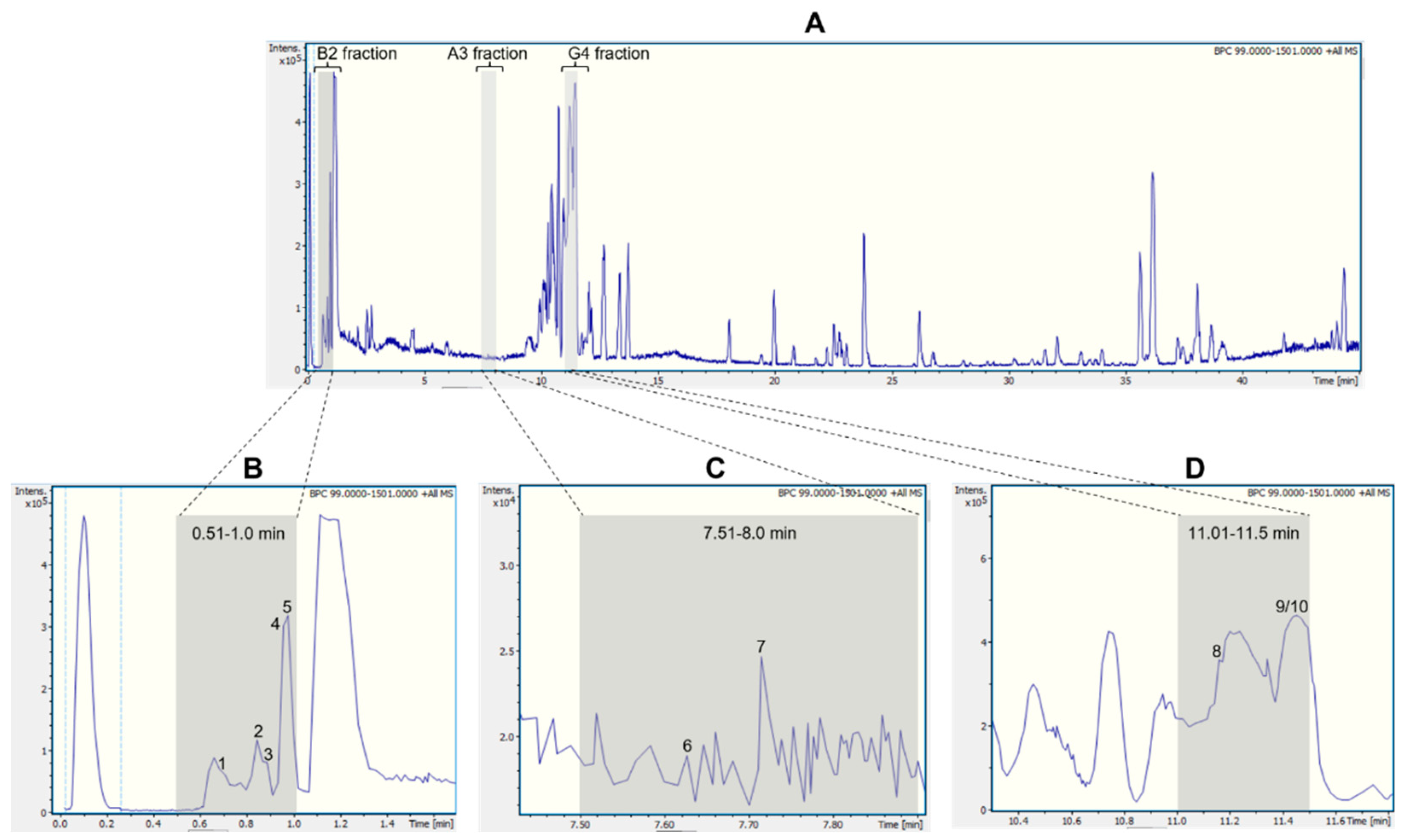
Figure 2.
Chemical structures of the annotated compounds: arginine (1), asparagine (2), glutamic acid (3), trigonelline (4), cathinone (5), tryptophan (6), bulbisine (7), tetrahydroharman-3-carboxylic acid (8), 2,7-dimethoxyhomolycorine (9), 3-O-acetylnarcissidine (10).
Figure 2.
Chemical structures of the annotated compounds: arginine (1), asparagine (2), glutamic acid (3), trigonelline (4), cathinone (5), tryptophan (6), bulbisine (7), tetrahydroharman-3-carboxylic acid (8), 2,7-dimethoxyhomolycorine (9), 3-O-acetylnarcissidine (10).
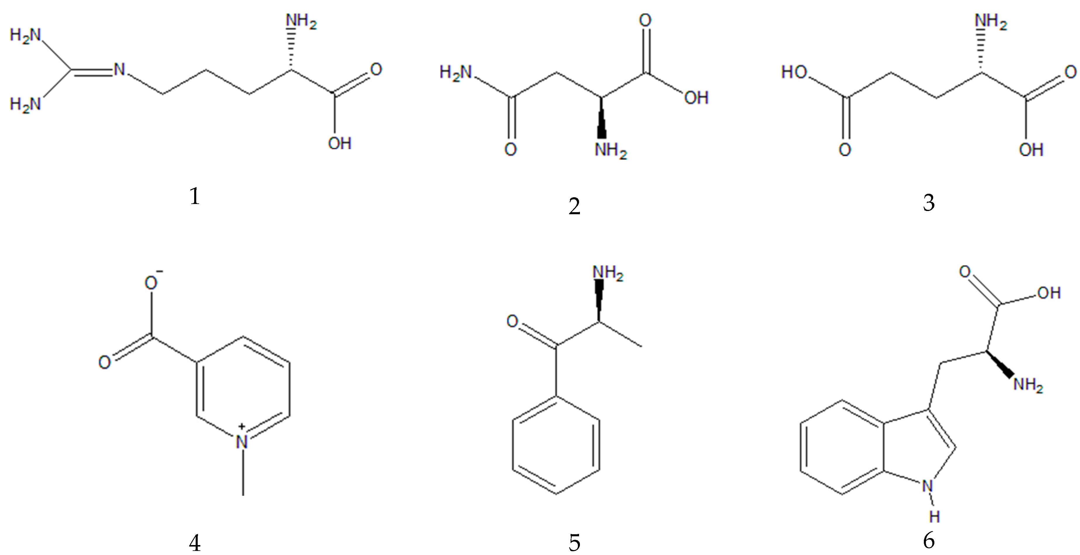

Table 1.
Ions detected by UHPLC-ESI-MS/MS in the active fractions derived from the bioassay-guided fractionation of the ethanolic extract of Hippeastrum puniceum bulbs.
Table 1.
Ions detected by UHPLC-ESI-MS/MS in the active fractions derived from the bioassay-guided fractionation of the ethanolic extract of Hippeastrum puniceum bulbs.
| Fraction | Peak ID | RT (min) |
Parent Ion [M+H]+ (m/z) | MS/MS Fragments (m/z, relative abundance - %) |
Molecular formula |
Annotation | |
|---|---|---|---|---|---|---|---|
| B2 (0.51-1.00 min) |
1 | 0.7 | 175.1190 | 175.1188 (100.0); 158.0922 (20.0); 130.0975 (9.7); 116.0698 (4.8) | C6H14N4O2 | Arginine a,b | |
| 2 | 0.9 | 133.0606 | 133.0607 (100.0); 116.0348 (10.1); 132.1022 (7.8); 130.0505 (5.0) | C4H8N2O3 | Asparagine a,b | ||
| 3 | 0.9 | 148.0603 | 148.0603 (33.3); 146.1177 (37.5); 130.0498 (100.0) | C5H9NO4 | Glutamic acid b | ||
| 4 | 1.0 | 138.0547 | 135.0676 (0.7); 136.0392(0.5); 136.0622 (0.5); 110.0899 | C7H7NO2 | Trigonelline a,b | ||
| 5 | 1.0 | 150.0911 | 135.0676 (24.3); 134.0600 (9.1); 132.0807 (2.0); 119.0491 (3.2); 117.0 (1.7) | C9H11NO | Cathinone b | ||
| A3 (7.51-8.00 min) |
6 | 7.6 | 205.0972 | 188.0705 (100.0); 146.0598 (43.1); 118.0650 (22.3) | C11H12N2O2 | Tryptophan a,b | |
| 7 | 7.7 | 320.1490 | 147.0438 (34.6); 119.0489 (9.2); 220.0750 (5.7) | C17H21NO5 | Bulbisine b | ||
| G4 (11.01-11.50 min) |
8 | 11.1 | 231.1126 | 158.0965 (100.0); 143.0723 (96.2); 130.0650 (52.8) | C13H14N2O2 | Tetrahydroharman-3-carboxylic acid b,c | |
| 9/10 | 11.4 | 376.1755 | 376.1755 (100.0); 377.1789 (21.0); 165.0912 (16.3); 124.0754 (3.2);139.0543 (0.2) | C20H25NO6 | 3-O-acetyl narcissidine b,c |
a Compounds annotated on the basis of their predicted molecular formula, MS/MS fragments and spectral comparison with public and in-house spectra libraries [25,26,27,28,29]. b Known to occur in the Amaryllidaceae family [30,31,32,33,34]. c Proposed by SIRIUS Software 5.5.7 (https://bio.informatik.uni-jena.de/sirius/) [35,36].
Table 2.
- Predicted pharmacokinetic and toxicity (ADMET) properties of the alkaloids present the bioactive fractions against wt-YFV .
Table 2.
- Predicted pharmacokinetic and toxicity (ADMET) properties of the alkaloids present the bioactive fractions against wt-YFV .
| PARAMETERS | COMPOUNDS | INDICATORS | ||||||||||
|---|---|---|---|---|---|---|---|---|---|---|---|---|
| Trigonelline | Cathinone | Bulbisine | Tetrahydroharman-3-carboxylic acid | 3-O-acetyl-narcissidine | 2,7-dimethoxy-homolycorine | |||||||
| MOL_WEIGHT | 137,138 | 149,193 | 319,357 | 230,267 | 375,421 | 375,421 | ||||||
| LOGP | -1.1254 | 1.2165 | 0.7652 | 1.8278 | 1.2328 | 1.9941 | Lipinski’s RO5: <5 Ideally between 1-4 | |||||
| #ROTATABLE_BONDS | 1 | 2 | 1 | 1 | 4 | 4 | ||||||
| #ACCEPTORS | 2 | 2 | 6 | 2 | 7 | 7 | ||||||
| #DONORS | 0 | 1 | 2 | 3 | 1 | 0 | ||||||
| SURFACE_AREA | 58,547 | 66,028 | 134,111 | 98,647 | 158,003 | 158,323 | ||||||
| Water solubility | -1.931 | -0.795 | -1.859 | -2.435 | -2.948 | -3.146 | The predicted water solubility of a compound is given as the logarithm of the molar concentration (log mol/L). | |||||
| Caco2 permeability | 1.124 | 1.237 | -0.138 | 0.832 | 0.705 | 1.472 | High CaCO-2 permeability would translate in predicted values >0.90 | |||||
| Intestinal absorption (human) | 96.44 | 76.876 | 70.972 | 94.534 | 64.054 | 97.738 | Poorly absorbed: < 30% | |||||
| Skin Permeability | -2.736 | -2.278 | -3.236 | -2.735 | -3.175 | -2.887 | Low skin permeability if it has a logKp > -2.5. | |||||
| P-glycoprotein substrate | Yes | No | Yes | Yes | Yes | No | Yes or No | |||||
| PARAMETERS | COMPOUNDS | INDICATORS | ||||||||||
| Trigonelline | Cathinone | Bulbisine | Tetrahydroharman-3-carboxylic acid | 3-O-acetylnarcissidine | 2,7-dimethoxyhomolycorine | |||||||
| P-glycoprotein I inhibitor | No | No | No | No | No | No | Yes or No |
|||||
| P-glycoprotein II inhibitor | No | No | No | No | No | No | Yes or No | |||||
| VDss (human) | -0.758 | 0.465 | 0.401 | -0.237 | 0.481 | 0.402 | Low if below 0.71 L/kg (log VDss < -0.15) and high if above 2.81 L/kg (log VDss > 0.45). | |||||
| Fraction unbound (human) | 0.857 | 0.469 | 0.436 | 0.481 | 0.413 | 0.411 | For a given compound the predicted fraction that would be unbound in plasma will be calculated. | |||||
| BBB permeability | -0.234 | -0.133 | -0.716 | 0.225 | -0.538 | -0.396 | Readily cross BBB >0.3; Poorly distributed in brain <-1 | |||||
| CNS permeability | -2.739 | -1.768 | -3.429 | -3.254 | -3.243 | -2.969 | Can penetrate, Log PS > -2 Cannot penetrate, Log PS < -3 | |||||
| CYP2D6 substrate | No | No | No | Yes | No | No | Yes or No | |||||
| CYP3A4 substrate | No | No | Yes | No | Yes | Yes | Yes or No | |||||
| CYP1A2 inhibitor | No | Yes | No | No | No | No | Yes or No | |||||
| CYP2C19 inhibitor | No | No | No | No | No | No | Yes or No | |||||
| CYP2C9 inhibitor | No | No | No | No | No | No | Yes or No | |||||
| CYP2D6 inhibitor | No | No | No | No | No | No | Yes or No | |||||
| CYP3A4 inhibitor | No | No | No | No | No | No | Yes or No | |||||
| Total Clearance | 0.378 | 0.811 | 1.137 | 0.694 | 0.902 | 0.636 | - | |||||
| Renal OCT2 substrate | No | No | No | No | No | No | Yes or No | |||||
| AMES toxicity | No | No | No | No | No | No | Yes or No | |||||
| Max. tolerated dose (human) | 0.743 | 0.779 | 0.025 | 0.323 | -0.558 | -0.099 | High is greater than 0.477 | |||||
| hERG I inhibitor | No | No | No | No | No | No | Yes or No | |||||
| hERG II inhibitor | No | No | No | No | No | No | Yes or No | |||||
| PARAMETERS | COMPOUNDS | INDICATORS | ||||||||||
| Trigonelline | Cathinone | Bulbisine | Tetrahydroharman-3-carboxylic acid | 3-O-acetylnarcissidine | 2,7-dimethoxyhomolycorine | |||||||
| Oral Rat Acute Toxicity (LD50) | 1.878 | 2.131 | 3.124 | 2.412 | 3.109 | 2.739 | The LD50 is the amount of a compound given all at once that causes the death of 50% of a group of test animals. | |||||
| Oral Rat Chronic Toxicity (LOAEL) | 0.454 | 1.542 | 2.614 | 1.115 | 1.610 | 2.689 | The LOAEL results need to be interpreted relative to the bioactive concentration and treatment lengths required. | |||||
| Hepatotoxicity | No | No | Yes | No | Yes | Yes | Yes or No | |||||
| Skin Sensitisation | No | No | No | No | No | No | Yes or No | |||||
Legend: LogP: logarithm of the partition coefficient; Caco2: Human colorectal adenocarcinoma cells; VDss: volume of distribution at steady state; BBB: Blood–Brain Barrier; CNS: Central nervous system. CY: Cytochrome P450 substrates; OCT2: Renal organic cation transporter 2; AMES: The name of the test used in mutagenicity assessment. hERG: Human ether-go-go gene; LD50: Lethal Dose for 50%; LOAEL: Lowest observed adverse effect level.
Disclaimer/Publisher’s Note: The statements, opinions and data contained in all publications are solely those of the individual author(s) and contributor(s) and not of MDPI and/or the editor(s). MDPI and/or the editor(s) disclaim responsibility for any injury to people or property resulting from any ideas, methods, instructions or products referred to in the content. |
© 2024 by the authors. Licensee MDPI, Basel, Switzerland. This article is an open access article distributed under the terms and conditions of the Creative Commons Attribution (CC BY) license (https://creativecommons.org/licenses/by/4.0/).
Copyright: This open access article is published under a Creative Commons CC BY 4.0 license, which permit the free download, distribution, and reuse, provided that the author and preprint are cited in any reuse.


