Submitted:
17 June 2024
Posted:
18 June 2024
You are already at the latest version
Abstract
Liquid biopsy, a non-invasive diagnosis that examines circulating tumor components in body fluids, is increasingly used in cancer management. An overview of relevant literature emphasizes the current state of liquid biopsy applications in cancer care. Liquid biopsy, particularly circulating tumor DNA (ctDNA), circulating tumor RNAs (ctRNA), circulating tumor cells (CTCs), extracellular vesicles, and other cell-free nucleic acids, offers promising opportunities for early cancer diagnosis, treatment selection, monitoring, and disease assessment. The implementation of liquid biopsy in precision medicine has shown significant potential in various cancer types, including lung cancer, colorectal cancer, breast cancer, and prostate cancer. Advances in genomic and molecular technologies, such as next generation sequencing (NGS) and digital polymerase chain reaction (dPCR) have expanded the utility of liquid biopsy, enabling the detection of somatic variants and actionable genomic alterations in tumors. Liquid biopsy has also demonstrated utility in predicting treatment responses, monitoring minimal residual disease (MRD), and assessing tumor heterogeneity. Nevertheless, standardizing liquid biopsy techniques, interpreting results, and integrating them into clinical routine remain challenges. Despite these challenges, liquid biopsy has significant clinical implications in cancer management, offering a dynamic and non-invasive approach to understanding tumor biology and guiding personalized treatment strategies.
Keywords:
liquid biopsy
; clinical management
; cancers
1. Introduction
Cancer is the leading cause of death in the world, accounting for nearly 20 million new cancer cases and 9.7 million deaths in 2022. Cancer is also referred to as a silent killer since its symptoms are vague and thus difficult to detect early. Cancer formation consists of 4 phases: initiation, promotion, progression, and metastasis. The first phase, initiation involves gene mutations in a cell. Promotion is the phase between a premalignant lesion and the development of invasive cancer, involving the accumulation of actively proliferating preneoplastic cells. The next phase is progression where genetic and phenotypic changes and cell proliferation occur. At this phase, the tumor size increases rapidly and the cells may undergo further mutations with invasive and metastatic potential [32]. The final stage is metastasis, which refers to the spread of cancer cells from a primary tumor to distant organs of the body. The metastasis process is a multi-step process, named metastatic cascade, broadly divided into 5 distinct steps (Figure 1). The malignant cells invades into adjacent tissues and penetrate into lymphatic and circulatory systems. These malignant cells are now known as circulating tumor cells (CTCs). These cells then exit the from the lymphatic and circulatory systems into adjacent normal tissue or organs which then survive and proliferate in in adjacent tissue or organs, leading to colonization [52].
Liquid biopsy has emerged as a revolutionary approach in the field of oncology, offering a minimally invasive and real-time method for the detection, monitoring, and characterization of cancer. Unlike traditional tissue biopsies, which are invasive and may not always capture the heterogeneity of tumors, liquid biopsy involves the analysis of biomarkers in biofluids such as blood, urine, saliva, sputum, stool, ascites, pleural effusion, seminal plasma or cerebrospinal fluid [104]. This approach holds great potential for transforming cancer management through the provision of clinicians with valuable insight into tumor dynamics, treatment response, and disease progression. In recent years, the field of liquid biopsy has made rapid advancements driven by technological innovations and a deeper understanding of cancer biology. The ability to detect CTC, ctDNA, ctRNA, extracellular vesicles, and metabolites in liquid biopsy samples has opened new avenues for personalized medicine and precision oncology [52] (Figure 2). With the aid of liquid biopsy, clinicians may be able to formulate tailor-made treatment strategies for individual cancers based on their individual molecular profiles, resulting in more effective and targeted therapies.
This review aims to explore the role of liquid biopsy in the clinical management of cancer, highlighting its potential applications, challenges, and future directions. By consolidating the current knowledge and research findings in this rapidly developing field, we seek to provide a comprehensive overview of the impact of liquid biopsy on cancer diagnosis, prognosis, treatment selection, and monitoring.
2. Background
Traditional methods of cancer diagnosis and monitoring have primarily relied on tissue biopsies, which involve the surgical removal of a sample of the tumor for analysis [155]. While tissue biopsies remain the gold standard for cancer diagnosis, they are limited by several factors, including their invasive nature, the possibility of tumor seeding [41], sampling errors, and a small risk of morbidity associated with tissue biopsy procedures [109]. These limitations underscore the need for non-invasive and dynamic approaches that can provide a more comprehensive picture of the disease. In addition, tissue biopsies typically obtain a sample of only a part of the tumor, thus only encompassing a part of tumor heterogeneity, limiting the information obtained on the levels of genetic and epigenetic variability of a patient’s cancer [55]. Liquid biopsy offers advantages over traditional tissue biopsies, providing a minimally invasive, dynamic, and comprehensive molecular analysis of tumors, aiding in early diagnosis, prognostication, and treatment response monitoring (Table 1) [2,32,47,90]. By analyzing biomolecules shed by tumors into the bloodstream, liquid biopsy provides valuable information about the genetic alterations, mutational profiles, and treatment response of cancer cells. Moreover, liquid biopsy can be performed after surgical resection and when there is no detectable metastatic mass for cancer treatment monitoring. Due to its minimally invasive nature, liquid biopsy can be performed multiple times or sequentially, which allows for continuous monitoring of disease progression as well as the emergence of treatment-resistant clones, offering insights that can be used to guide clinical decision-making. With the less invasive obtaining method and the various analytes that can be sampled in liquid biopsy, thus it can be used as a routine method for the real-time monitoring of cancer progression, early diagnosis of cancer, and assessing treatment response [2]. Liquid biopsy may help reveal intra-tumor heterogeneity (within tumors) and inter-tumor heterogeneity (between tumors), allowing the differences between the primary tumor and metastases, and the differences exposed during disease progression to be distinguished [112]. For example, Jin et al successfully identified three potential prognostic ctRNA biomarkers, which may be possible to apply MRD testing involving the identification of any remnants of cancer cells after operation [58]. Through the integration of liquid biopsy into clinical practice, cancer management will be revolutionized by early detection, personalized treatment selection, and real-time monitoring of treatment outcomes. Researchers are exploring liquid biopsy's utility in diverse cancer types and clinical settings, where the field has great potential to improve patient outcomes and expand our understanding of cancer biology.
Although liquid biopsy holds several advantages over tissue biopsy, more studies and advanced technologies are required to be conducted and invented in order for liquid biopsy to have the potential to replace tissue biopsy. The analytes in liquid biopsy are present in a very low concentration, thus the assays for the detection of analytes should be sensitive and specific enough in order to produce a valid result for the detection and monitoring of cancer. Moreover, tumor histology specification and staging cannot be determined by liquid biopsy, stating that liquid biopsy should be conducted with tissue biopsy for the treatment and diagnosis of cancer. Studies have shown that validation is still needed to increase the potential of liquid biopsy [57,58]. Therefore, implementing liquid biopsy assays in clinical practice is indeed facing significant challenges despite the promising potential of this technology.
The comparison between liquid biopsy and tissue biopsy is shown in Table 1.
3. Technology to Collect and Detect Liquid Biopsy
In liquid biopsy, biomarkers are identified in biofluids in order to provide valuable insights into the presence and characteristics of cancer. The main components of liquid biopsy include CTCs, ctDNA, ctRNA, extracellular vesicles (exosomes), and other circulating nucleic acids and proteins as shown in Figure 3. These biomarkers shed by tumors into the bloodstream or other bodily fluids can offer valuable insights into the genetic changes, mutations, and heterogeneity of cancer cells. Among different kinds of body fluids, blood is considered the most common material to analyze, but other body fluids may obtain some benefits over blood. For instance, the malignant pleural and peritoneal effusions may exhibit a higher concentration of CTCs than blood, or collecting CTCs in urine can enable the diagnosis of bladder cancer without the need for endoscopy [139]. Besides, many analyses are performed on ctDNA and CTCs isolated from blood. Therefore, the main focus would be on the isolation and analysis of ctDNA and CTCs in blood.
3.1. Circulating Tumor Cells (CTCs)
Cells that shed from a tumor and enter the circulatory or lymphatic system are referred to as circulating tumor cells (CTCs). They are able to travel through the bloodstream or lymphatic system to other areas of the body which have the potential to cause distant metastases. Different molecular markers are present on different CTCs depending on the type of cancer. As most of the cancers are of epithelial origin, a “universal” epithelial marker of cancer, EpCAM can be used as a common marker for CTCs [73]. The expression of EpCAM varies in different cancers with breast and prostate cancer being the highest and neurogenic cancers being the lowest. Cancer-specific CTC marks such as epidermal growth factor receptor-2 (HER2), prostate-specific membrane antigen, estrogen receptor, folate receptor, and surviving are in accordance with the specific molecular markers of the primary tumor [72]. CTCs are found to have a very low concentration in the blood even in patients with metastatic cancer [146], thus highly sensitive technologies are required to detect and isolate these cells.
3.1.1. Capture and Isolation of CTCs
Advanced Microfluidic Technologies
Microfluidic devices offer high-throughput and high-resolution isolation and analysis of CTCs from blood samples. These platforms use microscale channels to capture and isolate CTCs based on physical or biological properties, allowing for more sensitive and specific detection of rare CTCs (Figure 4).
Since CTCs are in a low concentration in blood, it is enriched from blood samples through various techniques that are based on the biological properties of CTCs such as the expression of specific protein markers, or based on the physical properties like size, density, deformability or electric charges [5].
In terms of biological properties, the epithelial cell adhesion molecule (EpCAM) serves as a surface antigen marker on CTCs so EPCAM-based enrichment for CTC detection is considered as a reliable method. However, in recent findings, CTCs can cease the expression of selected markers which allows markers to escape detection and result in false negative results [99,145]. In this case, negative selection rather than positive selection can be adopted. It refers to the depletion of non-malignant blood cells from blood by using antibodies such as targeting cell surface antigen CD45 on white blood cells [12]. It is noted that the drawbacks of negative selection include a lower purity of CTCs relative to the positive selection approach and the potential risk of depletion of CTCs owing to being trapped in a mass of blood cells [26,49,137]. Apart from EpCAM, cytokeratin family members such as CK8, CK18, and CK19 that are specific for epithelial cells also enable isolation of CTCs with epithelial phenotype by antibodies [96,97,98].
In terms of physical properties, CTCs show a larger size than normal blood cells. Size-based methods including isolation by size of epithelial tumor cells (ISET) have been developed, in which the blood passes through the pores to enable size exclusion of CTCs so larger CTCs are trapped but smaller CTCs may be lost and a lower purity of isolated CTCs [126]. Density gradient centrifugation is also available to separate CTCs which allows for fast separation but a lower sensitivity. Dielectrophoresis (DEP) can separate CTCs from normal blood cells by their unique dielectric properties which depend on their diameter, membrane area, density, conductivity, and volume, albeit the changes in dielectric properties during prolonged storage [139].
3.1.2. Strategies on CTCs Analysis
After the isolation of CTCs, they can be identified by immunological, molecular, or functional assays [5].
Immunological Technologies
In the immunological assay, CTCs are detected by antibodies against epithelial, mesenchymal, tissue-specific, or tumor-associated markers. For instance, EpCAM and cytokeratin members are the epithelial markers to be detected, or mesenchymal markers like N-cadherin or vimentin can also be targeted [65]. The tissue-specific markers like prostate-specific antigen (PSA) show a high specificity to particular tumor types and tumor-associated markers like HER-2 are important for targeted therapies.
Molecular Technologies (RNA-based)
Tumor cells have changed at genetic and transcriptomic levels making them able to escape from the primary tumor and survive as CTCs [50], hence RNA analysis of CTCs can be used for detection and quantification. CTCs are identified by quantitative reverse transcription polymerase chain reaction (RT-qPCR), RNA sequencing (RNA-seq), and RNA in situ hybridization (RNA-ISH). RT-qPCR is a frequently used technique to detect gene expression in CTCs due to its high sensitivity and cost-effectiveness when compared to other RNA-based methods [9]. Since the concentration of CTCs is often low in blood, droplet digital PCR (ddPCR) may also be applied to separate the CTCs’ RNA into many partitions by lipid droplets to enhance the detection of CTCs’ RNA in the early stage of cancer, despite only 1 gene can be quantified at a time [66]. RNA-ISH can allow the detection of localized RNA in CTCs without denaturing the cells, and it can be used without any enrichment process to reduce the risk of losing CTCs during processing while obtaining a high sensitivity, namely the CTCscope method [102]. RNA-seq can study a large number of genes simultaneously in either a single CTC or total CTCs to determine the gene expression profile so that identification of sub-populations of CTCs with various gene expressions from the same patient by single-cell sequencing [105], and the examination of comprehensive gene expression profiles of CTCs by whole genome sequencing [45] can be achieved, though low-level transcripts cause a reduced sensitivity [22].
Single-Cell Analysis of CTCs Using Microfluidic Devices
A study conducted by Aceto et al. (2014) demonstrated a high level of concordance between expression patterns of CTC clusters and single CTCs, highlighting the importance of single-cell resolution RNA sequencing [1]. Single-cell analysis of CTC clusters provided insight into tumor cell migration and metastatic properties [8]. Huang et al. (2023) and Cheng et al. (2023) developed microfluidic devices for the isolation and analysis of CTCs, emphasizing the importance of efficient capture and release of CTCs in cancer diagnosis and monitoring [25,56]. Cheng et al. (2023) introduced a poly(ethylene oxide) concentration gradient-based microfluidic device for the isolation of CTCs, highlighting the importance of efficient capture and release of these cells for cancer diagnosis and management [25]. Huang et al. (2023) developed a conductive nanofibers-enhanced microfluidic device for the efficient capture and electrical stimulation-triggered rapid release of CTCs [56]. This novel approach not only enables the effective detection of CTCs but also offers a mechanism for rapid release, which is crucial for cancer diagnosis and monitoring.
Functional Assays
Functional assays, such as drug sensitivity testing on isolated CTCs or CTC-derived xenograft models, can help predict treatment response and guide personalized therapeutic strategies based on the drug sensitivity profile of CTCs. Capturing viable CTCs can allow downstream culturing of CTCs cell lines derived from patients [20]. Some developed methods can maintain CTCs viability and culture possibility, such as density gradient centrifugation by Ficoll-PaqueTM but centrifugation leads to CTCs loss and lower purity due to a mixture of leukocytes and CTCs [40]. The microfluidic system is also a method to trap CTCs for characterization and purification such as coating the micro posts with anti-EpCAM antibodies to collect CTCs but it is challenging for high-throughput production [117]. Some other chips are created such as herringbone-chip (HB-Chip) to increase throughput but a trypsinization step is required which may affect the expression of the receptor on CTCs [124], or CTC-iChip to undergo magnetic sorting of CTCs but with a long preparation time [88]. All the mentioned methods require an extra step to illustrate the CTCs’ viability. Therefore, Epithelial Immuno-SPOT (EPISPOT) assay is developed to directly assess CTC viability in fewer steps.
EPISPOT is a method to detect viable CTCs only based on the specific tumor-associated proteins that are secreted, shed, and released by CTCs using an adaption of enzyme-linked immunospot (ELISPOT) technology [4]. A cell culture is required to accumulate enough released marker proteins amount because the immunospots are the protein fingerprint created by viable epithelial cells only so the dead cells which do not release sufficient marker proteins are not detected [7]. The CTCs are cultured on the nitrocellulose membrane coated with specific antibodies, and the released proteins from CTCs are captured during the incubation period. EPISPOT often combines leukocyte depletion negative enrichment step and it can identify CTCs without epithelial features such as EpCAM-negative CTCs [83]. EPISPOT method has been already validated for prostate cancer [6], breast cancer [108], head and neck squamous cell carcinoma [42] colorectal cancer [33], and melanoma [23], illustrating the clinical values of the assay.
3.2. Circulating Nucleic Acids
Circulating nucleic acids, including ctDNA and ctRNA, are fragments of nucleic acids released into the bloodstream by cells, including tumor cells. The release of circulating nucleic acids into the human circulatory system can originate from six sources, including apoptosis, necrosis, NETosis, erythroblast enucleation, extracellular vesicles, and exogenous sources [31].
3.3. Circulating Tumor DNA (ctDNA)
Circulating tumor DNA (ctDNA) is a portion of cell-free DNA (cfDNA) that is released from tumor cells and enters the circulatory system. The presence of epigenetic or genetic alterations, including tumor-specific methylation markers, rearrangements, copy number variations, and somatic point mutations allows ctDNA to be distinguished from normal cfDNA fragments [107]. The genetic and epigenetic modifications of ctDNA molecules may reflect the genome or epigenome of the cell of origin, thus it can used to estimate prognosis, for identification of residual disease and treatment selection, and/or indicate potential risk of relapse [28]. The half-life of ctDNA in the circulatory system varies from minutes to 2.5 hours [140] and the levels of ctDNA are generally very low which may require highly sensitive technologies for the detection of ctDNA.
3.4. Circulating Tumor RNA (ctRNA)
RNA released from cells through apoptosis, necrosis, and active secretion is referred to as cfRNA [59]. ctRNA, including microRNA (miRNA) and long non-coding RNA (lncRNA), are a portion of cfRNAs that are present in the body fluids of tumor patients. These cfRNAs can be isolated from various types of body fluids such as blood, urine, breast milk, and other fluids [64].
MiRNAs are small non-coding RNAs that are essential for the expression of post-translation genes. They are desirable biomarkers for non-invasive cancer diagnosis due to their stability in body fluids. Moreover, the expression patterns of miRNAs are tissue-specific, suggesting that miRNAs could be used as a biomarker for cancer diagnosis, prediction, and prognosis [153]. Numerous studies have shown that specific miRNA signatures are associated with various cancer types. miRNA-21 is an extensively studied miRNA that is found to be upregulated in a number of malignancies, including breast, lung, and colorectal cancer [141,142,152].
lncRNAs are transcripts with no protein-coding capacity. These RNAs act as regulatory factors that play an important role in different cellular processes such as cell growth, differentiation, proliferation, and apoptosis [64]. lncRNAs are bound to RNA-binding proteins and are released either inside apoptotic bodies or encapsulated in exosomes, making them resistant to RNase degradation. Thus, they can exist stably in the circulatory system and could be used as a non-invasive biomarker [110]. Studies have found that lncRNAs are expressed in a cell- and tissue-type specific pattern, allowing it to be used in the diagnosis of various cancers such as lung cancer, breast cancer, gastric cancer, liver cancer, and prostate cancer by measuring the expression level of lncRNAs [18].
3.4.1. Isolation of Circulating Cell-Free Nucleic Acids
After blood taking, it proceeds to plasma processing and DNA extraction. One of the main factors that affect the sensitivity of detecting cfDNA is the unintended release of wild-type DNA owing to the lysis of white blood cells occurring between the blood-drawing time and plasma processing [85]. Since circulating cell-free nucleic acids have a very low concentration in the peripheral blood which already demonstrates a significant challenge to any analytical system, the unwanted increase in normal DNA can further negatively disturb the detection. Besides, despite a higher concentration of cfDNA in the serum, the increased level is caused by the clotting process of white blood cells resulting in lysis which contaminates cfDNA [38], so using plasma sample can reduce the contamination. Therefore, the key to reducing the release of normal DNA from white blood cells is to process the blood sample rapidly after collection and undergo an additional high-speed centrifugation step after separating the plasma, which requires the preparation of plasma from whole blood within 1-2 hours after venipuncture [34]. In view of the rigorous requirements of rapid handling of blood samples, an alternative approach of inhibiting nuclease activity in blood and stabilizing white blood cells in the blood collection tube can also achieve the purpose [131].
In DNA extraction, there are several methods developed including the magnetic-based method, silica column-based enrichment, polymer-mediated enrichment (PME), and phenol-chloroform-based extraction. Comparing different types of DNA extraction methods, they illustrate a large variation in terms of the yield and fragment size of cfDNA [39]. For example, the phenol-chloroform-based extraction shows a higher yield but decreased cfDNA purity when compared to the magnetic-based method so the downstream analysis can be affected [60]. Also, the silica column-based enrichment may obtain a lower yield and partial loss of DNA that is smaller than 150bp [89,116]. Hence, the choice of DNA extraction method is also an important factor to consider.
3.4.2. Circulating Nucleic Acids Detection and Analysis
Digital PCR and Next-Generation Sequencing (NGS)
Ultra-sensitive sequencing technologies are crucial for the analysis of circulating tumor nucleic acids due to the low abundance of ctDNA in biological samples [132]. These technologies, such as ultra-deep massively parallel sequencing with unique molecular identifier tagging enable the detection and quantification of minute amounts of ctDNA, which are typically released into the bloodstream by tumor cells undergoing cell death [132]. By utilizing ultra-sensitive sequencing methods, such as Illumina and Thermo Fisher, researchers can achieve comparable performance to droplet digital PCR (ddPCR), like Bio-Rad's QX200 and Thermo Fisher's QuantStudio for the detection and quantification of ctDNA from cancer patients [114,132]. For accurate identification of and quantification of genetic changes in ctDNA, the ultrasensitive nature of these sequencing technologies is essential, providing insight into tumor dynamics, treatment response, and disease progression based on the molecular profile of ctDNA, which ultimately guides personalized therapeutic strategies.
Methylation Profiling
Methylation analysis of ctDNA has been used in cancer diagnostics and monitoring. Numerous studies have investigated the importance of DNA methylation patterns in ctDNA for early detection, treatment assessment, and personalized medicine approaches. Tamkovich et al. (2022) discuss the potential use of aberrantly methylated DNA in liquid biopsy for patients with breast cancer, emphasizing its early involvement in cancer cell transformation [156]. A recent study by Markou and colleagues (2022) explored the role of DNA methylation analysis in early-stage non-small-cell lung cancer (NSCLC) cases, demonstrating the potential for early cancer detection through DNA methylation analysis [80].
Fragmentomics Analysis
Fragmentomics analysis of cfDNA is essential for understanding the fragmentation properties of cfDNA, including size distribution and biochemical characteristics of free ends. The method enables the analysis of the size distribution and frequency of millions of naturally occurring cfDNA fragments across the genome, offering valuable insights into cfDNA "fragmentomes." [82]. Using ultrasensitive sequence technologies, such as nanopore sequencing, researchers can analyze previously understudied cfDNA populations by acquiring genomic and fragmentomic data [136]. A comprehensive view of cfDNA fragmentation patterns can be obtained by fragmentomic profiling within clinical settings, provided there is clear traceability of analytical and physiological factors, allowing for disease diagnosis and monitoring based on cfDNA fragmentation patterns [103]. Epigenetic profiling of cfDNA fragmentomes can also offer valuable information on DNA methylation patterns, aiding in predicting CpG methylation of cfDNA without the need for bisulfite treatment [154]. Using fragmentomics to examine cfDNA fragments offers the potential for enhancing early cancer detection, monitoring treatment response, and developing personalized therapeutic strategies based on the molecular characteristics of cfDNA fragments.
3.5. Extracellular Vesicles (EV)
Extracellular vesicles (EV), including exosomes and microvesicles, ectosomes, oncosomes microparticles, and many others are groups of membrane-closed vesicles containing DNA, RNA, proteins, and lipids enclosed in a phospholipid biolayer, which play a crucial role in intercellular communication and disease progression, including cancer [123]. In addition, EVs are secreted by all cell types and are present in various types of body fluids, allowing the collection of the biomarkers present in extracellular vesicles for cancer diagnosis and management. For example, exosomes are small extracellular vesicles that are emerging biomarkers for cancer diagnosis and guide treatments. Studies have found that exosomes are involved in the occurrence and development of various tumors, which act as carriers in tumor cells allowing them to escape immune system surveillance. Exosomes are also able to create a suitable microenvironment for tumor growth and guide the direction of tumor cell metastasis [48,94]. To improve understanding of circulating EVs, researchers are integrating multi-omics approaches, implementing machine learning algorithms, and standardizing isolation and analysis protocols [68].
3.5.1. EV Isolation and Characterization Technologies
The isolation and characterization of EVs are essential for understanding their functions and potential therapeutic applications. There have been several techniques developed and refined to address the challenges associated with EV isolation and characterization. The size exclusion chromatography (SEC) method is one of the most common methods for isolating EVs from plasma in recent years due to its ability to rapidly isolate relatively pure EVs [125]. Nevertheless, SEC is not suitable for all downstream applications, highlighting the need to develop a variety of isolation techniques in order to meet the needs of different research projects. An alternative method of EV isolation is ultracentrifugation, however, it can be time-consuming and difficult to implement in a clinical environment [17]. There have been recent advances in EV isolation efficiency and biomarker profiling by using membrane affinity-based methods and microfluidics [11,51]. These advancements are essential for improving diagnostic capabilities and advancing precision medicine initiatives. Further, the development of novel techniques such as the EVtrap [24] and the salting-out approach [118] offer alternatives for achieving high purity and efficiency in EV isolation. Characterizing EVs is equally important, and a variety of methods have been used for this purpose, including nanoparticle tracking analysis (NTA), transmission electron microscopy, and Western blotting.
EV Proteomic and RNA Profiling
The integration of proteomic and RNA profiling in EV research holds great potential for advancing precision medicine and improving disease diagnosis and monitoring. The development of EV proteomic profiling has been promising for the detection and monitoring of cancer, emphasizing the importance of understanding the protein cargo of EVs for clinical applications [19,148]. Through the use of high-throughput sequencing and NGS, comprehensive analyses of EV RNA content have been carried out, which have shed light on the multitude of RNA species present in EVs as well as the functional implications of their presence [130].
3.6. Metabolites
Metabolomics can be utilized to identify metabolites with a molecular weight of less than 1kDa present in body fluids, with serum being the best choice. These metabolites include vitamins, sugars, and lipids. The changes in metabolite levels highly relate to the pathophysiological state in an individual, thus they have the ability to become biomarkers for the screening and early diagnosis of cancer. For instance, a study has demonstrated that benzoic acid might have the potential to be a diagnostic marker of colorectal cancer [134] and significant differences in metabolite levels can be found in different stages of the same cancer type [44].
4. Applications in Cancer Management
Liquid biopsy is usually performed for monitoring cancer progression, early diagnosis of cancer, and assessing treatment response. Analyzing tumor-derived biomarkers in biofluids has revolutionized personalized medicine and precision oncology. some key applications of liquid biopsy will be discussed.
4.1. Early Detection and Diagnosis
A number of recent studies have demonstrated the potential of liquid biopsy for cancer screening, refinement of diagnosis, and early detection of cancer recurrences [43,61]. By detecting circulating tumor cells, nucleic acids, and exosomes, the method can be used for early cancer detection, diagnosis, and prognosis in various types of cancer [35,63,71,149]. Additionally, liquid biopsy can provide insights into cancer progression and treatment response by assessing cancer cachexia, muscle-derived microRNAs, and energy metabolism [16]. Recent advances in liquid biopsy techniques, including the detection of exosome microRNAs and protein biomarkers, have made them more useful for diagnosing and monitoring cancer, providing real-time feedback and non-invasive disease assessment [71,106]. Moreover, nanotechnology and antibody-conjugated signals in nanocavities have been proposed to detect cancer markers in liquid biopsy [128]. For instance, since fewer amounts of ctDNA are released for detecting mutations during the early stages of lung cancer, evaluating both ctDNA and circulating proteins such as carcinoembryonic antigen (CEA), cancer antigen 125 (CA-125), cancer antigen 19-9 (CA19-9) etc can allow higher sensitivity for early detection [147]. Therefore, the evaluation of both 16 driver genes mutation in ctDNA and 8 circulating proteins called CancerSEEK is conducted to improve early detection for multiple cancers including lung cancer, which is shown in Table 2 as demonstrated by the study from Cohen et al [27]. In addition, miRNA is the most sufficient cfRNA molecule in the body fluids so it also has the potential for early diagnosis of lung cancer. For instance, various types of miRNA in sputum such as miR-145, miR-126, and miR-7 have been shown to obtain a high sensitivity and specificity to detect NSCLC [14]. Apart from the identification of lung cancer, miRNA can also be used to distinguish different types of lung cancer. The use of a miRNA panel in plasma consisting of miR-17, miR-190b, and miR-375, shows high accuracy in distinguishing small cell lung cancer (SCLC) and NSCLC [78].
4.2. Prognostication and Predictive Biomarkers
Liquid biopsy, through the analysis of CTCs, ctDNA, ctRNA, and exosomes, provides prognostic and predictive information in various cancer types, including prostate cancer, bladder cancer, colorectal cancer, and NSCLC [79] [58,70,111]. In recent studies, liquid biopsy has been shown to have prognostic and predictive value in cancer management, offering molecular and phenotypic information about cancers [81]. The analysis of nucleic acids in liquid biopsies has demonstrated prognostic potential and predictive value for guiding treatment decisions in colorectal cancer and hepatocellular carcinoma [3,67]. In addition, clinical outcomes and cancer recurrence have consistently been predicted by liquid biopsy, particularly through the detection of CTCs [21].
4.3. Treatment Selection and Personalized Medicine
The use of liquid biopsy to identify specific biomarkers in body fluids has significantly influenced treatment selection and personalized medicine. Several recent studies have highlighted the importance of liquid biopsy when selecting targeted therapies and evaluating treatment responses. Personalized medicine has benefited tremendously from liquid biopsy, enabling the identification of tumor-specific biomarkers that are used to optimize treatment regimens and monitor therapeutic outcomes [37,101]. The use of liquid biopsy has enabled accurate prediction, monitoring, and rational selection of appropriate therapies for individual patients by analyzing ctDNA and CTCs as well [150,151]. Aside from this, liquid biopsy has also been demonstrated to have promise in guiding therapeutic decisions for many types of cancer, including lung cancer, breast cancer, and prostate cancer by providing real-time molecular information that is useful in guiding treatment choices and evaluating response to treatment.
4.4. Lung Cancer
Lung cancer is one of the most common cancers to cause mortality but there is a low rate of early detection so most patients are diagnosed at the advanced stage which leads to a lower survival rate. Pre-screening program is thus an important tool to enable early screening of the population and liquid biopsy can detect lung cancer in a safer and earlier way. On the other hand, tissue biopsy may not be available owing to the invasive procedure and the failure to have sufficient tumor tissue for further gene variations detection [120]. With the properties of non-invasiveness, simple accessibility, and great repeatability, the use of liquid biopsy is increased for molecular profiling testing and drug-resistance monitoring [147].
In the management of targeted therapy, NSCLC has achieved a targeted treatment according to molecular classification to increase overall survival, namely the use of EGFR tyrosine kinase inhibitors for patients with EGFR mutation. However, drug resistance can also occur decreasing the effectiveness of targeted therapy. Hence, testing gene variations is required for NSCLC patients but there are some problems. For instance, it is complicated to obtain tissue biopsy, especially for drug-resistant patients [93]. Tumor heterogeneity causes non-comprehensive genomic profiles by tissue biopsy obtained in 1 site only [127]. It is also difficult for continuous monitoring due to the invasive procedure of tissue biopsy. Therefore, liquid biopsy may be effective in tackling these problems. ctDNA is found to be mostly consistent with genomic variations in tumors so EGFR mutation detection can be performed by ctDNA in the plasma. EGFR T790M acquired drug resistance mutation can also be discovered by ctDNA instead of tumor tissue [113]. Therefore, ctDNA allows the early detection of drug resistance to the use of treatment which ensures the treatment is useful to the patient.
4.5. Breast Cancer
Breast cancer can be classified into 4 subtypes based on molecular information, including luminal A, luminal B, HER2-positive, and triple-negative. This molecular classification can categorize patients who are able to benefit from targeted therapy. The luminal A subtype is characterized by the presence of estrogen receptor (ER) positive, progesterone receptor (PR) positive but human epidermal growth factor receptor (HER2) negative, which shows low-grade and slow-growing characteristics to have the best prognosis result. The luminal B subtype, which is characterized by ER positive, PR negative, and HER2 positive, obtains a higher grade and worse prognosis when compared to the luminal A subtype. The HER2-positive subtype, which is characterized by ER negative, PR negative, and HER2 positive, grows faster than the luminal subtypes and improves after HER2-targeted therapies. The triple-negative subtype, which is characterized by ER negative, PR negative, and HER2 negative, is likely to benefit from chemotherapy.
When breast cancer becomes metastatic, it is hard to control. After confirming the status of hormone receptors and HER2 by a biopsy of metastatic breast cancer lesions, determination of the metastatic phenotype by repeated sampling can provide useful information on molecular changes and treatment customization [115], and thus liquid biopsies can achieve the purpose.
In breast cancer, there is an increase in both ctDNA and CTCs that correlate with cancer development. The analysis of these biomarkers is beneficial to manage advanced or early breast cancer patients. It is found that a high concentration of ctDNA is related to a more aggressive or relapsing disease [84]. CTCs are also useful in characterizing phenotypes and determining the possibility of metastatic spread and prognosis [119].
Detection of phosphatidylinositol-4,5-bisphosphate 3-kinase catalytic subunit alpha (PIK3CA) mutation is important for breast cancer because of the targeted therapies developed. PIK3CA mutation may result in resistance to HER2-targeted therapies. PIK3CA hotspot mutations such as E545K and H1047R mutations can be detected by ctDNA and CTCs but not detected by the DNA from the primary tumor (Formalin-Fixed Paraffin-Embedded samples) through the use of the same highly sensitive methods, indicating the importance of liquid biopsies [122].
Estrogen Receptor 1 (ESR1) mutation plays a significant role in endocrine therapy (ET) resistance for metastatic breast cancer, and it can also be detected by CTCs and ctDNA. Compared to tissue sequencing, ctDNA may be more superior because it can accurately reveal the molecular file of metastatic cancer [138]. Therefore, integrating liquid biopsies into the diagnostic workflow in breast cancer patients can help highlight the resistance to ET and guide treatment decisions such as changing to other ET or non-endocrine treatments.
4.6. Prostate Cancer
Prostate cancer is a prevalent cancer in men resulting in mortality. The treatment of metastatic prostate cancer involves androgen deprivation therapy combined with androgen receptor pathway inhibitors (ARPI), chemotherapy, radionuclides, and immunotherapy [15]. However, the treatment decisions informed by markers such as PSA, bone scintigraphy, computed tomography (CT), or positron emission tomography (PET) scans do not provide information on disease drivers or resistance mechanisms [133]. Therefore, technologies for providing a more comprehensive disease characterization are needed and liquid biopsy assays are developed to facilitate prognosis, assess treatment response, etc, in prostate cancer.
CTCs in the patient’s blood can represent the disease burden and act as precursors for metastasis so CTCs enumeration can be used for prognosis and treatment response. For instance, CellSearch assay is a CTCs enumeration platform to serve as a prognosis marker in metastatic prostate cancer because multiple studies demonstrate an obvious association between CTCs count and overall survival (OS) [30,87]. The CTCs count serves as a marker for treatment response based on several criteria for changes in CTCs count by the studies of treatment response, including decline percentage in CTC count relative to baseline level [53,75,76], complete decrease of CTCs from at least 1 at baseline to 0 in 7.5mL of blood [54], etc. Generally, the reduction in CTC count following treatment is proportional to the effect of treatment.
Besides, liquid biopsy can be used to guide treatment decisions. According to genomic studies of prostate cancer, it is identified that recurrent mutations may affect treatment response and somatic mutations can be used to predict response to ARPI therapy, indicating the potential of personalized treatment [10,13,29]. Liquid biopsy assays can provide a minimally invasive and comprehensive assessment of mutations that affect the response to therapies. For example, The androgen receptor splice variant 7 (AR-V7) is the most commonly detected variant associated with treatment response because it continuously activates downstream signaling and evades androgen receptor blockers due to retaining the DNA-binding domain but lacking the regulatory ligand-binding domain [91,143]. AR-V7 detection can be achieved in CTCs and ctDNA, such as using EPIC Sciences CTC AR-V7 nuclear localization assay [77] or AR-V7-modified AdnaTest followed by RT-qPCR amplification of AR-V7 mRNA [74]. Furthermore, mutations that cause a deficiency in DNA damage repair pathways have resistance to ARPI such as mutations in BRCA1, BRCA2, and ATM which are responsible for producing proteins for homologous recombination repair (HRR) [133]. It is observed that patients with homologous recombination deficiency (HRD) mutations are sensitive to poly(ADP-ribose) polymerase inhibitors (PARPi) treatment like olaparib or rucaparib. FoundationOne Liquid CDx test is an FDA-approved next-generation sequencing-based test to identify mutations in BRCA1, BRCA2, and ATM in cfDNA in order to guide the treatment of olaparib or rucaparib [144]. All in all, the integration of liquid biopsy assays into clinical investigations of prostate cancer remains the potential for prognosis and treatment guidance.
5. Key Clinical Trials Evaluating Liquid Biopsy for Cancer
There have been numerous clinical trials on liquid biopsy, including the detection of actionable mutations, evaluating treatment response, and predicting prognosis. For instance, an ongoing clinical trial at the Jewish General Hospital in Montreal, Canada, called TRICIA (TRIPLE Negative Breast Cancer Markers In Liquid Biopsies Using Artificial Intelligence) is developing a test or score based on ctDNA expression within a cohort of patients with triple-negative breast cancer, which demonstrates the potential of liquid biopsy to guide treatment decisions based on specific breast cancer subtypes. [121]. Another prospective phase II clinical trial in CRC utilized liquid biopsies to detect spatial and temporal heterogeneity of resistance to anti-EGFR monoclonal antibodies by combining sequential profiling of ctDNA with mathematical modeling and imaging of tumor progression, which showed that liquid biopsy can be used to monitor the progression of cancer and the response to treatment [135]. Furthermore, EGFR mutation status is being evaluated in NSCLC patients using the ddPCR method in ongoing clinical trials, suggesting liquid biopsy can play a major role in guiding treatment decisions for NSCLC patients [95]. As a result of these trials, it is evident that liquid biopsy can be useful for identifying specific mutations in lung cancer patients and guiding targeted therapies.
6. Liquid Biopsy Regulatory Considerations and Challenges.
Integrating liquid biopsy into clinical practice presents a number of regulatory challenges and considerations. Regulatory issues, standardization of methods, diagnostic performance, and further research are key factors affecting the integration of liquid biopsy in the clinic [92]. Challenges such as low amounts of CTCs, ctRNA, and ctDNA, lack of consensus in pre-analytical and analytical processes, clinical validation, and regulatory approval hinder the successful implementation of liquid biopsy in clinical settings. The success of regulatory pathways for liquid biopsy diagnostics has been attributed in part to the incremental value of FDA approval for tests developed under the Clinical Laboratory Improvement Amendment (CLIA), as well as their complexity, which can hinder their widespread replication in CLIA laboratories [46]. In addition to its clinical utility, liquid biopsy is used to monitor tumor genomes non-invasively and ensure appropriate treatment for patients. However, liquid biopsy remains a challenge in effective cancer management due to the lack of sensitive and specific biomarkers, the limitations of sampling single primary tumors, and the difficulty of monitoring cancer progression over time [100].
Among the challenges faced in integrating liquid biopsy into clinical practice are the need to standardize techniques, validate assays, and interpret results [62]. Considering the complexity of liquid biopsy approaches, robust validation processes are required to ensure accurate and reliable results [129]. Moreover, tumor heterogeneity, clonality, and dynamic nature of tumors pose challenges when interpreting liquid biopsy results for effective cancer management [69]. The implementation of liquid biopsy in clinical practice relies greatly on international standards, such as the International Organization for Standardization (ISO). It is essential that different laboratories and settings follow standardized protocols for sample collection, processing, and analysis. In cancer management, following international standards helps harmonize practices, improve data quality, and facilitate information exchange [62]. For example, the workflow and quality evaluation of sequencing data from massively parallel sequencing should be based on ISO20397-1:2021 and ISO20397-2:2021. To be used in clinical laboratories, assays should follow ISO15189 [62]. A standardized liquid biopsy method and workflow could further enhance the clinical usefulness of this approach in cancer management. Healthcare providers can ensure accurate, reproducible, and clinically relevant liquid biopsy results by following established guidelines and protocols [36]. Additionally, standardizing approaches facilitates the sharing of data, collaboration, and integration of liquid biopsy into routine clinical practice [62,86].
8. Conclusions
Liquid biopsy shows great potential in making early diagnoses and continuously monitoring disease development or therapeutic response with its less invasive property. Compared to tissue biopsy which is regarded as the only diagnostic method traditionally, the analysis of liquid biopsy can minimize the burden on patients. Despite the benefits of liquid biopsy, more studies and improvements are still required. For instance, the feasibility of extracting different biomarkers from samples except for blood, or the advancements in technologies to improve sensitivity and specificity for detecting biomarkers from the liquid biopsy. With more investigations, it is hoped to address the challenges associated with liquid biopsy, and eventually integrate liquid biopsy in the clinical settings.
References
- Aceto, N.; Bardia, A.; Miyamoto, D.T.; Donaldson, M.C.; Wittner, B.S.; Spencer, J.A.; Yu, M.; Pely, A.; Engstrom, A.; Zhu, H.; et al. Circulating Tumor Cell Clusters Are Oligoclonal Precursors of Breast Cancer Metastasis. Cell 2014, 158, 1110–1122. [Google Scholar] [CrossRef] [PubMed]
- Adhit, K.K.; Wanjari, A.; Menon, S.; K, S. Liquid Biopsy: An Evolving Paradigm for Non-invasive Disease Diagnosis and Monitoring in Medicine. Cureus 2023, 15, e50176. [Google Scholar] [CrossRef] [PubMed]
- Agashe, R.; Kurzrock, R. Circulating Tumor Cells: From the Laboratory to the Cancer Clinic. Cancers 2020, 12, 2361. [Google Scholar] [CrossRef] [PubMed]
- ALIX-PANABIèRES, C. 2012. EPISPOT Assay: Detection of Viable DTCs/CTCs in Solid Tumor Patients. In: IGNATIADIS, M., SOTIRIOU, C. & PANTEL, K. (eds.) Minimal Residual Disease and Circulating Tumor Cells in Breast Cancer. Berlin, Heidelberg: Springer Berlin Heidelberg.
- Alix-Panabières, C.; Pantel, K. Challenges in circulating tumour cell research. Nat. Rev. Cancer 2014, 14, 623–631. [Google Scholar] [CrossRef]
- Alix-Panabières, C.; Rebillard, X.; Brouillet, J.-P.; Barbotte, E.; Iborra, F.; Segui, B.; Maudelonde, T.; Jolivet-Reynaud, C.; Vendrell, J.-P. Detection of Circulating Prostate-Specific Antigen–Secreting Cells in Prostate Cancer Patients. Clin. Chem. 2005, 51, 1538–1541. [Google Scholar] [CrossRef] [PubMed]
- Alix-Panabières, C.; Riethdorf, S.; Pantel, K. Circulating Tumor Cells and Bone Marrow Micrometastasis. Clin. Cancer Res. 2008, 14, 5013–5021. [Google Scholar] [CrossRef]
- Amintas, S.; Bedel, A.; Moreau-Gaudry, F.; Boutin, J.; Buscail, L.; Merlio, J.-P.; Vendrely, V.; Dabernat, S.; Buscail, E. Circulating Tumor Cell Clusters: United We Stand Divided We Fall. Int. J. Mol. Sci. 2020, 21, 2653. [Google Scholar] [CrossRef] [PubMed]
- ANDERGASSEN, U. , KöLBL, A. C., HUTTER, S., FRIESE, K. & JESCHKE, U. 2013. Detection of Circulating Tumour Cells from Blood of Breast Cancer Patients via RT-qPCR. Cancers, 5, 1212-1220.
- Annala, M.; Vandekerkhove, G.; Khalaf, D.; Taavitsainen, S.; Beja, K.; Warner, E.W.; Sunderland, K.; Kollmannsberger, C.; Eigl, B.J.; Finch, D.; et al. Circulating Tumor DNA Genomics Correlate with Resistance to Abiraterone and Enzalutamide in Prostate Cancer. Cancer Discov. 2018, 8, 444–457. [Google Scholar] [CrossRef] [PubMed]
- Antounians, L.; Tzanetakis, A.; Pellerito, O.; Catania, V.D.; Sulistyo, A.; Montalva, L.; McVey, M.J.; Zani, A. The Regenerative Potential of Amniotic Fluid Stem Cell Extracellular Vesicles: Lessons Learned by Comparing Different Isolation Techniques. Sci. Rep. 2019, 9, 1–11. [Google Scholar] [CrossRef]
- Armakolas, A.; Kotsari, M.; Koskinas, J. Liquid Biopsies, Novel Approaches and Future Directions. Cancers 2023, 15, 1579. [Google Scholar] [CrossRef]
- AZAD, A. A. , VOLIK, S. V., WYATT, A. W., HAEGERT, A., LE BIHAN, S., BELL, R. H., ANDERSON, S. A., MCCONEGHY, B., SHUKIN, R., BAZOV, J., YOUNGREN, J., PARIS, P., THOMAS, G., SMALL, E. J., WANG, Y., GLEAVE, M. E., COLLINS, C. C. & CHI, K. N. 2015. Androgen Receptor Gene Aberrations in Circulating Cell-Free DNA: Biomarkers of Therapeutic Resistance in Castration-Resistant Prostate Cancer. Clin Cancer Res, 21, 2315-24.
- BAGHERI, A. , KHORSHID, H. R. K., TAVALLAIE, M., MOWLA, S. J., SHERAFATIAN, M., RASHIDI, M., ZARGARI, M., BOROUJENI, M. E. & HOSSEINI, S. M. 2019. A panel of noncoding RNAs in non-small-cell lung cancer. J Cell Biochem, 120, 8280-8290.
- Basch, E.; Loblaw, D.A.; Oliver, T.K.; Carducci, M.; Chen, R.C.; Frame, J.N.; Garrels, K.; Hotte, S.; Kattan, M.W.; Raghavan, D.; et al. Systemic Therapy in Men With Metastatic Castration-Resistant Prostate Cancer: American Society of Clinical Oncology and Cancer Care Ontario Clinical Practice Guideline. J. Clin. Oncol. 2014, 32, 3436–3448. [Google Scholar] [CrossRef] [PubMed]
- Belli, R.; Ferraro, E.; Molfino, A.; Carletti, R.; Tambaro, F.; Costelli, P.; Muscaritoli, M. Liquid Biopsy for Cancer Cachexia: Focus on Muscle-Derived microRNAs. Int. J. Mol. Sci. 2021, 22, 9007. [Google Scholar] [CrossRef] [PubMed]
- Benecke, L.; Chiang, D.M.; Ebnoether, E.; Pfaffl, M.W.; Muller, L. Isolation and analysis of tumor-derived extracellular vesicles from head and neck squamous cell carcinoma plasma by galectin-based glycan recognition particles. Int. J. Oncol. 2022, 61, 1–14. [Google Scholar] [CrossRef] [PubMed]
- Beylerli, O.; Gareev, I.; Sufianov, A.; Ilyasova, T.; Guang, Y. Long noncoding RNAs as promising biomarkers in cancer. Non-coding RNA Res. 2022, 7, 66–70. [Google Scholar] [CrossRef] [PubMed]
- James, K.; Bryl-Gorecka, P.; Olde, B.; Gidlof, O.; Torngren, K.; Erlinge, D. Increased expression of miR-224-5p in circulating extracellular vesicles of patients with reduced coronary flow reserve. BMC Cardiovasc. Disord. 2022, 22, 1–10. [Google Scholar] [CrossRef] [PubMed]
- BURR, R. , EDD, J. F., CHIRN, B., MISHRA, A., HABER, D. A., TONER, M. & MAHESWARAN, S. 2022. Negative-Selection Enrichment of Circulating Tumor Cells from Peripheral Blood Using the Microfluidic CTC-iChip. In: VIVANCO, M. D. (ed.) Mammary Stem Cells: Methods and Protocols. New York, NY: Springer US.
- Cabezas-Camarero, S.; Pérez-Segura, P. Liquid Biopsy in Head and Neck Cancer: Current Evidence and Future Perspective on Squamous Cell, Salivary Gland, Paranasal Sinus and Nasopharyngeal Cancers. Cancers 2022, 14, 2858. [Google Scholar] [CrossRef] [PubMed]
- Castro-Giner, F.; Scheidmann, M.C.; Aceto, N. Beyond enumeration: Functional and Computational Analysis of Circulating Tumor Cells to investigate Cancer Metastasis. Front. Med. 2018, 5, 34. [Google Scholar] [CrossRef] [PubMed]
- Cayrefourcq, L.; De Roeck, A.; Garcia, C.; Stoebner, P.-E.; Fichel, F.; Garima, F.; Perriard, F.; Daures, J.-P.; Meunier, L.; Alix-Panabières, C. S100-EPISPOT: A New Tool to Detect Viable Circulating Melanoma Cells. Cells 2019, 8, 755. [Google Scholar] [CrossRef] [PubMed]
- Zeid, F.A.; Charrier, H.; Beseme, O.; Michel, J.-B.; Mulder, P.; Amouyel, P.; Pinet, F.; Turkieh, A. Lim Domain Binding 3 (Ldb3) Identified as a Potential Marker of Cardiac Extracellular Vesicles. Int. J. Mol. Sci. 2022, 23, 7374. [Google Scholar] [CrossRef]
- Cheng, Y.; Zhang, S.; Qin, L.; Zhao, J.; Song, H.; Yuan, Y.; Sun, J.; Tian, F.; Liu, C. Poly(ethylene oxide) Concentration Gradient-Based Microfluidic Isolation of Circulating Tumor Cells. Anal. Chem. 2023, 95, 3468–3475. [Google Scholar] [CrossRef]
- Chu, P.-Y.; Hsieh, C.-H.; Wu, M.-H. The Combination of Immunomagnetic Bead-Based Cell Isolation and Optically Induced Dielectrophoresis (ODEP)-Based Microfluidic Device for the Negative Selection-Based Isolation of Circulating Tumor Cells (CTCs). Front. Bioeng. Biotechnol. 2020, 8, 921. [Google Scholar] [CrossRef] [PubMed]
- Cohen, J.D.; Li, L.; Wang, Y.; Thoburn, C.; Afsari, B.; Danilova, L.; Douville, C.; Javed, A.A.; Wong, F.; Mattox, A.; et al. Detection and localization of surgically resectable cancers with a multi-analyte blood test. Science 2018, 359, 926–930. [Google Scholar] [CrossRef] [PubMed]
- Connal, S.; Cameron, J.M.; Sala, A.; Brennan, P.M.; Palmer, D.S.; Palmer, J.D.; Perlow, H.; Baker, M.J. Liquid biopsies: the future of cancer early detection. J. Transl. Med. 2023, 21, 1–18. [Google Scholar] [CrossRef] [PubMed]
- Conteduca, V.; Wetterskog, D.; Sharabiani, M.T.A.; Grande, E.; Fernandez-Pérez, M.P.; Jayaram, A.; Salvi, S.; Castellano, D.; Romanel, A.; Lolli, C.; et al. Androgen receptor gene status in plasma DNA associates with worse outcome on enzalutamide or abiraterone for castration-resistant prostate cancer: a multi-institution correlative biomarker study. Ann. Oncol. 2017, 28, 1508–1516. [Google Scholar] [CrossRef] [PubMed]
- De Bono, J.S.; Scher, H.I.; Montgomery, R.B.; Parker, C.; Miller, M.C.; Tissing, H.; Doyle, G.V.; Terstappen, L.W.W.M.; Pienta, K.J.; Raghavan, D. Circulating Tumor Cells Predict Survival Benefit from Treatment in Metastatic Castration-Resistant Prostate Cancer. Clin. Cancer Res. 2008, 14, 6302–6309. [Google Scholar] [CrossRef]
- DE MIRANDA, F. S. , BARAUNA, V. G., DOS SANTOS, L., COSTA, G., VASSALLO, P. F. & CAMPOS, L. C. G. 2021. Properties and Application of Cell-Free DNA as a Clinical Biomarker. Int J Mol Sci, 22.
- DELMONICO, L. , ALVES, G. & BINES, J. 2020. Cell free DNA biology and its involvement in breast carcinogenesis. Adv Clin Chem, 97, 171-223.
- Denève, E.; Riethdorf, S.; Ramos, J.; Nocca, D.; Coffy, A.; Daurès, J.-P.; Maudelonde, T.; Fabre, J.-M.; Pantel, K.; Alix-Panabières, C. Capture of Viable Circulating Tumor Cells in the Liver of Colorectal Cancer Patients. Clin. Chem. 2013, 59, 1384–1392. [Google Scholar] [CrossRef] [PubMed]
- Diehl, F.; Schmidt, K.; Choti, M.A.; Romans, K.; Goodman, S.; Li, M.; Thornton, K.; Agrawal, N.; Sokoll, L.; Szabo, S.A.; et al. Circulating mutant DNA to assess tumor dynamics. Nat. Med. 2008, 14, 985–990. [Google Scholar] [CrossRef] [PubMed]
- Ding, J.; Zhao, W. The Application of Liquid Biopsy Techniques in High-Risk Population for Hepatocellular Carcinoma. Cancer Manag. Res. 2022, ume 14, 2735–2748. [Google Scholar] [CrossRef]
- Ding, Y.; Li, W.; Wang, K.; Xu, C.; Hao, M.; Ding, L. Perspectives of the Application of Liquid Biopsy in Colorectal Cancer. BioMed Res. Int. 2020, 2020, 1–13. [Google Scholar] [CrossRef]
- Eigeliene, N.; Saarenheimo, J.; Jekunen, A. Potential of Liquid Biopsies for Breast Cancer Screening, Diagnosis, and Response to Treatment. Oncology 2019, 96, 115–124. [Google Scholar] [CrossRef]
- EL MESSAOUDI, S. , ROLET, F., MOULIERE, F. & THIERRY, A. R. 2013. Circulating cell free DNA: Preanalytical considerations. Clinica Chimica Acta, 424, 222-230.
- Elazezy, M.; Joosse, S.A. Techniques of using circulating tumor DNA as a liquid biopsy component in cancer management. Comput. Struct. Biotechnol. J. 2018, 16, 370–378. [Google Scholar] [CrossRef]
- Eslami-S, Z.; Cortés-Hernández, L.E.; Thomas, F.; Pantel, K.; Alix-Panabières, C. Functional analysis of circulating tumour cells: the KEY to understand the biology of the metastatic cascade. Br. J. Cancer 2022, 127, 800–810. [Google Scholar] [CrossRef] [PubMed]
- Gao, W.; Chen, Y.; Yang, J.; Zhuo, C.; Huang, S.; Zhang, H.; Shi, Y. Clinical Perspectives on Liquid Biopsy in Metastatic Colorectal Cancer. Front. Genet. 2021, 12. [Google Scholar] [CrossRef] [PubMed]
- Garrel, R.; Mazel, M.; Perriard, F.; Vinches, M.; Cayrefourcq, L.; Guigay, J.; Digue, L.; Aubry, K.; Alfonsi, M.; Delord, J.-P.; et al. Circulating Tumor Cells as a Prognostic Factor in Recurrent or Metastatic Head and Neck Squamous Cell Carcinoma: The CIRCUTEC Prospective Study. Clin. Chem. 2019, 65, 1267–1275. [Google Scholar] [CrossRef] [PubMed]
- Gawel, S.H.; Jackson, L.; Jeanblanc, N.; Davis, G.J. Current and future opportunities for liquid biopsy of circulating biomarkers to aid in early cancer detection. J. Cancer Metastasis Treat. 2022, 8, 26. [Google Scholar] [CrossRef]
- Geijsen, A.J.; van Roekel, E.H.; van Duijnhoven, F.J.; Achaintre, D.; Bachleitner-Hofmann, T.; Baierl, A.; Bergmann, M.M.; Boehm, J.; Bours, M.J.; Brenner, H.; et al. Plasma metabolites associated with colorectal cancer stage: Findings from an international consortium. Int. J. Cancer 2019, 146, 3256–3266. [Google Scholar] [CrossRef]
- George, J.; Lim, J.S.; Jang, S.J.; Cun, Y.; Ozretić, L.; Kong, G.; Leenders, F.; Lu, X.; Fernández-Cuesta, L.; Bosco, G.; et al. Comprehensive genomic profiles of small cell lung cancer. Nature 2015, 524, 47–53. [Google Scholar] [CrossRef] [PubMed]
- Goodsaid, F.M. The Labyrinth of Product Development and Regulatory Approvals in Liquid Biopsy Diagnostics. Clin. Transl. Sci. 2019, 12, 431–439. [Google Scholar] [CrossRef]
- Grölz, D.; Hauch, S.; Schlumpberger, M.; Guenther, K.; Voss, T.; Sprenger-Haussels, M.; Oelmüller, U. Liquid Biopsy Preservation Solutions for Standardized Pre-Analytical Workflows—Venous Whole Blood and Plasma. Curr. Pathobiol. Rep. 2018, 6, 275–286. [Google Scholar] [CrossRef]
- Guo, W.; Li, Y.; Pang, W.; Shen, H. Exosomes: A Potential Therapeutic Tool Targeting Communications between Tumor Cells and Macrophages. Mol. Ther. 2020, 28, 1953–1964. [Google Scholar] [CrossRef]
- Habli, Z.; AlChamaa, W.; Saab, R.; Kadara, H.; Khraiche, M.L. Circulating Tumor Cell Detection Technologies and Clinical Utility: Challenges and Opportunities. Cancers 2020, 12, 1930. [Google Scholar] [CrossRef]
- Hassan, S.; Blick, T.; Williams, E.D.; Thompson, E.W. Applications of RNA characterisation in circulating tumour cells. Front. Biosci. 2020, 25, 874–892. [Google Scholar] [CrossRef]
- Havers, M.; Broman, A.; Lenshof, A.; Laurell, T. Advancement and obstacles in microfluidics-based isolation of extracellular vesicles. Anal. Bioanal. Chem. 2022, 415, 1265–1285. [Google Scholar] [CrossRef]
- HE, J. , XI, N., HAN, Z., LUO, W., SHEN, J., WANG, S., LI, J., GUO, Z. & CHENG, H. 2022. The Role of Liquid Biopsy Analytes in Diagnosis, Treatment and Prognosis of Colorectal Cancer. Front Endocrinol (Lausanne), 13, 875442.
- Heller, G.; Fizazi, K.; McCormack, R.; Molina, A.; MacLean, D.; Webb, I.J.; Saad, F.; de Bono, J.S.; Scher, H.I. The Added Value of Circulating Tumor Cell Enumeration to Standard Markers in Assessing Prognosis in a Metastatic Castration-Resistant Prostate Cancer Population. Clin. Cancer Res. 2017, 23, 1967–1973. [Google Scholar] [CrossRef] [PubMed]
- Heller, G.; McCormack, R.; Kheoh, T.; Molina, A.; Smith, M.R.; Dreicer, R.; Saad, F.; de Wit, R.; Aftab, D.T.; Hirmand, M.; et al. Circulating Tumor Cell Number as a Response Measure of Prolonged Survival for Metastatic Castration-Resistant Prostate Cancer: A Comparison With Prostate-Specific Antigen Across Five Randomized Phase III Clinical Trials. J. Clin. Oncol. 2018, 36, 572–580. [Google Scholar] [CrossRef] [PubMed]
- HIRAHATA, T. , UL QURAISH, R., QURAISH, A. U., UL QURAISH, S., NAZ, M. & RAZZAQ, M. A. 2022. Liquid Biopsy: A Distinctive Approach to the Diagnosis and Prognosis of Cancer. Cancer Inform, 21, 11769351221076062.
- Huang, Y.; Li, X.; Hou, J.; Luo, Z.; Yang, G.; Zhou, S. Conductive Nanofibers-Enhanced Microfluidic Device for the Efficient Capture and Electrical Stimulation-Triggered Rapid Release of Circulating Tumor Cells. Biosensors 2023, 13, 497. [Google Scholar] [CrossRef]
- ILIE, M. & HOFMAN, P. 2016. Pros: Can tissue biopsy be replaced by liquid biopsy? Transl Lung Cancer Res, 5, 420-3.
- Jin, N.; Kan, C.-M.; Pei, X.M.; Cheung, W.L.; Ng, S.S.M.; Wong, H.T.; Cheng, H.Y.-L.; Leung, W.W.; Ni Wong, Y.; Tsang, H.F.; et al. Cell-free circulating tumor RNAs in plasma as the potential prognostic biomarkers in colorectal cancer. Front. Oncol. 2023, 13. [Google Scholar] [CrossRef] [PubMed]
- Jin, N.; Kan, C.-M.; Pei, X.M.; Cheung, W.L.; Ng, S.S.M.; Wong, H.T.; Cheng, H.Y.-L.; Leung, W.W.; Ni Wong, Y.; Tsang, H.F.; et al. Cell-free circulating tumor RNAs in plasma as the potential prognostic biomarkers in colorectal cancer. Front. Oncol. 2023, 13. [Google Scholar] [CrossRef]
- Jorgez, C.J.; Dang, D.D.; Simpson, J.L.; Lewis, D.E.; Bischoff, F.Z. Quantity versus quality: Optimal methods for cell-free DNA isolation from plasma of pregnant women. Anesthesia Analg. 2006, 8, 615–619. [Google Scholar] [CrossRef]
- Kan, C.-M.; Pei, X.M.; Yeung, M.H.Y.; Jin, N.; Ng, S.S.M.; Tsang, H.F.; Cho, W.C.S.; Yim, A.K.-Y.; Yu, A.C.-S.; Wong, S.C.C. Exploring the Role of Circulating Cell-Free RNA in the Development of Colorectal Cancer. Int. J. Mol. Sci. 2023, 24, 11026. [Google Scholar] [CrossRef]
- Kan, C.-M.; Tsang, H.F.; Pei, X.M.; Ng, S.S.M.; Yim, A.K.-Y.; Yu, A.C.-S.; Wong, S.C.C. Enhancing Clinical Utility: Utilization of International Standards and Guidelines for Metagenomic Sequencing in Infectious Disease Diagnosis. Int. J. Mol. Sci. 2024, 25, 3333. [Google Scholar] [CrossRef]
- Khachfe, H.H. Use of liquid biopsies in gastrointestinal cancers. World J. Gastrointest. Oncol. 2021, 13, 1210–1212. [Google Scholar] [CrossRef]
- KOLENDA, T. , GUGLAS, K., BARANOWSKI, D., SOBOCINSKA, J., KOPCZYNSKA, M., TERESIAK, A., BLIZNIAK, R. & LAMPERSKA, K. 2020. cfRNAs as biomarkers in oncology - still experimental or applied tool for personalized medicine already? Rep Pract Oncol Radiother, 25, 783-792.
- Krawczyk, N.; Meier-Stiegen, F.; Banys, M.; Neubauer, H.; Ruckhaeberle, E.; Fehm, T. Expression of Stem Cell and Epithelial-Mesenchymal Transition Markers in Circulating Tumor Cells of Breast Cancer Patients. BioMed Res. Int. 2014, 2014, 1–11. [Google Scholar] [CrossRef] [PubMed]
- Kwan, T.T.; Bardia, A.; Spring, L.M.; Giobbie-Hurder, A.; Kalinich, M.; Dubash, T.; Sundaresan, T.; Hong, X.; LiCausi, J.A.; Ho, U.; et al. A Digital RNA Signature of Circulating Tumor Cells Predicting Early Therapeutic Response in Localized and Metastatic Breast Cancer. Cancer Discov. 2018, 8, 1286–1299. [Google Scholar] [CrossRef] [PubMed]
- Lampis, A.; Ghidini, M.; Ratti, M.; Mirchev, M.B.; Okuducu, A.F.; Valeri, N.; Hahne, J.C. Circulating Tumour DNAs and Non-Coding RNAs as Liquid Biopsies for the Management of Colorectal Cancer Patients. Gastrointest. Disord. 2020, 2, 212–235. [Google Scholar] [CrossRef]
- Li, J.; Guan, X.; Fan, Z.; Ching, L.-M.; Li, Y.; Wang, X.; Cao, W.-M.; Liu, D.-X. Non-Invasive Biomarkers for Early Detection of Breast Cancer. Cancers 2020, 12, 2767. [Google Scholar] [CrossRef]
- Liebs, S.; Nonnenmacher, A.; Klauschen, F.; Keilholz, U.; Vecchione, L. Liquid biopsy assessment of synchronous malignancies: a case report and review of the literature. ESMO Open 2019, 4, e000528. [Google Scholar] [CrossRef]
- Lim, M.; Kim, C.-J.; Sunkara, V.; Kim, M.-H.; Cho, Y.-K. Liquid Biopsy in Lung Cancer: Clinical Applications of Circulating Biomarkers (CTCs and ctDNA). Micromachines 2018, 9, 100. [Google Scholar] [CrossRef] [PubMed]
- Lin, B.; Jiang, J.; Jia, J.; Zhou, X. Recent Advances in Exosomal miRNA Biosensing for Liquid Biopsy. Molecules 2022, 27, 7145. [Google Scholar] [CrossRef]
- Lin, D.; Shen, L.; Luo, M.; Zhang, K.; Li, J.; Yang, Q.; Zhu, F.; Zhou, D.; Zheng, S.; Chen, Y.; et al. Circulating tumor cells: biology and clinical significance. Signal Transduct. Target. Ther. 2021, 6, 1–24. [Google Scholar] [CrossRef]
- Liu, Y.; Wang, Y.; Sun, S.; Chen, Z.; Xiang, S.; Ding, Z.; Huang, Z.; Zhang, B. Understanding the versatile roles and applications of EpCAM in cancers: from bench to bedside. Exp. Hematol. Oncol. 2022, 11, 1–19. [Google Scholar] [CrossRef] [PubMed]
- Lokhandwala, P.M.; Riel, S.L.; Haley, L.; Lu, C.; Chen, Y.; Silberstein, J.; Zhu, Y.; Zheng, G.; Lin, M.-T.; Gocke, C.D.; et al. Analytical Validation of Androgen Receptor Splice Variant 7 Detection in a Clinical Laboratory Improvement Amendments (CLIA) Laboratory Setting. J. Mol. Diagn. 2016, 19, 115–125. [Google Scholar] [CrossRef] [PubMed]
- Lorente, D.; Olmos, D.; Mateo, J.; Bianchini, D.; Seed, G.; Fleisher, M.; Danila, D.C.; Flohr, P.; Crespo, M.; Figueiredo, I.; et al. Decline in Circulating Tumor Cell Count and Treatment Outcome in Advanced Prostate Cancer. Eur. Urol. 2016, 70, 985–992. [Google Scholar] [CrossRef] [PubMed]
- Lozano, R.; Lorente, D.; Aragon, I.M.; Romero-Laorden, N.; Nombela, P.; Mateo, J.; Reid, A.H.M.; Cendón, Y.; Bianchini, D.; Llacer, C.; et al. Value of Early Circulating Tumor Cells Dynamics to Estimate Docetaxel Benefit in Metastatic Castration-Resistant Prostate Cancer (mCRPC) Patients. Cancers 2021, 13, 2334. [Google Scholar] [CrossRef] [PubMed]
- Lu, D.; Krupa, R.; Harvey, M.; Graf, R.P.; Schreiber, N.; Barnette, E.; Carbone, E.; Jendrisak, A.; Gill, A.; Orr, S.; et al. Development of an immunofluorescent AR-V7 circulating tumor cell assay – A blood-based test for men with metastatic prostate cancer. J. Circ. Biomarkers 2020, 9, 13–19. [Google Scholar] [CrossRef]
- Lu, S.; Kong, H.; Hou, Y.; Ge, D.; Huang, W.; Ou, J.; Yang, D.; Zhang, L.; Wu, G.; Song, Y.; et al. Two plasma microRNA panels for diagnosis and subtype discrimination of lung cancer. Lung Cancer 2018, 123, 44–51. [Google Scholar] [CrossRef] [PubMed]
- LU, Y. T. , DELIJANI, K., MECUM, A. & GOLDKORN, A. 2019. ≪p>Current Status of Liquid Biopsies for the Detection and Management of Prostate Cancer. Cancer Management and Research, Volume 11, 5271-5291.
- Markou, .; Londra, D.; Tserpeli, V.; Kollias, .; Tsaroucha, E.; Vamvakaris, I.; Potaris, K.; Pateras, I.; Kotsakis, .; Georgoulias, V.; et al. DNA methylation analysis of tumor suppressor genes in liquid biopsy components of early stage NSCLC: a promising tool for early detection. Clin. Epigenetics 2022, 14, 1–9. [Google Scholar] [CrossRef]
- Martins, I.; Ribeiro, I.P.; Jorge, J.; Gonçalves, A.C.; Sarmento-Ribeiro, A.B.; Melo, J.B.; Carreira, I.M. Liquid Biopsies: Applications for Cancer Diagnosis and Monitoring. Genes 2021, 12, 349. [Google Scholar] [CrossRef] [PubMed]
- Mathios, D.; Johansen, J.S.; Cristiano, S.; Medina, J.E.; Phallen, J.; Larsen, K.R.; Bruhm, D.C.; Niknafs, N.; Ferreira, L.; Adleff, V.; et al. Detection and characterization of lung cancer using cell-free DNA fragmentomes. Nat. Commun. 2021, 12, 1–14. [Google Scholar] [CrossRef]
- MAZARD, T., CAYREFOURCQ, L., PERRIARD, F., SENELLART, H., LINOT, B., DE LA FOUCHARDIèRE, C., TERREBONNE, E., FRANçOIS, E., OBLED, S., GUIMBAUD, R., MINEUR, L., FONCK, M., DAURèS, J. P., YCHOU, M., ASSENAT, E. & ALIX-PANABIèRES, C. 2021. Clinical Relevance of Viable Circulating Tumor Cells in Patients with Metastatic Colorectal Cancer: The COLOSPOT Prospective Study. Cancers (Basel), 13.
- Mazzitelli, C.; Santini, D.; Corradini, A.G.; Zamagni, C.; Trerè, D.; Montanaro, L.; Taffurelli, M. Liquid Biopsy in the Management of Breast Cancer Patients: Where Are We Now and Where Are We Going. Diagnostics 2023, 13, 1241. [Google Scholar] [CrossRef]
- MEDINA DIAZ, I. , NOCON, A., MEHNERT, D. H., FREDEBOHM, J., DIEHL, F. & HOLTRUP, F. 2016. Performance of Streck cfDNA Blood Collection Tubes for Liquid Biopsy Testing. PLOS ONE, 11, e0166354.
- Mikilps-Mikgelbs, R.; Pūpola, D.; Antone, E.; Kiršners, A.; Luguzis, A.; Salna, E.; Krams, A.; Ērglis, A. Liquid Biopsy — A Novel Diagnostic Tool for Management of Early-Stage Peripheral Lung Cancer. Proc. Latv. Acad. Sci. Sect. B. Nat. Exact, Appl. Sci. 2022, 76, 325–332. [Google Scholar] [CrossRef]
- Miller, M.C.; Doyle, G.V.; Terstappen, L.W.M.M. Significance of Circulating Tumor Cells Detected by the CellSearch System in Patients with Metastatic Breast Colorectal and Prostate Cancer. J. Oncol. 2009, 2010, 1–8. [Google Scholar] [CrossRef] [PubMed]
- Mishra, A.; Dubash, T.D.; F., J.; Jewett, M.K.; Garre, S.G.; Karabacak, N.M.; Rabe, D.C.; Mutlu, B.R.; Walsh, J.R.; Kapur, R.; et al. Ultrahigh-throughput magnetic sorting of large blood volumes for epitope-agnostic isolation of circulating tumor cells. Proc. Natl. Acad. Sci. USA 2020, 117, 16839–16847. [Google Scholar] [CrossRef] [PubMed]
- Müller, I.; Beeger, C.; Alix-Panabières, C.; Rebillard, X.; Pantel, K.; Schwarzenbach, H. Identification of Loss of Heterozygosity on Circulating Free DNA in Peripheral Blood of Prostate Cancer Patients: Potential and Technical Improvements. Clin. Chem. 2008, 54, 688–696. [Google Scholar] [CrossRef] [PubMed]
- Nagasaka, M.; Uddin, M.H.; Al-Hallak, M.N.; Rahman, S.; Balasubramanian, S.; Sukari, A.; Azmi, A.S. Liquid biopsy for therapy monitoring in early-stage non-small cell lung cancer. Mol. Cancer 2021, 20, 1–16. [Google Scholar] [CrossRef] [PubMed]
- NETWORK, C. G. A. R. 2015. The Molecular Taxonomy of Primary Prostate Cancer. Cell, 163, 1011-25.
- Nordgård, O.; Forthun, R.B.; Lapin, M.; Grønberg, B.H.; Kalland, K.H.; Kopperud, R.K.; Thomsen, L.C.V.; Tjensvoll, K.; Gilje, B.; Gjertsen, B.T.; et al. Liquid Biopsies in Solid Cancers: Implementation in a Nordic Healthcare System. Cancers 2021, 13, 1861. [Google Scholar] [CrossRef] [PubMed]
- Nosaki, K.; Satouchi, M.; Kurata, T.; Yoshida, T.; Okamoto, I.; Katakami, N.; Imamura, F.; Tanaka, K.; Yamane, Y.; Yamamoto, N.; et al. Re-biopsy status among non-small cell lung cancer patients in Japan: A retrospective study. Lung Cancer 2016, 101, 1–8. [Google Scholar] [CrossRef] [PubMed]
- Oliva, S.; D'Agostino, M.; Boccadoro, M.; Larocca, A. Clinical Applications and Future Directions of Minimal Residual Disease Testing in Multiple Myeloma. Front. Oncol. 2020, 10, 1. [Google Scholar] [CrossRef]
- Palacín-Aliana, I.; García-Romero, N.; Asensi-Puig, A.; Carrión-Navarro, J.; González-Rumayor, V.; Ayuso-Sacido. Clinical Utility of Liquid Biopsy-Based Actionable Mutations Detected via ddPCR. Biomedicines 2021, 9, 906. [Google Scholar] [CrossRef]
- Pantel, K.; Alix-Panabières, C. The clinical significance of circulating tumor cells. Nat. Clin. Pr. Oncol. 2007, 4, 62–63. [Google Scholar] [CrossRef]
- Pantel, K.; Alix-Panabières, C. Circulating tumour cells in cancer patients: challenges and perspectives. Trends Mol. Med. 2010, 16, 398–406. [Google Scholar] [CrossRef] [PubMed]
- PANTEL, K. , ALIX-PANABIèRES, C. & RIETHDORF, S. 2009. Cancer micrometastases. Nature Reviews Clinical Oncology, 6, 339-351.
- Paoletti, C.; Larios, J.M.; Muñiz, M.C.; Aung, K.; Cannell, E.M.; Darga, E.P.; Kidwell, K.M.; Thomas, D.G.; Tokudome, N.; Brown, M.E.; et al. Heterogeneous estrogen receptor expression in circulating tumor cells suggests diverse mechanisms of fulvestrant resistance. Mol. Oncol. 2016, 10, 1078–1085. [Google Scholar] [CrossRef] [PubMed]
- Paracchini, L.; D’incalci, M.; Marchini, S. Liquid Biopsy in the Clinical Management of High-Grade Serous Epithelial Ovarian Cancer—Current Use and Future Opportunities. Cancers 2021, 13, 2386. [Google Scholar] [CrossRef] [PubMed]
- Pardini, B.; Sabo, A.A.; Birolo, G.; Calin, G.A. Noncoding RNAs in Extracellular Fluids as Cancer Biomarkers: The New Frontier of Liquid Biopsies. Cancers 2019, 11, 1170. [Google Scholar] [CrossRef] [PubMed]
- E Payne, R.; Wang, F.; Su, N.; Krell, J.; Zebrowski, A.; Yagüe, E.; Ma, X.-J.; Luo, Y.; Coombes, R.C. Viable circulating tumour cell detection using multiplex RNA in situ hybridisation predicts progression-free survival in metastatic breast cancer patients. Br. J. Cancer 2012, 106, 1790–1797. [Google Scholar] [CrossRef] [PubMed]
- van der Pol, Y.; Moldovan, N.; Verkuijlen, S.; Ramaker, J.; Boers, D.; Onstenk, W.; de Rooij, J.; Bahce, I.; Pegtel, D.M.; Mouliere, F. The Effect of Preanalytical and Physiological Variables on Cell-Free DNA Fragmentation. Clin. Chem. 2022, 68, 803–813. [Google Scholar] [CrossRef] [PubMed]
- Ponti, G.; Manfredini, M.; Tomasi, A. Non-blood sources of cell-free DNA for cancer molecular profiling in clinical pathology and oncology. Crit. Rev. Oncol. 2019, 141, 36–42. [Google Scholar] [CrossRef] [PubMed]
- Powell, A.A.; Talasaz, A.H.; Zhang, H.; Coram, M.A.; Reddy, A.; Deng, G.; Telli, M.L.; Advani, R.H.; Carlson, R.W.; Mollick, J.A.; et al. Single Cell Profiling of Circulating Tumor Cells: Transcriptional Heterogeneity and Diversity from Breast Cancer Cell Lines. PLOS ONE 2012, 7, e33788. [Google Scholar] [CrossRef] [PubMed]
- Preethi, K.A.; Selvakumar, S.C.; Ross, K.; Jayaraman, S.; Tusubira, D.; Sekar, D. Liquid biopsy: Exosomal microRNAs as novel diagnostic and prognostic biomarkers in cancer. Mol. Cancer 2022, 21, 1–15. [Google Scholar] [CrossRef]
- Ramirez-Garrastacho, M.; Bajo-Santos, C.; Line, A.; Martens-Uzunova, E.S.; de la Fuente, J.M.; Moros, M.; Soekmadji, C.; Tasken, K.A.; Llorente, A. Extracellular vesicles as a source of prostate cancer biomarkers in liquid biopsies: a decade of research. Br. J. Cancer 2021, 126, 331–350. [Google Scholar] [CrossRef]
- Ramirez, J.-M.; Fehm, T.; Orsini, M.; Cayrefourcq, L.; Maudelonde, T.; Pantel, K.; Alix-Panabières, C. Prognostic Relevance of Viable Circulating Tumor Cells Detected by EPISPOT in Metastatic Breast Cancer Patients. Clin. Chem. 2014, 60, 214–221. [Google Scholar] [CrossRef] [PubMed]
- Rastogi, A. Changing role of histopathology in the diagnosis and management of hepatocellular carcinoma. World J. Gastroenterol. 2018, 24, 4000–4013. [Google Scholar] [CrossRef] [PubMed]
- Ratti, M.; Lampis, A.; Ghidini, M.; Salati, M.; Mirchev, M.B.; Valeri, N.; Hahne, J.C. MicroRNAs (miRNAs) and Long Non-Coding RNAs (lncRNAs) as New Tools for Cancer Therapy: First Steps from Bench to Bedside. Target. Oncol. 2020, 15, 261–278. [Google Scholar] [CrossRef] [PubMed]
- Rink, M.; Party, O.B.O.T.E.Y.A.U.C.W.; Schwarzenbach, H.; Vetterlein, M.W.; Riethdorf, S.; Soave, A. The current role of circulating biomarkers in non-muscle invasive bladder cancer. Transl. Androl. Urol. 2019, 8, 61–75. [Google Scholar] [CrossRef] [PubMed]
- RUSSANO, M. , NAPOLITANO, A., RIBELLI, G., IULIANI, M., SIMONETTI, S., CITARELLA, F., PANTANO, F., DELL'AQUILA, E., ANESI, C., SILVESTRIS, N., ARGENTIERO, A., SOLIMANDO, A. G., VINCENZI, B., TONINI, G. & SANTINI, D. 2020. Liquid biopsy and tumor heterogeneity in metastatic solid tumors: the potentiality of blood samples. J Exp Clin Cancer Res, 39, 95.
- SACHER, A. G. , PAWELETZ, C., DAHLBERG, S. E., ALDEN, R. S., O'CONNELL, A., FEENEY, N., MACH, S. L., JäNNE, P. A. & OXNARD, G. R. 2016. Prospective Validation of Rapid Plasma Genotyping for the Detection of EGFR and KRAS Mutations in Advanced Lung Cancer. JAMA Oncol, 2, 1014-22.
- SAHA, S. , ARAF, Y. & PROMON, S. K. 2022. Circulating tumor DNA in cancer diagnosis, monitoring, and prognosis. Journal of the Egyptian National Cancer Institute, 34, 8.
- Sanches, S.M.; Braun, A.C.; Calsavara, V.F.; Barbosa, P.N.V.P.; Chinen, L.T.D. Comparison of hormonal receptor expression and HER2 status between circulating tumor cells and breast cancer metastases. Clinics 2021, 76. [Google Scholar] [CrossRef] [PubMed]
- Schmidt, B.; Weickmann, S.; Witt, C.; Fleischhacker, M. Improved Method for Isolating Cell-Free DNA. Clin. Chem. 2005, 51, 1561–1563. [Google Scholar] [CrossRef] [PubMed]
- Sequist, L.V.; Nagrath, S.; Toner, M.; Haber, D.A.; Lynch, T.J. The CTC-Chip: An Exciting New Tool to Detect Circulating Tumor Cells in Lung Cancer Patients. J. Thorac. Oncol. 2009, 4, 281–283. [Google Scholar] [CrossRef] [PubMed]
- Serratì, S.; Palazzo, A.; Lapenna, A.; Mateos, H.; Mallardi, A.; Marsano, R.M.; Quarta, A.; Del Rosso, M.; Azzariti, A. Salting-Out Approach Is Worthy of Comparison with Ultracentrifugation for Extracellular Vesicle Isolation from Tumor and Healthy Models. Biomolecules 2021, 11, 1857. [Google Scholar] [CrossRef] [PubMed]
- Shegekar, T.; Vodithala, S.; Juganavar, A. The Emerging Role of Liquid Biopsies in Revolutionising Cancer Diagnosis and Therapy. Cureus 2023, 15, e43650. [Google Scholar] [CrossRef]
- Shih, J.; Gow, C.; Yu, C.; Yang, C.; Chang, Y.; Tsai, M.; Hsu, Y.; Chen, K.; Su, W.; Yang, P. Epidermal growth factor receptor mutations in needle biopsy/aspiration samples predict response to gefitinib therapy and survival of patients with advanced nonsmall cell lung cancer. Int. J. Cancer 2005, 118, 963–969. [Google Scholar] [CrossRef]
- Shoukry, M.; Broccard, S.; Kaplan, J.; Gabriel, E. The Emerging Role of Circulating Tumor DNA in the Management of Breast Cancer. Cancers 2021, 13, 3813. [Google Scholar] [CrossRef] [PubMed]
- Stergiopoulou, D.; Markou, A.; Strati, A.; Zavridou, M.; Tzanikou, E.; Mastoraki, S.; Kallergi, G.; Georgoulias, V.; Lianidou, E. Comprehensive liquid biopsy analysis as a tool for the early detection of minimal residual disease in breast cancer. Sci. Rep. 2023, 13, 1–13. [Google Scholar] [CrossRef]
- Stevic, I.; Buescher, G.; Ricklefs, F.L. Monitoring Therapy Efficiency in Cancer through Extracellular Vesicles. Cells 2020, 9, 130. [Google Scholar] [CrossRef]
- Stott, S.L.; Hsu, C.-H.; Tsukrov, D.I.; Yu, M.; Miyamoto, D.T.; Waltman, B.A.; Rothenberg, S.M.; Shah, A.M.; Smas, M.E.; Korir, G.K.; et al. Isolation of circulating tumor cells using a microvortex-generating herringbone-chip. Proc. Natl. Acad. Sci. USA 2010, 107, 18392–18397. [Google Scholar] [CrossRef]
- Stranska, R.; Gysbrechts, L.; Wouters, J.; Vermeersch, P.; Bloch, K.; Dierickx, D.; Andrei, G.; Snoeck, R. Comparison of membrane affinity-based method with size-exclusion chromatography for isolation of exosome-like vesicles from human plasma. J. Transl. Med. 2018, 16, 1–9. [Google Scholar] [CrossRef] [PubMed]
- Sun, N.; Li, X.; Wang, Z.; Li, Y.; Pei, R. High-purity capture of CTCs based on micro-beads enhanced isolation by size of epithelial tumor cells (ISET) method. Biosens. Bioelectron. 2018, 102, 157–163. [Google Scholar] [CrossRef]
- Sundaresan, T.K.; Sequist, L.V.; Heymach, J.V.; Riely, G.J.; Jänne, P.A.; Koch, W.H.; Sullivan, J.P.; Fox, D.B.; Maher, R.; Muzikansky, A.; et al. Detection of T790M, the Acquired Resistance EGFR Mutation, by Tumor Biopsy versus Noninvasive Blood-Based Analyses. Clin. Cancer Res. 2016, 22, 1103–1110. [Google Scholar] [CrossRef] [PubMed]
- Takeuchi, T.; Mori, K.; Sunayama, H.; Takano, E.; Kitayama, Y.; Shimizu, T.; Hirose, Y.; Inubushi, S.; Sasaki, R.; Tanino, H. Antibody-Conjugated Signaling Nanocavities Fabricated by Dynamic Molding for Detecting Cancers Using Small Extracellular Vesicle Markers from Tears. J. Am. Chem. Soc. 2020, 142, 6617–6624. [Google Scholar] [CrossRef]
- Temilola, D.O.; Wium, M.; Coulidiati, T.H.; Adeola, H.A.; Carbone, G.M.; Catapano, C.V.; Zerbini, L.F. The Prospect and Challenges to the Flow of Liquid Biopsy in Africa. Cells 2019, 8, 862. [Google Scholar] [CrossRef]
- Tesovnik, T.; Bizjan, B.J.; Šket, R.; Debeljak, M.; Battelino, T.; Kovač, J. Technological Approaches in the Analysis of Extracellular Vesicle Nucleotide Sequences. Front. Bioeng. Biotechnol. 2021, 9, 787551. [Google Scholar] [CrossRef]
- Toro, P.V.; Erlanger, B.; Beaver, J.A.; Cochran, R.L.; VanDenBerg, D.A.; Yakim, E.; Cravero, K.; Chu, D.; Zabransky, D.J.; Wong, H.Y.; et al. Comparison of cell stabilizing blood collection tubes for circulating plasma tumor DNA. Clin. Biochem. 2015, 48, 993–998. [Google Scholar] [CrossRef] [PubMed]
- Tran, L.S.; Pham, H.-A.T.; Tran, V.-U.; Tran, T.-T.; Dang, A.-T.H.; Le, D.-T.; Nguyen, S.-L.; Nguyen, N.-V.; Nguyen, T.-V.; Vo, B.T.; et al. Ultra-deep massively parallel sequencing with unique molecular identifier tagging achieves comparable performance to droplet digital PCR for detection and quantification of circulating tumor DNA from lung cancer patients. PLOS ONE 2019, 14, e0226193. [Google Scholar] [CrossRef]
- Tulpule, V.; Morrison, G.J.; Falcone, M.; Quinn, D.I.; Goldkorn, A. Integration of Liquid Biopsies in Clinical Management of Metastatic Prostate Cancer. Curr. Oncol. Rep. 2022, 24, 1287–1298. [Google Scholar] [CrossRef] [PubMed]
- Uchiyama, K.; Yagi, N.; Mizushima, K.; Higashimura, Y.; Hirai, Y.; Okayama, T.; Yoshida, N.; Kamada, K.; Handa, O.; Ishikawa, T.; et al. Serum metabolomics analysis for early detection of colorectal cancer. J. Gastroenterol. 2016, 52, 677–694. [Google Scholar] [CrossRef] [PubMed]
- Vacante, M.; Ciuni, R.; Basile, F.; Biondi, A. The Liquid Biopsy in the Management of Colorectal Cancer: An Overview. Biomedicines 2020, 8, 308. [Google Scholar] [CrossRef]
- van der Pol, Y.; Tantyo, N.A.; Evander, N.; E Hentschel, A.; Wever, B.M.; Ramaker, J.; Bootsma, S.; Fransen, M.F.; Lenos, K.J.; Vermeulen, L.; et al. Real-time analysis of the cancer genome and fragmentome from plasma and urine cell-free DNA using nanopore sequencing. EMBO Mol. Med. 2023, 15, e17282. [Google Scholar] [CrossRef] [PubMed]
- van der Toom, E.E.; Verdone, J.E.; Gorin, M.A.; Pienta, K.J. Technical challenges in the isolation and analysis of circulating tumor cells. Oncotarget 2016, 7, 62754–62766. [Google Scholar] [CrossRef] [PubMed]
- Venetis, K.; Cursano, G.; Pescia, C.; D'Ercole, M.; Porta, F.M.; Blanco, M.C.; Frascarelli, C.; Ivanova, M.; Rocco, E.G.; Fusco, N. Liquid biopsy: Cell-free DNA based analysis in breast cancer. J. Liq. Biopsy 2023, 1. [Google Scholar] [CrossRef]
- Vidlarova, M.; Rehulkova, A.; Stejskal, P.; Prokopova, A.; Slavik, H.; Hajduch, M.; Srovnal, J. Recent Advances in Methods for Circulating Tumor Cell Detection. Int. J. Mol. Sci. 2023, 24, 3902. [Google Scholar] [CrossRef]
- Wan, J.C.M.; Massie, C.; Garcia-Corbacho, J.; Mouliere, F.; Brenton, J.D.; Caldas, C.; Pacey, S.; Baird, R.; Rosenfeld, N. Liquid biopsies come of age: towards implementation of circulating tumour DNA. Nat. Rev. Cancer 2017, 17, 223–238. [Google Scholar] [CrossRef]
- Wang, H.; Tan, Z.; Hu, H.; Liu, H.; Wu, T.; Zheng, C.; Wang, X.; Luo, Z.; Wang, J.; Liu, S.; et al. microRNA-21 promotes breast cancer proliferation and metastasis by targeting LZTFL1. BMC Cancer 2019, 19, 1–13. [Google Scholar] [CrossRef]
- Wang, W.; Li, X.; Liu, C.; Zhang, X.; Wu, Y.; Diao, M.; Tan, S.; Huang, S.; Cheng, Y.; You, T. MicroRNA-21 as a diagnostic and prognostic biomarker of lung cancer: a systematic review and meta-analysis. Biosci. Rep. 2022, 42. [Google Scholar] [CrossRef] [PubMed]
- Watson, P.A.; Arora, V.K.; Sawyers, C.L. Emerging mechanisms of resistance to androgen receptor inhibitors in prostate cancer. Nat. Rev. Cancer 2015, 15, 701–711. [Google Scholar] [CrossRef] [PubMed]
- WOODHOUSE, R. , LI, M., HUGHES, J., DELFOSSE, D., SKOLETSKY, J., MA, P., MENG, W., DEWAL, N., MILBURY, C., CLARK, T., DONAHUE, A., STOVER, D., KENNEDY, M., DACPANO-KOMANSKY, J., BURNS, C., VIETZ, C., ALEXANDER, B., HEGDE, P. & DENNIS, L. 2020. Clinical and analytical validation of FoundationOne Liquid CDx, a novel 324-Gene cfDNA-based comprehensive genomic profiling assay for cancers of solid tumor origin. PLoS One, 15, e0237802.
- Wu, L.-L.; Tang, M.; Zhang, Z.-L.; Qi, C.-B.; Hu, J.; Ma, X.-Y.; Pang, D.-W. Chip-Assisted Single-Cell Biomarker Profiling of Heterogeneous Circulating Tumor Cells Using Multifunctional Nanospheres. Anal. Chem. 2018, 90, 10518–10526. [Google Scholar] [CrossRef] [PubMed]
- Wu, T.-M.; Liu, J.-B.; Liu, Y.; Shi, Y.; Li, W.; Wang, G.-R.; Ma, Y.-S.; Fu, D. Power and Promise of Next-Generation Sequencing in Liquid Biopsies and Cancer Control. Cancer Control. 2020, 27. [Google Scholar] [CrossRef] [PubMed]
- Wu, Z.; Yang, Z.; Dai, Y.; Zhu, Q.; Chen, L.-A. Update on liquid biopsy in clinical management of non-small cell lung cancer. OncoTargets Ther. 2019, ume 12, 5097–5109. [Google Scholar] [CrossRef]
- Xue, V.W.; Yang, C.; Wong, S.C.C.; Cho, W.C.S. Proteomic profiling in extracellular vesicles for cancer detection and monitoring. Proteomics 2021, 21, 2000094. [Google Scholar] [CrossRef] [PubMed]
- Yan, T.-B.; Huang, J.-Q.; Huang, S.-Y.; Ahir, B.K.; Li, L.-M.; Mo, Z.-N.; Zhong, J.-H. Advances in the Detection of Pancreatic Cancer Through Liquid Biopsy. Front. Oncol. 2021, 11, 801173. [Google Scholar] [CrossRef] [PubMed]
- Yang, J.-C.; Hu, J.-J.; Li, Y.-X.; Luo, W.; Liu, J.-Z.; Ye, D.-W. Clinical Applications of Liquid Biopsy in Hepatocellular Carcinoma. Front. Oncol. 2022, 12, 781820. [Google Scholar] [CrossRef]
- Ye, S.; You, Q.; Song, S.; Wang, H.; Wang, C.; Zhu, L.; Yang, Y. Nanostructures and Nanotechnologies for the Detection of Extracellular Vesicle. Adv. Biol. 2022, 7, e2200201. [Google Scholar] [CrossRef]
- You, C.; Jin, L.; Xu, Q.; Shen, B.; Jiao, X.; Huang, X. Expression of miR-21 and miR-138 in colon cancer and its effect on cell proliferation and prognosis. Oncol. Lett. 2018, 17, 2271–2277. [Google Scholar] [CrossRef] [PubMed]
- Zhang, C.; Sun, C.; Zhao, Y.; Wang, Q.; Guo, J.; Ye, B.; Yu, G. Overview of MicroRNAs as Diagnostic and Prognostic Biomarkers for High-Incidence Cancers in 2021. Int. J. Mol. Sci. 2022, 23, 11389. [Google Scholar] [CrossRef] [PubMed]
- Zhou, Q.; Kang, G.; Jiang, P.; Qiao, R.; Lam, W.K.J.; Yu, S.C.Y.; Ma, M.-J.L.; Ji, L.; Cheng, S.H.; Gai, W.; et al. Epigenetic analysis of cell-free DNA by fragmentomic profiling. Proc. Natl. Acad. Sci. 2022, 119. [Google Scholar] [CrossRef] [PubMed]
- Zhu, S.; Fang, Y.; Guo, K.; Ni, Z.; Xiang, N. Next-generation liquid biopsy instruments: Challenges and opportunities. Electrophoresis 2023, 44, 775–783. [Google Scholar] [CrossRef]
- Tamkovich, S.; Tupikin, A.; Kozyakov, A.; Laktionov, P. Size and Methylation Index of Cell-Free and Cell-Surface-Bound DNA in Blood of Breast Cancer Patients in the Contest of Liquid Biopsy. Int. J. Mol. Sci. 2022, 23, 8919. [Google Scholar] [CrossRef]
Figure 1.
Metastasis cascade This figure was created with BioRender (https://biorender.com) (accessed on 10 June 2024).
Figure 1.
Metastasis cascade This figure was created with BioRender (https://biorender.com) (accessed on 10 June 2024).
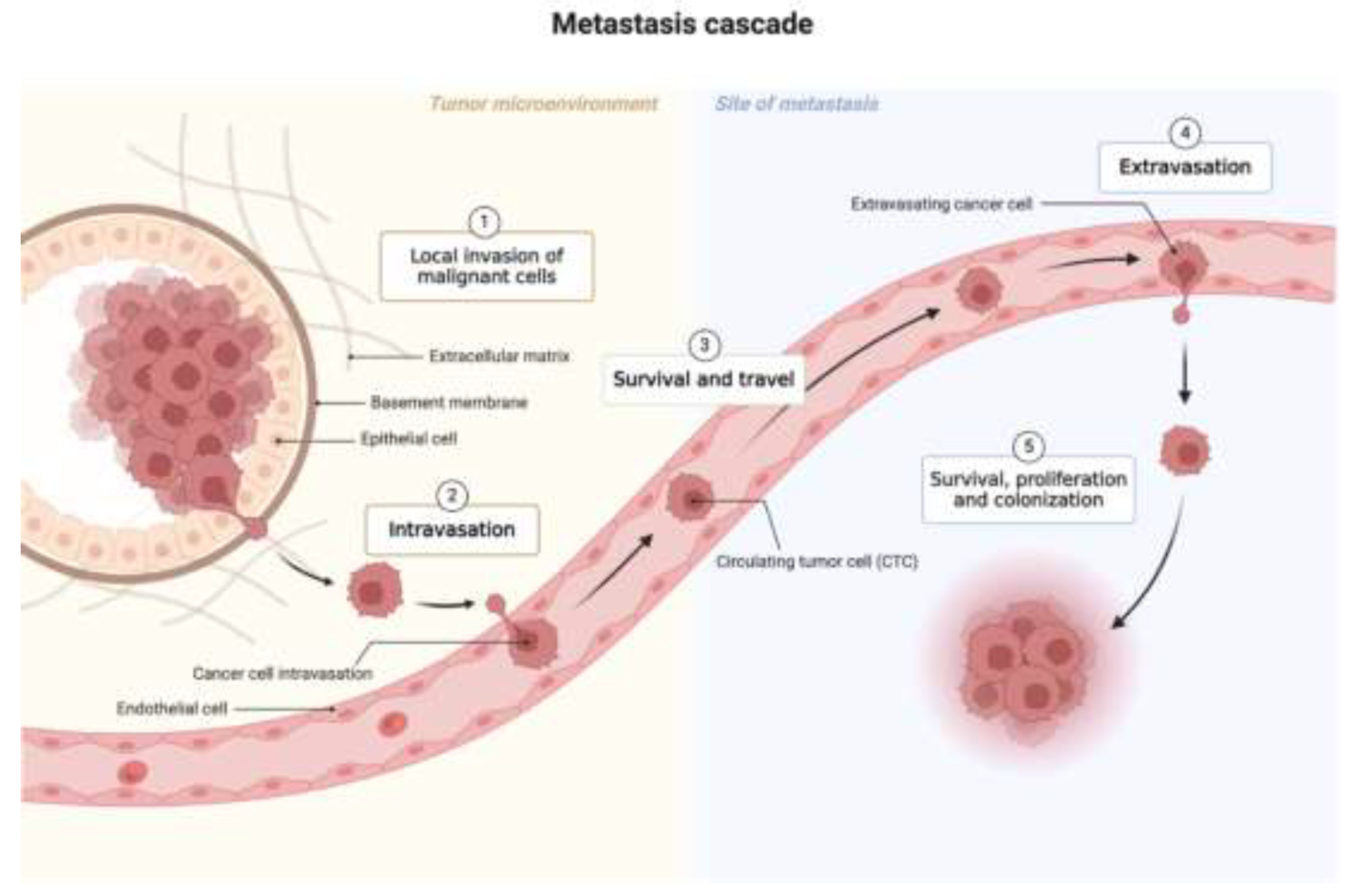
Figure 2.
Biomarkers present in liquid biopsy. This figure was created with BioRender (https://biorender.com) (accessed on 10 June 2024).
Figure 2.
Biomarkers present in liquid biopsy. This figure was created with BioRender (https://biorender.com) (accessed on 10 June 2024).
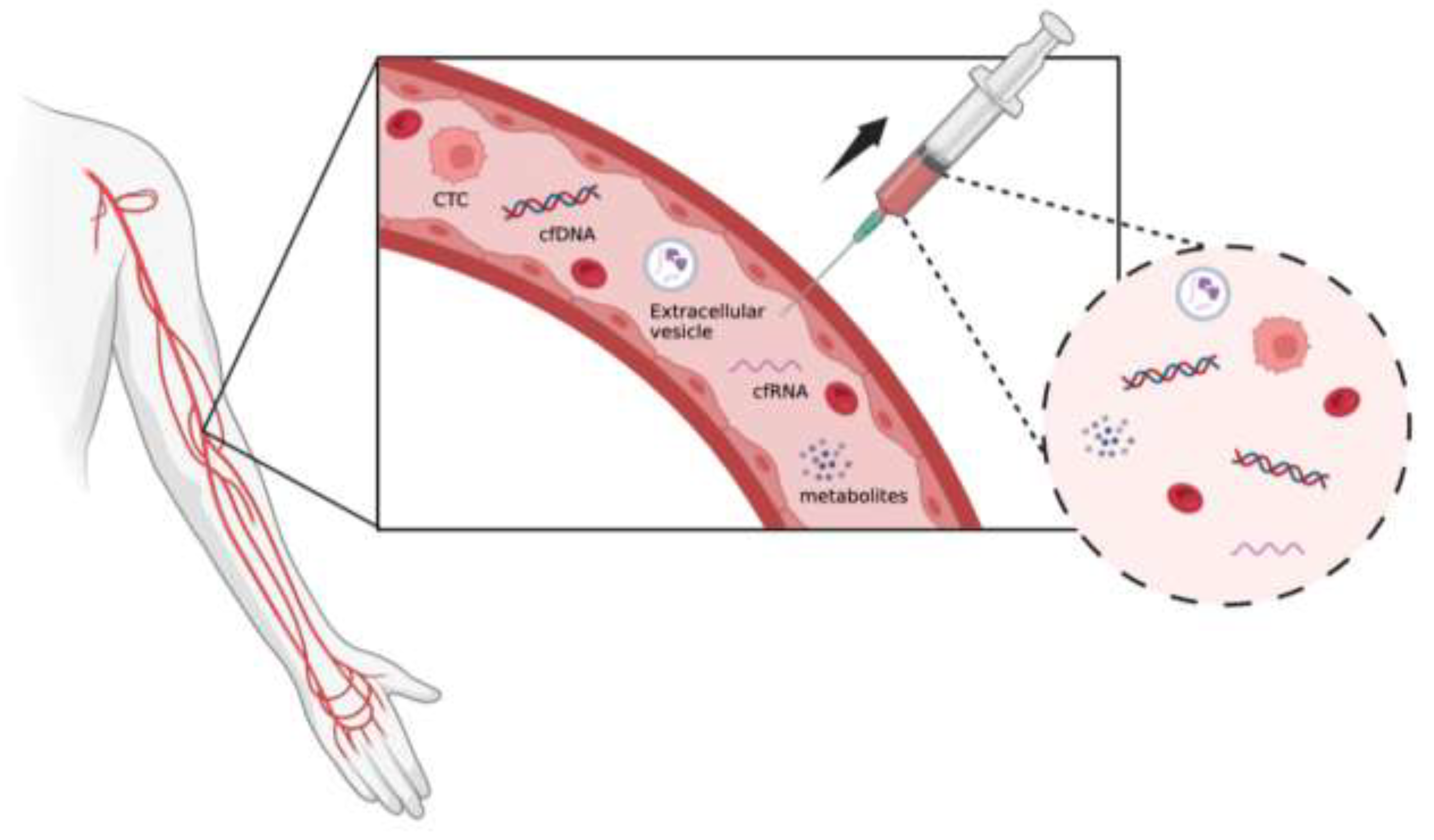
Figure 3.
Biomarkers in blood. This figure was created with BioRender (https://biorender.com) (accessed on 10 June 2024).
Figure 3.
Biomarkers in blood. This figure was created with BioRender (https://biorender.com) (accessed on 10 June 2024).
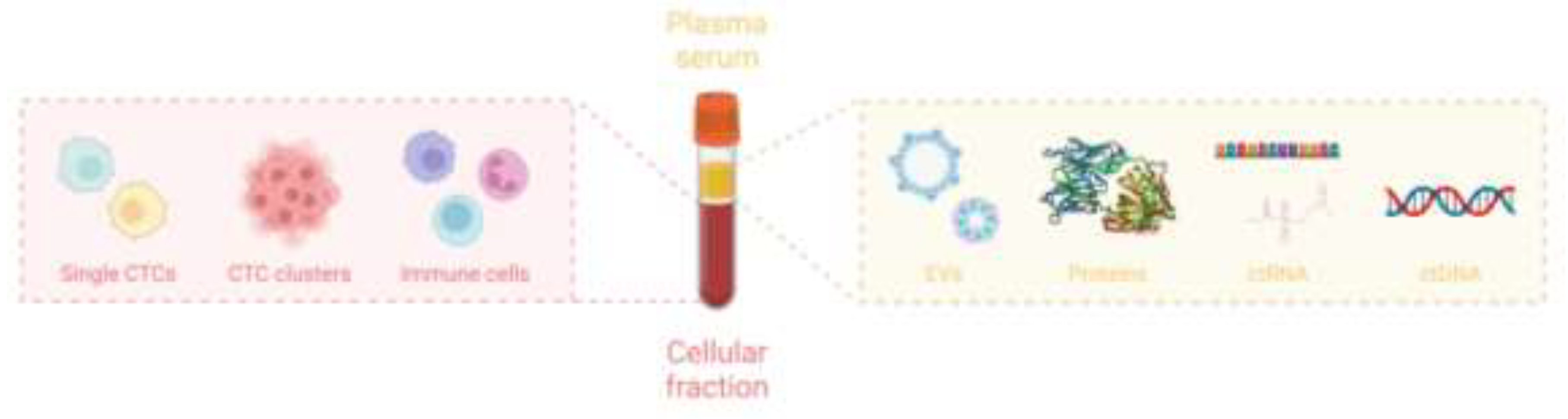
Figure 4.
The isolation methods of CTCs. This figure was created with BioRender (https://biorender.com) (accessed on 9 June 2024).
Figure 4.
The isolation methods of CTCs. This figure was created with BioRender (https://biorender.com) (accessed on 9 June 2024).
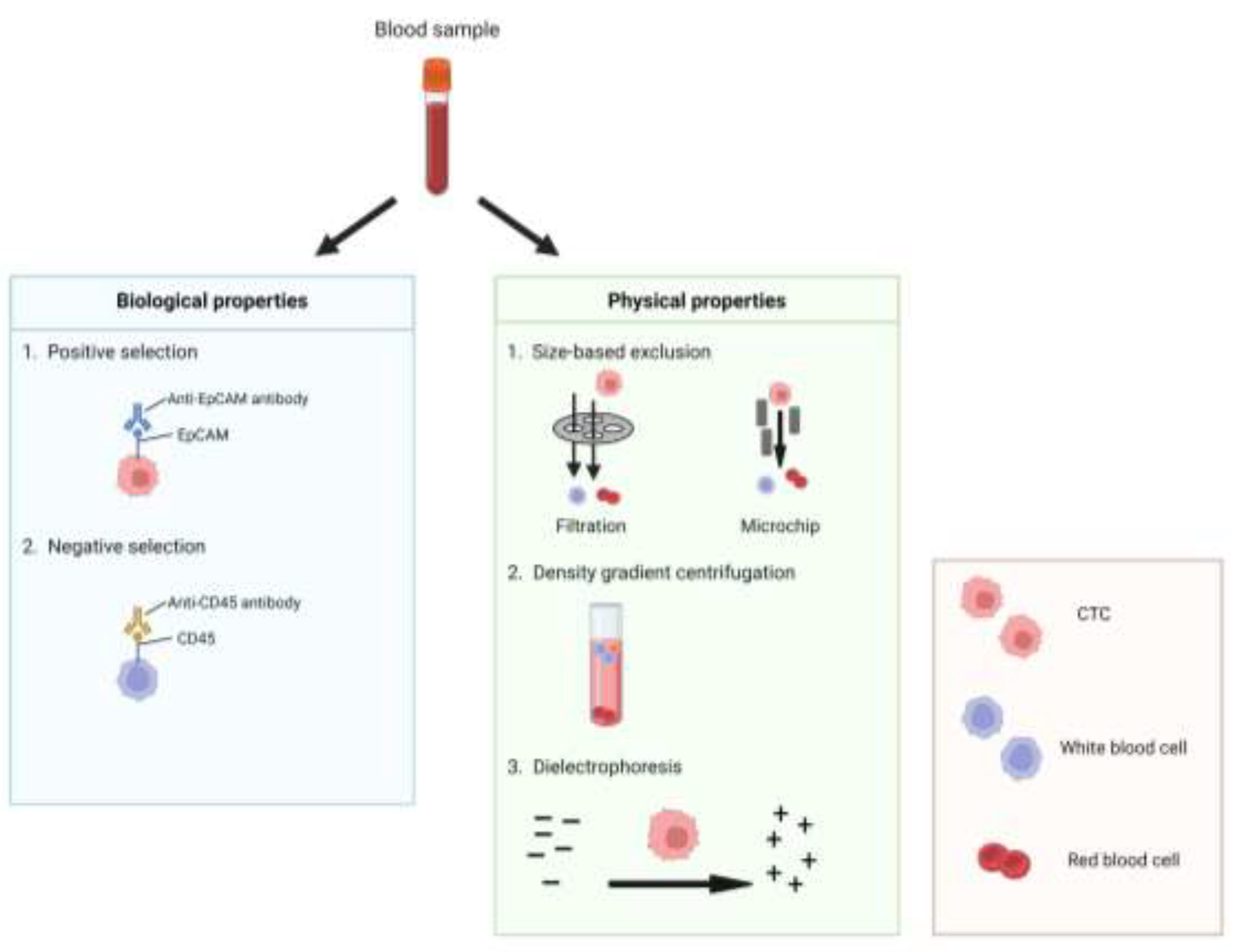
Figure 5.
The detection methods of CTCs. This figure was created with BioRender (https://biorender.com) (accessed on 9 June 2024).
Figure 5.
The detection methods of CTCs. This figure was created with BioRender (https://biorender.com) (accessed on 9 June 2024).
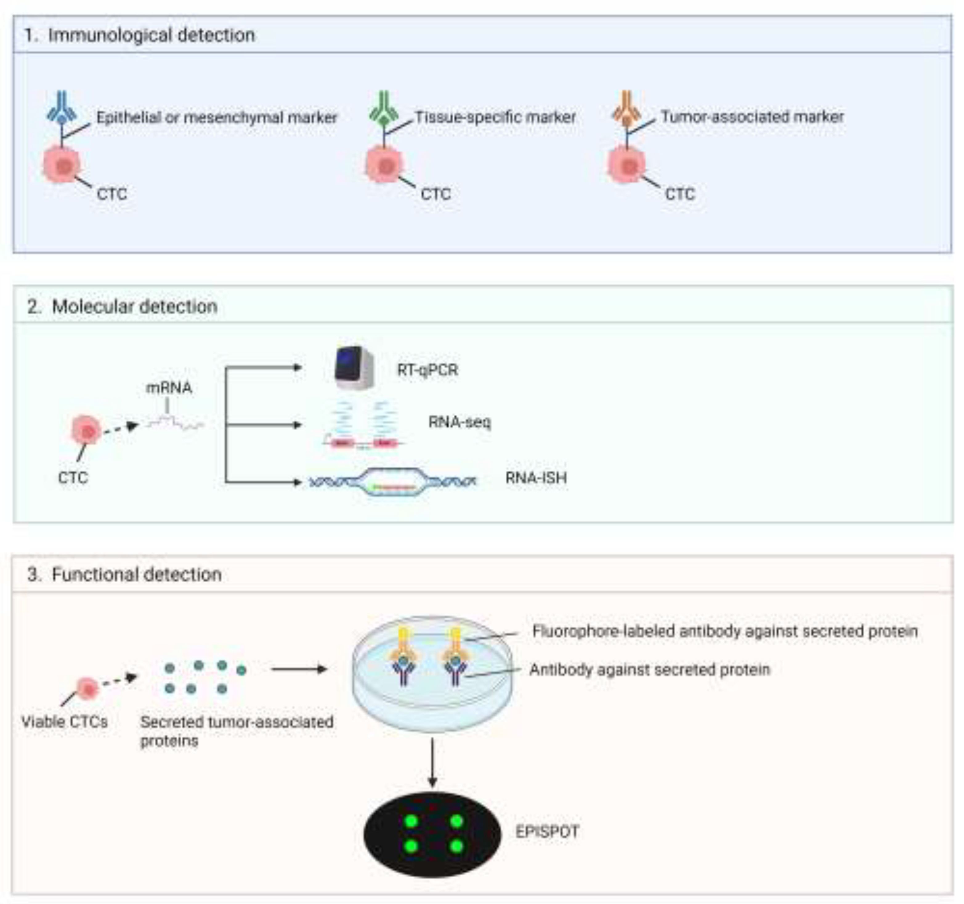

Table 1.
The comparison between liquid biopsy and tissue biopsy.
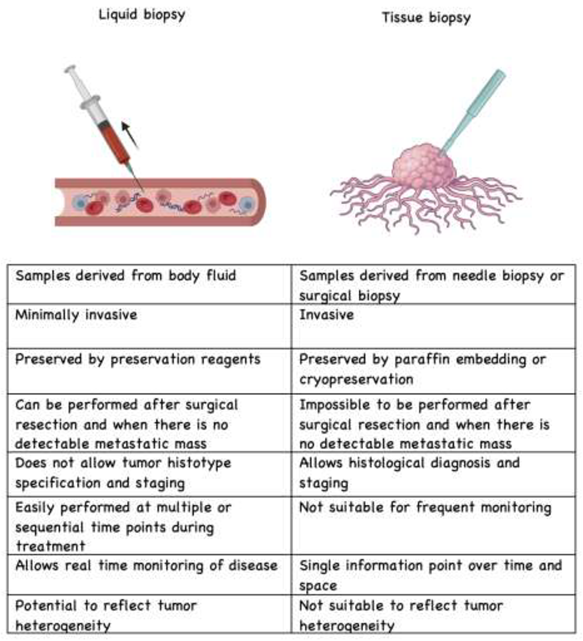 |
Table 2.
Gene and protein biomarkers in CancerSEEK.
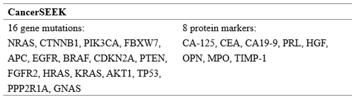 |
Disclaimer/Publisher’s Note: The statements, opinions and data contained in all publications are solely those of the individual author(s) and contributor(s) and not of MDPI and/or the editor(s). MDPI and/or the editor(s) disclaim responsibility for any injury to people or property resulting from any ideas, methods, instructions or products referred to in the content. |
© 2024 by the authors. Licensee MDPI, Basel, Switzerland. This article is an open access article distributed under the terms and conditions of the Creative Commons Attribution (CC BY) license (http://creativecommons.org/licenses/by/4.0/).
Copyright: This open access article is published under a Creative Commons CC BY 4.0 license, which permit the free download, distribution, and reuse, provided that the author and preprint are cited in any reuse.


