

Submitted:
11 June 2024
Posted:
12 June 2024
You are already at the latest version
Abstract
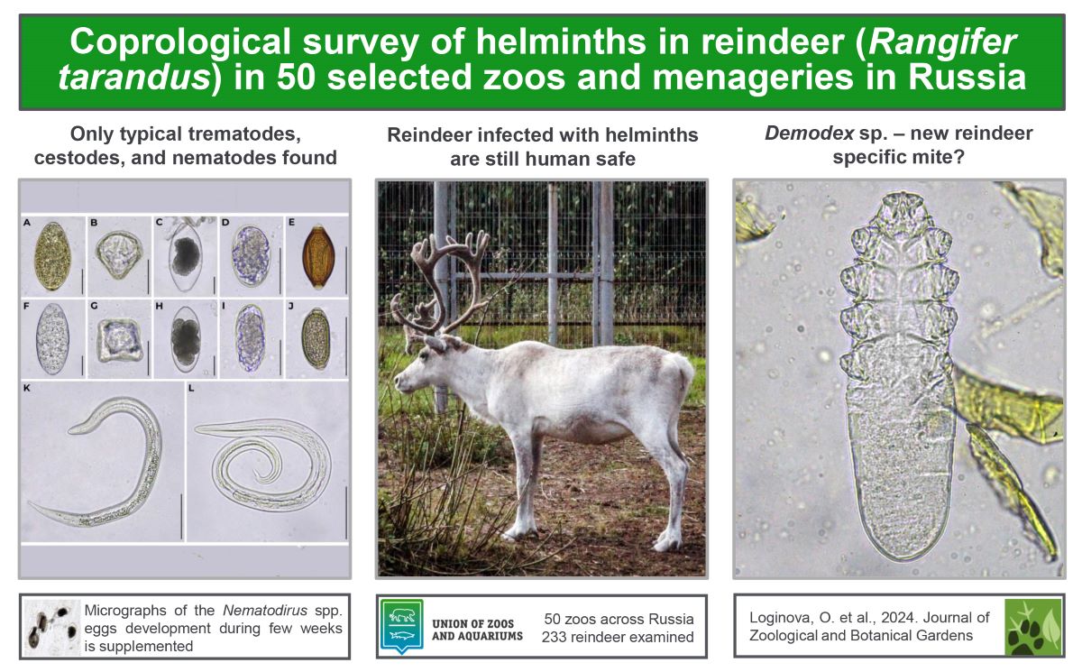
Zoo conditions are unique for reindeer, since even domestic reindeer are not kept in captive facilities like cattle. In the zoo, reindeer are usually surrounded by many different animals they would never encounter naturally. Thus they might pick up some new helminths. Numerous petting zoos raise an issue of safety of tactile communications for human visitors. Out study is the first large-scale one. Qualitative and quantitative fecal analysis was carried out for 233 reindeer distributed over 50 Russian zoos according to National Standard of the Russian Federation (GOST R 54627-2011) Ruminant animals—Methods of Laboratory Helminthological Diagnostics. Where possible, DNA analyses of helminths were performed targeting internal transcribed spacer region. In result, F. hepatica, Paramphistomum sp., Moniezia sp. (including M. expansa), gastrointestinal strongylids (including Nematodirus spp.), Dictyocaulus sp., E. rangiferi, Trichuris sp., and Capillaria sp. were found in 106 (45%) zoo reindeer. All these helminths were previously reported for reindeer and pose no direct danger for humans. Intensity of invasions was mostly low. Fecal examination might be considered as an indirect method for mange diagnostics, as Chorioptes and Demodex mites were found in reindeer fecal samples. The latter may represent a novel species of mite specific for reindeer.
Keywords:
reindeer
; Rangifer tarandus
; helminth
; trematode
; cestode
; nematode
; mite
; zoo
1. Introduction
Helminths of reindeer (Rangifer tarandus) have already been studied quite thoroughly, and the knowledge on them was summarized in thematic books [1,2,3] and chapters [4,5,6,7,8]. Ongoing studies expand our views on many aspects of reindeer helminths [9,10,11,12,13], and sometimes even add new species to the checklist of parasitic worms of Rangifer [14,15].
Reindeer exist in domestic and wild forms totaling in more than 4 mln animals worldwide [16]. Their popularity as zoo cervids in the 21st century is unprecedentedly growing. According to Species360 ZIMS database, there are more than 500 Rangifer spp. distributed in more than 150 zoos around the world [17]. Some research on reindeer helminths in captive facilities were published [3,16,18,19,20], but they are still too rare and fragmentary.
Meanwhile zoo conditions are unique for reindeer, because even domestic reindeer mostly are not kept in farms and captive facilities of the kind like cattle. In the zoo, reindeer are usually surrounded by many different animals they would never encounter naturally. It might pose a threat of picking up some new helminths. Numerous petting zoos raise another issue: the safety of such tactile communications for human visitors.
Therefore, the aim of our study was to conduct a large-scale survey of reindeer helminths in Russian zoos. Fecal examination being non-invasive method (an essential trait for zoo animals) offers wide diagnostic capabilities, scoping gastrointestinal, lung, brain, and muscle parasitic worms. This study was done to find out, what kind of helminths inhabit zoo reindeer across Russia, how widespread they are, how typical they are, and how dangerous they are both for reindeer and humans.
2. Materials and Methods
2.1. Helminths Recovery
Our study collected fecal samples from 233 reindeer kept in 50 selected zoos and menageries located in Russia (Table 1). Those include: all the state zoos in which collections reindeer are present (17); other state organizations having live reindeer for exhibition (Children's ecological station, Natural-ethnographic complex, and Arctic Tourism Center); private organizations-members of the Union of Zoos and Aquariums of Russia (5 private zoos and an urban farm); and 25 private menageries. The latter constitute of “clubs”, “parks: (“recreation-“, “ethnic-“, “fauna-“, and “safari park”), “reserve” (self-proclaimed), “mini zoo”, “ranch”, “farm”, “farmstead”, “pets' corner” and no name menageries. Many of these organizations participated in our research anonymously; therefore, for uniformity, we do not provide the actual names of all the 50 of them. However, their location combined with the type of ownership makes the data quite transparent. For brevity, further on all of these organizations would be referred to as zoos.
Qualitative and quantitative fecal analyses, light microscopic study, and measurements of obtained eggs and larvae of helminths were performed as described by Loginova et al. [21].
2.2. DNA Analysis
For trematodes, the QIAamp DNA Accessory Set, Micro kit (Quiagen, Netherlands) was used to extract DNA from embryonated eggs as described by Loginova et al. [22]. For nematodes, DNA was extracted either via digestion of first larval stages (L1) in presence of Proteinase K [23] as described by Loginova et al. [24] or by using the QIAamp DNA Accessory Set, Micro kit (Quiagen, Netherlands). We targeted the region of the internal transcribed spacer (ITS rDNA). For trematodes, we used BD1, BD2 primers [25] and corresponding protocols [26]. For nematodes we used either NC1, NC2 primers and protocols as described by Gasser et al. [27] or 18S, 26S primers [28] and protocols as described by Loginova et al. [24]. Visual check of PCR, cleaning of the PCR products, sequencing, and analysis of obtained chromatograms were performed as described by Loginova et al. [21].
3. Results
Flukes, tapeworms and round worms were found at the stage of egg or larva in feces of 106 reindeer (45%) kept in 31 zoos (62%). Identification of helminths was based on their morphology, morphometric data, and (where possible) DNA analysis (Table 2).
Appearance of eggs and larvae of helminths obtained from zoo reindeer feces is shown in Figure 1.
Prevalence rates and distribution of helminths obtained from zoo reindeer feces are summarized in Table 3. For brevity, only helminth-positive zoos are included.
Prevalence rates for total 233 reindeer examined are shown in Figure 2.
Intensity of invasion was mostly low in terms of National Standard of the Russian Federation (GOST R 54627-2011) Ruminant animals—Methods of Laboratory Helminthological Diagnostics [29]. Nematode eggs / gram of feces ranged from 1 to 56 in all reindeer samples. Nematode larvae / gram varied from 1 to 98. Cestode eggs / gram ranged from 1 to 17. Trematode eggs / gram varied from 1 to 8. Exceptions were: fecal sample of reindeer from the zoo #9 that showed 537 E.rangiferi larvae / gram; fecal sample of reindeer from the zoo #30 that showed 56 Paramphistomum sp. eggs / gram. Those numbers are estimated as medium intensity (GOST R 54627-2011).
4. Discussion
Our study revealed trematodes (Fasciola hepatica and Paramphistomum sp.), cestodes (Moniezia expansa and Moniezia sp.), and nematodes (so called small strongylids, Nematodirus spp., Dictyocaulus sp., Elaphostrongylus rangiferi, Trichuris sp., and Capillaria sp.) in zoo reindeer via fecal examination. All of these helminths were previously reported for reindeer [3,8].
In many samples, only one kind of helminths was found. However, we have also discovered combinations of two or three kinds of helminths (Table 4).
Common liver fluke F. hepatica inhabits bile ducts of its host. Previously it was reported for reindeer only in Eurasia, and one of the cases also involved zoo reindeer [3]. It is a rare finding for reindeer, and the latter is considered as an accidental host. Infestation of reindeer with F. hepatica usually is associated with other ruminants in contact and captive facilities [3]. In two private zoos where F. hepatica was found in reindeer, there were sheep, goats and sika deer (Cervus nippon) also infected.
Rumen fluke Paramphistomum sp. is widespread in Russian reindeer. When numerous, they cause atrophy of the rumen papillae and may lead to death [3,8]. Reindeer from the private zoo #30 with medium level of invasion with rumen flukes died within 6 month afterwards.
Tapeworms found were present with M. expansa (easily recognizable by its triangular eggs) and Moniezia sp. These helminths inhabit small intestine and may be quite pathogenic, especially for calves [3,8]. However, in our study it was found only in adults. Reindeer is considered an accidental host for M. expansa, and its infestation is also associated with sympatric livestock and captive facilities [3].
Supposedly, small strongylids found in zoo reindeer are of two different genera (Figure 1D,I) which may belong to fam. Chabertiidae, Cooperiidae, Haemonchidae or Trichostrongylidae. Coproculture is needed to obtain larvae and clarify the diagnosis. In any case, small strongylids inhabit gastrointestinal tract, and their impact depends on many circumstances [8,10].
Eggs of Nematodirus obtained from the reindeer fecal sample from the zoo #40 certainly belong to two different species. Apart from their differences in size and shape, their development rates were also different. Figure 1H shows Nematodirus sp. with 8 blastomeres, whereas Figure 1C shows Nematodirus sp. with gastrulated embryo. These two types of eggs were found in the same fecal sample. In total, 4 eggs (two per type) were obtained from that sample. We made an attempt to get third larval stages (L3s) for DNA analysis, but did not succeed. However, light micrographs (taken daily during three weeks after the finding of these eggs) show their embryonic development, and are available as Supplementary Materials. Helminths of Nematodirus sp. inhabit small intestine. In reindeer, they are often accompanied by Nematodirella longissimespiculata [3], but in this study the latter was not found.
Lung worm Dictyocaulus sp. is the most concerning finding, because its presence may be life threatening. In the only zoo (#30) where it was found, there was also a sika deer infected with Dictyocaulus sp. It is hard to tell, which animal was the source of invasion, or was it a coincidental infestation.
Brain worm E. rangiferi was the most widespread helminth in our research. Its pathogenicity is tricky, because it may as well cause lethal epizootics or have no manifestation of its presence [3,8,30]. Due to localization of adult worms in the nervous system or between the muscles, there is no effective treatment. On the other hand, authors observed few reindeer that were severely infected in 2018 and eventually outlived their brain worms. At least, no E. rangiferi larvae have been found in their fecal samples since 2023. Probably, it would be impossible in the wild, but in human care reindeer can live much longer and, apparently, get rid of some of their parasites naturally [Loginova, unpublished].
Whipworms Trichuris sp. inhabit large intestine and can cause hemorrhagic diarrhoea [8]. They are more expected in captive animals. There are reindeer specific species and those common to ruminants that can also infect reindeer [3]. Egg morphology is not sufficient for the identification up to the species level.
Round worm Capillaria sp. inhabits intestine and is often associated with young animals [3,8]. Still the impact of this parasite is not known. Infection with Capillaria sp. was previously reported for zoo reindeer as well [3].
Thus, via fecal examination we managed to find liver and rumen flukes, GINs, lung worms, brain worms, whipworms, and capillarids. However, this method is not suitable for those helminths that inhabit ligaments (like Onchocerca), abdominal cavity (like Setaria), muscles, lungs, liver and brain (like larval stages of Taeniidae). Therefore, despite the large scale of our study, the helminth status of zoo reindeer is still incomplete.
Another bias of this work concerns different seasons of fecal sampling (which, probably, affected reproductive activity of helminths) and differences in age and sex of studied animals [3,8]. These circumstances should be kept in mind while comparing obtained results between zoos.
As we said earlier, popularity of reindeer as zoo animals in 21 century is outstanding. Only one zoo (#17) has 100 years of its history of keeping reindeer in captivity. It is not surprising that fecal samples from their reindeer were helminth negative not only in this study, but years prior to it. Many other zoos got their first reindeer only in 2023. Majority of the private zoos purchase domestic reindeer from agricultural husbandries. The latter are all private and run by families of indigenous peoples of the North. Zoos are able to buy only those reindeer that indigenous people are willing to sell. Reindeer herders want to keep the best individuals in the herd, and reindeer for sale may be less resistant to helminths.
Domestic reindeer in contrast to wild reindeer can be pure white (leucism, not albinism) throughout the year [31,32]. White reindeer attract most people [33], and numerous private zoos tend to have exactly the white reindeer. However, reindeer herders noticed that white reindeer are not good survivors [33,34]. It might add another disadvantage to such zoo reindeer in terms of helminth resistance, and requires special research.
Apart from husbandry “heritage”, how can zoo reindeer become infected with helminths? In particular, through the birds. Helminths eggs were reported to survive through sparrow digestive system and remain invasive [35]. Thus, birds can be vectors for helminths within the zoo and out of its boundaries.
It is noteworthy that even in helminth positive zoos, about 30% (0 to 83%) reindeer fecal samples were helminth free, probably, due to the different immune status of reindeer.
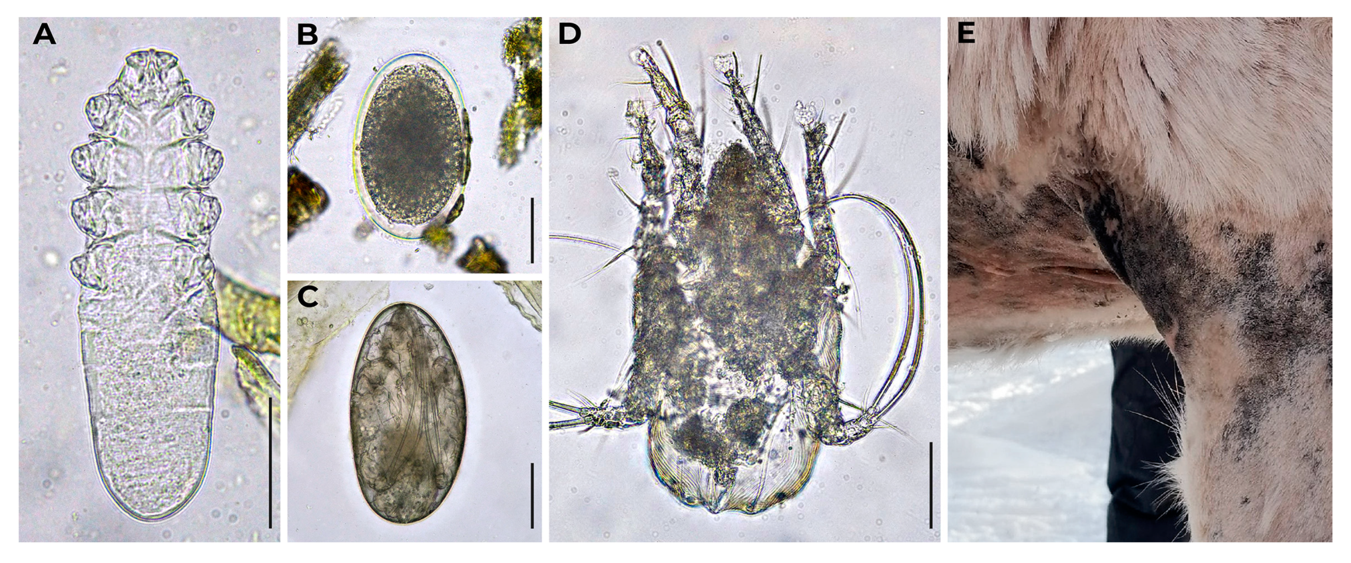
Besides helminths, we found mites in reindeer fecal samples (Figure A1). They were either ingested during autogruming and passed digestive tract or contaminated fecal samples afterwards. Having found a mite egg in the sample from the zoo #30, we contacted that zoo and shared our considerations. It turned out that the zoo indeed had problems with mange caused by mites (Figure A1E). One adult mite found in a sample from the zoo #40 was identified as Chorioptes sp. (Figure A1D). Chorioptes mites were reported for reindeer in Canada, Norway, and Finland [8]. Another adult mite found in reindeer fecal sample (zoo #12) met morphological criteria for Demodex sp. To authors’ knowledge, Demodex has never been reported for reindeer [3,8,36]. This finding also requires additional research which may result in the description of the new species.
Tactile contact between humans and animals in petting zoos may pose a risk of helminth infestation. For instance, red fox (Vulpes vulpes) and Arctic fox (V. lagopus) are recognized definitive hosts for Echinococcus spp. The latter produces numerous eggs which are shed with feces. Those eggs are very light and flying, and easily contaminate animal’s fur. Stroking such sick fox or even breathing closely to it may result in ingestion of Echinococcus eggs which are instantly invasive [37].
Among helminths found in zoo reindeer in our research, there are few genera, reported for humans. Those are Fasciola, GINs (Trichostrongylus, Oesophagostomum), Trichuris, and Capillaria [37,38,39,40]. However, infected reindeer pose no direct threat for human visitors or zoo employees for the following reasons. Trematode F. hepatica is indeed the species, that can infect both cervids and humans, but its life cycle requires intermediate host (fresh water mollusk) [3]. Eggs shed by reindeer are not the infective stage for any vertebrate host. Moreover, eggs are excreted without miracidium, and it takes time for it to develop (that is possible only in water). Species of Trichostrongylus, Oesophagostomum, Trichuris, and Capillaria that can infect humans are not the same species that infect reindeer [3,8,37,40]. Besides, all these nematodes shed eggs that are not embryonated by the moment of their excretion with feces. Larvae development also requires few days or weeks. Thus, if reindeer dung is regularly removed, even reindeer infected with helminths are safe for humans.
5. Conclusions
In our study F. hepatica, Paramphistomum sp., Moniezia spp. (including M. expansa), gastrointestinal strongylids (including Nematodirus spp.), Dictyocaulus sp., E. rangiferi, Trichuris sp., and Capillaria sp. were found in 106 Russian zoo reindeer out of 233 ones via fecal examination. All these helminths were previously reported for reindeer and pose no direct danger for humans. Intensity of invasions was mostly low. Fecal examination might be considered as an indirect method for mange diagnostics, as Chorioptes and Demodex mites were found in reindeer fecal samples. The latter may represent a novel species of mite specific for reindeer.
Supplementary Materials
The following supporting information can be downloaded at the website of this paper posted on Preprints.org, Figures S1–S25: Light microscopic images of the two types of Nematodirus eggs showing the development of embryos from 13 December 2023 to 04 January 2024 respectively. Eggs were obtained from the same fecal sample from a reindeer from the zoo #40. Eggs were placed in a watch glass filled with tap water. Pictures were taken via photo camera of the smart phone Xperia2 DS Black (H4113) (Sony, China) and the optical microscope Micmed-6 (LOMO-MA, Russia).
Author Contributions
Conceptualization, O.A.L. and S.V.A.; methodology, O.A.L., D.N.E., S.E.S., and L.M.B.; formal analysis, O.A.L.; investigation, O.A.L. and S.E.S.; resources, O.A.L., S.V.A., D.N.E., N.S.E., S.E.S., Iu.K.P., L.M.B., Yu.E.K., D.I.Ch., A.A.K., Iu.V.V., D.A.G., Yu.A.Shch., I.A.M., D.V.P., M.G.B., T.P.S.; writing—original draft preparation, O.A.L.; writing—review and editing, S.V.A., D.N.E., N.S.E., S.E.S., Iu.K.P., L.M.B., Yu.E.K., D.I.Ch., A.A.K., Iu.V.V., D.A.G., Yu.A.Shch., I.A.M., D.V.P., M.G.B., T.P.S.; visualization, O.A.L.; supervision, L.M.B. and S.E.S.; project administration, S.V.A.; funding acquisition, I.A.M. All authors have read and agreed to the published version of the manuscript.
Funding
Part of this article was prepared within the framework of the State assignment for the N. Laverov Federal Center for Integrated Arctic Research of the Ural Branch of the Russian Academy of Sciences “Scientific foundations and social and cultural factors for the conservation and use of the potential of biological diversity in the European North and the Arctic” (registration number – 122011400382-8)”. Another part of this study was performed under State order of IB KRC RAS (project FMEN-2022–0003).
Institutional Review Board Statement
Not applicable.
Data Availability Statement
Data are contained within the article.
Generative AI in Scientific Writing Statement: During the preparation of this work the authors did not use any AI-tool / service.
Acknowledgments
Authors are deeply grateful to all the zoo administrators, veterinarians, keepers, and workers (as well as to people not involved in zoo work directly) who assisted in fecal sampling and made this assistance possible. In particular, we would like to thank Alexander Makhov (Moscow), Kakhramon Khuzhanov (Moscow), Nelly Khafizova (Moscow), Andrey Soludkov (Moscow), Igor Tsaryov (Moscow), Andrey Kajdalov (Saint Petersburg), Lyudmila Zakharova (Saint Petersburg), Kristina Zabarina (Samara), Nataliya Chervyakova (Moscow), Vera Shiryaeva (Saint Petersburg), Irina Pavlova (Saint Petersburg), Daria Zimina (Izhevsk), Elena Kolesnikova (Khabarovsk), Vitaliy Soroka (Saint Petersburg), Irina Chuprak (Saint Petersburg), Irina Suvorova (Moscow), Evgeniy Popov and Alexander Sokolov with the Veterinary service of the Yamalo-Nenets Autonomous Okrug (Salekhard), and Vladimir Vasilev with the Northern Forum (Yakutsk). We also extend our gratitude to our colleagues who assisted in mite identification: Dr. Sergey Mironov with the Zoological Institute of the Russian Academy of Sciences (Saint Petersburg) and Dr. Nadezhda Gavrilova with the Saint Petersburg State University of Veterinary Medicine (Saint Petersburg). Special thanks to Dr. Dmitry Kuznetsov for his suggestion to write this article.
Conflicts of Interest
The authors declare no conflict of interest.
Appendix A
Mites found in the feces of zoo reindeer and manifestation of mange caused by mites in reindeer is shown in Figure A1.
Figure A1.
Mites of reindeer. (A) Demodex sp. obtained from feces of reindeer from the zoo #12; (B) Egg of mite at early developmental stage obtained from the feces of reindeer from the zoo #30; (C) Egg of mite at late developmental stage obtained from the feces of reindeer from the zoo #48; (D) Chorioptes sp. obtained from the feces of reindeer from the zoo #40; (E) Manifestation of mange caused by mites in reindeer from the zoo #30 (hairless front leg and chest are shown); photo courtesy: Kristina Zabarina. Light micrographs were made via bright field microscopy, 400× magnification. Scale bar equals 50 μm.
Figure A1.
Mites of reindeer. (A) Demodex sp. obtained from feces of reindeer from the zoo #12; (B) Egg of mite at early developmental stage obtained from the feces of reindeer from the zoo #30; (C) Egg of mite at late developmental stage obtained from the feces of reindeer from the zoo #48; (D) Chorioptes sp. obtained from the feces of reindeer from the zoo #40; (E) Manifestation of mange caused by mites in reindeer from the zoo #30 (hairless front leg and chest are shown); photo courtesy: Kristina Zabarina. Light micrographs were made via bright field microscopy, 400× magnification. Scale bar equals 50 μm.
References
- Skrjabin, K.I. Glistnye invazii severnogo olenya (Helminth Diseases of Reindeer); Selkhozgiz: Moscow – Leningrad, USSR, 1931; 88 pp. (In Russian).
- Polyanskaya, M.V. Gel'mintosy severnykh oleney (Helminthiases of Reindeer); Knizhnoe Izdatelstvo: Murmansk, USSR, 1963; 47 pp. (In Russian).
- Mizkewitsch, V.Yu. Gel'minty severnogo olenya i vyzyvayemyye imi zabolevaniya (Reindeer Helminths and the Diseases They Cause); Kolos: Leningrad, USSR, 1967; 308 pp. (In Russian).
- Golosov, I.M.; Mizkewitsch, V.Yu. Helminthiases. In Parazitarnye bolezni severnykh oleney (Parasitic Diseases of Reindeer); Lyzhin, K., Ed.; Krasnoyarskoe Knizhnoe Izdatelstvo: Krasnoyarsk, USSR, 1964; pp. 57–141. (In Russian).
- Laaksonen, S. Trematoda. Cestoda. Nematoda. In TUNNE PORO. Poron sairaudet ja terveydenhoito (FEEL THE REINDEER. Reindeer Diseases and Health Care); Livonia Print: Riga, Latvia, 2016, pp. 220–251. (In Finnish).
- Belova, L.M.; Loginova, O.A. Helminthiases. In Bolezni severnykh oleney (Diseases of Reindeer); Zabrodin, V.A., Laishev, K.A., Eds.; Knizhnoe Izdatelstvo: Saint Petersburg – Pushkin, Russia, 2019; pp. 94–113. (In Russian).
- Kutz, S.J.; Ducrocq, J.; Verocai, G.G.; Hoar, B.M.; Colwell, D.D.; Beckmen, K.B.; Polley, L.; Elkin, B.T.; Hoberg, E.P. Parasites in ungulates of Arctic North America and Greenland: A view of contemporary diversity, ecology, and impact in a world under change. Adv. Parasitol. 2012, 79, 99–252. [CrossRef]
- Kutz, S.J.; Laaksonen, S.; Asbakk, K.; Nilssen, A.C. Helminths. In Reindeer and Caribou. Health and Disease; Tryland, M., Kutz, S.J., Eds.; c, 2019, pp. 180–205.
- Hrabok, J.T.; Oksanen, A.; Nieminen, M.; Waller, P.J. Population dynamics of nematode parasites of reindeer in the sub-arctic. Vet. Parasitol. 2006, 142, 301–311. [CrossRef]
- Jokelainen, P.; Moroni, B.; Hoberg, E.; Oksanen, A.; Laaksonen, S. Gastrointestinal parasites in reindeer (Rangifer tarandus tarandus): A review focusing on Fennoscandia. Vet. Parasitol. Reg. Stud. Rep. 2019, 17, 100317. [CrossRef]
- Emelyanova, A.; Savolainen, A.; Oksanen, A.; Nieminen, P.; Loginova, O.; Abass, K.; Rautio, A. c. Int. J. Environ. Res. Public Health 2022, 19, 11260:1–11260:20. [CrossRef]
- Dickinson, E.R.; Orsel, K.; Cuyler, C.; Kutz, S.J. Life history matters: Differential effects of abomasal parasites on caribou fitness, Int. J. Parasitol. 2023, 53, 221–231. [CrossRef]
- Andrew, C.L.; Wagner, B.; Harms, N.J.; Jenkins, E.J.; Jung, T.S. Comparative Prevalence and Intensity of Endoparasites in a Dynamic Boreal Ungulate Community. Diversity 2024, 16, 230. [CrossRef]
- Verocai, G.G.; Kutz, S.J.; Simard, M.; Hoberg, E.P. Varestrongylus eleguneniensis sp. n. (Nematoda: Protostrongylidae): A widespread, multi-host lungworm of wild North American ungulates, with an emended diagnosis for the genus and explorations of biogeography. Parasites Vectors 2014, 7, 1–22. [CrossRef]
- Loginova, O.A.; Kolpashchikov, L.A.; Spiridonov, S.E. First report of Orthostrongylus sp. (Nematoda: Protostrongylidae) in wild reindeer (Rangifer tarandus) from the Taimyr, Russia: Nearctic parasites in a Palearctic host. Parasitol. Res. 2023, 122, 685–689. [CrossRef]
- Tryland, M.; Kutz, S.J. Reindeer and Caribou. Health and Disease; CRC Press (Taylor and Francis Group): Boca Raton, USA – London, GB – New York, USA, 2019; p. 3. ISBN 978-1-4822-5068-8.
- Species 360. Data science for zoos and aquariums. Available online:https://species360.org/products-services/zoo-aquarium-animal-management-software-2/ (accessed on 31 May 2024).
- Haigh, J.C.; Mackintosh, C.; Griffin, F. Viral, parasitic and prion diseases of farmed deer and bison. Revue Scientifique et Technique (International Office of Epizootics) 2002, 21, 219–248. [CrossRef]
- Flach, E. Cervidae and Tragulidae. In Zoo and Wild Animal Medicine, 5th ed.; Fowler, M.E., Miller, E.R, Eds.; Elsevier Science: Amsterdam, Netherlands, 2003, pp. 634–649.
- Goossens, E.; Vercruysse, J.; Boomker, J.; Vercammen, F.; Dorny, P. A 12-month survey of gastrointestinal helminth infections of cervids kept in two zoos in Belgium. J. Zoo Wildl. Med. 2005, 36, 470–478. [CrossRef]
- Loginova, O.A.; Rozenfeld, S.B.; Sipko, T.P., Mizin, I.A.; Panchenko, D.V.; Laishev, K.A.; Bondar, M.G.; Kolpashchikov, L.A.; Gruzdev, A.R.; Kulemeev, P.S.; Litovka, D.A.; Semerikova, M.N.; Mamontov, V.N.; Mamaev, E.G.; Spiridonov, S.E. Diversity and Distribution of Helminths in Wild Ruminants of the Russian Arctic: Reindeer (Rangifer tarandus), Muskoxen (Ovibos moschatus), and Snow Sheep (Ovis nivicola). Diversity 2023, 15, 672:1–672:15. [CrossRef]
- Loginova, O.; Efeykin, B.; Krutikova, A.; Mizin, I.; Spiridonov, S. Fasciola hepatica: updates on egg morphology, host range, and distribution. Food Waterborne Parasitol. 2024, submitted.
- Holterman, M.; van der Wurff, A.; van den Elsen, S.; van Megen, H.; Bongers, T.; Holovachov, O.; Bakker, J.; Helder, J. Phylum-Wide Analysis of SSU rDNA Reveals Deep Phylogenetic Relationships among Nematodes and Accelerated Evolution toward Crown Clades. Mol. Biol. Evol. 2006, 23, 1792–1800. [CrossRef]
- Loginova, O.A; Belova, L.M.; Spiridonov, S.E. The First Report on Elaphostrongylus rangiferi (Reindeer Invasive Parasite) in Leningrad Oblast. Russ. J. Biol. Invasions 2022, 13, 232–244. [CrossRef]
- Luton, K.; Walker, D.; Blair, D. Comparisons of ribosomal internal transcribed spacers from two congeneric species of flukes (Platyhelminthes: Trematoda: Digenea). Mol. Biochem. Parasitol. 1992, 56, 323–327. [CrossRef]
- Mahami-Oskouei, M.; Dalimi, A.; Forouzandeh-Moghadam, M.; Rokni, M. B. Molecular identification and differentiation of Fasciola isolates using PCR-RFLP method based on internal transcribed spacer (ITS1, 5.8 S rDNA, ITS2). Iranian J. Parasitol. 2011, 6, 35–42.
- Gasser, R.B.; Chilton, N.B.; Hoste, H.; Beveridge, I. Rapid sequencing of rDNA from single worms and eggs of parasitic helminths. Nucleic Acids Res. 1993, 21, 2525–2526. [CrossRef]
- Vrain, T.C.; Wakarchuk, D.A.; Levesque, A.C.; Hamilton, R.I. Intraspecific rDNA restriction fragment length polymorphism in the Xiphinema americanum group. Fundam. Appl. Nematol. 1992, 15, 563–573.
- Latypov, D.G.; Timerbayeva, R.R.; Kirillov, E.G. Parazitologiya i Invazionnyye Bolezni Zhvachnykh Zhivotnykh (Parasitology and Invasive Diseases of Ruminants); Lan: St. Petersburg, Russia, 2019; pp. 59–67. ISBN 978-5-8114-3561-6. (In Russian).
- Handeland, K.; Slettbakk, T. Outbreaks of Clinical Cerebrospinal Elaphostrongylosis in Reindeer (Rangifer tarandus tarandus) in Finnmark, Norway, and their Relation to Climatic Conditions. J. Vet. Med., Series B 1994, 41, 407–410. [CrossRef]
- Baskin, L.M. Odomashnivaniye severnogo olenya. Ot okhotnika do pastukha i ranchevoda (Reindeer domestication. From hunter to herdsman and rancher); Tovarishchestvo nauchnykh izdaniy KMK: Moscow, Russia, 2021; p. 280. ISBN 978-5-907372-89-4. (In Russian).
- Holand, Ø.; Mizin, I.; Weladji, R.B. Reindeer Rangifer tarandus (Linnaeus, 1758). In Handbook of the Mammals of Europe; Hackländer, K., Zachos, F.E., Eds.; Springer: Cham, Switzerland, 2022, pp. 247–277. ISBN 978-3-030-24474-3. [CrossRef]
- Smith, T. Velvet Antlers, Velvet Nose. The Story of a Reindeer Family; Coronet Books, Hodder and Stoughton: London, UK, 1995; p. 90. ISBN 0-340-66003-1.
- Wella, Yu. Azbuka Olenevoda (Reindeer Herder's ABC); Studiya O.K.: Surgut, Russia, 2011, pp. 43. (In Russian).
- Mosgovoy, A.A. Sparrows as agent distributing helminthic infections among domestic animals. In Papers on Helminthology; Schultz, R.-E.S., Gnedina, M.P., Eds.; All-Union Lenin Academy of Agricultural Sciences: Moscow, USSR, 1937, pp. 398–402. (In Russian).
- Izdebska, J.N.; Fryderyk, S. Demodex acutipes Bukva et Preisler, 1988 (Acari, Demodecidae) – a rare parasite of red deer (Cervus elaphus L.). Ann. Parasitol. 2012, 58, 161–166.
- Vasilevich, F.I., Belova, L.M., Burmistrova, M.I. Parazitarnye zoonozy (Parasitic Zoonoses); ZooVetKniga: Moscow, Russia, 2020, pp. 21–28. ISBN 978-5-6045650-5-6. (In Russian).
- Mas-Coma, S.; Bargues, M.D.; Valero, M.A. Human fascioliasis infection sources, their diversity, incidence factors, analytical methods and prevention measures. Parasitology 2018, 145, 1665–1699. [CrossRef]
- Lattes, S.; Ferte, H. Delaunay, P.; Depaquit, J.; Vassallo, M.; Vittier, M.; Kokcha, S.; Coulibaly, E.; Marty, P. Trichostrongylus colubriformis Nematode Infections in Humans, France. Emerg. Inf. Dis. 2011, 17, 1301–1302. [CrossRef]
- Dhaliwal, B.B.S.; Juyal, P.D. Textbook of Parasitic Zoonoses; Springer Nature: Singapore, Republic of Singapore, 2022, pp. 46–110. ISBN 978-81-322-1550-9.
Figure 1.
Diagnostic stages of helminths obtained from feces of zoo reindeer. (A) Fasciola hepatica egg; (B) Moniezia expansa egg; (C) Nematodirus sp. egg at late stage of embryonic development; (D) Rounded Strongyle-type egg; (E) Trichuris sp. egg; (F) Paramphistomum sp. egg; (G) Moniezia sp. egg; (H) Nematodirus sp. egg at the early stage of embryonic development; (I) Elongated Strongyle-type egg; (J) Capillaria sp. egg; (K) Dictyocaulus sp. L1; (L) Elaphostrongylus rangiferi L1. Bright field microscopy, 400× magnification. Scale bar equals 50 μm.
Figure 1.
Diagnostic stages of helminths obtained from feces of zoo reindeer. (A) Fasciola hepatica egg; (B) Moniezia expansa egg; (C) Nematodirus sp. egg at late stage of embryonic development; (D) Rounded Strongyle-type egg; (E) Trichuris sp. egg; (F) Paramphistomum sp. egg; (G) Moniezia sp. egg; (H) Nematodirus sp. egg at the early stage of embryonic development; (I) Elongated Strongyle-type egg; (J) Capillaria sp. egg; (K) Dictyocaulus sp. L1; (L) Elaphostrongylus rangiferi L1. Bright field microscopy, 400× magnification. Scale bar equals 50 μm.
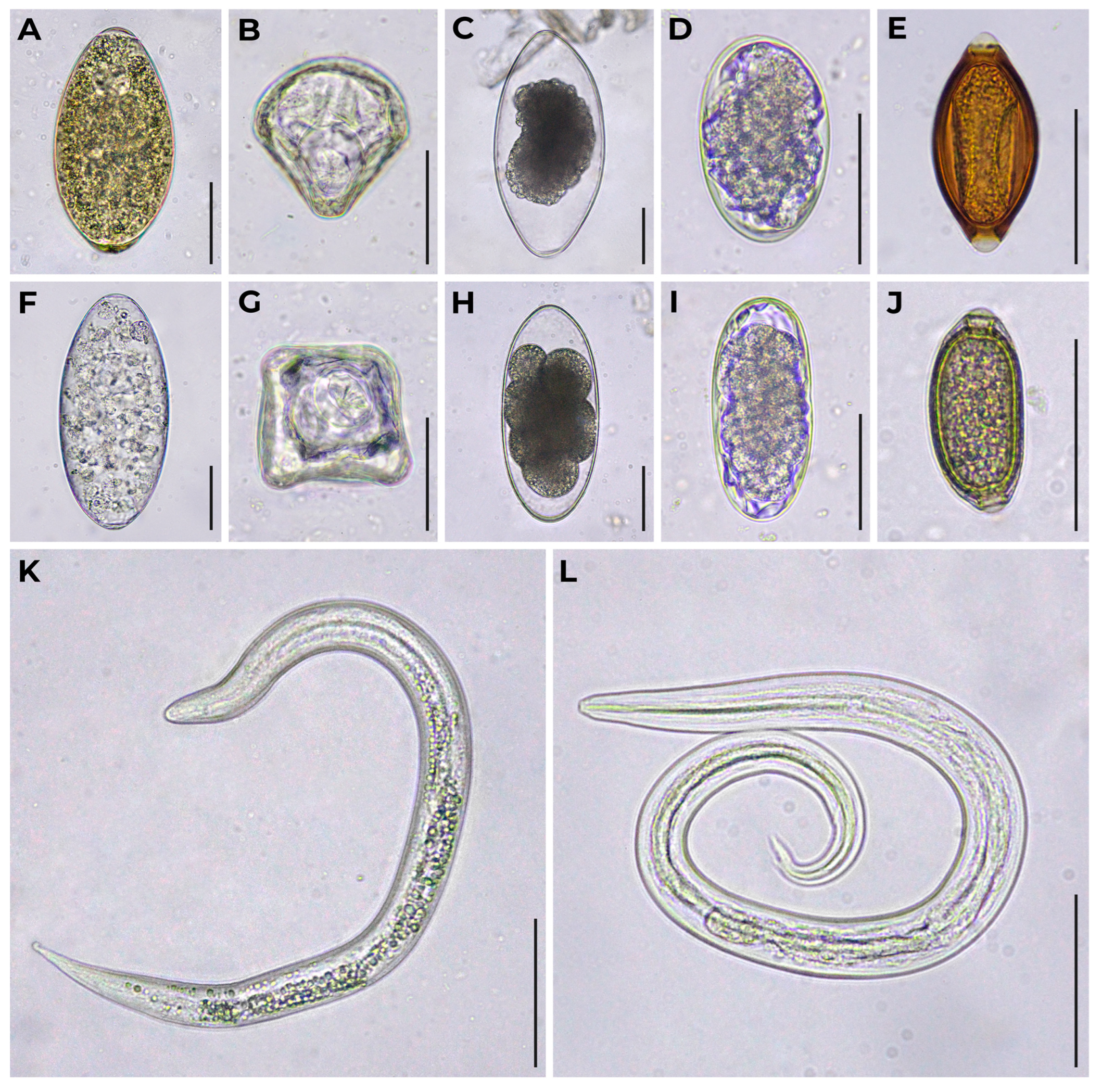
Figure 2.
Prevalence rates (%) of helminths found in feces of zoo reindeer.
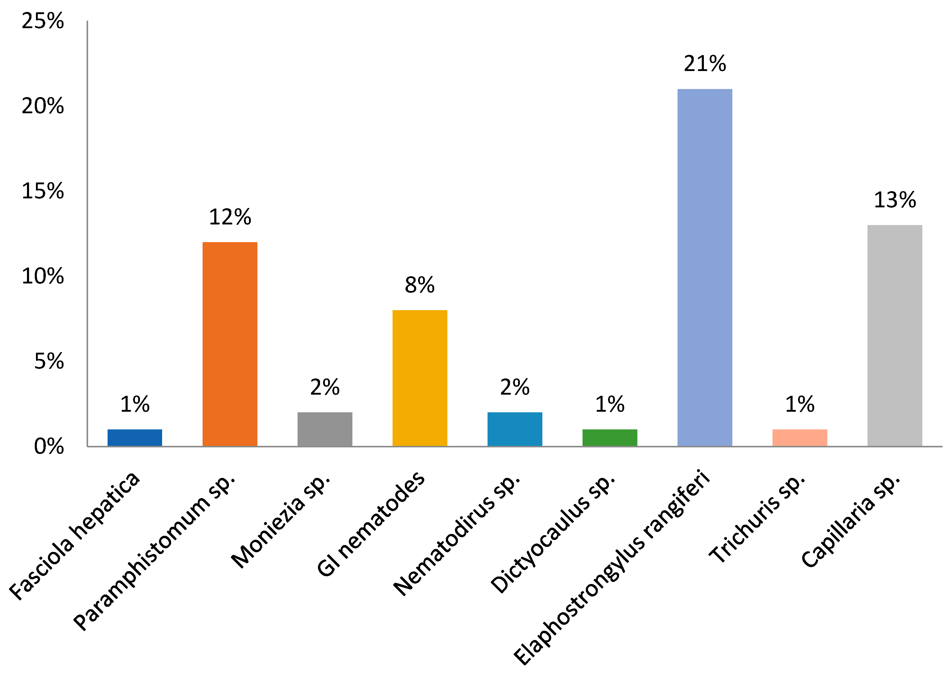

Table 1.
Collection data for fecal samples from reindeer (Rangifer tarandus) in the Russian zoos (Federal subjects arranged from the West to the East).
Table 1.
Collection data for fecal samples from reindeer (Rangifer tarandus) in the Russian zoos (Federal subjects arranged from the West to the East).
| Zoo ID | Ownership Type (S/P)1 | Location (Federal Subject of Russia) | Coordinates (Decimal Degrees) |
Number of Fecal Samples3 | Date Collected |
|---|---|---|---|---|---|
| 1 | P | Murmansk Oblast | 68.98716 33.07261 |
2 | November 2023 |
| 2 | P | Murmansk Oblast | 68.85722 33.19556 |
12 | November 2023 |
| 3 | P | Murmansk Oblast | 67.56317 33.36571 |
2 | November 2023 |
| 4 | P | Murmansk Oblast | 67.65276 33.66051 |
11 | November 2023 |
| 5 | P | Murmansk Oblast | 69.16835 35.13280 |
3 | November 2023 |
| 6 | P | Republic of Karelia | 65.76340 31.07419 |
1 | March 2024 |
| 7 | P | Republic of Karelia | 66.43669 32.85459 |
7 | June 2022 |
| 8 | P | Republic of Karelia | 62.33297 34.00604 |
3 | March 2024 |
| 9 | P | Republic of Karelia | 61.87838 34.07819 |
9 | March 2024 |
| 10 | P | Leningrad Oblast | 60.59067 30.00422 |
1 | July 2020 |
| 11 | P2 | Leningrad Oblast | 60.59161 30.11204 |
8 | May 2019 |
| 12 | P | Leningrad Oblast | 60.14195 30.32823 |
11 | August 2018 |
| 13 | P | Leningrad Oblast | 59.94762 30.68122 |
3 | May 2023 |
| 14 | P | Saint Petersburg | 59.84108 30.06470 |
2 | June 2019 |
| 15 | P | Saint Petersburg | 59.98017 30.24438 |
3 | March 2024 |
| 16 | P | Saint Petersburg | 59.97054 30.25672 |
4 | January 2020 |
| 17 | S2 | Saint Petersburg | 59.95210 30.30891 |
9 | March 2024 |
| 18 | P | Saint Petersburg | 59.67605 30.42401 |
2 | February 2018 |
| 19 | P | Tver Oblast | 56.75051 36.36719 |
1 | June 2020 |
| 20 | S2 | Moscow Oblast | 55.94012 36.21055 |
7 | January 2024 |
| 21 | P | Moscow Oblast | 56.13343 36.50051 |
2 | March 2024 |
| 22 | S2 | Moscow | 55.76347 37.57537 |
3 | December 2023 |
| 23 | P2 | Moscow | 55.83306 37.62197 |
1 | October 2023 |
| 24 | S2 | Yaroslavl Oblast | 57.67709 39.90005 |
6 | November 2023 |
| 25 | P2 | Nizhny Novgorod Oblast |
56.33468 43.85420 |
6 | November 2023 |
| 26 | P | Nizhny Novgorod Oblast |
56.92384 45.40185 |
4 | August 2022 |
| 27 | S2 | Republic of Mordovia | 54.17500 45.18599 |
3 | November 2023 |
| 28 | S2 | Vologda Oblast | 60.74784 46.17739 |
5 | November 2023 |
| 29 | P2 | Ulyanovsk Oblast | 54.35485 48.52409 |
3 | February 2024 |
| 30 | P | Samara Oblast | 53.34493 50.22240 |
21 | August 2021 |
| 31 | S2 | Udmurt Republic | 56.86555 53.17413 |
2 | November 2023 |
| 32 | S | Nenets Autonomous Okrug | 67.63436 53.24135 |
1 | February 2024 |
| 33 | S2 | Perm Krai | 58.01672 56.23728 |
4 | November 2023 |
| 34 | P | Sverdlovsk Oblast | 57.17585 60.65764 |
11 | February 2024 |
| 35 | S2 | Chelyabinsk Oblast | 55.16894 61.36764 |
5 | November 2023 |
| 36 | P | Tyumen Oblast | 56.99540 65.73485 |
7 | February 2024 |
| 37 | S | Yamalo-Nenets Autonomous Okrug |
66.59257 66.85846 |
3 | January 2024 |
| 38 | S2 | Yamalo-Nenets Autonomous Okrug |
66.07485 76.65427 |
2 | November 2023 |
| 39 | S2 | Omsk Oblast | 56.08978 74.64219 |
5 | November 2023 |
| 40 | S2 | Novosibirsk Oblast | 55.05612 82.88010 |
7 | December 2023 |
| 41 | P2 | Altai Krai | 53.35593 83.68230 |
2 | November 2023 |
| 42 | S2 | Tomsk Oblast | 56.60427 84.86807 |
1 | March 2024 |
| 43 | P | Krasnoyarsk Krai | 69.42091 88.26126 |
1 | February 2024 |
| 44 | S2 | Krasnoyarsk Krai | 55.96669 92.73100 |
5 | November 2023 |
| 45 | S2 | Republic of Sakha (Yakutia) | 61.67818 129.35184 |
5 | April 2024 |
| 46 | P | Republic of Sakha (Yakutia) | 62.03243 129.72416 |
5 | August 2020 |
| 47 | S2 | Khabarovsk Krai | 48.62218 135.06819 |
6 | December 2019 |
| 48 | S2 | Sakhalin Oblast | 46.96788 142.75403 |
2 | December 2019 |
| 49 | S2 | Kamchatka Krai | 53.18850 158.38604 |
1 | April 2019 |
| 50 | P | Kamchatka Krai | 55.92095 158.69500 |
3 | March 2024 |
1 S is for State ownership, P is for Private ownership. 2 Member of the Union of Zoos and Aquariums of Russia. 3 Number of fecal samples equals the number of reindeer in the zoo.
Table 2.
Species of helminths recovered from the feces of zoo reindeer and identified genetically.
| Zoo ID | Species | GenBank 1 | Vouchers 2 |
|---|---|---|---|
| 13 | Fasciola hepatica3 | PP328913 | IPEE_Parasites 14320 |
| 4 | Elaphostrongylus rangiferi 4 | PP843608 | IPEE_Parasites 14319 |
| 5 | Elaphostrongylus rangiferi 4 | PP843600 | IPEE_Parasites 14339 |
| 6 | Elaphostrongylus rangiferi 4 | PP843598 | IPEE_Parasites 14343 |
| 7 | Elaphostrongylus rangiferi 4 | PP843592 | IPEE_Parasites 14344 |
| 8 | Elaphostrongylus rangiferi 4 | PP845195 | IPEE_Parasites 14345 |
| 9 | Elaphostrongylus rangiferi 4 | PP845196 | IPEE_Parasites 14346 |
| 12 | Elaphostrongylus rangiferi 5 | MW848820 | IPEE_Parasites 14282 |
| 13 | Elaphostrongylus rangiferi 4 | PP843584 | IPEE_Parasites 14347 |
| 25 | Elaphostrongylus rangiferi 4 | PP845193 | IPEE_Parasites 14348 |
| 29 | Elaphostrongylus rangiferi 4 | PP845194 | IPEE_Parasites 14349 |
| 30 | Elaphostrongylus rangiferi 4 | PP845192 | IPEE_Parasites 14350 |
1 GenBank accession numbers for sequences from 65 embryonated eggs of F. hepatica and sets of 30 first larval stages (L1) of E. rangiferi. 2 Voucher specimens with definitive identifications and accession numbers archived in the Museum of Helminthological Collections of the Parasitology Center at the A. N. Severtsov Institute of Ecology and Evolution of the Russian Academy of Sciences (Moscow, Russia). 3 Sequence previously in GenBank as reported by Loginova et al. [22]. 4 Sequence information reported here for the first time. 5 Sequence previously in GenBank as reported by Loginova et al. [24].
Table 3.
Helminths found in the feces of zoo reindeer.
| Zoo ID | Trematodes | Cestodes | Nematodes | ||||||
|---|---|---|---|---|---|---|---|---|---|
| Fasciola hepatica | Paramphistomum sp. | Moniezia spp. | Strongyle-type | Nematodirus spp. | Dictyocaulus sp. | Elaphostrongylus rangiferi | Trichuris sp. | Capillaria sp. | |
| 2 | – | – | – | – | – | – | – | – | 4 (33%) |
| 4 | – | 2 (18%) | – | – | – | – | 4 (36%) | – | 2 (18%) |
| 5 | – | 2 (67%) | – | – | – | – | 3 (100%) | – | – |
| 6 | – | – | – | – | – | – | 1 (100%) | – | – |
| 7 | – | – | – | 3 (43%) | – | – | 4 (57%) | – | 1 (14%) |
| 8 | – | – | – | – | – | – | 1 (33%) | – | 1 (33%) |
| 9 | – | 3 (33%) | 1 (11%) | – | – | – | 6 (67%) | – | 1 (11%) |
| 10 | – | – | – | – | – | – | – | – | 1 (100%) |
| 11 | – | 4 (50%) | – | – | – | – | – | – | – |
| 12 | – | – | – | 4 (37%) | 2 (18%) | – | 7 (63%) | – | 5 (45%) |
| 13 | 1 (33%) | 2 (67%) | – | – | – | – | 1 (33%) | – | – |
| 14 | – | – | 1 (50%) | 2 (100%) | – | – | – | 1 (50%) | – |
| 16 | – | – | – | – | – | – | – | – | 2 (50%) |
| 18 | – | – | – | – | – | – | – | – | 1 (50%) |
| 21 | 2 (100%) | – | – | – | – | – | – | – | – |
| 22 | – | – | – | 3 (100%) | – | – | – | 1 (33%) | – |
| 24 | – | – | 1 (17%) | 1 (17%) | – | – | – | – | – |
| 25 | – | – | – | – | – | – | 1 (17%) | – | 1 (17%) |
| 26 | – | – | 1 (25%) | – | – | – | – | – | 2 (50%) |
| 28 | – | – | – | – | – | – | – | – | 1 (20%) |
| 29 | – | – | – | – | – | – | 2 (67%) | – | – |
| 30 | – | 8 (38%) | – | 1 (5%) | – | 1 (5%) | 6 (29%) | – | 3 (14%) |
| 34 | – | – | 1 (9%) | 2 (18%) | – | – | – | – | 2 (18%) |
| 36 | – | – | – | – | 2 (29%) | – | 4 (57%) | – | – |
| 37 | – | 1 (33%) | – | – | – | – | 3 (100%) | – | – |
| 38 | – | 2 (100%) | – | 2 (100%) | – | – | – | – | – |
| 39 | – | 4 (80%) | – | – | – | – | – | – | – |
| 44 | – | – | – | – | 1 (20%) | – | – | – | – |
| 46 | – | – | – | – | – | – | 5 (100%) | – | – |
| 47 | – | – | – | – | – | – | – | – | 1 (17%) |
| 48 | – | – | – | – | – | – | – | – | 2 (100%) |
Table 4.
Combinations of helminths found in the feces of zoo reindeer.
| Helminth 1 | Helminth 2 | Helminth 3 |
|---|---|---|
| Fasciola hepatica | Paramphistomum sp. | – |
| Paramphistomum sp. | small strongylids | – |
| Paramphistomum sp. | Dictyocaulus sp. | – |
| Paramphistomum sp. | Elaphostrongylus rangiferi | – |
| Paramphistomum sp. | Elaphostrongylus rangiferi | Capillaria sp. |
| Moniezia expansa | Moniezia sp. | – |
| Moniezia sp. | small strongylids | – |
| Moniezia sp. | Elaphostrongylus rangiferi | – |
| Moniezia sp. | Trichuris sp. | – |
| Moniezia sp. | Capillaria sp. | – |
| small strongylids | Elaphostrongylus rangiferi | – |
| small strongylids | Trichuris sp. | – |
| small strongylids | Capillaria sp. | – |
| Nematodirus sp. type 1 | Nematodirus sp. type 2 | – |
| Elaphostrongylus rangiferi | Nematodirus sp. | – |
| Elaphostrongylus rangiferi | Capillaria sp. | – |
Disclaimer/Publisher’s Note: The statements, opinions and data contained in all publications are solely those of the individual author(s) and contributor(s) and not of MDPI and/or the editor(s). MDPI and/or the editor(s) disclaim responsibility for any injury to people or property resulting from any ideas, methods, instructions or products referred to in the content. |
© 2024 by the authors. Licensee MDPI, Basel, Switzerland. This article is an open access article distributed under the terms and conditions of the Creative Commons Attribution (CC BY) license (http://creativecommons.org/licenses/by/4.0/).
Copyright: This open access article is published under a Creative Commons CC BY 4.0 license, which permit the free download, distribution, and reuse, provided that the author and preprint are cited in any reuse.