
Submitted:
29 May 2024
Posted:
29 May 2024
You are already at the latest version
Abstract
In this study, we analyzed the microbial community of traditional fermented foods in Jeju Island to identify the distribution of useful microorganisms and confirm their anti-inflammatory and anti-melanogenic effects to determine their potential use as cosmetic ingredients. Firstly, we examined the microbial communities of Omphalius rusticus Jeotgal (OR), Spratelloides gracilis Jeotgal (SG), Chromis notata Jeotgal (CN), Turbo cornutus Jeotgal (TC), Trichiurus lepturus intestine Jeotgal (TL), Branchiostegus japonicus Sweet Rice Punch (BJ), Salted Anchovy Sauce (SA), Jeju Soy Sauce (JSS), and Jeju Soybean Paste (JSP). We found that Latilactobacillus sakei (87.2%), Tetragenococcus halophilus (37.7%), T. halophilus (96.8%), Bacillus subtilis (23.4%), T. halophilus (71.3%), L. sakei (53.7%), Lentibacillus sp. (42.9%), Enterococcus durans (14.6%), and E. durans (32.8%) were the dominant species. Secondly, to study the nine Jeju fermented foods’ anti-inflammatory and anti-melanogenic effects, we employed RAW 264.7 and B16F10 cells, classic cell models for inflammation and melanogenesis studies. Ethyl acetate extracts of the nine Jeju fermented foods all inhibited nitric oxide (NO) and melanin production in a concentration-dependent manner. Thirdly, to test the applicability of the nine Jeju fermented foods to human skin, we used the MTT assay to assess their cytotoxic effects on human keratinocytes (HaCaT cells). Finally, the topical applicability of the nine Jeju fermented foods was tested through primary skin irritation, and it was found that they did not cause any adverse effects. Therefore, extracts from the nine Jeju fermented foods have potential applications as ingredients in anti-inflammatory and anti-melanogenic products and can be used in the cosmetic industry.
Keywords:
B16F10 melanoma
; Jeju traditional fermented foods
; macrophage
; melanin
; nitric oxide
; skin irritation
1. Introduction
Jeju Island became the first UNESCO World Heritage Site in Korea in 2007, with about 10% of its total area being volcanic islands and lava tubes. In the past, volcanic islands had little fertile agricultural land and inconvenient transportation to land, forcing islanders to rely on food produced by the island itself. Securing food was a major problem, especially when storms or famines cut inhabitants off from land for a long time. In addition, the lack of refrigeration facilities made storing food in the hot and humid summer months difficult, and leftover food was often kept fermented or dried [1,2]. Due to this local environment, Jeju Island’s fermented foods are also unique compared to those of other regions: grain wine, fermented from field-grown grains with barley nuruk; Jeotgal, salted fish and shellfish; and Doenjang (soybean paste) and Ganjang (soy sauce), fermented soybeans. Jeotgal was used as a side dish rather than a seasoning, and unusual fish and shellfish such as Omphalius rusticus, Spratelloides gracilis, Chromis notata, Turbo cornutus, and Trichiurus lepturus intestines were used. Also, while the mainland is known for its soybean-fermented paste, Cheonggukjang (rich soybean paste) or Gochujang (chili pepper paste), Jeju Island relies on blue soybean-fermented Doenjang, which is unique in that it is seasoned unripe without heat treatment. Jeju’s unique fermented foods clearly entail scarce microbial resources [3,4,5,6,7].
All over the world, traditional fermented foods representing countries and regions have been handed down by the wisdom of their ancestors for hundreds of years. Typical traditional fermented foods include Germany’s ’sauerkraut’, which is made by fermenting vegetables like Korean kimchi, Indonesian témpé made by fermenting beans like Korean soy sauce and soybean paste, and Sweden’s ’surströmming’, fermented herring from the Baltic Sea which is famous as the world’s strongest-smelling food. Additionally, narezushi, a fermented food that is the beginning of today’s Japanese sushi, is a natural mixture of salted fish meat and rice, and Thailand has a traditional fermented food called Prik nam pla, which is fish fermented with salt. Microorganisms adapt to their surroundings to form communities through interactions with other microorganisms, leading microbial communities to have a wide range of expression characteristics, so it is very important to understand microbial communities rather than individual organisms [8,9,10,11]. For this reason, microbial community analysis has also been reported in various fermented foods from around the world [12,13,14,15,16].
In recent years, there has been an explosion of new strategies for disease diagnosis, environmental diagnostics, biomarker discovery, drug discovery, and drug development based on high-throughput next-generation sequencing (NGS) technologies with high throughput and the ability tothat can generate thousands or millions of sequences simultaneously. As such, these NGS technologies enable the accurate identification of microbial taxa, including those not cultured in the laboratory or containing small numbers of organisms. Advances in NGS technology have enabled researchers to study a wide range of the microbial world, ultimately creating new industries based on a stronger understanding of the microbial world. In fact, many studies have applied NGS technology to investigate the microbial communities of fermented foods such as cheese, kimchi, and sausage, which has had a major impact on applied microbiology by continuously improving quality and cost on an industrial scale [17,18].
In an ongoing screening program to discover new cosmeceuticals and nutraceuticals based on traditional fermented foods from Jeju Island, we reported the novel finding that Shindari, a traditional fermented grain beverage called Jeju yogurt or low-alcohol wine enjoyed by the indigenous people who lived on the island, is a treasure trove of beneficial microorganisms and has anti-inflammatory properties, breaking new ground in the study of ancestral fermented foods [19].
Therefore, this study focused on further analyzing the microbial communities of nine traditional Jeju fermented foods using NGS technology. In addition, extracts of traditional Jeju fermented foods were prepared to determine their anti-inflammatory and anti-melanin effects using RAW 264.7 macrophages and B16F10 melanoma cells. Finally, skin safety was evaluated using human keratinocytes (HaCaT cells) and a human skin irritation test.
2. Materials and Methods
2.1. Chemicals and Reagents
Lipopolysaccharide (LPS) from Escherichia coli, modified Griess reagent, protease inhibitor cocktail used to prevent proteolysis after cell disruption, α-melanocyte-stimulating hormone (α-MSH), sodium hydroxide (NaOH), and L-DOPA were purchased from Sigma-Aldrich (St. Louis, MO, USA). 3-(4,5-dimethylthiazol-2-yl)-2,5-diphenyltetrazolium bromide (MTT) and dimethyl sulfoxide (DMSO) were purchased from Biosesang (Seongnam, Gyeonggi-do, Korea), Dulbecco’s Modified Eagle’s Medium (DMEM) and penicillin/streptomycin were purchased from Thermo Fisher Scientific (Waltham, MA, USA), and fetal bovine serum (FBS) was purchased from Merck Millipore (Burlington, MA, USA). All reagents were of the highest-quality analytical grade.
2.2. Analysis of Microbial Communities
Microbial communities were analyzed by paired-end metagenome amplicon sequencing using the Illumina system through Macrogen, Inc. (Seoul, Korea). Libraries were prepared using the Herculase II Fusion DNA Polymerase Nextera XT Index V2 Kit (Illumina, Inc., San Diego, CA U.S.A.). Samples were prepared according to an NGS library preparation workflow and sequenced using the Illumina platform.
2.3. Preparation of Ethyl Acetate (EtOAc) Fraction
Five species of Jeotgal (Omphalius rusticus Jeotgal (OR), Spratelloides gracilis Jeotgal (SG), Chromis notata Jeotgal (CN), Turbo cornutus Jeotgal (TC), and Trichiurus lepturus intestines Jeotgal (TL)), Branchiostegus japonicus Sweet Rice Punch (BJ), Jeju Soy Sauce (JSS), and Jeju Soybean Paste (JSP) were purchased from the Jeju Dongmun Traditional Market. Salted Anchovy Sauce (SA) was purchased from the National Federation of Fisheries Cooperatives on Chuja Island. An 800 mL volume of absolute ethanol was added to 200g of the nine types of traditional Jeju fermented foods, including OR, JS SG, CN, BJ, TC, SA, TL, and JSS, and extracted at room temperature for 24 h. An 80% ethanol extract was prepared through concentration under reduced pressure and lyophilization. The EtOAc extract was then prepared by adding the same amount of water and EtOAc.
2.4. Cell Cultures
RAW 264.7 mouse macrophages and B16F10 mouse melanoma cells were purchased from the Korean Cell Line Bank (Seoul, Korea) and (Manassas, VA, USA), respectively. RAW 264.7 and B16F10 cells were cultured in DMEM supplemented with 1% penicillin/streptomycin and 10% FBS in a humidified incubator (NB-203XL, N-BIOTEK, Inc., Bucheon, South Korea) at 37°C in 5% CO2. RAW 264.7 and B16F10 cells were subcultured every 2 and 3 days, respectively, and experiments were performed when the cell density reached 90%.
2.5. Measurement of Cell Viability
To determine cell viability using the MTT assay, RAW 264.7 and B16F10 cells were seeded in 24-well plates at 1.5 × 10⁵ cells/well and 1.0 × 10⁴ cells/well, respectively, and incubated for another 24 h. Next, RAW 264.7 and B16F10 cells were treated with different sample concentrations (0.78 to 800 μM) for 24 or 72 h, and MTT reagent (0.2 mg/mL) was incubated with the medium for 4 h. Finally, the medium was removed, DMSO was added to each well to dissolve the purple formazan crystals, and the absorbance was measured at 570 nm using a spectrophotometric microplate reader (Epoch, Biotech Instruments, Vermont IL, USA).
2.6. Measurement of Nitric Oxide Production
The inhibition of NO production was measured through nitrite in the cell culture medium using a modified Griess reagent. RAW 264.7 cells were seeded in 24-well plates at 1.5 x 10⁵ cells/well for 24 h and further treated with LPS (1 μg/mL) and various sample concentrations (12.5, 25.0, 50.0, 100 and 200 μM) for 24 h. Then, 100 μL of the supernatant and 100 μL of Griess reagent were mixed in a 96-well plate and the absorbance was measured at 540 nm using a microplate reader (Epoch, Biotech Instruments, Vermont IL, USA). L-NIL (40 μM), an inducible nitric oxide synthase inhibitor, was used as a positive control.
2.7. Measuring Melanin Contents
B16F10 cells were seeded at 7.0 x 10⁴ cells/dish in 60 mm cell culture dishes and cultured for 24 h, followed by treatment with α-MSH (100 nM) and various sample concentrations (0.20 to 200 μM) for 72 h. After incubation, cells were washed with 1 x PBS buffer and lysed using lysis buffer (RIPA buffer, 1% protease inhibitor cocktail) for 10 min at 4°C. Cells were then scraped with a cell scraper, and the lysate was vortexed three times at 5 min intervals. Finally, after centrifugation at 15,000 rpm for 20 min at -8°C, the supernatant was removed to obtain a cell pellet, which was dissolved in 1N NaOH with 10% DMSO for 10 min at 80°C. This cell lysate was transferred back to a 96-well plate, and the melanin content was measured at 405 nm using a microplate reader (Epoch, Biotech Instruments, Vermont IL, USA).
2.8. Measuring Intracellular Tyrosinase Activity
To determine intracellular tyrosinase activity, B16F10 cell culture, sample preparation, and cell lysis with lysis buffer were performed under the same conditions used for melanin content determination. Lysates were centrifuged at 15,000 rpm for 20 min at -8°C to obtain supernatants, and protein levels were quantified using the BCA protein assay kit. Then, 20 μL of the adjusted protein sample and 80 μL of L-DOPA (final 8 mM) were added to 96-well plates to measure tyrosinase activity. Correction for L-DOPA autoxidation in the control was also performed. Tyrosinase activity was determined by absorbance at 490 nm using a microplate reader (Epoch, Biotech Instruments, Vermont IL, USA) after incubation at 37°C for 1 h.
2.9. Human Skin Irritation Test
The skin patch test involved 33 volunteers aged 20 to 50 years, with a mean age of 47.1 years and a range of 24 to 59 years, who had no history of irritant and/or allergic contact dermatitis. Samples were prepared with squalane and applied at different concentrations (50-100 μM). The primary skin irritation response was assessed according to PCPC guidelines, and the skin reaction results for each test substance were calculated according to the formula below. This study was approved by the Institutional Review Board (IRB) of the Korea Dermatology Research Institute, in accordance with the ethical principles of medical research in the Declaration of Helsinki, and written informed consent was obtained from each volunteer (IRB number: KDRI-IRB-230425).
2.10. Statistical Analyses
All experiment results were expressed as the mean ± standard deviation (SD) of at least three independent experiments. Statistical analyses were performed using Student’s t-tests or one-way ANOVA using IBM SPSS (v. 20, SPSS Inc., Armonk NY, USA). p-values < 0.05 (*) or 0.01 (**) were marked as statistically significant.
3. Results and Discussion
3.1. Microbial Community Analysis
Jeju Island’s barren basalt soil makes it impossible to grow rice, so nuruk, a traditional Korean fermentation starter, uses barley instead of wheat or rice to make traditional fermented foods such as Ganjang (soy sauce) and Doenjang (soy paste). Jeju Island, the southernmost island on the Korean Peninsula, also has a unique geographical environment that makes Jeotgal, a fermented food made by pickling fish and shellfish with salt, unique compared to its mainland counterparts. In this study, we analyzed the microbial communities of nine traditional Jeju fermented foods: Omphalius rusticus Jeotgal (OR), Spratelloides gracilis Jeotgal (SG), Chromis notata Jeotgal (CN), Turbo cornutus Jeotgal (TC), Trichiurus lepturus intestine Jeotgal (TL), Branchiostegus japonicus Sweet Rice Punch (BJ), Salted Anchovy Sauce (SA), Jeju Soy Sauce (JSS), and Jeju Soybean Paste (JSP). As shown in the Supplementary Materials, we found that the Latilactobacillus sakei group (87.2%) was the dominant species in OA, followed by the Leuconostoc mesenteroids group (4.9%), and the Weissella minor group (4.3%). Similar to OA, the dominant species of BJ was the L. sakei group (53.7%), followed by the Tetragenococcus halophilus group (29.6%). T. osmophilus and T. solitarius, which belong to the Tetragenococcus phylum, also constituted the microbial community, albeit in a small proportion. Interestingly, CN and TL had extremely large microbial communities with the T. halophilus group. The T. halophilus group comprised 96.8% and 71.3% of the microbiome in CN and TL, respectively. Meanwhile, the microbiome of TC was a haven for the Bacillus phylum. As shown in the supplementary data, TC was dominated by the B. subtilis group (23.4%), followed by the B. sonorensis group (19.3%), B. amyloliquefaciens group (5.6%), B. licheniformis group (5. 12%), B. pumilus group (1.8%), B. altitudinis group (1.6%), B. clausii group (0.4%), B. coagulans (0.3%), B. mojavensis group (0.2%), and other Bacillus species. SA, a famous Salted Anchovy Sauce in Korea, is a traditional fermented food from Chuja Island, an annexed island of Jeju Island. The SA microbiome was dominated by the Lentibacillus genus (42.9%) and Tetragenococcus muriaticus (10.8%), rather than T. halophilus. Jeju blue beans are indigenous seed beans that grow only in the Seogwipo area, and unlike the yellow meju beans used in fermented foods on the mainland of the Korean Peninsula, they have a blue color. Inevitably, Jeju’s traditional soy sauce, JSS, and soybean paste, JSP, have the distinction of being made with blue beans. Surprisingly, according to the results of the microbial community analysis of JSS and JSP, the Enterococcus durans group, which was not encountered in the seven fermented foods analyzed earlier, was the dominant species at 14.6% and 32.8%. Enterococci can be found in fermented food products such as cheeses, sausages, olives, and vegetables. Although E. durans is a Gram-positive coccus implicated as the cause of enteritis in some studies, it has been reported as a potentially safe strain with desirable probiotic and antimicrobial properties. It can also adhere to Caco-2 cells and has cholesterol-lowering effects, DPPH-scavenging activities, and antimicrobial activities against several Gram-positive pathogenic bacteria [20,21,22,23]. Finally, the dominant species in the SG microbiome were found to be the T. halophilus group (37.7%) and Halomonas garicola (32.2%).
3.2. Nitric Oxide (NO) Inhibitory Effect of Jeju Fermented Foods
LPS is a major component of the outer membrane of Gram-negative bacteria and a potent initiator of inflammation. LPS activates monocytes and macrophages to produce pro-inflammatory mediators, such as NO and cytokines. Therefore, in this study, LPS-stimulated RAW 264.7 macrophages were used as an in vitro inflammation experimental model [24]. NO is a reactive free radical that plays an important role in inflammatory response regulation and is released in high amounts during inflammation. Overproduction of NO can cause numerous inflammatory diseases, such as cardiovascular disease, hypotension, vasodilation, and apoptosis induction, as well as joint, visceral, and lung-related inflammation [25,26,27]. Therefore, NO levels can be a useful indicator for monitoring chronic inflammation and evaluating the effectiveness of anti-inflammatory treatments [24,25,26,27]. To determine whether different concentrations of nine Jeju fermented food extracts had cytotoxic effects on LPS-stimulated RAW 264.7 macrophages, the 3-(4,5-dimethylthiazol-2-yl)-2,5-diphenyltetrazolium bromide (MTT) assay was used. For further anti-inflammatory analysis, we selected Jeju fermented food extract concentrations with a cell survival rate of over 90%, and as shown in Figure 1, we confirmed that the cell survival rate was over 90% up to 100 μg/mL for OR and SG; 200 μg/mL for JSP, CN, BJ, TC, and TL; and 800 μg/mL for SA, the highest concentration. The anti-inflammatory effects of the nine kinds of Jeju fermented food extracts were evaluated by assessing the inhibition of NO production in LPS-induced RAW 264.7 macrophages using the Griess reagent. The results showed that the treatments with nine kinds of Jeju fermented food extracts suppressed NO production in LPS-stimulated RAW 264.7 macrophages in a concentration-dependent manner (Figure 2). These results indicate that OR, JSP SG, CN, BJ, TC, SA, TL, and JSS extracts inhibit LPS-induced inflammatory responses in macrophages, supporting the use of these extracts as therapeutic anti-inflammatory agents.
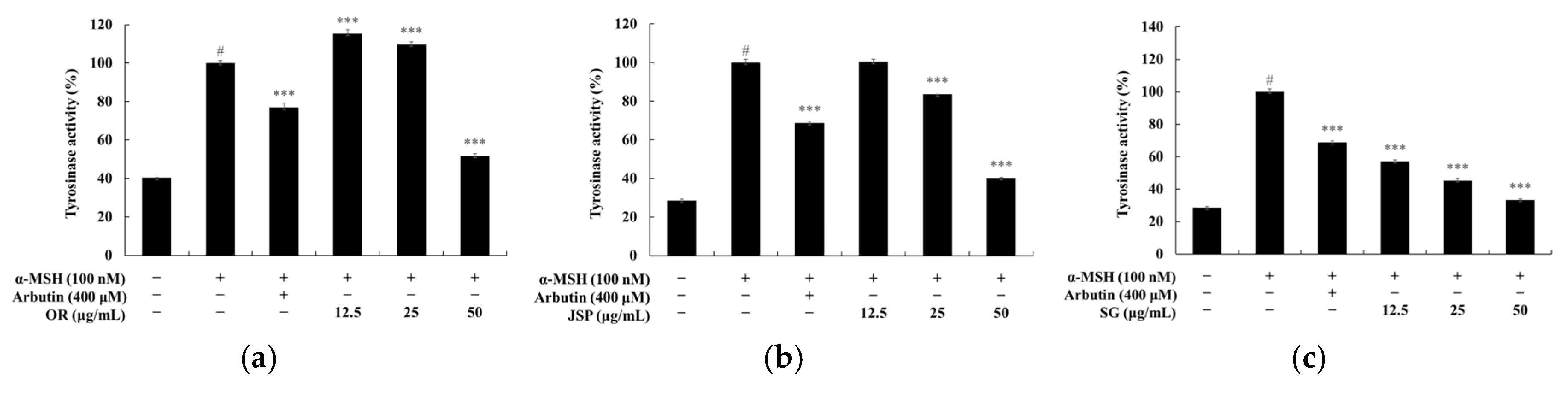
3.3. Melanin and Tyrosinase Inhibitory Effects of Jeju Fermented Foods
Melanin is the primary pigment in human skin and is crucial in protecting the skin from ultraviolet radiation. Changes in melanin production can lead to hyperpigmentation along with cosmetic and health-related consequences. Therefore, melanin inhibitors are considered valuable tools in medical and cosmetic treatments [28]. Tyrosinase is a rate-limiting enzyme in melanin synthesis and has been widely studied as a regulator of melanin production. It is a multifunctional copper-containing enzyme responsible for melanin synthesis and browning in both plants and animals. This enzyme catalyzes two melanin formation reactions: the hydroxylation of tyrosine to o-dopaquinone by monophenolase and the oxidation of L-DOPA to o-dopaquinone by diphenolase. These reactive o-quinones then undergo non-enzymatic polymerization to form melanin [29,30,31]. Therefore, melanin production is thought to be primarily controlled by the expression and activation of tyrosinase. Consequently, a major strategy is targeting tyrosinase, given the increasing interest in the use of natural products as tyrosinase inhibitors [28,29,30,31]. Established murine B16 melanoma cells (B16F10 cells) have easily quantifiable differentiation characteristics, including melanin production. Melanin production in B16F10 melanoma cells can be induced using UV and α-MSH, and the proliferation of these cells is also significant [32,33,34].
Thus, we monitored the survival and melanin production levels of these cells when treated with nine Jeju fermented food extracts at low cytotoxic concentrations. To determine whether Jeju fermented food extracts exhibited cytotoxic effects on B16F10 melanoma cells, we first treated the cells with various concentrations of the extracts for 72 hours and investigated their survival using MTT analysis. As shown in Figure 3, we confirmed that the cell survival rate was over 90% up to 50 μg/mL for OR, JSP, and SG; 100 μg/mL for CN, BJ, TC, and TL; and 200 μg/mL for SA, the highest concentration. Meanwhile, JSS showed the lowest survival rate. To evaluate the effect of the nine Jeju fermented food extracts on melanin production, cells were treated with each extract for 72 h at a concentration that had a cell survival rate above 90%. α-MSH was used as a positive control. The results suggested that melanin levels decreased in a dose-dependent manner when treated with OR, JSP, SG, CN, BJ, TC, and TL extracts, excluding SA and JSS, possibly for use in the treatment of hyperpigmentation disorders. The effect of the nine Jeju fermented food extracts on tyrosinase activity in B16F10 cells is depicted in Figure 4. Cellular tyrosinase activity was significantly reduced compared to untreated controls when treated with OR, JSP, SG, CN, BJ, TC, and TL, excluding SA and JSS, consistent with the melanogenesis inhibitory effect.
Figure 3.
Viability of Jeju fermented food extracts in B16F10 melanoma cells. The cells were treated with Jeju fermented food extracts for 72 h. The cytotoxicity of Jeju fermented food extracts was evaluated using the MTT assay. Cell viability is expressed as percentages relative to untreated cells. The results are presented as the mean ± SD from three repeated experiments. * p < 0.05, ** p < 0.01, *** p < 0.001 vs. unstimulated control group.
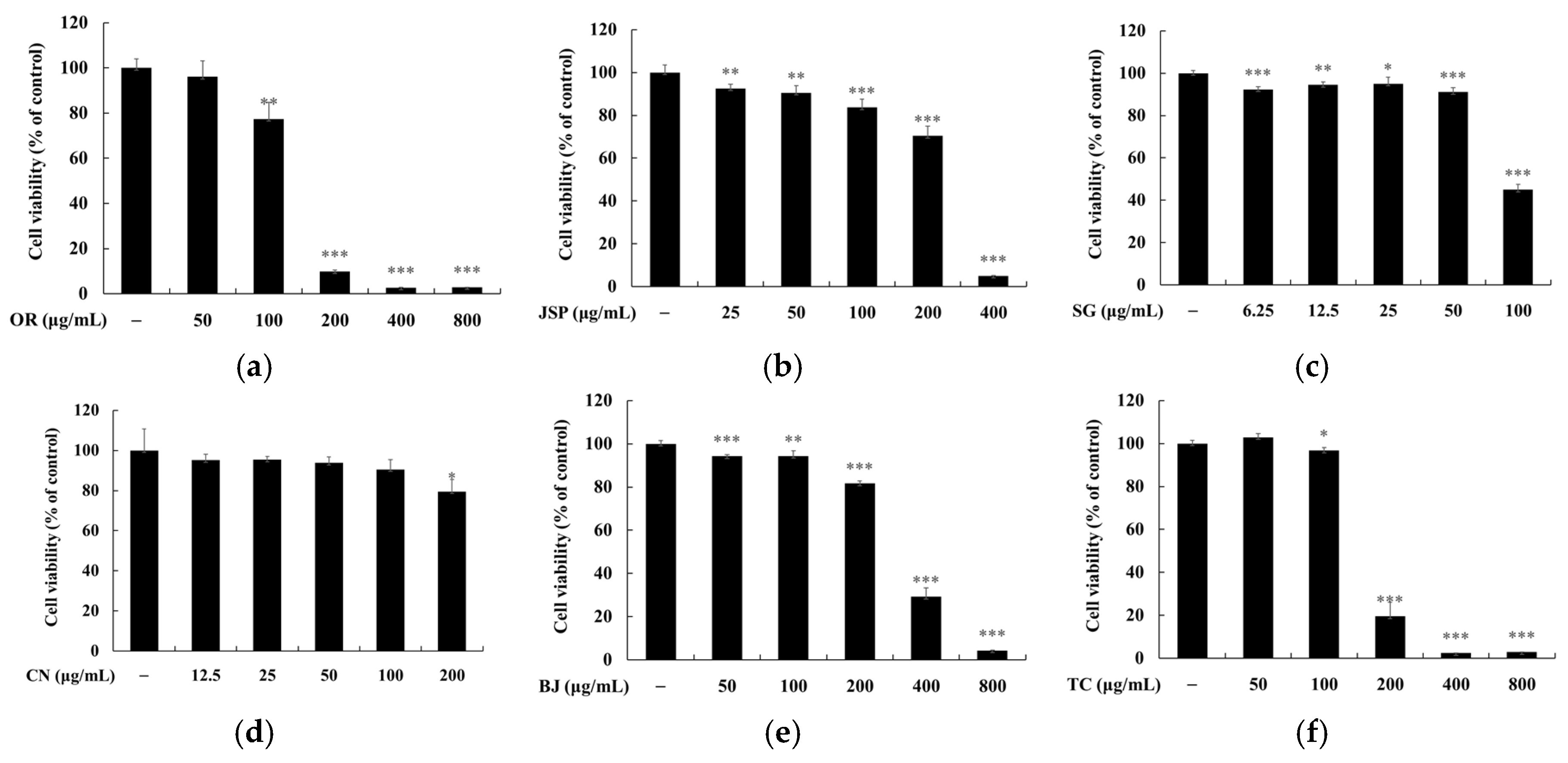
Figure 4.
The effect of Jeju fermented food extracts on melanin contents in B16F10 melanoma cells. The cells were treated with Jeju fermented food extracts for 72 h. α−MSH was used as the negative control and arbutin (200 μM) was used as the positive control. The results are presented as the mean ± SD from three repeated experiments. # p < 0.001 vs. unstimulated control group. * p < 0.05, ** p < 0.01, *** p < 0.001 vs. α−MSH alone.

Figure 5.
The effect of Jeju fermented food extracts on tyrosinase activity in B16F10 melanoma cells. The cells were treated with Jeju fermented food extracts for 72 h. α−MSH was used as the negative control and arbutin (200 μM) was used as the positive control. The results are presented as the mean ± SD from three repeated experiments. # p < 0.001 vs. unstimulated control group. * p < 0.05, ** p < 0.01, *** p < 0.001 vs. α−MSH alone.
3.4. Skin Safety of Jeju Fermented Foods
Finally, using human keratinocytes (HaCaT cells) and human skin application on subjects, we confirmed the safety of Jeju fermented food extracts, which have been proven to have anti-inflammatory and whitening effects. To analyze the effect of Jeju fermented foods on the viability of HaCaT human keratinocytes, we assessed cytotoxicity using the MTT assay. As shown in Figure 6, OR, JSP, SG, and TL showed no cytotoxicity (≥ 90% cell survival) up to 12.5 μg/mL, and CN, BJ, and TC showed no cytotoxicity up to 25 μg/mL; the best cell viability was identified with SA and the lowest viability with JSS treatments.
We conducted a skin irritation test to determine the safety of OR, SG, JSP, BJ, TC, CN, SA, TL, and JSS on human skin. Specifically, patches containing OR, SG, JSP, BJ, TC, CN, SA, TL, and JSS dissolved in squalene solvent at 50 µg/mL to 200 µg/mL concentrations were applied to the backs of 33 volunteers and left in contact for 24 h, and then the areas were observed 24, 30, and 48 h after the patches were removed. As a result, the test materials BJ, JSP, SA, TC, TL, OR, SG, CN, and JSS were determined to be non-irritating ingredients, with a skin irritation index of 0 (Table 1).
4. Conclusions
Traditional fermented foods and beverages are crucial in diverse human diets and have demonstrated potential positive impacts on human health in numerous experimental studies. Research worldwide has revealed strong associations between the microorganisms present in certain fermented foods, such as Kombucha, and various health benefits including weight maintenance, improvement of cardiovascular disease and diabetes, regulation of glucose and lipid levels, enhancement of the immune system, anticancer effects, and, most significantly, reductions in mortality [35]. Therefore, the aim of this study was to analyze the microbial communities of traditional fermented foods on Jeju Island, where the soil is predominantly volcanic ash, and to explore their association with human skin health.
Specifically, to investigate the relationship between the nine Jeju fermented foods and skin health, including anti-inflammatory and anti-melanogenic effects, we utilized RAW 264.7 and B16F10 cells, classic cell models in inflammation and melanogenesis studies. Most extracts from the nine Jeju fermented foods inhibited NO production and melanogenesis in a concentration-dependent manner. Finally, the human safety of Jeju fermented foods was confirmed through human patch tests and cytotoxicity assays on human keratinocytes (HaCaT cells). Based on the findings of these studies, the nine Jeju fermented food extracts have potential applications as ingredients in anti-inflammatory and anti-melanogenic products, thereby contributing to human health, including in the cosmetics industry.
Supplementary Materials
The following supporting information can be downloaded at the website of this paper posted on Preprints.org.
Author Contributions
Conceptualization, C.G.H.; methodology, S.B. and S.E.B.; software, S.B.; validation, S.B. and S.E.B.; formal analysis, S.B. and S.E.B.; investigation, S.B. and S.E.B.; writing—original draft preparation, C.G.H.; writing—review and editing, C.G.H.; supervision, S.J.P.; project administration, C.G.H.; funding acquisition, C.G.H. All authors have read and agreed to the published version of the manuscript.
Funding
This research was financially supported by the Ministry of Trade, Industry and Energy, Korea, under the “Regional Innovation Cluster Development Program (Non-R&D, P0024160)” supervised by the Korea Institute for Advancement of Technology (KIAT).
Institutional Review Board Statement
The study was conducted in accordance with the Declaration of Helsinki and was approved by the ethics committee of the Korea Dermatology Research Institute for studies involving humans (IRB no. KDRI-IRB-230425).
Informed Consent Statement
Not applicable.
Data Availability Statement
Not applicable.
Conflicts of Interest
The authors declare no conflicts of interest.
References
- Byun, K.H.; Kang, E.J.; Kim, K.H. Environment Management for Sustainability of Hallasan National Park in Jeju Island, Korea. AMR 2014, 905, 334–338. [Google Scholar] [CrossRef]
- Lee, J. Research of the Food Culture Comparison between the Tamra & Mongolia. Trans Humanit. 2012, 5, 211–243. [Google Scholar] [CrossRef]
- Jung, W.Y.; Jung, J.Y.; Lee, H.J.; Jeon, C.O. Functional Characterization of Bacterial Communities Responsible for Fermentation of Doenjang: A Traditional Korean Fermented Soybean Paste. Front. Microbiol. 2016, 31, 7:827. [Google Scholar] [CrossRef] [PubMed]
- Lee, S.H.; Jung, J.Y.; Jeon, C.O. Bacterial community dynamics and metabolite changes in myeolchi-aekjeot, a Korean traditional fermented fish sauce, during fermentation. Int. J. Food Microbiol. 2015, 203, 15–22. [Google Scholar] [CrossRef] [PubMed]
- Kim, M.S.; Park, E.J. Bacterial communities of traditional salted and fermented seafoods from Jeju Island of Korea using 16S rRNA gene clone library analysis. J. Food Sci. 2014, 79, M927–934. [Google Scholar] [CrossRef] [PubMed]
- Kim, Y.S.; Kim, M.C.; Kwon, S.W.; Kim, S.J.; Park, I.C.; Ka, J.O.; Weon, H.Y. Analyses of bacterial communities in meju, a Korean traditional fermented soybean bricks, by cultivation-based and pyrosequencing methods. J. Microbiol. 2011, 49, 340–348. [Google Scholar] [CrossRef] [PubMed]
- Jung, J.Y.; Lee, H.J.; Chun, B.H.; Jeon, C.O. Effects of Temperature on Bacterial Communities and Metabolites during Fermentation of Myeolchi-Aekjeot, a Traditional Korean Fermented Anchovy Sauce. PLoS One 2016, 11, e0151351. [Google Scholar] [CrossRef]
- Kobayashi, T.; Kimura, B.; Fujii, T. Strictly Anaerobic Halophiles Isolated from Canned Swedish Fermented Herrings (Surströmming). Int. J. Food Microbiol. 2000, 54, 81–89. [Google Scholar] [CrossRef] [PubMed]
- Barrangou, R.; Yoon, S.S.; Breidt, F., Jr.; Fleming, H.P.; Klaenhammer, T.R. Characterization of Six Leuconostoc fallax Bacteriophages Isolated from an Industrial Sauerkraut Fermentation. Appl. Environ. Microbiol. 2002, 68, 5452–5458. [Google Scholar] [CrossRef]
- Rizzo, G. Soy-Based Tempeh as a Functional Food: Evidence for Human Health and Future Perspective. Front. Biosci. (Elite Ed). 2024, 16, 3. [Google Scholar] [CrossRef]
- Feng, C.H. The Tale of Sushi: History and Regulations. Compr. Rev. Food Sci. Food Saf. 2012, 11, 205–220. [Google Scholar]
- Valentino, V.; Magliulo, R.; Farsi, D.; Cotter, P.D.; O’Sullivan, O.; Ercolini, D.; De Filippis, F. Fermented foods, their microbiome and its potential in boosting human health. Microb. Biotechnol. 2024, 17, e14428. [Google Scholar] [CrossRef] [PubMed]
- Chen, H.; Kang, X.; Wang, X.; Chen, X.; Nie, X.; Xiang, L.; Liu, D.; Zhao, Z. Potential Correlation between Microbial Diversity and Volatile Flavor Substances in a Novel Chinese-Style Sausage during Storage. Foods. 2023, 12, 3190. [Google Scholar] [CrossRef] [PubMed]
- Zhao, L.; Liu, Y.; Xu, Q.; Yu, Y.; Zheng, G.; Wang, Y.; Zhang, Q.; Xu, X.; Zhang, N.; Chu, J.; Zhang, Y.; Sun, Y.; Zhao, Q.; Zhang, Y.; Qu, Q.; Zhong, J. Microbial Community Succession and Its Correlation with Quality Characteristics during Gray Sufu Fermentation. Foods. 2023, 12, 2767. [Google Scholar] [CrossRef] [PubMed]
- Wu, Y.; Li, A.; Cheng, L.; Chen, Q.; Li, J.; Xu, Y.; Huo, D. Deep Shotgun metagenomic and 16S rRNA analysis revealed the microbial diversity of lactic acid bacteria in traditional fermented foods of eastern Hainan, China. Food Funct. 2022, 13, 12938–12952. [Google Scholar] [CrossRef] [PubMed]
- de Jong, M.; Alekseeva, A.Y.; Miraji, K.F.; Phiri, S.; Linnemann, A.R.; Schoustra, S.E. Environmental Selection Shapes Bacterial Community Composition in Traditionally Fermented Maize-Based Foods from Benin, Tanzania and Zambia. Microorganisms. 2022, 10, 1354. [Google Scholar] [CrossRef] [PubMed]
- Cao, Y.; Fanning, S.; Proos, S.; Jordan, K.; Srikumar, S. A Review on the Applications of Next Generation Sequencing Technologies as Applied to Food-Related Microbiome Studies. Front. Microbiol. 2017, 8, 1829. [Google Scholar] [CrossRef] [PubMed]
- Laudadio, I.; Fulci, V.; Palone, F.; Stronati, L.; Cucchiara, S.; Carissimi, C. Quantitative Assessment of Shotgun Metagenomics and 16S rDNA Amplicon Sequencing in the Study of Human Gut Microbiome. OMICS. 2018, 22, 248–254. [Google Scholar] [CrossRef] [PubMed]
- Hyun, S.B.; Hyun, C.G. Anti-Inflammatory Effects and Their Correlation with Microbial Community of Shindari, a Traditional Jeju Beverage. Fermentation 2020, 6, 87. [Google Scholar] [CrossRef]
- Jeong, D.W.; Kim, H.R.; Jung, G.; Han, S.; Kim, C.T.; Lee, J.H. Bacterial Community Migration in the Ripening of Doenjang, a Traditional Korean Fermented Soybean Food. J. Microbiol. Biotechnol. 2014, 24, 648–660. [Google Scholar] [CrossRef]
- Jang, M.; Jeong, D.W.; Lee, J.H. Identification of the Predominant Bacillus, Enterococcus, and Staphylococcus Species in Meju, a Spontaneously Fermented Soybean Prod. Microbiol. Biotechnol. Lett. 2019, 47, 359–363. [Google Scholar] [CrossRef]
- Hussein, W.E.; Abdelhamid, A.G.; Rocha-Mendoza, D.; García-Cano, I.; Yousef, A.E. Assessment of Safety and Probiotic Traits of Enterococcus durans OSY-EGY, Isolated From Egyptian Artisanal Cheese, Using Comparative Genomics and Phenotypic Analyses. Front. Microbiol. 2020, 11, 608314. [Google Scholar] [CrossRef] [PubMed]
- Carasi, .;, Racedo, S.M.; Jacquot, C.; Elie, A.M.; Serradell, M.L.; Urdaci, M.C. Enterococcus durans EP1 a Promising Anti-inflammatory Probiotic Able to Stimulate sIgA and to Increase Faecalibacterium prausnitzii Abundance. Front. Immunol. 2017, 8, 88. [CrossRef] [PubMed]
- Facchin, B.M.; Dos Reis, G.O.; Vieira, G.N.; Mohr, E.T.B.; da Rosa, J.S.; Kretzer, I.F.; Demarchi, I.G.; Dalmarco, E.M. Inflammatory biomarkers on an LPS-induced RAW 264.7 cell model: a systematic review and meta-analysis. Inflamm. Res. 2022, 71, 741–758. [Google Scholar] [CrossRef] [PubMed]
- Kang, J.K.; Hyun, C.G. 4-Hydroxy-7-Methoxycoumarin Inhibits Inflammation in LPS-activated RAW264.7 Macrophages by Suppressing NF-κB and MAPK Activation. Molecules 2020, 25, 4424. [Google Scholar] [CrossRef] [PubMed]
- Rodríguez-Negrete, E.V.; Morales-González, Á.; Madrigal-Santillán, E.O.; Sánchez-Reyes, K.; Álvarez-González, I.; Madrigal-Bujaidar, E.; Valadez-Vega, C.; Chamorro-Cevallos, G.; Garcia-Melo, L.F.; Morales-González, J.A. Phytochemicals and Their Usefulness in the Maintenance of Health. Plants (Basel). 2024, 13, 523. [Google Scholar] [CrossRef] [PubMed]
- Kamalian, A.; Sohrabi Asl, M.; Dolatshahi, M.; Afshari, K.; Shamshiri, S.; Momeni Roudsari, N.; Momtaz, S.; Rahimi, R.; Abdollahi, M.; Abdolghaffari, A.H. Interventions of natural and synthetic agents in inflammatory bowel disease, modulation of nitric oxide pathways. World J. Gastroenterol. 2020, 26, 3365–3400. [Google Scholar] [CrossRef] [PubMed]
- Malaspina, P.; Catellani, E.; Burlando, B.; Brignole, D.; Cornara, L.; Bazzicalupo, M.; Candiani, S.; Obino, V.; De Feo, V.; Caputo, L.; Giordani, P. Depigmenting potential of lichen extracts evaluated by in vitro and in vivo tests. PeerJ. 2020, 8, e9150. [Google Scholar] [CrossRef]
- Goelzer Neto, C.F.; do Nascimento, P.; da Silveir,a V.C.; de Mattos, A.B.N.; Bertol, C.D. Natural sources of melanogenic inhibitors: A systematic review. Int. J. Cosmet. Sci. 2022, 44, 143-153. [CrossRef]
- Qu, Y.; Zhan, Q.; Du, S.; Ding, Y.; Fang, B.; Du, W.; Wu, Q.; Yu, H.; Li, L.; Huang, W. Catalysis-based specific detection and inhibition of tyrosinase and their application. J. Pharm. Anal. 2020, 10, 414–425. [Google Scholar] [CrossRef] [PubMed]
- Saghaie, L.; Pourfarzam, M.; Fassihi, A.; Sartippour, B. Synthesis and tyrosinase inhibitory properties of some novel derivatives of kojic acid. Res. Pharm. Sci. 2013, 8, 233–242. [Google Scholar]
- Oh, S.Y.; Hyun, C.G. Chrysoeriol Enhances Melanogenesis in B16F10 Cells Through the Modulation of the MAPK, AKT, PKA, and Wnt/β-Catenin Signaling Pathways. Nat. Prod. Commun. 2022, 17. [Google Scholar] [CrossRef]
- Potez, M.; Trappetti, V.; Bouchet, A.; Fernandez-Palomo, C.; Güç, E.; Kilarski, W.W.; Hlushchuk, R.; Laissue, J.; Djonov, V. Characterization of a B16-F10 melanoma model locally implanted into the ear pinnae of C57BL/6 mice. PLoS One 2018, 13, e0206693. [Google Scholar] [CrossRef]
- Kim, T.; Hyun, C.G. Imperatorin Positively Regulates Melanogenesis through Signaling Pathways Involving PKA/CREB, ERK, AKT, and GSK3β/β-Catenin. Molecules. 2022, 27, 6512. [Google Scholar] [CrossRef]
- Cuamatzin-García, L.; Rodríguez-Rugarcía, P.; El-Kassis, E.G.; Galicia, G.; Meza-Jiménez, M.L.; Baños-Lara, M.D.R.; Zaragoza-Maldonado, D.S.; Pérez-Armendáriz, B. Traditional Fermented Foods and Beverages from around the World and Their Health Benefits. Microorganisms. 2022, 10, 1151. [Google Scholar] [CrossRef] [PubMed]
Figure 1.
Viability of Jeju fermented food extracts in RAW 264.7 cells. The cells (1.5 × 10⁵ cells/well) were seeded for 24 h, and then in the presence of LPS, they were treated with varying concentrations of Jeju fermented food extracts for 24 h. The survival rate of RAW 264.7 cells treated with Jeju fermented food extracts was measured by the MTT assay and is represented as a percentage compared to the non-treated group. The results are expressed as the mean ± SD of data obtained from three independent experiments. ### P < 0.001 compared with the control group; *** P < 0.001 and **P < 0.01 compared with the LPS-treated group.
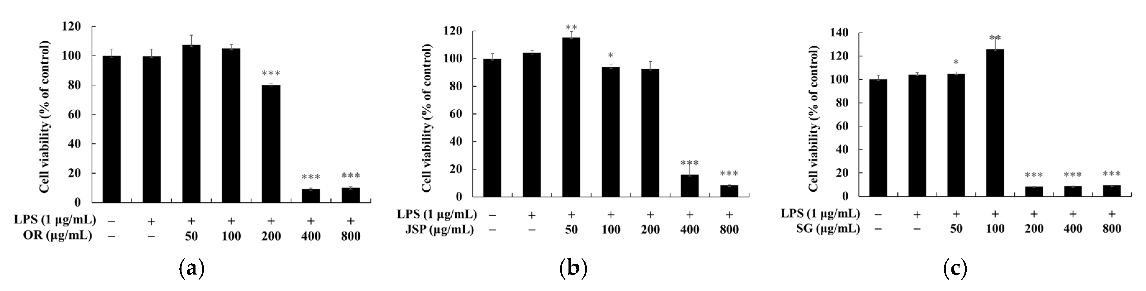
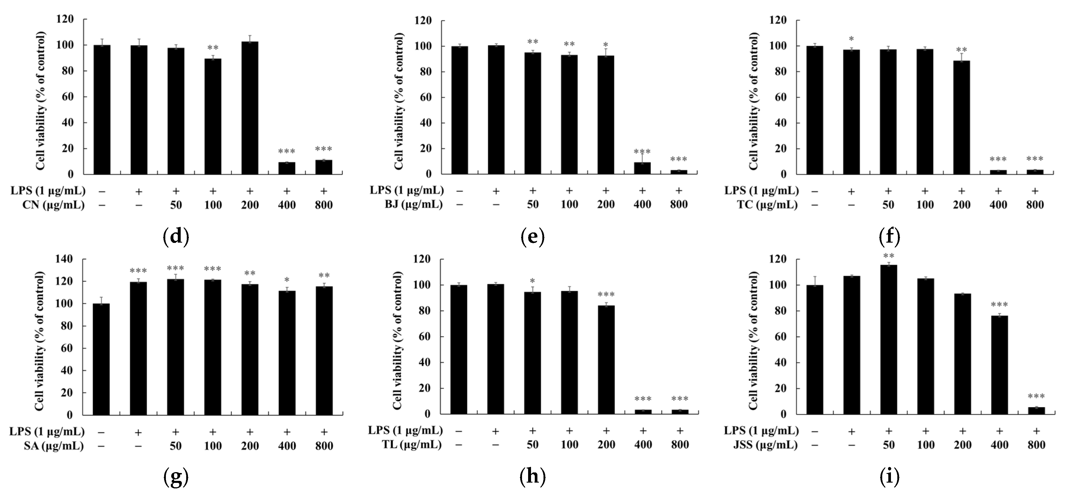
Figure 2.
Inhibition of nitric oxide (NO) production by Jeju fermented food extracts in RAW 264.7 cells. The cells (1.5 × 10⁵ cells/well) were seeded for 24 h, and then in the presence of LPS, they were treated with varying concentrations of Jeju fermented food extracts for 24 h. The supernatant of the culture medium was analyzed for NO using Griess reagent. The results are expressed as the mean ± SD of data obtained from three independent experiments. ### P < 0.001 compared with the control group; *** P < 0.001 and **P < 0.01 compared with the LPS-treated group.
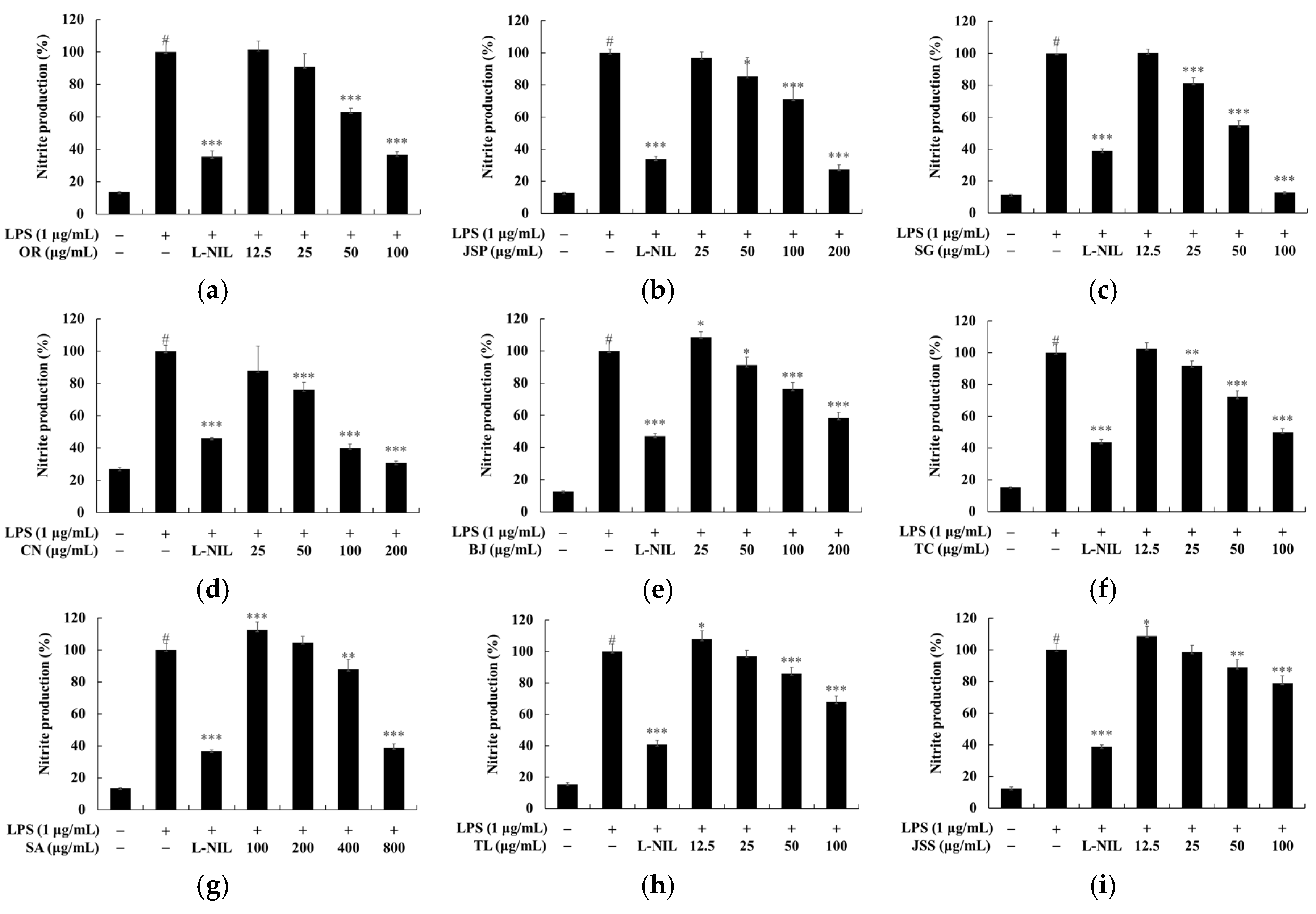
Figure 6.
Viability of Jeju fermented food extracts in human keratinocytes (HaCaT cells). The cells were treated with Jeju fermented food extracts for 72 h. The cytotoxicity of Jeju fermented food extracts was evaluated using the MTT assay. Cell viability is expressed as percentages relative to untreated cells. The results are presented as the mean ± SD from three repeated experiments. * p < 0.05, ** p < 0.01, *** p < 0.001 vs. unstimulated control group.
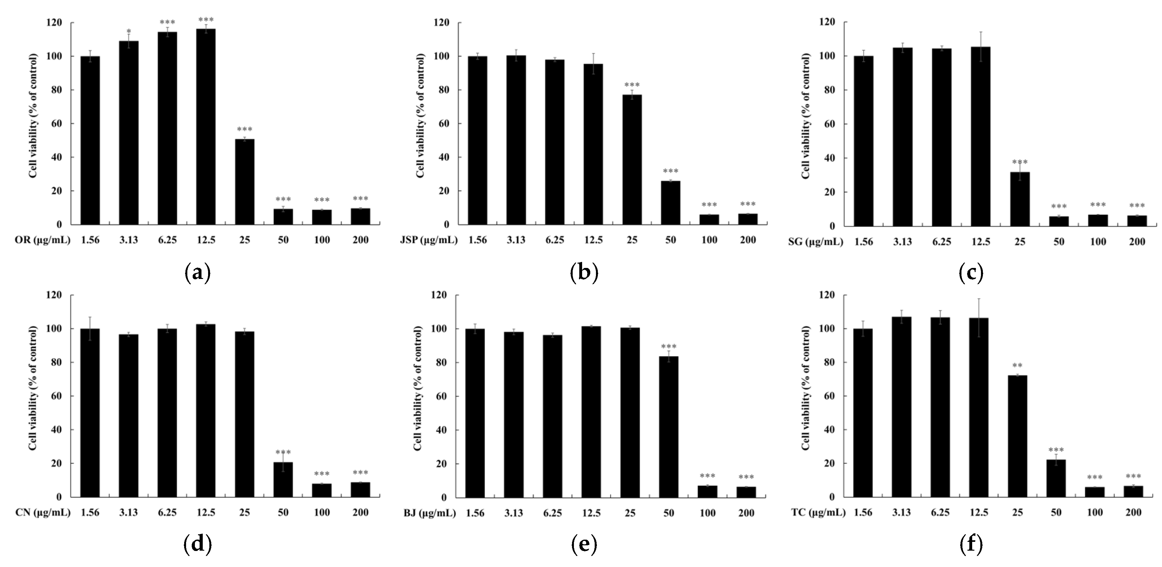
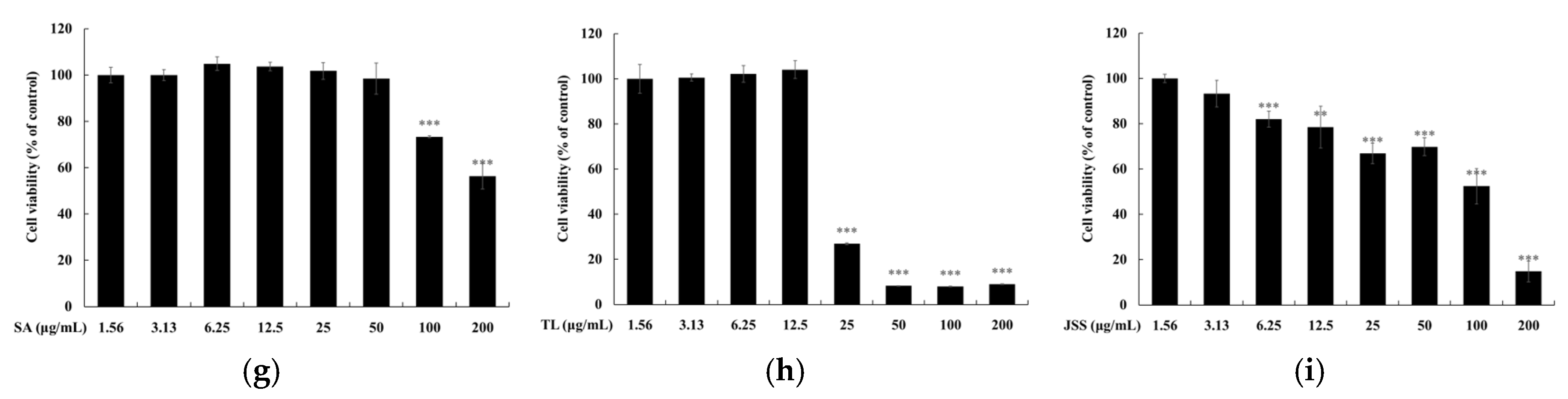

Table 1.
Results of human skin primary irritation test (n = 33).
| No | Test Samples | 30 min | 24 h | 48 h | Reaction Grade | |||
|---|---|---|---|---|---|---|---|---|
| 30 min | 24 h | 48 h | Mean | |||||
| 1 | BJ (100 µg/mL) | 0 | 0 | 0 | 0 | 0 | 0 | 0 |
| 2 | BJ (200 µg/mL) | 0 | 0 | 0 | 0 | 0 | 0 | 0 |
| 3 | JSP (100 µg/mL) | 0 | 0 | 0 | 0 | 0 | 0 | 0 |
| 4 | JSP (200 µg/mL) | 0 | 0 | 0 | 0 | 0 | 0 | 0 |
| 5 | SA (100 µg/mL) | 0 | 0 | 0 | 0 | 0 | 0 | 0 |
| 6 | SA (200 µg/mL) | 0 | 0 | 0 | 0 | 0 | 0 | 0 |
| 7 | TC (50 µg/mL) | 0 | 0 | 0 | 0 | 0 | 0 | 0 |
| 8 | TC (100 µg/mL) | 0 | 0 | 0 | 0 | 0 | 0 | 0 |
| 9 | TL (50 µg/mL) | 0 | 0 | 0 | 0 | 0 | 0 | 0 |
| 10 | TL (100 µg/mL) | 0 | 0 | 0 | 0 | 0 | 0 | 0 |
| 11 | OR (50 µg/mL) | 0 | 0 | 0 | 0 | 0 | 0 | 0 |
| 12 | OR (100 µg/mL) | 0 | 0 | 0 | 0 | 0 | 0 | 0 |
| 13 | SG (50 µg/mL) | 0 | 0 | 0 | 0 | 0 | 0 | 0 |
| 14 | SG (100 µg/mL) | 0 | 0 | 0 | 0 | 0 | 0 | 0 |
| 15 | CN (50 µg/mL) | 0 | 0 | 0 | 0 | 0 | 0 | 0 |
| 16 | CN (100 µg/mL) | 0 | 0 | 0 | 0 | 0 | 0 | 0 |
| 17 | JSS (50 µg/mL) | 0 | 0 | 0 | 0 | 0 | 0 | 0 |
| 18 | JSS (100 µg/mL) | 0 | 0 | 0 | 0 | 0 | 0 | 0 |
Disclaimer/Publisher’s Note: The statements, opinions and data contained in all publications are solely those of the individual author(s) and contributor(s) and not of MDPI and/or the editor(s). MDPI and/or the editor(s) disclaim responsibility for any injury to people or property resulting from any ideas, methods, instructions or products referred to in the content. |
© 2024 by the authors. Licensee MDPI, Basel, Switzerland. This article is an open access article distributed under the terms and conditions of the Creative Commons Attribution (CC BY) license (http://creativecommons.org/licenses/by/4.0/).
Copyright: This open access article is published under a Creative Commons CC BY 4.0 license, which permit the free download, distribution, and reuse, provided that the author and preprint are cited in any reuse.


