
Submitted:
29 April 2024
Posted:
30 April 2024
You are already at the latest version
Abstract
Litsea martabanica root's antioxidant and AChE activity showed promise as a pesticide detoxification agent in our previous study. In addition to roots, its leaves can help mitigate pesticide exposure, although there is limited scientific evidence supporting their efficacy. However, the use of roots in many countries, such as Thailand, could contribute to environmental degradation, as highland communities traditionally cut down whole trees to obtain them. This study aims to evaluate the antioxidant activity and anti-pesticide potential of water extracts from L. martabanica leaves instead of its roots through in vitro and in vivo investigations. The findings showed that L. martabanica water extracts and fractions demonstrated varied antioxidant activity. The water extract, obtained using traditional extraction methods, was chosen for in vivo experiments. Rats received the extract followed by chlorpyrifos exposure for 16 days. Results indicated reduced acetylcholinesterase activity in chlorpyrifos-exposed rats, while treatment with leaf extract led to increased activity, suggesting its potential as a detoxifying agent. Histopathological examination revealed no liver cell necrosis and showed liver cell regeneration, indicating potential protection against free radical-induced liver damage without affecting rat liver weight or biochemical profiles. Utilizing L. martabanica leaves for healthcare purposes holds promise without negative environmental impact.
Keywords:
L. martabanica leaf
; water extraction
; antioxidant
; anti-pesticide
; medicinal plant
; acetylcholinesterase activity
; hepatoprotective
; access to healthcare.
1. Introduction
Pesticides play a crucial role in agriculture globally, where they are extensively employed to enhance and protect crop yields by controlling insects, weeds, mollusks, and fungi [1]. Changing or straying from these traditional practices can be challenging, especially in regions where agriculture is crucial for supporting rural economies [2,3]. However, the long-term use of chemical pesticides which has been shown to have adverse effects on ecosystems. There is also increasing evidence of negative impacts on human health, including acute neurologic poisoning, chronic neurodevelopmental impairment, cancers, liver diseases, and potential immune dysfunction [4,5,6,7,8]. Farm workers who apply chemical pesticides in particular are at high risk of health complications as those substances can enter the body through the skin, lungs, and mucous membranes, as well as through diet [9]. Most pesticides, such as chlorpyrifos, are synthetic compounds consisting primarily of organophosphates, which irreversibly inhibit the synthesis of acetylcholinesterase (AChE) [10,11]. This inhibition results in excessive stimulation of both muscarinic and nicotinic receptors, leading to symptoms such as cramps, lacrimation, paralysis, muscular weakness, muscular fasciculation, diarrhea, and blurred vision [12,13]. Reduced AChE concentrations in capillary blood can indicate exposure to organophosphates. Symptoms of acute organophosphate poisoning include heightened salivation, diarrhea, vomiting, muscle tremors, gastrointestinal discomfort, and confusion [14]. Symptoms can begin within minutes or hours of exposure and can persist for days to weeks. Long-term exposure to lower doses of organophosphates has been associated with polyneuropathy and cardiovascular diseases [15]. While reducing overall pesticide use can be problematical, means of mitigating the severity of adverse reactions and diseases caused by exposure to these substances are worth investigating.
If a body is exposed to harmful chemicals from insecticides or pesticides, it can result in oxidative damage by malondialdehyde (MDA) and hydrogen peroxide (H2O2) to various organs, notably the liver [16]. Despite the liver's vital role in shielding the body from such chemical exposure, excessive pesticide exposure can diminish the liver’s detoxification ability and can lead to liver cell damage [17]. Additionally, reports indicate that organophosphates can induce the generation of reactive oxygen species (ROS), disrupting the antioxidant system and causing oxidative damage to cells [18]. Increased lipid peroxidation and decreased AChE activity are consequences of occupational pesticide exposure in humans [19,20]. Such exposure also reduces the concentrations of reduced glutathione, a non-enzymatic antioxidant, and antioxidant enzymes catalase, superoxide dismutase, and glutathione peroxidase [21,22]. Antioxidants have been proposed as a potential treatment for acute organophosphate pesticide poisoning because of their ability to counteract ROS damage in the liver [23,24].
Continual exposure to pesticides has been linked with the onset of liver disorders, such as hepatitis and fibrosis [25,26]. These chemicals, such as organophosphates, can disrupt hepato-biliary functions, signaling liver injury [27]. Subsequent to liver injury, hepatic stellate cells (HSCs), which are key players in fibrosis, become activated and undergo a transformation into a myofibroblast-like state to engage in the process of injury repair [28]. The activated HSCs express α-smooth muscle actin (αSMA) and produce type-I collagen, which serves as a major component of the extracellular matrix (ECM) [29]. Therefore, the activation of HSCs plays a pivotal role in the formation of liver fibrosis. Strategies aimed at inhibiting hepatic stellate cell proliferation and promoting apoptosis hold promise in the treatment of fibrosis [30].
Plants are recognized as valuable resources for mitigating reactive oxygen species (ROS), potentially serving as food ingredients to counteract the adverse effects of pesticides [31,32]. An in vivo experimental study found that green tea extracts, abundant in polyphenols, notably decreased pesticide-induced oxidative stress and pulmonary fibrosis [33]. Olive leaf extract has also been shown to have notable antioxidant and antiapoptotic effects in rats exposed to chlorpyrifos-induced neurotoxicity and reproductive toxicity [34].
The Lauraceae family encompasses 52 genera, totaling around 2,550 species which are distributed primarily in tropical and warm regions of southeastern Asia and Brazil [35]. Among these, Litsea martabanica (Kurz) Hook. f. is found in Thailand, China, and Myanmar [36]. The traditional utilization of this plant is deeply ingrained in the traditional wisdom of highland communities. Various parts of the plant, including roots, leaves, and stems, have been used as medicinal remedies in the highland areas of northern Thailand. These applications extend to treating kidney disease, mitigating toxic allergy symptoms, and facilitating detoxification [37]. In a previous study, we highlighted the root of L. martabanica for its potential as an anti-pesticide detoxifying agent which was attributed to its substantial antioxidant and AChE activity [25]. That investigation also discovered, drawing on the wisdom of highland communities, that in addition to boiling the plant's roots for consumption to mitigate pesticide exposure, the leaves can also be utilized for the same purpose. The present study assessed the impacts of leaf water extract derived from L. martabanica, including the desirable properties described in traditional knowledge and wisdom, for developing products for use by farmers in highland communities. The use of L. martabanica leaf water extract could potentially play a role in promoting both environmental sustainability and human health by enhancing healthcare accessibility through its traditional uses to help protect people in lowland areas from toxicity caused by chemical residues in food, potable water, and food crops.
2. Materials and Methods
2.1. Reagents
Dulbecco’s Modified Eagle Medium - high glucose (DMEM-HG) (12800-58), fetal bovine serum (FBS), phosphate-buffered saline (PBS), and trypsin-EDTA solution - were purchased from Gibco (Grand Island, NY, USA). Chlorpyrifos, dimethyl sulfoxide (DMSO), and sulforhodamine B (SRB) were purchased from Sigma Chemical, Inc. (St Louis, MO, USA). Penicillin, streptomycin, and L-glutamine were obtained from Invitrogen (Thermo Fisher Scientific Inc., Waltham, MA, USA).
2.2. Quality Control of Raw Materials
Leaf powder of L. martabanica was evaluated for its physico-chemical properties following the methods described in Thai Herbal Pharmacopoeia 2018 [38].
2.2.1. Determination of Loss on Drying
Powdered drug samples were accurately weighed at 2-5 grams and placed into weighing bottles. The samples were then dried in a hot air oven at 105°C until they reached a constant weight. After that, the samples were cooled down in a desiccator. The weight loss was then measured as the moisture content.
2.2.2. Determination of Ash Values
Determination of total ash
Approximately 2-3 grams of the powdered drug were weighed and added to a crucible that had been thoroughly dried and weighed. The test sample was gradually heated in an electrical muffle furnace (Thermo Fisher, Waltham, MA, USA) until it reached a temperature of 500°C and was determined to be free of carbon. The sample was then cooled in a desiccator and reweighed to measure the total ash content.
Determination of acid-insoluble ash
Approximately 25 mL of 2 M hydrochloric acid (HCl) was added to the crucible that contained the total ash. The crucible was then covered with a watch glass and gently boiled for 5 minutes in a water bath. Then the insoluble matter was filtered using No. 41 filter paper and washed with hot water until the filtrate reached a neutral pH. The filter paper containing the insoluble matter was then transferred to a crucible, and gradually ignited in an electrical muffle furnace until a constant weight was achieved. Finally, the test samples were cooled in desiccators and weighed to measure the acid-insoluble content.
2.2.3. Determination of Extractive Value
Ethanol soluble extractive value
Initially, 5.0 g of the powdered drug was transferred to a glass-stopper conical flask, macerated with 100.0 mL of 95% ethanol for 6 h, frequently shaken, and then allowed to stand for 18 h. The sample was filtered, and the marc was washed with ethanol to adjust the filtrate to 100.0 mL. Subsequently, 20.0 mL of the filtrate was transferred to a pre-weighed evaporating dish. The test sample was then evaporated to dryness in a water bath, dried at 105°C, cooled in a desiccator, and weighed.
A total of 5.0 grams of the powdered drug was added to a conical flask with a glass stopper. The sample was then mixed with 100.0 mL of 95% ethanol and left to soak for 6 hours. During that time, the mixture was shaken frequently. After soaking, the mixture was allowed to stand for 18 hours and was then filtered. The marc was washed with ethanol to make 100 mL of the filtrate. From that filtrate, 20.0 mL was transferred to an evaporating dish that had been pre-weighed. The sample was then evaporated to dryness using a water bath after which it was dried at 105°C, cooled in a desiccator, and weighed.
Water-soluble extractive value
Approximately 5.0 grams of powdered drug was mixed with 100 mL of water and shaken for 6 hours. Then the mixture was allowed to stand for 18 hours. The sample was filtered, and the solid residue was washed with water to make 100 mL of filtrate. From that filtrate, 20 mL was transferred to an evaporating dish that had been pre-weighed. The sample was then evaporated to dryness in a water bath, dried at 105°C, cooled in a desiccator, and weighed.
2.3. Extract Preparation
The powder of L. martabanica leaves was extracted by decoction using water as a solvent, with a ratio of 1 L of water to 100 g of leaves (soaked for 30 minutes) over 1.5 hours. The filtrate was obtained by filtering the extract through filter paper, and then the marc of plant material was re-extracted twice. Subsequently, the filtrate was gathered and concentrated to achieve a concentration of approximately 3% Brix, and the acid-base balance was measured. The extract was dried using a spray dryer (BUCHI Mini Spray Dryer B-290) at an inlet temperature of 140°C and an outflow temperature of 85 ± 5°C, with the aspirator set at 100% and the pump at 25%, resulting in a water-soluble powder.
2.4. Plant Material
Litsea martabanica was collected from Chiang Mai province, Thailand. The plant material was identified by a taxonomist. The voucher specimen was deposited in the Queen Sirikit Botanical Garden (No. WP 7185). The leaves of L. martabanica were selected, cut into small pieces, and dried in the hot air oven until the moisture content was less than 10%, after which they were pulverized. The powder of the plant material was evaluated for the quality of the raw material following the methods described in the Thai Herbal Pharmacopoeia 2018 [38].
2.5. Chemical Profile by Thin Layer Chromatography
The water extracts of L. martabanica leaves (5 mg) were dissolved in 1 mL of aqueous ethanol as a test solution. Standards of apigenin, caffeic acid, ellagic acid, gallic acid, kaempferol, quercetin, and rutin were prepared separately in concentrations of 1 mg/1 mL. A CAMAG (Muttenz, Switzerland) HPTLC system, comprised of a Linomat 5 automatic applicator with a 10 mL syringe, CAMAG Automatic Developing Chamber 2 (ADC 2), Camag TLC scanner 4, and winCATS software version 1.4 were used. For HPTLC fingerprinting analysis, 2 µL of the test solution and 2 µL of the standard solution were loaded as an 8 mm long band on the Silica Gel GF254 TLC plate. The plate was kept in a TLC twin trough developing chamber (after saturation with solvent vapor) with the mobile phase (ethyl acetate: methanol: water = 80:20:2). Densitometric scanning was performed with a TLC scanner equipped with winCATS software. The plate was scanned at 254, 280, and 320 nm. The plate was kept in a photo-documentation chamber (CAMAG TLC Visualizer 2) and images were captured at white light, UV 254 nm, and UV 366 nm. The developed plate was sprayed with respective spray reagents (anisaldehyde, natural product, and DPPH spraying reagent) and dried at 100 °C in a hot air oven.
2.6. DPPH Assay
The antioxidant capacity of L. martabanica was assessed using the DPPH (2,2-diphenyl-1-picrylhydrazyl) radical scavenging assay [39]. In that process, DPPH was dissolved in methanol to achieve a final concentration of 80 µg/mL. Subsequently, the extracts, dissolved in DMSO, underwent dilution to varying concentrations. Each diluted extract (20 µL) was then dispensed into separate wells. Following this, 180 µL of DPPH solution was added to each well. The plate was then left to incubate at room temperature in darkness for 30 minutes. Absorbance readings were taken at 517 nm using a microplate reader. Vitamin C and methanol served as the reference standard and control, respectively. The percentage of DPPH scavenging activity was determined, and the concentration of the sample necessary to inhibit 50% of DPPH radicals was quantified as IC50 values.
2.7. Superoxide Radical Assay
Assay was conducted to assess the antioxidant efficacy of the test sample against superoxide free radicals. Superoxide radicals were generated through the action of phenazine methosulfate (PMS) and nicotinamide adenine dinucleotide (NADH), subsequently reducing nitroblue tetrazolium (NBT) to yield purple formazan [40,41]. Reagents, including PMS (25 µM), NADH (0.5 mM), and NBT (0.2 mM), were prepared by dissolving them in a phosphate buffer solution (pH 7.4). The experimental procedure involved adding 50 µL each of NBT solution, NADH solution plus varying concentrations of the test sample to individual wells of a 96-well plate, followed by the addition of 50 µL of PMS solution. After a 10-minute incubation at room temperature, the optical density (OD) was measured at 560 nm using a microplate reader. Gallic acid and phosphate buffer solutions were used as the reference standard and control, respectively. Subsequently, the percentage of superoxide radical scavenging and IC50 values were determined using the same formula as the DPPH assay [25].
2.8. Cell Culture
Human hepatic stellate cell line (LX-2) was purchased from Merck Millipore (Burlington, MA, USA) and cultured in Dulbecco’s Modified Eagle Medium with 100 Units/mL penicillin and 100 µg/mL streptomycin supplemented with 2% heat-inactivated fetal bovine serum and 2 mM L-Glu. The cells were incubated at 37°C in a 5% CO2 atmosphere. When the cells reached a confluence of 70–80%, they were harvested, plated, or sub-cultured for preservation or for use in subsequent passages.
2.9. Cell Viability Assay
The SRB assay procedure commenced with the seeding of cells at a density of 10,000 cells per well in a 96-well plate, each well containing 100 µL of complete medium. Following this, the extract treatments, also prepared in 100 µL of complete medium, were administered and incubated for either 24 or 48 hours at 37°C under 5% CO2 conditions. Subsequently, cell viability was evaluated utilizing the SRB assay, with comparisons made against untreated cells. Post-treatment, 40 µL of 50% trichloroacetic acid (TCA) was added to each well and the plates were incubated at 4°C for one hour after which the plates underwent four washes with tap water and were subsequently dried. Following that, 100 µL of 0.057% SRB solution was added to each well for a 30-minute incubation period, followed by four washes with 1% acetic acid to eliminate unbound dye. After drying, 200 µL of 10 mM Tris-based solution (pH 10.5) was introduced, and the absorbance at 510 nm was measured using a microplate reader (BioTek, Winooski, VT, USA) [42].
2.10. Apoptosis Assay by Flow Cytometry
The LX-2 cells were plated at a density of 3 x 10⁵ cells in 6-well plates and exposed to each extract ranging from 25 to 100 µg/mL for 24 hours. Following treatment, the cells were collected and washed twice with PBS, after which they were stained with 50 µL of binding buffer containing annexin V-FITC and propidium iodide (PI) for 15 minutes. Following this, 250 µL of binding buffer was added to the stained cells, and the samples were analyzed using a BD FACScan™ flow cytometer (BD Biosciences, San Jose, CA, USA). The results were evaluated based on the percentage of cells exhibiting positive signals for annexin V-FITC and PI, or only annexin V-FITC [43].
2.11. Assay of AChE Activity
An AChE activity kit was used following the manufacturer’s instructions (Sigma Chemical, Inc., St. Louis, MO, USA). Briefly, 10 µL of diluted whole blood samples were dispensed into each well of a 96-well plate, followed by the addition of 190 µL of the working reagent to all wells. The reaction mixtures were then left to incubate at room temperature, and absorbance readings were recorded after 2 and 10 minutes using a microplate reader set at 412 nm [25].
2.12. Anti-Pesticide Potential
2.12.1. Animals
Male Sprague-Dawley rats, weighing 180–200 g, were obtained from the Nomura Siam International (Nomura Siam International Co., Ltd., Bangkok, Thailand). Standard environmental conditions were adhered to, including a temperature of 24 ± 1oC and a 12-hour dark-light cycle for their housing. All animals were provided with unrestricted access to drinking water and a regular pellet diet. They were acclimated for a minimum of one week before commencing the studies. The Animal Ethics Committee of the Faculty of Medicine, Chiang Mai University, approved all experimental protocols (approval number 49/2559).
2.12.2. Experimental Groups
The anti-pesticide potential of L. martabanica leaf water extract was assessed using a previously reported method [25]. Male rats were divided into five groups of six animals each. Group 1 (normal group) was orally administered 2 mL/kg of distilled water daily for 16 days. Group 2 (control group) received the same dose of distilled water plus chlorpyrifos (Sigma Chemical, Inc., St. Louis, MO, USA) at a dosage of 16 mg/kg daily for the same 16-day period. The rats in groups 3 to 5 were orally administered varying doses of L. martabanica leaf water extract 30 minutes before receiving chlorpyrifos at a dosage of 16 mg/kg daily for 16 days. The administration of the leaf extract acted as an imitation of the concoction based on traditional knowledge used in highland communities. In group 3, rats were subjected to a cyclic dosing regimen of L. martabanica leaf water extract, receiving a cyclical daily administration of 7.5 mg/kg for 2 days followed by 2.5 mg/kg for 2 days over a period of 16 days (four rounds). Group 4 received a cyclic dose of 75 mg/kg for 2 days followed by 25 mg/kg for 2 days, and group 5 received a cyclic dose of 750 mg/kg for 2 days followed by 250 mg/kg for 2 days, also with daily administration over 16 days (four rounds). Behavioral changes following administration of chlorpyrifos and L. martabanica leaf water extract were observed in the rats. Signs of toxicity, especially nicotinic- (muscular fasciculation and weakness) and muscarinic-related signs (small pupils, tremors, diarrhea, salivation, lacrimation, and others), were observed and recorded. Additionally, the rats' body weight was measured once daily throughout the experiment.
Following the end of the period of daily administration, on the 17th day all rats were anesthetized via an intraperitoneal injection of 120 mg/kg of thiopental sodium. Vital signs, pulse, and reflexes were assessed to confirm the animals' death. Blood samples were obtained for hematological and biochemical analysis, and their serums were determined for malondialdehyde (MDA), superoxide dismutase (SOD), and reduced glutathione in accordance with previous methods; Placer et al. [44], Sun et al. [45], and Sedlak and Lindsay [46], respectively. Liver and kidneys were removed for weighing and gross pathological examination. The liver and kidney specimens were fixed in 10% formalin for histopathological analysis.
2.13. Statistical Analysis
Results are expressed as mean ± S.E.M. The initial analysis involved the Shapiro–Wilk test to check for normality. If the data showed no significant deviation from a normal distribution, ANOVA followed by Tukey’s multiple comparison tests were conducted. Conversely, if the Shapiro–Wilk test indicated a non-normal distribution, the Kruskal–Wallis nonparametric ANOVA test followed by Dunn's test were applied. Statistical significance was set at p < 0.05. Statistical analyses were performed using IBM SPSS Statistics, version 22.0 (International Business Machines Corporation, Armonk, NY, USA).
3. Results
3.1. Quality Control of Raw Materials and Extracts
Physico-chemical examinations, loss on drying, total ash, acid-insoluble ash, ethanol-soluble extractive value, and water-soluble extractive value of the raw material are presented in Table 1 along with their mean values.
The leaves of L. martabanica were extracted using traditional methods that involved decoction using water as the solvent. Although the leaf of L. martabanica is not included in any pharmacopeia or textbook, this study developed the specification of L. martabanica’s leaf alignment following Thai Herbal Pharmacopeia methods for quality control when handling raw materials could assist future research.
3.2. Chemical Profile by Thin Layer Chromatography
Chemical profiles of the water extracts of L. martabanica leaf were determined by HPTLC with the use of a detector under UV light at 254 and 366 nm. The plates were sprayed with a freshly prepared anisaldehyde-sulfuric acid reagent, DPPH, or natural product spraying reagent. The major component was determined at Rf 0.46, 0.58, 0.66, 0.80 at 254 nm. Based on comparison with chemical standards, the extract of L. martabanica was found not to contain apigenin (Rf 0.91), caffeic acid (Rf 0.77), ellagic acid (Rf 0.03), gallic acid (Rf 0.75), kaempferol (Rf 0.91), quercetin (Rf 0.88), or rutin (Rf 0.23).
3.3. Antioxidant Activity by DPPH Assay and Superoxide Radical Scavenging Activity
The half maximal inhibitory concentration (IC50) values for both DPPH and superoxide free radical scavenging activity are presented in Table 2. Among these fractions, the ethyl acetate fraction demonstrated greater antioxidant properties compared to both other fractions and the crude extract. Additionally, the reference standard gallic acid exhibited the highest efficacy in scavenging free radicals.
3.4. Cytotoxic Effects of L. martabanica Leaf Water Extract and Its Fractions on Human Hepatic Stellate Cells
To evaluate the impact on cell viability, various concentrations (12.5 – 800 µg/mL) of water extracts from L. martabanica leaf and its fractions were prepared and then incubated with LX-2 cells for 24 and 48 hours, as shown in Figure 1. The IC50 value of L. martabanica leaf water extract exceeded 1,000 µg/mL and was 564.70 ± 40.95 µg/mL at 24 and 48 hours as depicted in Table 3. Interestingly, the ethyl acetate fraction exhibited toxicity towards LX-2 cells, with IC50 values of 154.7 ± 11.29 µg/mL at 24 hours and 539.90 ± 38.26 µg/mL at 48 hours (Table 3). Other fractions, including butanol and residue, showed relatively low toxicity, with IC50 values exceeding 500 µg/mL at both 24 and 48 hours.
To confirm apoptosis induction by L. martabanica leaf water extract, apoptosis cell death was assessed using annexin V-fluorescein isothiocyanate (FITC)/propidium iodide (PI) staining and analyzed through flow cytometry. The results revealed that L. martabanica leaf water extract tended to induce apoptosis in LX-2 cells. Interestingly, ethyl acetate at a high concentration (800 µg/mL) was capable of inducing apoptosis significantly, while butanol and residue extracts did so at concentrations of 400 and 800 µg/mL, respectively, induced apoptosis with statistical significance (Figure 2).
3.5. Anti-Pesticide Potential in Rat
3.5.1. Acetylcholinesterase Activity
The normal group of rats showed an acetylcholinesterase activity value of 2,008 ± 182 U/L. In contrast, rats in the control group treated with chlorpyrifos exhibited significantly decreased average acetylcholinesterase activity levels compared to normal rats. Conversely, rats receiving L. martabanica leaf water extract at all concentrations displayed significantly increased acetylcholinesterase activity levels compared to the control group that received only chlorpyrifos (Figure 3A).
3.5.2. Malondialdehyde (MDA) and Superoxide Dismutase (SOD), and Reduced Glutathione in Serum
Regarding serum MDA levels, rats treated with chlorpyrifos showed a tendency for increased levels (0.057 ± 0.005 nmol/mg protein), though not significantly different from the normal rat group. However, rats receiving L. martabanica leaf water extract at all concentrations along with chlorpyrifos exhibited significantly decreased MDA levels compared to the control group that received chlorpyrifos alone (Figure 3B).
The experiment also revealed elevated SOD levels in the serum of rats treated with chlorpyrifos, to be statistically significantly different from the normal group. Conversely, rats receiving L. martabanica leaf water extract at all doses along with chlorpyrifos showed a statistically significant increase in SOD enzyme levels compared to the control group that received only chlorpyrifos (Figure 3C). Regarding reduced glutathione levels in serum, the control group receiving chlorpyrifos displayed a level of 0.159 ± 0.010 nmol/mg protein, was significantly different from the normal group. Rats receiving medium doses of L. martabanica leaf water extract (75 and 25 mg/kg) along with chlorpyrifos showed a tendency toward increased reduced glutathione levels, although the values were not statistically significantly different. However, rats receiving high doses of L. martabanica leaf water extract (750 and 250 mg/kg) along with chlorpyrifos exhibited significantly higher levels of reduced glutathione in serum than the chlorpyrifos-treated group (p<0.05), with values similar to those in normal rats receiving distilled water (Figure 3D).
Overall, the experimental results from the acetylcholinesterase activity assay, superoxide dismutase (SOD) assay, and malondialdehyde (MDA) assay indicated antioxidant enzyme activity has the potential to reverse organophosphate poisoning.
3.5.3. Body Weight and Organ Weight Change
This experiment involved administering the insecticide chlorpyrifos at a dose of 16 mg/kg body weight daily for 16 days to rats in all three groups receiving L. martabanica leaf water extract. The results revealed a higher body weight in rats from the group receiving low and middle doses of the extracts combined with the insecticide compared to that of the control group on the first day, as depicted in Table 4. Additionally, the kidney weight of only the group receiving the extract combined with the insecticide significantly increased compared to the normal and control groups.
3.5.4. Hematological Analysis
In the hematological analysis investigating the anti-pesticide effect, it was found that the rats in the groups receiving L. martabanica leaf water extract in middle doses (75 and 25 mg/kg) and high doses (750 and 250 mg/kg) showed a statistically significant decrease in red blood cell count, hemoglobin, and hematocrit levels compared to the normal groups as presented in Table 5. The rats in the groups receiving L. martabanica leaf water extract in the high doses (750 and 250 mg/kg) showed statistically significant decreases in hemoglobin and hematocrit levels compared to the control groups. The white blood cell count parameters were not significantly different from the control group (Table 6).
3.5.5. Blood Chemistry Analysis
In the blood chemistry analysis to assess liver and kidney functions, as shown in Table 7, it was found that the rats receiving L. martabanica leaf water extract at high doses (750 and 250 mg/kg) had significantly decreased BUN and alkaline phosphatase levels compared to the normal group. Additionally, ALT levels in chlorpyrifos-treated rats tended to first increase and then revert to normal levels following administration of L. martabanica leaf water extract. Moreover, the control group rats receiving distilled water along with the insecticide had a statistically significant decrease in total protein levels compared to normal rats. Rats in the group receiving L. martabanica leaf water extract in all three conditions had a statistically significant decrease in total protein levels compared to the normal group.
3.5.6. Histopathological Examination
In the histopathology examination of rats receiving chlorpyrifos (control group), dilation or widening of the sinusoids with liver hypertrophy was observed but was not observed in the normal group. However, no signs of scattered cell necrosis or lymphocytic infiltration were observed. The cells displayed varying shapes and sizes, with hyperchromatic and hypertrophied nuclei (Figure 4B). Interestingly, the dilation or widening of sinusoids was notably reduced, but no hepatic necrosis was observed in the liver of rats receiving L. martabanica leaf water extract at high doses (750 and 250 mg/kg) (Figure 4C). For the kidneys, histopathological examination revealed normal characteristics in rats of the control group receiving distilled water and the insecticide (Figure 5A-C).
4. Discussion
An investigation was performed to study the detoxifying and antioxidant properties of the root of L. martabanica in reducing the toxicity induced by chlorpyrifos in rats [25] which is consistent with the traditional approach to therapeutic use applied by Thai highland communities. Using leaves rather than roots could help reduce the environmental degradation caused by highland communities which traditionally cut down whole trees to obtain the roots. In this study, the identification and authentication of herbal drug substances was crucial to ensuring their authenticity before proceeding to further steps. For that reason, physico-chemical examination, total ash, acid-insoluble ash, ethanol-soluble extractive value, water-soluble extractive value, and water content of the crude drugs (Table 1) will be among the criteria used to evaluate the quality of the raw material used in future studies. To that end, we examined the microscopic characteristics and chemical compositions to establish L. martabanica’s monograph to help ensure appropriate quality control of raw material and extracts. Additionally, we investigated the antioxidant effects and anti-pesticide capabilities of L. martabanica as an aid to the further development of products for human use. This effort aligns with the project’s intention to employ traditional knowledge and wisdom and potentially transforming L. martabanica leaf water extract into various forms including granules, infusions, and effervescent tablets for possible wider application.
Previous research of bioactive compounds has shown that phenolic and flavonoid compounds are abundant in L. martabanica roots, suggesting that phenolic and flavonoid compounds may serve as active ingredients [25]. Furthermore, it is possible that a cluster of terpenoids could serve as the active component in preventing pesticide toxicity [47]. Moreover, phenolic, and flavonoid compounds possess antioxidant capabilities, which aid in the reduction of oxidative stress [48]. Consequently, it is plausible that these chemicals are the active components in L. martabanica leaf water extract that are responsible for the reduction of liver fibrosis activity.
In this study, the chemical composition of the water extracts of L. martabanica leaf were analyzed using HPTLC with detection under UV light at 254 and 366 nm. An anisaldehyde-sulfuric acid, a universal reagent for natural products, and DPPH spraying reagent were also used. Evaluation using accepted chemical standards found the extract of L. martabanica to be free of caffeic acid, ellagic acid, gallic acid, kaempferol, quercetin, and rutin.
Chromatographic techniques can establish chemical profiles that are useful for quality control and for determining the authenticity of raw materials and herbal products. These specifications can provide guidance for quality control and can serve as a reference for future research on the repeatability of the extract preparation process. Phytochemical screening revealed the presence of phenolics, flavonoids, saponins, and terpenoids. The bioactive compounds found in this plant will be used in future studies using bioassay-guided isolation.
Antioxidants are crucial for neutralizing free radicals and safeguarding cells against oxidative damage [49]. To evaluate antioxidant capabilities, it is necessary to utilize three models, encompassing the DPPH and superoxide radical assays, i.e., employing multiple models is necessary to comprehensively evaluate antioxidant activity and mechanisms, including DPPH assay and superoxide radical assay [50,51]. The outcomes from all antioxidant tests in this study demonstrated that the leaf water extract of L. martabanica displayed considerable antioxidant properties in both DPPH and superoxide radical assays. Moreover, previous research and literature reviews have highlighted the antioxidant activity present in plants of the genus Litsea, with this attribute being observed across different parts of the plants, including leaves, roots, and stems [52,53,54]. For example, the methanolic extract of L. glutinosa has demonstrated antioxidant properties including hydrogen peroxide scavenging activity, total antioxidant capacity, nitric oxide scavenging activity assay, and reducing power test [55]. The 80% ethanol extract and solvent fractions of L. japonica leaves have radical-scavenging antioxidant activity [56]. Our own earlier experiments have produced consistent results, confirming that the water extract of L. martabanica leaf water extract also exhibits antioxidant effects.
The liver is the major organ that deals with drugs and hazardous substances that can cause inflammation and oxidative stress [57]. Free radicals drive liver cell fibrosis, disrupting normal cellular processes like growth, division, and death regulation. This oxidative stress triggers Kupffer cells to release fibrosis-promoting factors, including TNFα, IL-1β, IL-6, and TGFβ, while hepatic stellate cells produce collagen, leading to fibrosis [58]. Targeting hepatic stellate cell proliferation and promoting apoptosis offers potential in fibrosis treatment [30]. Compounds like luteolin inhibit fibrosis-related genes, block cytokine pathways, and induce hepatic stellate cell apoptosis [59]. In our study, we utilized the LX-2 cell line, which possesses characteristics resembling hepatic stellate cells and is commonly used for investigating human hepatic fibrosis. The present study observed that the water extracts ethyl acetate and butanol as well as the residue of L. martabanica leaf at concentrations ranging from 200 – 800 µg/mL significantly inhibited cell growth compared to control cells. The results also showed that L. martabanica leaf water extract tended to induce apoptosis, but only at very high concentrations. Ethyl acetate (800 µg/mL), butanol, and residue (400 and 800 µg/mL), were able to induce apoptosis cell death, demonstrating early apoptosis. The identification of ethyl acetate fraction as an active ingredient presents an intriguing possibility for further investigation.
The objective of this test, however, was to verify the efficacy of the selected water extract. Its administration is intended to imitate traditional concoctions based on local knowledge, with the goal of expanding upon traditional knowledge and developing new products for future use. The present animal research study used conventional extraction methods, including water extraction, in order to conduct a more comprehensive examination of the impacts of L. martabanica leaf water extract.
The toxicity of organophosphate pesticides largely arises from inhibiting AChE, which results in an accumulation of acetylcholine [10]. L. martabanica root water extracts have previously been shown to have anti-pesticide effects, such as improving AChE activity in rats exposed to chlorpyrifos [25]. In our study, the rats exposed to chlorpyrifos experienced a decrease in AChE activity. Treatment with L. martabanica leaf water extract demonstrated promise in reversing this effect, indicating potential as an anti-pesticide agent. Additionally, organophosphate pesticides are involved in oxidative stress and reactive oxygen species (ROS) production [18]. In our study, chlorpyrifos increased MDA levels while decreasing reduced glutathione, but treatment with L. martabanica leaf water extract reversed these effects. Furthermore, the L. martabanica leaf water extract was found to elevate SOD levels. According to prior studies, the methanol leaf extract of L. glutinosa, including neophytadiene (sesquiterpenoids), increased SOD, GSH, and GPx concentrations substantially while decreasing MDA in comparison to the control group. Furthermore, it exhibited the potential to exert hepatoprotective effects via TGF-β1 signaling pathways [47]. Based on our study evaluating the quality of the extracts, antioxidant effects in vitro, and anti-pesticide properties in the animal study, it is plausible that the active ingredients belong to the terpenoids group.
Long-term exposure to high doses of organochloride and organophosphate pesticides commonly used in agriculture has been linked to persistent hematotoxicity and a higher incidence of aplastic anemia in humans [60,61]. Additionally, studies have demonstrated a significant decrease in hemoglobin concentration, RBC count, and hematocrit with increased exposure to chlorpyrifos [62]. The reduction in red blood cell count could potentially be attributed to an elevated rate of erythrocyte destruction within the hematopoietic organ, erythropoiesis inhibition, hemosynthesis impairment, or osmoregulatory dysfunction [63]. In our investigation, we observed a decrease in RBCs, hematocrit, and hemoglobin levels across all chlorpyrifos-exposed groups. Despite some decreases in values such as RBCs, hematocrit, and hemoglobin after receiving various doses of the extract, they remained within the normal range [64,65,66,67]. Interestingly, a previous study reported that chlorpyrifos triggers a rise in white blood cells [62]. An increase in the total number of white blood cells (WBCs) caused by exposure to toxic substances, including pesticides, is a response to stress conditions [68]. Our investigation confirmed this relationship, showing an increase in white blood cells after chlorpyrifos exposure, with specific increases observed in neutrophils, lymphocytes, monocytes, and eosinophils, although without reaching statistical significance. Exposure to L. martabanica leaf water extract appeared to restore these white blood cell counts to normal levels, with all white blood cell parameters remaining within normal ranges [69].
To assess the potential impact on renal and hepatic functions, we conducted thorough clinical blood chemistry assessments. The kidneys, due to their high blood perfusion, are particularly susceptible to toxic substances. Such toxins can be actively filtered by the kidneys, potentially leading to their accumulation in renal tubules, emphasizing the importance of monitoring BUN and creatinine levels as sensitive indicators of renal health [70,71]. Most studies have reported that chlorpyrifos increases BUN and creatinine levels [72]. In our study, we observed a decrease in chlorpyrifos-induced BUN levels, while creatinine levels remained normal. This change does not imply abnormality from chlorpyrifos at this concentration. Despite some decreases in values such as BUN after administering the water extracts at various doses, all values remained within normal ranges [66,67,70]. These findings suggest that the administration of L. martabanica leaf water extract had no discernible effect on kidney function or increase in kidney toxicity.
Chlorpyrifos leads to hepatotoxic changes by elevating AST, ALT, and ALP levels while reducing total protein and albumin [73,74]. Moreover, chlorpyrifos may cause liver dysfunction due to liver membrane permeability changes [75]. Our findings were in line with this, showing that chlorpyrifos increased AST levels but without statistical significance and significantly decreased total protein, with a trend toward decreasing albumin. Although L. martabanica leaf water extract returned AST to normal levels, it did not impact albumin levels. For this reason, L. martabanica leaf water extract might reduce ROS-induced cellular damage by enhancing antioxidant enzyme activity in liver tissues, thus reversing AST levels.
Serum alkaline phosphatase activity (ALP) is a valuable indicator of liver disease, especially cholestatic disease. Additionally, ALP levels can serve as an indicator of bone mineral density loss [76]. Exposure to chlorpyrifos has been documented to affect chondrogenesis in the growth plate cartilage of long bones in chick embryos, disrupting ossification [77]. Our research showed a reduction in ALP levels following chlorpyrifos administration compared to the normal group. However, co-administration of L. martabanica leaf water extract with chlorpyrifos failed to normalize ALP levels. Thus, it is plausible that L. martabanica leaf water extract does not restore normal ossification or other bone processes. Furthermore, the extract does not induce toxicity in the liver or bones, as evidenced by the ALP value remaining within the normal range.
Due to the short 16-day duration of chlorpyrifos exposure and the potentially low concentration of chlorpyrifos, it is possible that the experimental animals did not experience pathological effects on red blood cell production, kidney, or liver functions as a result. Additional long-term, high-dose chlorpyrifos exposure studies will be necessary to confirm whether chlorpyrifos has an effect on laboratory animals and whether these specific L. martabanica leaf water extracts can alleviate the condition.
Due to the presence of abnormalities, in particular blood chemistry analyses related to kidney and liver function, it was essential to conduct a histological examination to identify potential cellular-level abnormalities in these organs. Upon examination, the kidney cells appeared normal, while abnormalities were evident in the liver cells. In our study, rats administered chlorpyrifos (control group) exhibited widened sinusoids and liver enlargement compared to the normal group. The cells displayed hyperchromatic and hypertrophied nuclei with variations in size. No dispersed cell necrosis or lymphocytic infiltration was observed. These pathology changes in liver cells could be both a regenerative and an early inflammatory process [78]. The duration of chlorpyrifos treatment in our study was 16 days, consistent with previous studies [79].
The administration of high concentrations of L. martabanica water extracts led to the restoration of normal liver tissue characteristics. Regarding the pathology of the liver, L. martabanica potentially inhibits oxidative stress by facilitating the action of antioxidants, including MDA, which restore the liver to its normal state [80]. This is consistent with previous studies which have reported that neophytadiene, one type of sesquiterpenoids (terpenoids) was found to have protective effects [47]. Additionally, flavonoids and phenolic substances typically have antioxidant properties that aid in the reduction of liver fibrosis [81]. Our findings suggest that the water extracts of L. martabanica may protect liver cells from oxidative damage caused by chlorpyrifos and improve liver function.
5. Conclusions
Based on the results of this study, it can be inferred that L. martabanica leaf water extract has anti-pesticide properties, possibly due to its antioxidant characteristics. Although the extract results in an augmentation of AChE activity and enhances liver function, it is imperative to address the renal condition as well. The results of this study offer scientific evidence supporting the traditional medicinal use of L. martabanica as beneficial. However, further investigation is required using chlorpyrifos and L. martabanica over longer periods and at higher concentrations of chlorpyrifos to determine its ability to protect animals' vital organs from the effects of pesticides. A safety evaluation of the potential of L. martabanica leaf water extract to mitigate side effects of pesticides, e.g., acute and chronic toxicity tests will also be needed. It is also crucial to explore the essence or active ingredients in the ethyl acetate fraction of L. martabanica, as it might demonstrate superior efficacy in vitro compared to other fractions. Furthermore, the use of L. martabanica leaf instead of its root has the potential to contribute to environmental sustainability and human health by facilitating healthcare access by capitalizing on its traditional uses.
Author Contributions
Conceptualization, S.Se. and P.K.; methodology, W.T., S.Sa., S.I., T.C., S.C., and K.J.; software, W.T., S.Sa., and S.I.; validation, A.W. and P.Y.; formal analysis, W.T., S.Sa., and S.Se.; investigation, W.T., S.Sa., S.I., P.K.,T.C., S.C., and K.J.; resources, S.C., A.W., and P.Y.; writing—original draft preparation and writing—review and editing, W.T., S.Sa., S.C., and S.Se.; visualization, W.T. and S.Sa.; supervision, S.C. and S.Se.; project administration and funding acquisition, S.Se.; All authors have read and agreed to the published version of the manuscript.
Funding
This research was funded annually from 2015 to 2022 by the Highland Research and Development Institute (Public Organization) or “HRDI”.
Institutional Review Board Statement
The animal study protocol was approved by the Research Ethics Committee for Animal Studies of the Faculty of Medicine at Chiang Mai University, Thailand (Protocol code 49/2559 was approved on December 7, 2016).
Informed Consent Statement
Not applicable.
Data Availability Statement
Data is available upon request.
Acknowledgments
This research was partially supported by Chiang Mai University. Special thanks are also due to Dr. G. Lamar Robert, Ph.D., for his editing of the manuscript to help ensure linguistic accuracy and clarity.
Conflicts of Interest
The authors declare no conflicts of interest.
References
- Aktar, M.W.; Sengupta, D.; Chowdhury, A. Impact of pesticides use in agriculture: their benefits and hazards. Interdiscip Toxicol 2009, 2, 1–12. [Google Scholar] [CrossRef] [PubMed]
- Panuwet, P.; Siriwong, W.; Prapamontol, T.; Ryan, P.B.; Fiedler, N.; Robson, M.G.; Barr, D.B. Agricultural Pesticide Management in Thailand: Situation and Population Health Risk. Environ Sci Policy 2012, 17, 72–81. [Google Scholar] [CrossRef] [PubMed]
- Lamichhane, J.R.; Bischoff-Schaefer, M.; Bluemel, S.; Dachbrodt-Saaydeh, S.; Dreux, L.; Jansen, J.P.; Kiss, J.; Köhl, J.; Kudsk, P.; Malausa, T.; et al. Identifying obstacles and ranking common biological control research priorities for Europe to manage most economically important pests in arable, vegetable and perennial crops. Pest Manag Sci 2017, 73, 14–21. [Google Scholar] [CrossRef] [PubMed]
- Mostafalou, S.; Abdollahi, M. Pesticides and human chronic diseases: evidences, mechanisms, and perspectives. Toxicol Appl Pharmacol 2013, 268, 157–177. [Google Scholar] [CrossRef] [PubMed]
- Nicolopoulou-Stamati, P.; Maipas, S.; Kotampasi, C.; Stamatis, P.; Hens, L. Chemical Pesticides and Human Health: The Urgent Need for a New Concept in Agriculture. Front Public Health 2016, 4, 148. [Google Scholar] [CrossRef] [PubMed]
- Kumar, V.; Kumar, R.; Singh, J.; Kumar, P. Contaminants in agriculture and environment: health risks and remediation; Agro Environ Media, Publication Cell of AESA, Agriculture and Environmental Science Academy: 2019; Volume 1.
- Weichenthal, S.; Moase, C.; Chan, P. A review of pesticide exposure and cancer incidence in the Agricultural Health Study cohort. Environmental health perspectives 2010, 118, 1117–1125. [Google Scholar] [CrossRef] [PubMed]
- Al-Eryani, L.; Wahlang, B.; Falkner, K.; Guardiola, J.; Clair, H.; Prough, R.; Cave, M. Identification of environmental chemicals associated with the development of toxicant-associated fatty liver disease in rodents. Toxicologic pathology 2015, 43, 482–497. [Google Scholar] [CrossRef] [PubMed]
- Curl, C.L.; Spivak, M.; Phinney, R.; Montrose, L. Synthetic Pesticides and Health in Vulnerable Populations: Agricultural Workers. Curr Environ Health Rep 2020, 7, 13–29. [Google Scholar] [CrossRef]
- Vale, A.; Lotti, M. Organophosphorus and carbamate insecticide poisoning. Handb Clin Neurol 2015, 131, 149–168. [Google Scholar] [CrossRef]
- Colović, M.B.; Krstić, D.Z.; Lazarević-Pašti, T.D.; Bondžić, A.M.; Vasić, V.M. Acetylcholinesterase inhibitors: pharmacology and toxicology. Curr Neuropharmacol 2013, 11, 315–335. [Google Scholar] [CrossRef]
- Samada, L.H.; Tambunan, U.S.F. Biopesticides as promising alternatives to chemical pesticides: A review of their current and future status. Online J. Biol. Sci 2020, 20, 66–76. [Google Scholar] [CrossRef]
- Pakvilai, N.; Prapamontol, T.; Thavornyutikarn, P.; Mangklabruks, A.; Chantara, C.; Hongsibsong, S.; Santasup, C. A simple and sensitive GC-ECD method for detecting synthetic pyrethroid insecticide residues in vegetable and fruit samples. Chiang Mai J. Sci 2015, 42, 196–207. [Google Scholar]
- Eddleston, M. Patterns and problems of deliberate self-poisoning in the developing world. Qjm 2000, 93, 715–731. [Google Scholar] [CrossRef] [PubMed]
- Hung, D.Z.; Yang, H.J.; Li, Y.F.; Lin, C.L.; Chang, S.Y.; Sung, F.C.; Tai, S.C. The Long-Term Effects of Organophosphates Poisoning as a Risk Factor of CVDs: A Nationwide Population-Based Cohort Study. PLoS One 2015, 10, e0137632. [Google Scholar] [CrossRef] [PubMed]
- El-Nagar, M.M.F.; Elsisi, A.E. Exposure to bromoxynil octanoate herbicide induces oxidative stress, inflammation, and apoptosis in testicular tissue via modulating NF-кB pathway. Food and Chemical Toxicology 2023, 180, 114008. [Google Scholar] [CrossRef] [PubMed]
- Grant, D.M. Detoxification Pathways in the Liver. In Journal of Inherited Metabolic Disease, Harkness, R.A., Pollitt, R.J., Addison, G.M., Eds.; Springer Netherlands: Dordrecht, 1991; pp. 421–430. [Google Scholar]
- Al-Gubory, K.H. Environmental pollutants and lifestyle factors induce oxidative stress and poor prenatal development. Reprod Biomed Online 2014, 29, 17–31. [Google Scholar] [CrossRef] [PubMed]
- Milatovic, D.; Gupta, R.C.; Aschner, M. Anticholinesterase toxicity and oxidative stress. The Scientific World Journal 2006, 6, 295–310. [Google Scholar] [CrossRef]
- Akhgari, M.; Abdollahi, M.; Kebryaeezadeh, A.; Hosseini, R.; Sabzevari, O. Biochemical evidence for free radicalinduced lipid peroxidation as a mechanism for subchronic toxicity of malathion in blood and liver of rats. Human and experimental toxicology 2003, 22, 205–211. [Google Scholar] [CrossRef]
- Mecdad, A.A.; Ahmed, M.H.; ElHalwagy, M.E.; Afify, M.M. A study on oxidative stress biomarkers and immunomodulatory effects of pesticides in pesticide-sprayers. Egyptian Journal of Forensic Sciences 2011, 1, 93–98. [Google Scholar] [CrossRef]
- Soltaninejad, K.; Abdollahi, M. Current opinion on the science of organophosphate pesticides and toxic stress: a systematic review. Medical science monitor: international medical journal of experimental and clinical research 2009, 15, RA75–90. [Google Scholar]
- Nurulain, S.M.; Szegi, P.; Tekes, K.; Naqvi, S.N. Antioxidants in organophosphorus compounds poisoning. Arh Hig Rada Toksikol 2013, 64, 169–177. [Google Scholar] [CrossRef] [PubMed]
- Ranjbar, A.; Solhi, H.; Mashayekhi, F.J.; Susanabdi, A.; Rezaie, A.; Abdollahi, M. Oxidative stress in acute human poisoning with organophosphorus insecticides; a case control study. Environ Toxicol Pharmacol 2005, 20, 88–91. [Google Scholar] [CrossRef] [PubMed]
- Kunnaja, P.; Chansakaow, S.; Wittayapraparat, A.; Yusuk, P.; Sireeratawong, S. In Vitro Antioxidant Activity of Litsea martabanica Root Extract and Its Hepatoprotective Effect on Chlorpyrifos-Induced Toxicity in Rats. Molecules 2021, 26. [Google Scholar] [CrossRef] [PubMed]
- Abbasi, A.; Memon, S.; Kazi, S.A.F. ANTIFIBROTIC EFFECTS OF ANGIOTENSIN RECEPTOR BLOCKERS IN ORGANOPHOSPHATE (CHLORPYRIFOS) INDUCED LIVER FIBROSIS. Journal of Population Therapeutics and Clinical Pharmacology 2024, 31, 1302–1315. [Google Scholar] [CrossRef]
- Karami-Mohajeri, S.; Ahmadipour, A.; Rahimi, H.-R.; Abdollahi, M. Adverse effects of organophosphorus pesticides on the liver: a brief summary of four decades of research. Arhiv za higijenu rada i toksikologiju 2017, 68, 261–275. [Google Scholar] [CrossRef] [PubMed]
- Zhang, C.-Y.; Yuan, W.-G.; He, P.; Lei, J.-H.; Wang, C.-X. Liver fibrosis and hepatic stellate cells: Etiology, pathological hallmarks and therapeutic targets. World journal of gastroenterology 2016, 22, 10512. [Google Scholar] [CrossRef] [PubMed]
- Liu, X.; Xu, J.; Brenner, D.A.; Kisseleva, T. Reversibility of liver fibrosis and inactivation of fibrogenic myofibroblasts. Current pathobiology reports 2013, 1, 209–214. [Google Scholar] [CrossRef]
- Weiskirchen, R. Hepatoprotective and Anti-fibrotic Agents: It's Time to Take the Next Step. Front Pharmacol 2015, 6, 303. [Google Scholar] [CrossRef] [PubMed]
- Zafar, F.; Asif, H.M.; Shaheen, G.; Ghauri, A.O.; Rajpoot, S.R.; Tasleem, M.W.; Shamim, T.; Hadi, F.; Noor, R.; Ali, T.; et al. A comprehensive review on medicinal plants possessing antioxidant potential. Clin Exp Pharmacol Physiol 2023, 50, 205–217. [Google Scholar] [CrossRef]
- Jabłońska – Trypuć, A.; Wiater, J. Protective effect of plant compounds in pesticides toxicity. Journal of Environmental Health Science and Engineering 2022, 20, 1035–1045. [Google Scholar] [CrossRef]
- Kim, H.-R.; Park, B.-K.; Oh, Y.-M.; Lee, Y.-S.; Lee, D.-S.; Kim, H.-K.; Kim, J.-Y.; Shim, T.-S.; Lee, S.-D. Green tea extract inhibits paraquat-induced pulmonary fibrosis by suppression of oxidative stress and endothelin-l expression. Lung 2006, 184, 287–295. [Google Scholar] [CrossRef] [PubMed]
- Hassan, A.A.; Bel Hadj Salah, K.; Fahmy, E.M.; Mansour, D.A.; Mohamed, S.A.M.; Abdallah, A.A.; Ashkan, M.F.; Majrashi, K.A.; Melebary, S.J.; El-Sheikh, E.A.; El-Shaer, N. Olive Leaf Extract Attenuates Chlorpyrifos-Induced Neuro- and Reproductive Toxicity in Male Albino Rats. Life (Basel) 2022, 12. [Google Scholar] [CrossRef] [PubMed]
- Mabberley, D.J. Mabberley's plant-book: a portable dictionary of plants, their classification and uses; Cambridge university press: 2017.
- Ngearnsaengsaruay, C.; Middleton, D.J.; Chayamarit, K. A revision of the genus Litsea Lam.(Lauraceae) in Thailand. Thai Forest Bulletin (Botany).
- Institute, T.H.R.a.D. Local Plant Utilization Guide. 2018, 118.
- Thai Herbal Pharmacopoeia. Available online: https://bdn.go.th/thp/home (accessed on 10 April 2024).
- Brand-Williams, W.; Cuvelier, M.-E.; Berset, C. Use of a free radical method to evaluate antioxidant activity. LWT-Food science and Technology 1995, 28, 25–30. [Google Scholar] [CrossRef]
- Jing, L.; Ma, H.; Fan, P.; Gao, R.; Jia, Z. Antioxidant potential, total phenolic and total flavonoid contents of Rhododendron anthopogonoides and its protective effect on hypoxia-induced injury in PC12 cells. BMC Complement Altern Med 2015, 15, 287. [Google Scholar] [CrossRef]
- Bajpai, V.K.; Agrawal, P.; Bang, B.H.; Park, Y.-H. Phytochemical analysis, antioxidant and antilipid peroxidation effects of a medicinal plant, Adhatoda vasica. Frontiers in Life Science 2015, 8, 305–312. [Google Scholar] [CrossRef]
- Vichai, V.; Kirtikara, K. Sulforhodamine B colorimetric assay for cytotoxicity screening. Nat Protoc 2006, 1, 1112–1116. [Google Scholar] [CrossRef] [PubMed]
- Wlodkowic, D.; Skommer, J.; Darzynkiewicz, Z. Flow cytometry-based apoptosis detection. Methods Mol Biol 2009, 559, 19–32. [Google Scholar] [CrossRef] [PubMed]
- Placer, Z.A.; Cushman, L.L.; Johnson, B.C. Estimation of product of lipid peroxidation (malonyl dialdehyde) in biochemical systems. Analytical biochemistry 1966, 16, 359–364. [Google Scholar] [CrossRef] [PubMed]
- Sun, Y.; Oberley, L.W.; Li, Y. A simple method for clinical assay of superoxide dismutase. Clinical chemistry 1988, 34, 497–500. [Google Scholar] [CrossRef]
- Sedlak, J.; Lindsay, R.H. Estimation of total, protein-bound, and nonprotein sulfhydryl groups in tissue with Ellman's reagent. Analytical biochemistry 1968, 25, 192–205. [Google Scholar] [CrossRef]
- Sahoo, S.; Rath, D.; Kar, D.M.; Pattanaik, S. Hepatoprotective potency of Litsea glutinosa (L.) C.B. Rob. leaf methanol extract on H2O2-induced toxicity in HepG2 cells. Journal of Ethnopharmacology 2023, 304, 116076. [Google Scholar] [CrossRef] [PubMed]
- Rudrapal, M.; Khairnar, S.J.; Khan, J.; Dukhyil, A.B.; Ansari, M.A.; Alomary, M.N.; Alshabrmi, F.M.; Palai, S.; Deb, P.K.; Devi, R. Dietary Polyphenols and Their Role in Oxidative Stress-Induced Human Diseases: Insights Into Protective Effects, Antioxidant Potentials and Mechanism(s) of Action. Front Pharmacol 2022, 13, 806470. [Google Scholar] [CrossRef] [PubMed]
- Nimse, S.B.; Pal, D. Free radicals, natural antioxidants, and their reaction mechanisms. RSC advances 2015, 5, 27986–28006. [Google Scholar] [CrossRef]
- Dudonné, S.; Vitrac, X.; Coutière, P.; Woillez, M.; Mérillon, J.M. Comparative study of antioxidant properties and total phenolic content of 30 plant extracts of industrial interest using DPPH, ABTS, FRAP, SOD, and ORAC assays. J Agric Food Chem 2009, 57, 1768–1774. [Google Scholar] [CrossRef] [PubMed]
- Kähkönen, M.P.; Hopia, A.I.; Vuorela, H.J.; Rauha, J.P.; Pihlaja, K.; Kujala, T.S.; Heinonen, M. Antioxidant activity of plant extracts containing phenolic compounds. J Agric Food Chem 1999, 47, 3954–3962. [Google Scholar] [CrossRef] [PubMed]
- Kamle, M.; Mahato, D.K.; Lee, K.E.; Bajpai, V.K.; Gajurel, P.R.; Gu, K.S.; Kumar, P. Ethnopharmacological Properties and Medicinal Uses of Litsea cubeba. Plants (Basel) 2019, 8. [Google Scholar] [CrossRef] [PubMed]
- Jamaddar, S.; Raposo, A.; Sarkar, C.; Roy, U.K.; Araújo, I.M.; Coutinho, H.D.M.; Alkhoshaiban, A.S.; Alturki, H.A.; Saraiva, A.; Carrascosa, C.; Islam, M.T. Ethnomedicinal Uses, Phytochemistry, and Therapeutic Potentials of Litsea glutinosa (Lour.) C. B. Robinson: A Literature-Based Review. Pharmaceuticals (Basel) 2022, 16. [Google Scholar] [CrossRef] [PubMed]
- Wang, Y.S.; Wen, Z.Q.; Li, B.T.; Zhang, H.B.; Yang, J.H. Ethnobotany, phytochemistry, and pharmacology of the genus Litsea: An update. J Ethnopharmacol 2016, 181, 66–107. [Google Scholar] [CrossRef] [PubMed]
- Rumzhum, N.; Rahman, M.; Sharukh, A.; Chowdhury, S.; Pervin, M. In vitro antioxidant and antinociceptive potentialities of methanolic extract of Litsea glutinosa. Bangladesh Journal of Scientific and Industrial Research 2012, 47, 401–406. [Google Scholar] [CrossRef]
- Yoon, W.-J.; Kang, S.C.; Ham, Y.-M.; Kim, K.-N.; Hyuk Yang, W.; Kim, H.-J.; Park, S.-Y.; Jung, Y.-H. Antioxidative and anti-inflammatory activities of Litsea japonica leaves. Journal of the Korean Society for Applied Biological Chemistry 2010, 53, 27–32. [Google Scholar] [CrossRef]
- Malaguarnera, G.; Cataudella, E.; Giordano, M.; Nunnari, G.; Chisari, G.; Malaguarnera, M. Toxic hepatitis in occupational exposure to solvents. World J Gastroenterol 2012, 18, 2756–2766. [Google Scholar] [CrossRef] [PubMed]
- Sánchez-Valle, V.; Chávez-Tapia, N.C.; Uribe, M.; Méndez-Sánchez, N. Role of oxidative stress and molecular changes in liver fibrosis: a review. Curr Med Chem 2012, 19, 4850–4860. [Google Scholar] [CrossRef] [PubMed]
- Li, J.; Li, X.; Xu, W.; Wang, S.; Hu, Z.; Zhang, Q.; Deng, X.; Wang, J.; Zhang, J.; Guo, C. Antifibrotic effects of luteolin on hepatic stellate cells and liver fibrosis by targeting AKT/mTOR/p70S6K and TGFβ/Smad signalling pathways. Liver Int 2015, 35, 1222–1233. [Google Scholar] [CrossRef] [PubMed]
- Aksoy, M. Hematotoxicity and carcinogenicity of benzene. Environ Health Perspect 1989, 82, 193–197. [Google Scholar] [CrossRef] [PubMed]
- Chatterjee, S.; Basak, P.; Chaklader, M.; Das, P.; Pereira, J.A.; Chaudhuri, S.; Law, S. Pesticide induced alterations in marrow physiology and depletion of stem and stromal progenitor population: an experimental model to study the toxic effects of pesticide. Environ Toxicol 2014, 29, 84–97. [Google Scholar] [CrossRef] [PubMed]
- Hossain, M.A.; Sutradhar, L.; Sarker, T.R.; Saha, S.; Iqbal, M.M. Toxic effects of chlorpyrifos on the growth, hematology, and different organs histopathology of Nile tilapia, Oreochromis niloticus. Saudi J Biol Sci 2022, 29, 103316. [Google Scholar] [CrossRef] [PubMed]
- Vani, T.; Saharan, N.; Mukherjee, S.C.; Ranjan, R.; Kumar, R.; Brahmchari, R.K. Deltamethrin induced alterations of hematological and biochemical parameters in fingerlings of Catla catla (Ham.) and their amelioration by dietary supplement of vitamin C. Pesticide Biochemistry and Physiology 2011, 101, 16–20. [Google Scholar] [CrossRef]
- diferelanko, M.J. The Toxicologist's Pocket Handbook; Taylor & Francis: 2017.
- Butadej, D.; Duangchanchot, M.; Inpunkaew, R.; Kengkoom, K. Chemical parameters in healthy Sprague-Dawley and Wistar rats from National Laboratory Animal Center, Mahidol University. In Proceedings of the 49. Kasetsart University Annual Conference, Bangkok (Thailand), 2011., 1-4 Feb 2011. [Google Scholar]
- Marzouk, M.; Sayed, A.A.; Soliman, A.M. Hepatoprotective and antioxidant effects of Cichorium endivia L. leaves extract against acetaminophen toxicity on rats. Journal of Medicine and Medical Sciences 2011, 2, 1273–1279. [Google Scholar]
- Shirazi, M.G.; Toosi, V. Effect of aqueous extract of Rheum ribes on cisplatin-induced nephrotoxicity in rat. J Pharm Bioallied Sci 2013, 5, 81. [Google Scholar]
- El-Sayed, Y.S.; Saad, T.T.; El-Bahr, S.M. Acute intoxication of deltamethrin in monosex Nile tilapia, Oreochromis niloticus with special reference to the clinical, biochemical and haematological effects. Environmental Toxicology and Pharmacology 2007, 24, 212–217. [Google Scholar] [CrossRef]
- Sireeratawong, S.; Lertprasertsuke, N.; Srisawat, U.; Thuppia, A.; Ngamjariyawat, A.; Suwanlikhid, N.; Jaijoy, K. Acute and subchronic toxicity study of the water extract from root of Sida rhombifolia Linn. in rats. Songklanakarin Journal of Science & Technology 2008, 30. [Google Scholar]
- Akindele, A.J.; Adeneye, A.A.; Salau, O.S.; Sofidiya, M.O.; Benebo, A.S. Dose and time-dependent sub-chronic toxicity study of hydroethanolic leaf extract of Flabellaria paniculata Cav. (Malpighiaceae) in rodents. Front Pharmacol 2014, 5, 78. [Google Scholar] [CrossRef] [PubMed]
- Edelstein, C.L. Biomarkers of acute kidney injury. Adv Chronic Kidney Dis 2008, 15, 222–234. [Google Scholar] [CrossRef] [PubMed]
- Mansour, S.A.; Mossa, A.-T.H. Oxidative damage, biochemical and histopathological alterations in rats exposed to chlorpyrifos and the antioxidant role of zinc. Pesticide Biochemistry and Physiology 2010, 96, 14–23. [Google Scholar] [CrossRef]
- Tanvir, E.; Afroz, R.; Chowdhury, M.; Gan, S.; Karim, N.; Islam, M.; Khalil, M. A model of chlorpyrifos distribution and its biochemical effects on the liver and kidneys of rats. Human and Experimental Toxicology 2016, 35, 991–1004. [Google Scholar] [CrossRef] [PubMed]
- Ashoush, Y.; Abozid, M.; Mansour, S.; Morgan, A. Effect of Chlorpyrifos on Liver Function of Albino Rates. Menoufia Journal of Agricultural Biotechnology 2020, 5, 83–92. [Google Scholar] [CrossRef]
- Uzun, F.G.; Kalender, Y. Chlorpyrifos induced hepatotoxic and hematologic changes in rats: The role of quercetin and catechin. Food and Chemical Toxicology 2013, 55, 549–556. [Google Scholar] [CrossRef] [PubMed]
- Tariq, S.; Tariq, S.; Lone, K.P.; Khaliq, S. Alkaline phosphatase is a predictor of Bone Mineral Density in postmenopausal females. Pak J Med Sci 2019, 35, 749–753. [Google Scholar] [CrossRef] [PubMed]
- Chandra Sekaran, S.P.; Thotakura, B.; Jyothi, A.K.; Manickam, S.; Chanemougavally, J.; Prabhu, K.; Gopalan, D.H. Effect of chlorpyrifos and its metabolites on skeletal system development of chick embryo. Birth Defects Res 2023, 115, 1063–1078. [Google Scholar] [CrossRef]
- Michalopoulos, G.K. Liver regeneration. J Cell Physiol 2007, 213, 286–300. [Google Scholar] [CrossRef]
- Tripathi, S.; Srivastav, A.K. Liver profile of rats after long-term ingestion of different doses of chlorpyrifos. Pesticide Biochemistry and Physiology 2010, 97, 60–65. [Google Scholar] [CrossRef]
- Zhang, B.J.; Xu, D.; Guo, Y.; Ping, J.; Chen, L.b.; Wang, H. Protection by and anti-oxidant mechanism of berberine against rat liver fibrosis induced by multiple hepatotoxic factors. Clinical and experimental pharmacology and physiology 2008, 35, 303–309. [Google Scholar] [CrossRef] [PubMed]
- Pan, X.; Ma, X.; Jiang, Y.; Wen, J.; Yang, L.; Chen, D.; Cao, X.; Peng, C. A comprehensive review of natural products against liver fibrosis: flavonoids, quinones, lignans, phenols, and acids. Evidence-based complementary and alternative medicine 2020, 2020. [Google Scholar] [CrossRef] [PubMed]
Figure 1.
Cytotoxic effects of L. martabanica leaf water extract and its fractions on LX-2 cell lines. Cells were treated with various concentrations (12.5 – 800 µg/mL) of either L. martabanica leaf water extract or its fractions for 24 and 48 hours. Cell viability was assessed by comparison with 0.1% DMSO-treated control cells after 24 and 48 hours of incubation. Results are presented as mean ± S.E.M. from three independent experiments. **p < 0.01, and ***p < 0.001 vs control.
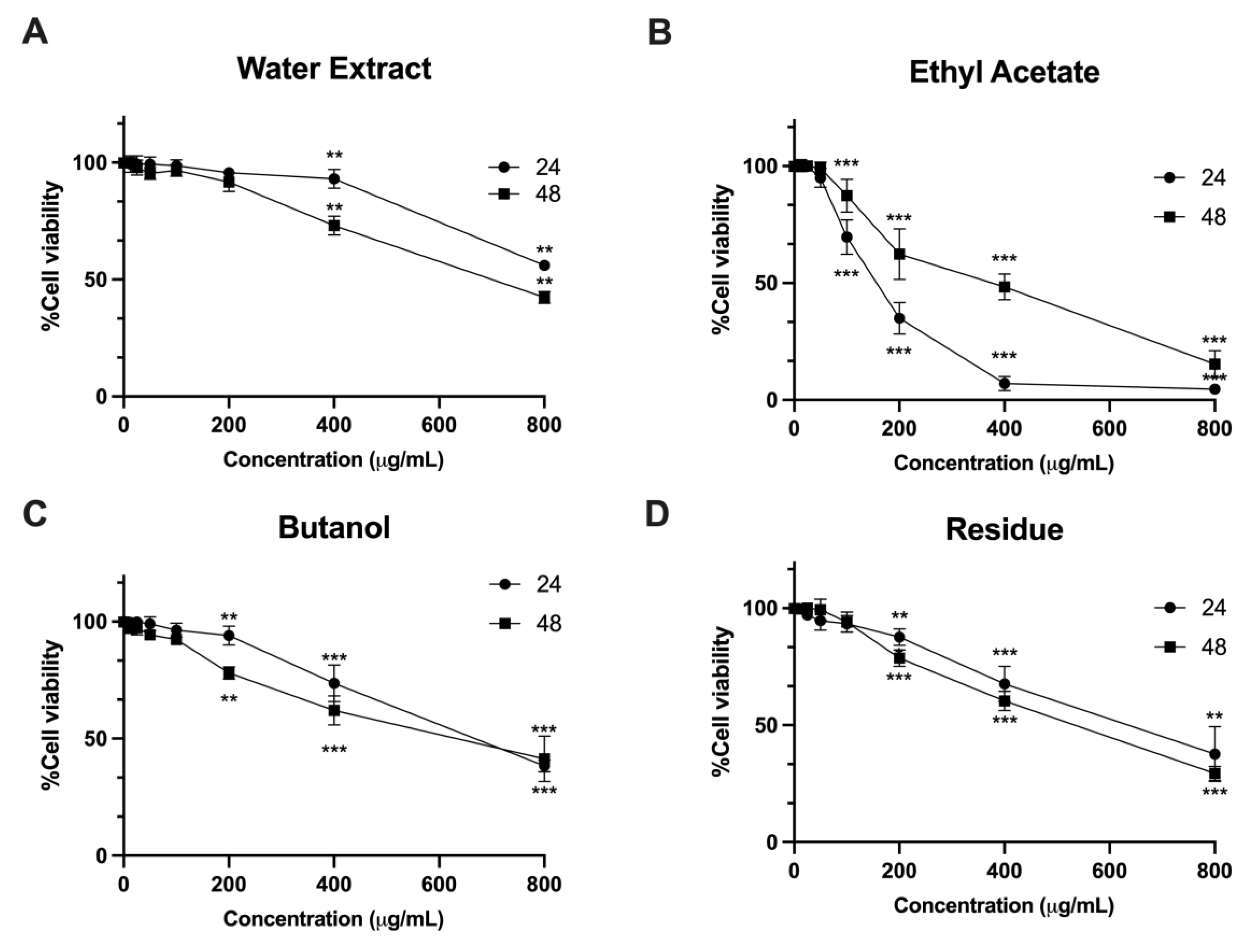
Figure 2.
Effects of L. martabanica leaf water extract on apoptosis induction in LX-2 cell lines. Dot plot (A) and bar graph (B) show the flow cytometric analysis of apoptosis induction on LX-2 cells treated with various concentrations (12.5 – 800 µg/mL) of L. martabanica leaf water extract. Results are presented as mean ± S.E.M. from three independent experiments.
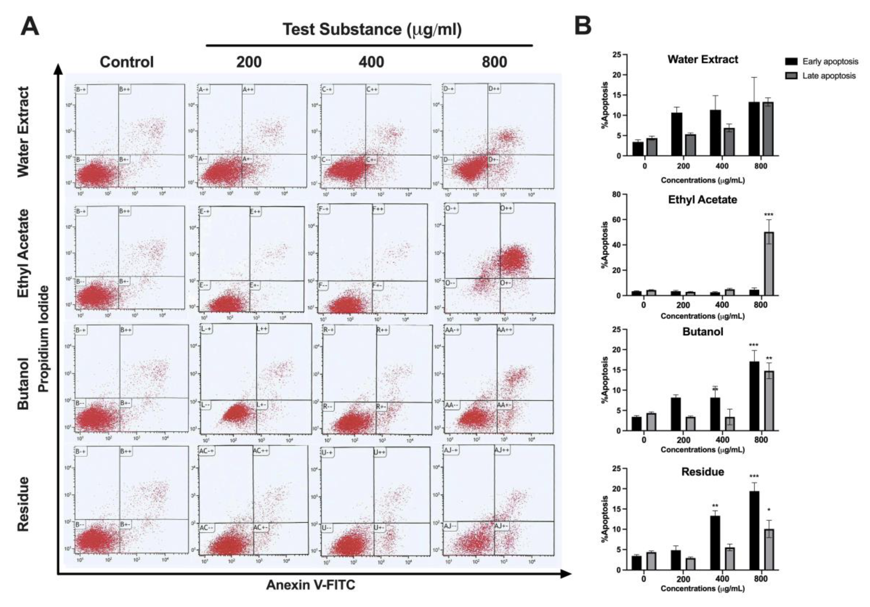
Figure 3.
Effects of L. martabanica leaf water extract on AChE activity: (A) Malondialdehyde, (B) Superoxide dismutase, (C) and Reduced glutathione (D). Results are presented as mean ± S.E.M. * Statistically significantly different from the normal rats (distilled water), p < 0.05, # *Statistically significantly different from the control group (distilled water + chlorpyrifos), p < 0.05.
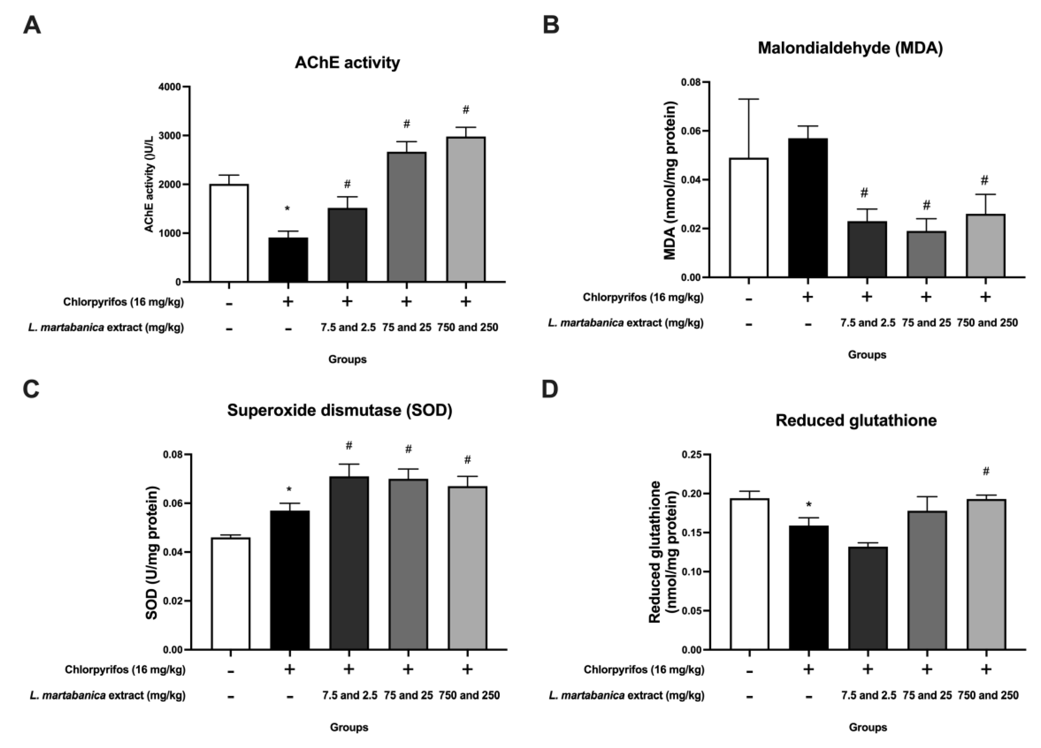
Figure 4.
Histopathologic results of rat liver. (A): Histology results of the normal rat liver in the normal group. (B): Histology results of the rat liver in the control group receiving chlorpyrifos. (C): Histology results of the rat liver in the group that received high doses of L. martabanica leaf water extract (750 and 250 mg/kg). The images indicate the hepatic portal vein (star) and sinusoidal dilatation (arrow).
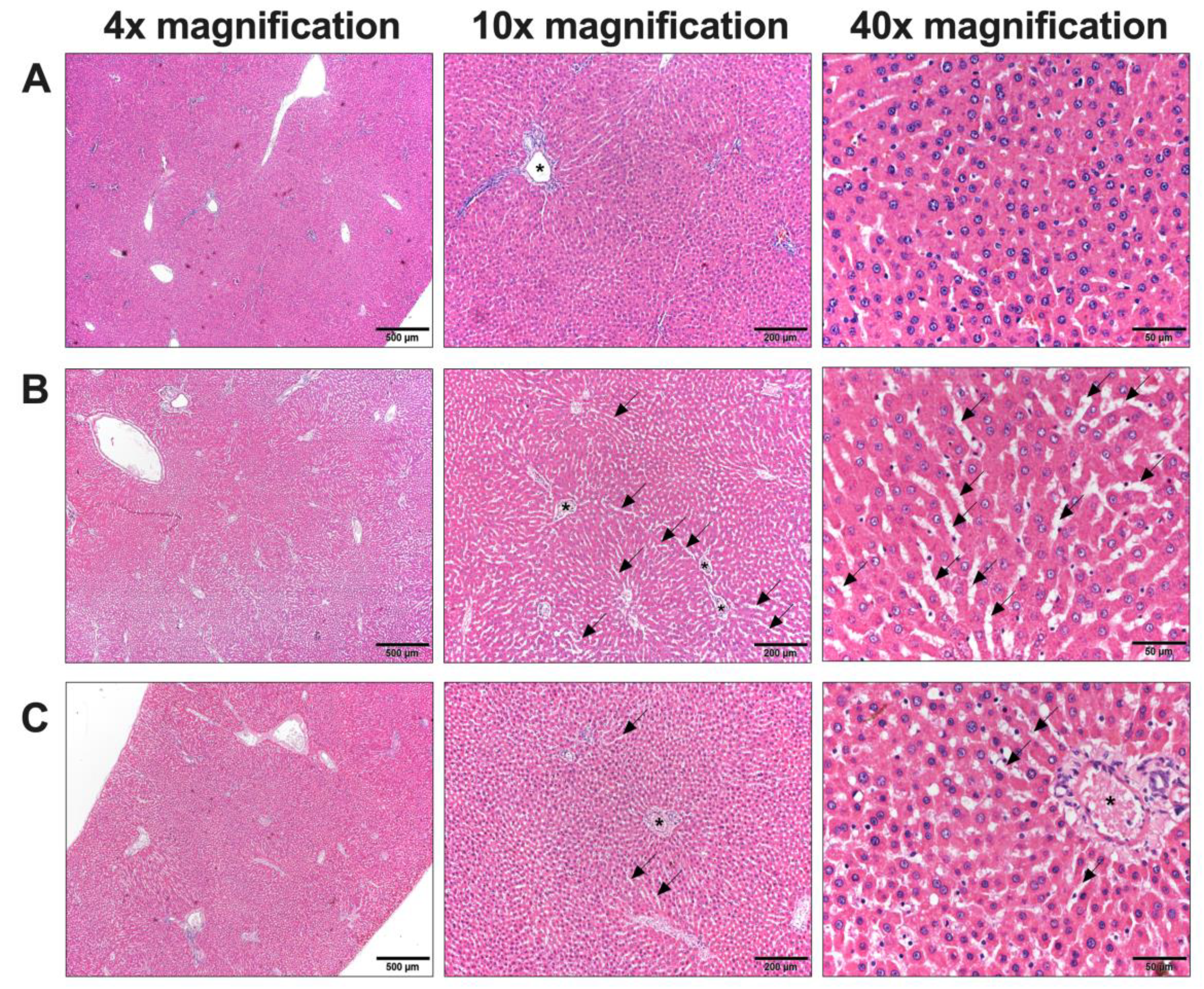
Figure 5.
Histopathologic results of rat kidney examination. (A): Histology results of rat kidneys in the normal group. (B): Histology results of kidneys in the control group receiving chlorpyrifos. (C): Histology results of kidneys in the group that received high doses of L. martabanica leaf water extract (750 and 250 mg/kg).
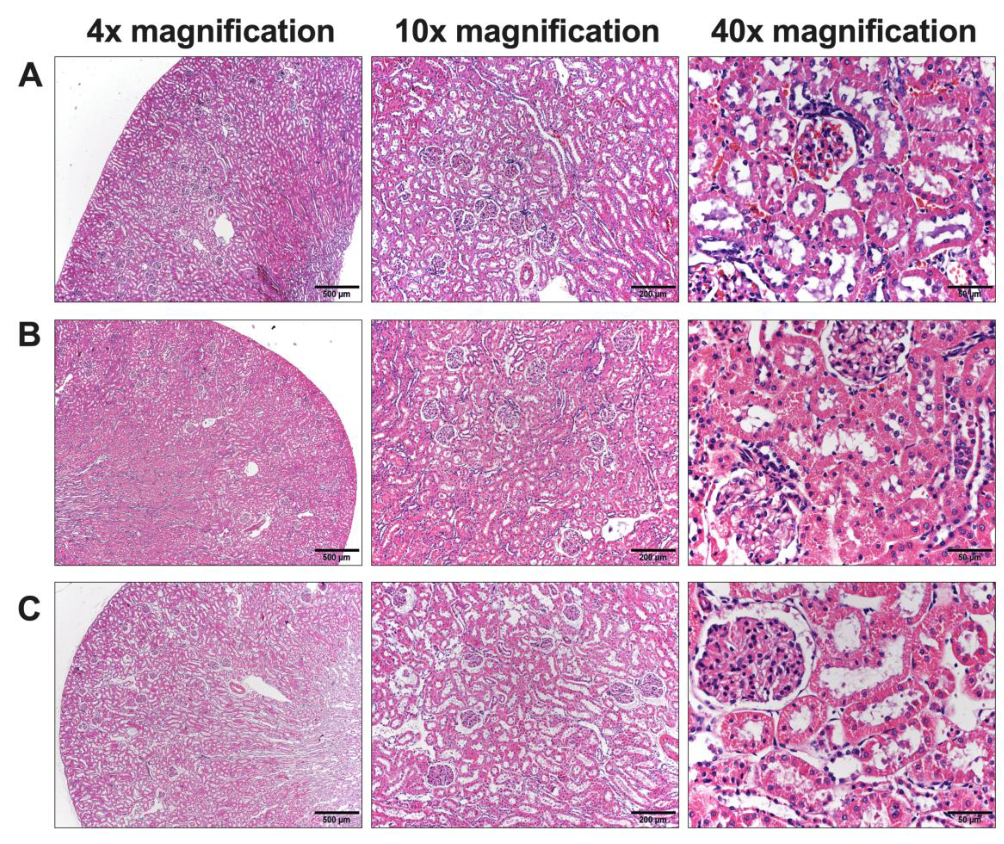

Table 1.
Pharmacognostic characters of L. martabanica leaves.
| Specification | Content (%) by dried weight |
|---|---|
| Loss on drying | 6.39 ± 0.04 |
| Total ash | 5.23 ± 0.01 |
| Acid-insoluble ash | 0.14 ± 0.01 |
| Ethanol-soluble extractive value | 9.57 ± 0.02 |
| Water–soluble extractive value | 13.19 ± 0.04 |
| Phytochemical group | Phenolics, flavonoids, saponins, terpenoids |
Values are expressed as mean ± S.E.M. from three independent experiments.
Table 2.
The IC50 values of L. martabanica Leaf Water Extract in DPPH assay and superoxide radical assay.
Table 2.
The IC50 values of L. martabanica Leaf Water Extract in DPPH assay and superoxide radical assay.
| Sample | IC50 (μg/mL) | |
|---|---|---|
| DPPH | Superoxide | |
| L. martabanica Leaf Water Extract | 56.17± 3.5 | 48.86 ± 4.9 |
| Ethyl acetate fraction | 35.3 ± 2.1 | 49.7 ± 3.0 |
| Butanol fraction | 45.7± 0.7 | 47.4 ± 2.4 |
| Residue fraction | 68.8± 10.9 | 80.6 ± 9.7 |
| Gallic acid | 2.70 ± 0.01 | 23.18 ± 3.9 |
Values are expressed as mean ± S.E.M. from three independent experiments. IC50, the half maximal inhibitory concentration; DPPH, 2,20-diphenyl-1-picrylhydrazyl.
Table 3.
Determination of IC50 values of L. martabanica Leaf Water Extract and its fractions.
| Sample | IC50 (μg/mL) | |
|---|---|---|
| 24 hours | 48 hours | |
| L. martabanica Leaf Water Extract | > 1,000 | 564.70 ± 40.95 |
| Ethyl acetate fraction | 154.7 ± 11.29 | 539.90 ± 38.26 |
| Butanol fraction | 572.90 ± 69.58 | 437.60 ± 22.57 |
| Residue fraction | 762.30 ± 10.74 | 690.00 ± 7.94 |
Values are expressed as mean ± S.E.M. from three independent experiments.
Table 4.
Effect of L. martabanica leaf water extract on body, liver, and kidney weight of rats.
| Parameters | Normal | Control | L. martabanica leaf water extract (mg/kg) | ||
| 7.5 and 2.5 | 75 and 25 | 750 and 250 | |||
| Body weights | |||||
| Day 1st | 242.50 ± 5.88 | 240.83 ± 3.27 | 243.33 ± 4.59 | 249.17 ± 4.73*,a | 250.00 ± 2.89*,a |
| Day 8th | 270.00 ± 9.22 | 281.67 ± 4.22 | 270.00 ± 9.04 | 279.17 ± 6.88 | 279.17 ± 7.35 |
| Day 16th | 263.33 ± 11.52 | 263.33 ± 8.53 | 275.00 ± 15.00 | 290.00 ± 8.16 | 295.00 ± 14.20 |
| Organ weights | |||||
| Liver | 11.60 ± 0.78 | 12.07 ± 0.31 | 14.32 ± 1.54 | 11.07 ± 0.46 | 10.83 ± 0.63 |
| Kidney | 1.34 ± 0.04 | 1.33 ± 0.06 | 1.62 ± 0.08*,a | 1.30 ± 0.04 | 1.45 ± 0.07 |
Values are expressed as mean ± S.E.M. from three independent experiments. n = 6. * Significantly different from the normal rats (distilled water), p < 0.05. a Significantly different from the control group (distilled water + chlorpyrifos), p < 0.05.
Table 5.
The hematological analysis of rats in the study of the anti-pesticide of L. martabanica leaf extract.
Table 5.
The hematological analysis of rats in the study of the anti-pesticide of L. martabanica leaf extract.
| Parameters | Normal | Control | L. martabanica leaf water extract (mg/kg) | ||
| 7.5 and 2.5 | 75 and 25 | 750 and 250 | |||
| Red blood cell (x106/µL) | 8.58 ± 0.22 | 8.20 ± 0.15 | 8.17 ± 0.23 | 7.59 ± 0.11* | 7.64 ± 0.08* |
| Hemoglobin (g/dL) | 15.42 ± 0.36 | 14.70 ± 0.20 | 14.85 ± 0.31 | 13.68 ± 0.19* | 13.57 ± 0.15*,a |
| Hematocrit (%) | 48.00 ± 1.23 | 45.33 ± 0.75 | 46.02 ± 0.92 | 42.32 ± 0.72* | 41.53 ± 0.29*,a |
| Mean corpuscular volume (fL) | 56.00 ± 0.93 | 55.38 ± 1.08 | 56.43 ± 0.70 | 55.78 ± 0.74 | 54.38 ± 0.38 |
| Mean corpuscular hemoglobin (pg) | 17.98 ± 0.35 | 17.95 ± 0.31 | 18.20 ± 0.20 | 18.05 ± 0.11 | 17.73 ± 0.23 |
| Mean corpuscular hemoglobin concentration (g/dL) | 32.15 ± 0.24 | 32.47 ± 0.18 | 32.27 ± 0.22 | 32.37 ± 0.27 | 32.65 ± 0.32 |
| Platelet (x105/ µL) | 8.26 ± 0.29 | 7.55 ± 0.29 | 8.12 ± 0.30 | 7.90 ± 0.27 | 9.59 ± 0.50a |
Values are expressed as mean ± S.E.M. from three independent experiments. n = 6. * Significantly different from the normal rats (distilled water), p < 0.05. a Significantly different from the control group (distilled water + chlorpyrifos), p < 0.05.
Table 6.
White blood cell count analysis of rats in the study of the anti-pesticide effect of L. martabanica leaf extract.
Table 6.
White blood cell count analysis of rats in the study of the anti-pesticide effect of L. martabanica leaf extract.
| Parameters | Normal | Control | L. martabanica leaf water extract (mg/kg) | ||
| 7.5 and 2.5 | 75 and 25 | 750 and 250 | |||
| White blood cell (x103 cells/µL) | 6.24 ± 0.89 | 8.64 ± 0.49 | 7.45 ± 0.39 | 7.55 ± 0.56 | 7.88 ± 0.61 |
| Neutrophil (cells/µL) | 1.48 ± 0.25 | 1.78 ± 0.35 | 1.92 ± 0.16 | 1.79 ± 0.25 | 1.75 ± 0.36 |
| Lymphocyte (cells/µL) | 4.03 ± 0.75 | 5.72 ± 0.37 | 4.99 ± 0.31 | 5.13 ± 0.34 | 5.36 ± 0.41 |
| Monocyte (cells/µL) | 0.64 ± 0.12 | 0.96 ± 0.16 | 0.45 ± 0.05 | 0.54 ± 0.15 | 0.65 ± 0.15 |
| Eosinophil (cells/µL) | 0.10 ± 0.02 | 0.19 ± 0.03 | 0.09 ± 0.01 | 0.10 ± 0.04 | 0.13 ± 0.04 |
| Basophil (cells/µL) | 0.00 ± 0.00 | 0.00 ± 0.00 | 0.00 ± 0.00 | 0.00 ± 0.00 | 0.00 ± 0.00 |
Values are expressed as mean ± S.E.M. from three independent experiments. n = 6. * Significantly different from the normal rats (distilled water), p < 0.05. a Significantly different from the control group (distilled water + chlorpyrifos), p < 0.05.
Table 7.
The blood chemical analysis of rats in the study of the anti-pesticide of L. martabanica leaf extract.
Table 7.
The blood chemical analysis of rats in the study of the anti-pesticide of L. martabanica leaf extract.
| Parameters | Normal | Control | L. martabanica leaf water extract (mg/kg) | ||
| 7.5 and 2.5 | 75 and 25 | 750 and 250 | |||
| BUN (mg/dL) | 21.48 ± 3.03 | 17.00 ± 0.45 | 18.75 ± 0.67 | 14.48 ± 0.94 | 12.02 ± 1.08* |
| Creatinine (mg/dL) | 0.68 ± 0.04 | 0.62 ± 0.02 | 0.64 ± 0.03 | 0.64 ± 0.02 | 0.62 ± 0.01 |
| Total protein (g/dL) | 6.23 ± 0.18 | 5.62 ± 0.18* | 5.25 ± 0.08* | 5.10 ± 0.07* | 5.32 ± 0.08* |
| Albumin (g/dL) | 2.92 ± 0.06 | 2.80 ± 0.06 | 2.73 ± 0.03 | 2.80 ± 0.04 | 2.77 ± 0.03 |
| Total bilirubin (mg/dL) | 0.20 ± 0.02 | 0.19 ± 0.01 | 0.20 ± 0.01 | 0.19 ± 0.01 | 0.21 ± 0.00 |
| Direct bilirubin (mg/dL) | 0.06 ± 0.01 | 0.06 ± 0.00 | 0.06 ± 0.00 | 0.05 ± 0.00 | 0.06 ± 0.00 |
| Aspartate aminotransferase (AST) (U/L) | 94.83 ± 5.31 | 102.83 ± 4.55 | 107.33 ± 13.04 | 78.33 ± 3.00 | 77.00 ± 3.07 |
| Alanine aminotransferase (ALT) (U/L) | 33.00 ± 3.71 | 29.00 ± 1.21 | 30.50 ± 4.57 | 30.33 ± 2.35 | 25.67 ± 1.99 |
| Alkaline phosphatase (U/L) | 116.83 ± 6.95 | 83.17 ± 7.66* | 85.67 ± 11.13 | 76.50 ± 7.33 | 70.17 ± 6.54* |
Values are expressed as mean ± S.E.M. from three independent experiments. n = 6. * Significantly different from the normal rats (distilled water), p < 0.05.
Disclaimer/Publisher’s Note: The statements, opinions and data contained in all publications are solely those of the individual author(s) and contributor(s) and not of MDPI and/or the editor(s). MDPI and/or the editor(s) disclaim responsibility for any injury to people or property resulting from any ideas, methods, instructions or products referred to in the content. |
© 2024 by the authors. Licensee MDPI, Basel, Switzerland. This article is an open access article distributed under the terms and conditions of the Creative Commons Attribution (CC BY) license (http://creativecommons.org/licenses/by/4.0/).
Copyright: This open access article is published under a Creative Commons CC BY 4.0 license, which permit the free download, distribution, and reuse, provided that the author and preprint are cited in any reuse.


