Submitted:
03 April 2024
Posted:
04 April 2024
You are already at the latest version
Abstract
Environmental pollution is one of today's main ecological and social concerns. Among these pollutants are the heavy metals present in the burning of fossil fuels, mining, smelting of metallic minerals, fertilizers and pesticides. Billions of tons of heavy metals are emitted every year from industrial chimneys and sewers, contaminating the hydrosphere and polluting rivers, lakes and seas. In this context, freshwater fish are the most affected due to their proximity to the source of contamination and are therefore used as bioindicators in assessing environmental impact following exposure to metals. However, our understanding of the effects of heavy metals on detoxification organs - such as the liver and gills - in freshwater fish in situ is still limited. Therefore, we conducted a systematic review to determine the main changes and mechanisms involved in morphological modifications in the liver and gills of cichlid fish following exposure to heavy metals in situ. Based on a structured search, 36 original articles were selected from the PubMed and SCOPUS databases. The selection of studies and the identification of duplicates were identified by Rayyan, and the study selection process was presented in the PRISMA statement diagram. Bias analysis was carried out using the Software Review Mananger (RevMan) 5.3, from the Cochrane Collaboration. Thirty-six studies were selected which showed negative effects attributed to environmental contamination by heavy metals, ranging from bioaccumulation to morphophysiological alterations. Almost all the heavy metals analyzed were found in the liver and gill tissues, especially cadmium, copper, lead, zinc, iron and manganese. In view of the continuous morphophysiological changes identified in the liver and gill studies, it is possible to state that bioaccumulation analyses, combined with these configurations, are emerging as crucial tools for assessing and guiding environmental monitoring. This approach aims not only to preserve but also to strengthen the integrity of the aquatic ecosystem and the ichthyofauna.
Keywords:
Toxicology
; Aquatic ecosystem
; Bioaccumulation
; Histopathology
; Fish
1. Introduction
Heavy metals pollution influences the lives of diverse aquatic organisms and humans who consume contaminated aquatic products. However, its potential impacts on aquatic organism health and, thus, ecological health, have been neglected in many regions [1]. Chemical and solid waste discharges by industries, oil spills, untreated sewage and urban run-off are responsible by contamination of rivers and oceans compromising all the entire aquatic ecosystem [2,3,4]. Among these compounds are the heavy metals from anthropogenic sources, mainly due to intense mining activity and environmental disasters, which release large amounts of chemical compounds in the aquatic environment [5,8]. Heavy metals can have a high bioaccumulation and bio magnification capacity along the food chain and accumulate in water and sediments [5,9]. In addition, they are an important protein resource and are part of human food composition, so fish represent one main sources of heavy metal intake for humans via food chain [10].
The mechanisms the action of the heavy metals in organisms of the aquatic ecosystem is very broad and it is not yet clear. However, currently it is known that the effects of heavy metals in fishes are dependent on the concentrations and properties of the metal [11,12,13]. The access of heavy metals in the organism can be limited by the anatomical structures of the animal, the physical and chemical characteristics of the environment and by the competition for binding sites for metal ions [14,15]. For these reasons, there are often considerable differences in sensitivity between organs and tissues [16,17]. The main organs affected by heavy metals exposure are liver, muscles and gills and the toxic effects alter the physiological activities and biochemical parameters in fish tissue and blood besides affect negatively their growth rate [5,18]. In addition, the accumulation of heavy metals - such as zinc and copper - can lead to gonadal abnormalities. In addition, the combination of metals with other chemical products present in the aquatic environment may have led to these abnormalities [6]
Among the freshwater fish, the family Cichlidae (order Perciformes) are the most studied [19,20,21]. Cichlids are widely distributed in Africa and Central and South America [22]. The morphological characteristics this Family are: lateral line divided into two branches, protractile mouth with variable position (lower, subterminal, terminal and upper) different eating habits (insectivore, omnivore, carnivore and detritivore/ insectivore), and morphological structures adapted to the trophic categories they occupy [21]. In addition, it presents important behavioral aspects, such as parental care [22]. These characteristics allow these animals to survive in different and varied environmental conditions and to be used as indicators of environmental pollution.
In the current manuscript, we systematically review of the scientific literature to determine the main changes and mechanisms involved in the morphological modifications in liver and gills on Cichlid fishes after exposure to heavy metals in situ. In addition, to present a general view of heavy metals exposure in environmental and to identify recommendations and perspectives for future and help understanding distribution of these pollutants in freshwater aquatic system. The methodological quality of each study identified was also evaluated, and the main sources of bias that undermine the quality of evidence were pointed out.
2. Method
This systematic review was carried out following the guidelines of The Preferred Reporting Items for Systematic reviews and Meta-Analyses (PRISMA) Supplement A (PAGE et al., 2021). The review was registered and published in PROSPERO (515999).
The systematic review is carried out in eight stages: (1) drafting the research question; (2) searching the literature; (3) selecting articles; (4) extracting data; (5) assessing methodological quality; (6) synthesizing the data; (7) assessing the quality of the evidence; and (8) writing up and publishing the results [23].
The research question was developed using the acronym PECO (P: population; E: exposure; C: comparator; O: outcome), adapted from the acronym PICOS for the inclusion of exposure, rather than intervention, in observational studies [23].
The guiding question of the review was: “what are the effects of exposure to heavy metals on the liver and gills of cichlids collected from the environment?”
2.1. Search Strategy
This systematic review followed the Preferred Reporting Items for Systematic Reviews and Meta-Analyses (PRISMA) guidelines [26]. The studies were selected through an advanced search on the platforms PubMed (https://www.ncbi.nlm.nih.gov/pubmed) and Scopus (https://www.scopus.com/home.uri) on october 14, 2023. Based on four search parameters, we developed a comprehensive search strategy for retrieving all relevant studies: “fish (cichlids)”, “heavy metals”, “gills” and “liver”. For PubMed, use MeSH terms and related keywords in title / summary (TiaB). The descriptors were separated by ligands OR within the same filter and AND as ligands of different filters. For the Scopus platform, use the same descriptors, but adapted for this platform. After a search on Scopus, use the platform’s own filter using the term “Fish” to make the selection in the two databases (Supplementary File S1). Secondary studies (literature reviews, letters to the editor, case studies, comments, and editorials) were also excluded.
After screening, all relevant studies were retrieved in full text and selected by eligibility. Eligibility was reviewed by the researchers and disagreements resolved by consensus. The reference list of each included study was reviewed, identified in all databases, manually crawled for additional articles [27]. If any documents have been added, your reference list has also been reviewed by the end of this cycle.
2.2. Study Selection
Studies were independently selected and duplicates identified using the Rayyan selection platform. Initially, the articles were selected after reading the title and abstract, and those that met the eligibility criteria were read in full for inclusion or exclusion in the review.
To discard the subjectivity in data collection and selection process, the information was independently extracted by three reviewers (L.V.B, A.L.F.D, M.M.S) and analyzed in four descriptive levels separately:
- (1)
- (2)
- (3)
- Studies in which at least one of the analyzes mentioned below were performed: quantification of the metal in the organ (bioaccumulation), histopathological analysis and oxidative status.
- (4)
- Studies that analyzed the effect of heavy metals on liver or gills or both.
2.3. Data Extraction and Management
A specific initial selection was conducted by three independent reviewers (L.V.B, A.L.F.D, M.M.S and C.D.L.R), who removed duplicate articles and elected titles and abstracts with respect to the eligibility criteria. After the initial screening, full-text articles from potentially relevant studies were independently assessed for eligibility. The Kappa test was performed to select and extract data (Kappa = 0.946). In cases of disagreement, another group of reviewers (R.V.G, S.S.R.S, M.B.F) selects the complete study from all included inclusion records. The data for each study was extracted with standardized information, such as: characteristics of the publication (author, year, country); animal model (species, weight, size, experimental groups) and methodological analyzes. The bibliographic references of the selected studies were analyzed and works that meet the inclusion requirements were added.
2.4. Bias Risk Assessment in the Included Studies
The risk of analysis was analyzed using the Software Review Mananger (RevMan) 5.3, from the Cochrane Collaboration, which aims to assess the methodological quality of the studies. To increase transparency and apply it, the selection criteria include 1. Generation of random sequence, 2. Concealment of allocation, 3. Concealment of caregivers and / or researchers with knowledge about the interventions that each animal received, 4. Concealment of the result evaluation, 5. Incomplete result data and 6. Selective result report [28]. The items of the RevMan tool were scored with “yes” (low risk of bias), “no” (high risk of bias); or “unclear” (indicating that the item was not reported and, therefore, the risk of bias was unknown). Two review authors (A.L.F.D, L.V.B) independently assessed the risk of bias for each study, using the adapted criteria described in the Cochrane Handbook for Systematic of Interventions. Any differences were resolved by the discussion between the authors (R.V.G., M.M.S and C.D.L.R). The risk of bias was created using Review Manager 5.3 [29].
In this study, this instrument was configured for specific bias aspects considering field studies, with animal intervention in situ. In order to establish consistency and avoid discrepancies in the evaluation of the methodological quality of the works, three topics were removed from the original RevMan tool: 1-Was the allocation to the different groups adequately concealed? 2-Were the animals randomly housed during the experiment? 3-Were the caregivers and/or investigators blinded from knowledge which intervention each animal received during the experiment?
3. Results
3.1. Characteristics of Publications
From 2.129 records identified in all eletronic databases, 506 were duplicates and 36 studies were recovered in full text (primary search 31; secondary search 5) and included in the systematic review (Figure 1). The filters applied in each database and the flowchart indicating the search strutucture are shown in online Supplementary Table S1 and Figure 1, respectively. Most studies identified (30,5%, n=11) originated from Egypt, followed by six studies (16,66%) from Mexico, four from Brazil and Saudi Arabia (11,11%), then by India, Ivory Coast (5,55%, n=2 each) and finally Zimbabwe, Zambia, Nigeria, Sri Lanka, Sudan and Vietname (2,77%, n=1).
3.2. Characteristics of Fish and Collecting Environments
The characteristics of fish and collecting environment of the selected articles can be seen in Table 1. Ten species from the Cichlidae family were studied. There was a predominance of studies with Oreochromis niloticus (52.77%, n=19), a species commonly used in ecotoxicological studies. The species Micropterus salmoides and O. aureus were present in three studies (8.33%). Other species analyzed were Cyprinus carpio, O. mossambicus, Geophagus brasiliensis and Sarotherodon melanotheron (5.55%, n=2) and, finally, S. galilaues, Tilapia andersonii, T. rendalli, Pelmatochromi quentheri, P. pulcher, Etroplus suratensis, Hypothalmic molitrix, Braquiplatystoma filamentosum (2.77%, n=1, each), in addition to these other four undefined: Oreochromis sp., Tilapia sp., Semaprochilodus sp and Cichla sp (2.77%, n=1, each). Therefore, the genus Oreochromis is the most represented Cichlidae, being reported in 77.77% (n=28) of the articles reviewed in this study.
The total number of animals was reported in almost all the articles analyzed (88,88%, n=32). Weight and length of the animals were related in most studies (55,55%, n=20), some studies related only weight (16,66%, n=6) and one study related only to length (2,77%, n=1). There are studies not related to any of the biometric parameters (25%, n=9). Some studies used biometric parameters for calculating condition factor (n=3) and hepatosomatic index (n=2).
Few studies used the control group (33,33%, n=12), the majority (69,44%, n=25) did not report or use any type of control. When used, the control animals were collected in contamination free rivers and lakes, or in fish farms, being considered as wild animals or experimental animals. Some studies compared different groups of animals, according to species (n=8), age (n=3), weight/size (n=3), gender (n=1), period (n=8) or site (n=16) of collection. Regarding the collection of animals, many studies (55,55%, n=20) mention collection periods and/or stations, but some (22,22%, n=8) neglected any information about this. The different collection sites were determined by their proximity to polluting sources, downstream and upstream, and sometimes also by the type of aquatic ecosystem, lentic and lotic (n=9). To determine the pollution of the environment by heavy metals, the contamination factor and the pollution index were calculated in only one of the studies.
3.3. Characteristics of the Analyses
The metals analyzed are listed in Table 1, and those analyzed in the liver and gill tissues are shown in Figure 2.
The heaviest metals analyzed in the liver were cadmium (n=20); copper (n=19); zinc (n=15) lead (n=13); ; manganese (n=12); iron (n=11); mercury (n=10); chromium (n=8); nickel (n=7); arsenic (n=5); cobalt (n=4); selenium (n-3); aluminum and silver (n=2 each); strontium (n=1); molybdenum and barium were not analyzed in the tissues. On the other hand, the heavy metals most analyzed in the gills were cadmium and copper (n=14 each); zinc (n=12); lead (n=10); iron (n=9); manganese and chromium (n=8 each); nickel (n=7); cobalt, mercury and arsenic (n=4 each); aluminum, strontium, silver and selenium (n=2 each); molybdenum and barium were not analyzed in the tissues.
The main characteristics of the methodological analyses carried out by the selected studies are in Table 1.
Of the methodological analyzes carried out in these ecotoxicological studies, the analysis of metal bioaccumulation in fish tissues was the one that had the greatest representation (n=26; 72,22%); followed by quantification of metals in water (n=25; 69,44%) and sediment (n = 9; 25%), as well as physical-chemical parameters of water (n=11; 30,55%); histopathological analyzes, whether structural or ultrastructural (n=10; 27,77%); analysis of oxidative stress, through the activity of antioxidant enzymes and quantification of stress markers (n=11; 30,55%); in addition to other biological analyzes, biochemical markers such as metabolic enzymes and metallothioneins (n=11; 30,55%), and genetic markers (n=3; 8,33%).
A variety of tissues has been used to quantity heavy metals in Cichlidae, in addition to liver (91,66%, n=33) and gills (66,66%, n=24), includes muscles (75%, n=27), kidneys (27,77%, n=10), brain (21.4%, n=6), digestive tract (22,22%, n=8), gonads (16,66%, n=6), scales and spine (7.1%, n=2 each). The water-tissue transfer factor was calculated in some studies (n = 6), as well as the sediment-tissue transfer factor (n = 3).In addition to the ethical issue in the use of animals for research, procedures involving collection, handling and euthanasia of fish are important because they affect some analyzes such as those of oxidative status. Few studies (n=12; 33,33%) reported any information about the euthanasia of the analyzed fish. The studies that informed the place of euthanasia (n=9; 25%) said they had carried out in the field (n=5; 13,88%) or laboratory (n=4; 11,11%), indicating the methods of maintaining and transporting the animals. Studies that specified euthanasia methods used clove oil (n=2; 5,55%), tricaine mesylate (MS-222) or spinal cord transection (n=1; 2,77% each).
3.4. Ecotoxicological Effects of Heavy Metals on Fish
All 36 selected studies showed negative effects attributed to contamination of the environment by heavy metals, from bioaccumulation to morphophysiological changes.
Virtually all heavy metals analyzed were found in the liver and branchial tissues, especially cadmium, copper, lead, zinc, iron and manganese (Rashed 2001a,b; Abdel-Monein 2013; Ajuima 2015; Coulibaly 2012; AlKahtani 2009; Doria 2017; Armel-Cyrile 2012; Elgobashy 2001; Mbewe 2016; Munoz-Najera 2018; Abdel-Baki 2011; Abdel-Khalek 2014; Yossef 2004; Voigt 2014; Pettamanna 2020; Martínez-Durazo 2020; Salem 2021; Ngo 2022; Páez-Osuna 2022; Vieira 2023; Martínez-Durazo 2023; Páez Osuna 2023), with bioaccumulation evidenced by concentration factors, water-tissue and/or sediment-tissue (Rashed 2001a; Ajima 2015; Mbewe 2016; Munoz-Najera 2018; Abdel-Baki 2011; Voigt 2014; Pettamanna 2020; Martínez-Durazo 2020; Páez-Osuna 2022; Vieira 2023).
Many heavy metals were generally found in higher concentrations in the liver than in gills (Voigt 2014; Jinadasa 2014, Rashed 2001ª; Doria 2017; Muisa 2011; Martínez-Durazo 2020; Ngo 2022; Páez-Osuna 2022; Vieira 2023; Martínez-Durazo 2023; Páez Osuna 2023), and the deleterious effects were often greater on the liver than in gills (Abdel-Khalek 2014, 2015; Abdel-Monein 2012b; Doria 2017).
There were ten studies that analyzed the liver morphology of fish and they all found alterations (Abdel Monein 2012b; 2013a, b; Doria 2017; Kumar 2016; Abdel-Khalek 2014; Adham 2001; Pettamanna 2020; Martínez-Durazo 2023). The main changes described were degenerations and cellular lesions such as nuclear and cytoplasmic vacuolization, cell rupture, pycnotic nucleus and necrosis; circulatory changes such as vascular congestion and dilation of the sinusoids; proliferative cell changes such as nuclear hypertrophy; and ultramorphological changes such as nuclear erythrocyte alteration, increase in the nucleolus, increase in the nuclear envelope, swelling of mitochondria, production of peroxisomes and proliferation of lipid drops (Table 2 and Figure 3). Of these works, only two (Kumar 2016; Pettamanna 2020) did not quantify the observed changes.
In relation to the gills, there were seven studies (Kumar 2016; Abdel-Monein 2012a, b; Abdel-Khalek 2014; Doria 2017; Adham 2001; Martínez-Durazo 2023) with morphological analyses and they also found alterations such as: proliferative cell changes such as epithelial hyperplasia and multifocal mucous cells; cell degenerations and injuries such as epithelial cell edema, epithelial cell lifting and peeling, lamellar fusion, rupture of cells and necrosis; circulatory changes such as hemorrhage, blood congestion and aneurysm; and ultramorphological changes such as decreased epithelial cell microvilli (Table 2 and Figure 3). Among these works, only one (Kumar 2016) did not quantify the alterations found.
Eleven studies (Eldemerdash 1999; Abdel Monein 2013a; Carvalho 2012; Kumar 2016, 2017; Abdel-Khalek 2015; Salem 2021; Ngo 2022; Vieira 2023) analyzed the oxidative status in the hepatic tissue of fish and six (Carvalho 2012; Kumar 2016, 2017; Abdel-Khalek 2015; Abdel-Monein 2012a; Ngo 2022) in the gill tissue, and different changes were found in relation to antioxidants enzymes, but Lipoperoxidation –LPO with increase of malondyaldeide (MDA) was unanimous (Table 2 and Figure 3).
Although the focus of this systematic review was the liver and gills, it is worth mentioning the large number (n=25) of studies with the muscle, considering its importance for human consumption and consequent health risks. Heavy metal analysis was used in muscle studies, and sometimes analyses of oxidative status (n=4), cellular stress (n=4) and/or meat quality (n=2) were also carried out. Heavy metals were almost always detected in lower concentration than in the liver and/or the gills (n=24). Of the studies that evaluated the risk to health by meat consumption (n=14), seven reported that the levels of metals in the muscle were above the limits considered safe by the legislation, and five reported that the levels were within the safe limits.
Studies that performed comparative analyzes between cichlid species did not report significant differences in the bioaccumulation (Ajima 2015; Mbewe 2016; Yossef 2004). As for the age of the fish, two studies (Rashed 2001a, b) showed a positive correlation of some metals (Pb, Cd, Sr) in the gills and a negative correlation of some metals (Pb, Cd, Sr, Ni, Fe) in the liver, and one study (Armel Cyrille 2012) showed no correlation between metal levels and age. A study (Voight 2014) with adult individuals showed that there are no differences in the bioaccumulation according to size, weight and gender.
The works that carried out comparative analyzes between different periods of the year showed that seasonality (or rainfall) influenced concentration and bioaccumulation (Al-Kahtani 2009; Coulibaly 2012; Carvalho 2012; Muisa 2011; Armel Cyrille 2012; Munoz Najera 2018; Ruelas Inzuenza 2015; Martínez-Durazo 2020; Ngo 2022; Martínez-Durazo 2023;), with varied results for different metals and tissues.
As for the differences regarding the collection sites, the closer to the contaminating source and the higher the levels of metals in the environment, the greater the bioaccumulation and the morphophysiological damage, showing a direct relationship of heavy metal contamination on the health of the fish (Doria 2017 ; Kumar 2016; Muisa 2011; Abdel-Monein 2012a, 2013a; Kumar 2016, 2017; Abdel-Khalek 2014, 2015; Adhan 2001; Mbewe 2016; Al-Khatani 2009; Jinadasa 2014; Yossef 2014; Ngo 2022; Páez-Osuna 2022; Vieira 2023; Marínez-Durazo 2023; Paez-Osuna 2023). In addition, there was a tendency to dilute metals downstream (Muisa 2011; Abdel-Khalek 2014, 2015; Mbewe 2016; Elghobash 2001) and their concentration in lentic environments, particularly in sediment and vegetation (Elgobash 2001). Water characteristics seem to influence bioaccumulation, especially salinity, alkalinity and hardness, as they affect the solubility and availability of metals in the environment, as well as the permeability of the gill membrane and the entry of metals into the fish organism (Elgobash 2001; Muisa 2011; Abdel-Khalek 2015; Carvalho 2012, Pettamanna 2020; Salem 2021; Ngo 2022; Páez-Osuna 2022).
3.5. Bias Analysis
The percentages of each risk of bias of the analyzed studies were shown in Figure 4, constructed by the Software Review Manager 5.3 [29]. This analysis took into account the adaptations necessary for the adequacy of animal studies in the environment. Regarding selection bias, the sequence generation process (Q1) was not clear in more than half of the studies (n = 16; 57.1%). In terms of animal characteristics, i.e., the similarities between them (Q2), more than half of the studies (n = 15; 53.6%) did not clearly report this information. Regarding detection bias, the vast majority of studies (n = 25; 89.3%) did not explicitly report on the random evaluation of results for relevant measures (Q3). In addition, the results evaluator was not blind in all studies (100%; Q4). Data from incomplete results were not adequately addressed (Q5) in almost all studies (n = 27; 96.4%). Regarding reporting bias (Q6), a low risk was identified in almost all studies (n = 26; 93.0%), and a high risk in two studies. Other potential sources of bias were not clearly pointed out in 25 studies (89.3%; Q7).
4. Discussion
This systematic review showed the main tissue changes caused by heavy metals in the liver and gills of cichlid fish collected in situ. Furthermore, the main methodological parameters that are used to analyze the toxicity of these metals were identified.
Through the results presented, this study demonstrated that cichlid fish found in environments contaminated by heavy metals can bioaccumulate these compounds in the liver and gills, as well as in other tissues. As a consequence, there are pathological changes in these organs, which certainly compromise their functionality and the organism’s homeostasis, which can affect the quality of life and survival of the animal, as well as that of humans, when ingesting fish contaminated by the metal bioaccumulation process.
The selected articles showed that Egypt is the country with the largest number of works in this area, mainly with Nilotic tilapia (Oreochromis niloticus), which is a species native to the region. Possibly the predominance of studies with this species is due to its characteristics, as it is a prolific fish, fast growing, resistant to diseases and easy to adapt and manage, being cultivated for commercial purposes and found throughout the world, during all year round [62,63]. Thus, tilapia have been used as indicators of environmental pollution, and intensively used in toxicological studies [53,64,65,66,67].
In addition to identifying the best bioindicator model for toxic effects, it is necessary to adopt some care when evaluating changes, especially oxidative stress, as other parameters can influence the results [41]. The form and type of anesthetic used to euthanize fish can affect the organism as a whole [68,69,70]. Thus, the correct type and use of anesthesia ensures that the possible changes observed are due to the effect of toxic compounds and not other factors [70]. In our review, few studies reported any information on euthanasia. Only three studies reported using anesthetic to perform euthanasia, namely clove oil (Eugenol) and tricaine mesylate (MS-222), in accordance with what is indicated in the literature and in compliance with the Ethics Committees [70]. MS-222 is an isomer of benzocaine used directly in water, and is widely used in the development of research with fish, being identified as the safest and most effective for various productive and scientific purposes [60,63,64]. Eugenol, the distillation product of the extract from parts of the clove Syzygium aromaticum (Eugenia aromaticum or Eugenia caryophyllata), needs to be diluted in alcohol, due to its hydrophobic characteristic [73]. In addition to having anesthetic properties, it is also widely used because it has bactericidal properties, as it is low cost and comes from a natural source and, in addition, it is a safe compound for the environment and can be used to reduce stress in situations that require the immediate release of the fish [68,74,75]. Wagner et al., (2003) demonstrated that a concentration of 60 mg.L-1 of clove oil reduced oxidative stress in rainbow trout (Oncorhynchus mykiss), while the same amount of MS-222 resulted in higher levels of cortisol, indicating greater stress. Thus, although both anesthetics are highly efficient, they present some differences in their anti-stress effects [68,75]. Furthermore, the place of euthanasia, whether in the field or laboratory, must also be considered, as it involves moving the animals, as well as the place where the animals were housed and the time elapsed until the moment of euthanasia. Data in the literature indicate that capture and transport are important factors in inducing stress in fish [76,77]. However, approximately 75% of the studies neglected this information about the place of euthanasia and the need to transport or accommodation of animals.
Regarding the season in which the collections were carried out, most of the studies evaluated both the rainy and dry seasons. When analyzing the results of these studies in terms of bioaccumulation of heavy metals in tissues, there were varied results depending on the metal and organ evaluated. It is possible that in the rainy season exposure to metals is greater [78], mainly due to the disturbance of contaminated sediments [79]. On the other hand, in the rainy season there is greater dilution of metals due to the greater rainfall, and therefore the concentrations of these metals would be lower [80,81]. During the dry period, high temperatures would lead to greater fish activity, with an increase in ventilation, metabolic rate and feeding sessions, causing greater absorption and accumulation of these toxic compounds [82].
When we analyzed the methodologies used to quantify heavy metals, we observed that the analysis of bioaccumulation in tissues was quite significant (around 72%). However, some studies only carried out analyzes to quantify heavy metals in water and sediment, inferring the bioaccumulation of metals in tissues and relating it to the effects found. In general, the accumulation of heavy metal in organs may indicate the high availability of these compounds in the environment, as animals have a higher uptake and accumulation rate than the excretion and detoxification rate [83]. However, the complementarity of the analyzes increases the reliability of the results, providing a relationship of cause (bioaccumulation) and effects (histopathologies), with more assertive conclusions.
The main organs used as bioindicators in fish are the gills and liver [84,85]. The gills, as they are in direct contact with the pollutant and due to their primary function of filtration and oxygenation, and the liver, as it is a detoxification gland, are potentially target organs for toxic compounds and are therefore essential in ecotoxicological studies [86,87]. In our study we found that muscle is also widely used in research because, although it does not accumulate as much metals as the liver and gills, it is extremely important as it is for human consumption, and even low concentrations of metals can be a health risk [60].
Bioaccumulation of heavy metals in organs triggers structural responses to protect them. Therefore, some biomarkers can be used to explain the cause and effect relationship in the toxicological assessments of organisms [88]. In this context, along with bioaccumulation analyses, histopathological assessments or other biological parameters are important in ecotoxicological studies with fish [89,90,91].
With regard to the contaminant and its histopathological changes, the metals most analyzed were cadmium (Cd) and copper (Cu), around 77% of the studies. Cadmium is present in several anthropogenic activities, of which we can highlight the refining of ore, the production of herbicides and other fertilizers, and the manufacture of batteries [18,92]. It has high cytotoxic activity in fish organs [18,92], and its accumulation and consequent effects have shown that the gills and liver are organs greatly affected by contamination by this metal. The liver and especially the gills accumulated cadmium. The gills have chloride cells, responsible for ionic exchange, and therefore can bind and accumulate heavy metals such as cadmium [92,93]. Cadmium can cause hyperplasia and cellular damage such as partial and total lamellar fusion, aneurysm and necrosis, compromising the entire respiratory function of the fish [86]. In the liver, this compound can cause responses related to inflammation, such as hyperemia and macrophage intrusion, as well as vacuolization, congestion and necrosis [92,94].
Copper is used in the manufacture of industrial parts, such as wires and TV parts [95], and in agricultural products such as fungicides, which, if released into the environment, can compromise aquatic organisms [61,84]. It is considered an essential metal in small concentrations, as it has specific interactions with proteins and enzymes in physiological processes such as cellular respiration [96], however, in high concentrations it can cause damage [88,98]. Our findings showed that exposure to this metal causes histopathological changes in the gills, such as epithelial cell hyperplasia, hypertrophy, shortening, congestion, necrosis and lamellar fusion [54,98]. Epithelial cell hyperplasia, increased chloride cells and mucus secretion are defense mechanisms that act as a barrier against the diffusion of contaminants such as heavy metals [99,100]. However, these processes compromise gas exchange and consequently cause respiratory imbalance. In the liver, this metal causes vacuolization of hepatocytes and a decrease in the number of hepatocyte nuclei, changes that are often associated with degenerative necrosis [96].
Zinc (Zn) was the third most analyzed metal in the studies. Like copper, it is also considered an essential element in low concentrations, participating in processes in the nervous system, in protein synthesis, but at high levels it causes damage to health. Contamination occurs through the use of galvanized pipes and iron, and the direct disposal of products, such as paint, that contain this compound [92,98]. In fish, the presence of zinc can cause fusion of the lamellae, partial or total lamellar deletion, in addition to reduction of the interlamellar space, cell rupture, deformities in the lamellae structures and aneurysm, consequently making it difficult for the animal to breathe [55,101,102]. In the liver, where it was found most, when considering our frequency analysis, zinc accumulation can cause degeneration, vacuolization and necrosis [56,102].
Iron (Fe) was the fourth most studied metal, found in nature in the form of ore and used in the production of steel and the manufacture of fungicides, electrodes and dyes. It is fundamental for the survival of individuals, as it is essential for multiple metabolic processes such as oxygen transport, DNA synthesis, electron transport and as a cofactor for many enzymes [105]. However, in large quantities it can cause damage to the body [106]. Greater iron accumulation has been found in the liver [105], which corroborates our study, in which the frequency of accumulation was higher in the liver compared to the gills. This metal can cause, in the liver, necrosis, steatosis, lipidosis, hyperemia, hemorrhage, congestion, neoplasia, tissue differentiation, cioplasmic and nuclear vacuolization, cytoplasmic and nuclear degeneration, pycnotic nucleus. In the gills, iron overload predisposes to harmful deposition, which can generate the formation of iron flakes in the fish’s gills, causing their obstruction and consequent respiratory disorders. Branchial histopathologies such as hyperplasia, detachment of the epithelium, hypertrophy and branchial congestion have also been reported [92,105,106].
Lead (Pb), the fifth most analyzed metal in the studies, is widely found in various activities and products, mainly in mining and the manufacture of pesticides, paints and batteries; and is considered a dangerous heavy metal [92]. This pollutant also has toxic effects on fish organs, especially the liver, such as necrosis, steatosis, lipidosis, hyperemia, hemorrhage, congestion, neoplasia, tissue differentiation, cytoplasmic and nuclear vacuolization. Among the changes in the gills, hyperplasia and partial and total lamellar fusion, aneurysm, congestion and necrosis stand out. These gill responses reduce oxygen diffusion as they increase the water-to-blood distance [92,99,103]. This compound can also cause the proliferation and displacement of chloride cells, since under normal conditions chloride cells are located in the filaments or interlamellar spaces, and when the fish is exposed to some type of stress, such as the presence of heavy metals, these cells tend to increase and migrate to the lamellae, in order to increase ion uptake and reestablish osmotic and respiratory balance [104].
Among the metals analyzed, chromium (Cr) is also observed, as it is a transition metal, abundant in the Earth’s crust. It is mainly obtained from chromite mining, where its main uses are in chemicals and the metallurgical industry [95]. According to reports in the literature, chromium accumulates mainly in the gills and liver of fish, and has the toxic effects of hyperplasia, fusion and lamellar edema in the gills, and in the liver, intense vacuolation of hepatocytes and congestion [92,107]. Gill epithelial proliferation is a sensitive marker of chromium concentration in water [108].
In addition to tissue modifications in organs [84,109], the daily discharge of metals into water bodies can trigger the production of reactive oxygen and nitrogen species (ROS/RNS), impacting antioxidant capacity in fish [90,1110]. In the context of oxidative stress, literature analyzes indicate an underexploitation in analyzes aimed at evaluating the effects of metals on fish tissues. Heavy metals generate free radicals that, when interacting with the body’s tissues, trigger the formation of reactive species, resulting in specific changes in cells. In the proposed review, all studies that addressed oxidative stress found an increase in lipid peroxidation (LPO), evidenced by an increase in malondialdehyde (MDA) in the gills and liver of fish in contaminated sites. Therefore, high levels attribute characteristics related to the intensity of contamination and oxidative damage to organs [57,111].
On the other hand, as a response activity, the organism produces antioxidant enzymes in order to protect the organ [90]. Among the enzymes we can highlight superoxide dismutase (SOD), catalase (CAT) and glutathione peroxidase GPX [43,49,52,90]. Some studies in this review reported an increase in the activity of antioxidant enzymes such as SOD, CAT, GPx and GSH in fish from sites contaminated by heavy metals. Since SOD is the enzyme responsible for the production of hydrogen peroxide (H2O2), and CAT has the function of reducing and removing H2O2 [104], the SOD-CAT system participates in the first line of defense against free radicals [36,40]. Peroxidases (GPx) are groups of enzymes responsible for the catalysis of H2O2 in water, dependent on GSH. It has the function of protecting membranes against the action of lipid peroxidation [90]. The increase in GPx may be a reflection of lipid peroxidation induced by oxidative stress, a result found especially in the liver [41]. However, controversial results were common among the studies in this review, which may indicate different environmental and physiological conditions, which is perfectly acceptable considering the heterogeneity of the studies, but may also be due to methodological problems, such as those involving capture, transport and animal euthanasia.
Thus, it is plausible to state that heavy metals present in the environment are absorbed by the organism, triggering histological changes and oxidative stress in the gills and liver of free-living cichlid fish. Considering bioaccumulation throughout the food chain, the health integrity of other animals, including humans, may be compromised. Given this scenario, it is imperative to carry out environmental biomonitoring, using indicator species such as tilapia and biological markers, such as histological changes and oxidative stress in target organs, such as liver and gills [113].
5. Limitations
One of the limitations observed in the studies lies in the partial aspects present in the methodological specifications, which compromise the application in future research.
The omission or lack of disclosure of crucial information, such as the number of animals, the presence of control groups in studies, biometric data and complementary analyses, are factors that contribute to the quality of individual studies.
Thus, it is clear that, in ecotoxicology research carried out in situ, protocols aimed at reducing bias are still incipient and poorly followed. The high degree of relevance of a review is intrinsically linked to the quality of the individual studies incorporated. Thus, any methodological bias can have a significant impact on interpretations and implications, especially with in situ research, where there are many parameters to consider.
6. Conclusions
The systematic review answered the guiding question by highlighting the main tissue changes caused by heavy metals in the liver and gills of cichlid fish collected in situ.
There is a growing focus on conducting research on heavy metal contamination in the aquatic environment, with the aim of analyzing bioaccumulation and its toxicological effects on aquatic organisms. In this scenario, the heavy metals cadmium, copper, zinc and lead are highlighted due to their wide use in industrial, commercial and agricultural products. The tilapia Oreochromis niloticus was the most used model among cichlid fish, possibly due to its wide distribution, commercialization, survivability, easy management and suitability for studies.
Considering the constant hepatic and gill morphophysiological changes found in the studies, we can say that bioaccumulation analyzes associated with these parameters are important tools to evaluate and direct environmental monitoring, in order to preserve the aquatic ecosystem and ichthyofauna.
This review also showed that there is a gap in the literature regarding the lack of important methodological information and the lack of complementary analysis.
Finally, it is anticipated that this work will offer significant assistance and contribute to improving future projects, reducing possible methodological bias and, therefore, increasing both the reproducibility and reliability of the data obtained.
7. Highlights
- Nile tilapia (Oreochromis niloticus) is the most used animal model among cichlids;
- Heavy metals accumulate in the liver and gills, causing several morphofunctional changes, with greater bioaccumulation and greater liver damage;
- Of the metals most analyzed in the works, we highlight the bioaccumulation of cadmium, copper, lead, zinc, iron and manganese.
Supplementary Materials
The following supporting information can be downloaded at the website of this paper posted on Preprints.org.
Author Contributions
Not applicable.
Funding
The authors are grateful to the support provided by Universidade Federal de Viçosa, Fundação do Amparo à Pesquisa do Estado de Minas Gerais (FAPEMIG, processes APQ-01895-16, PPM-00687-17 and PPM-00077-18), Conselho Nacional de Desenvolvimento Científico e Tecnológico (CNPq, processes 303972/2017-3, 423594/2018-4, 305093/2017-7 and MCTIC 408503/2018-1), and Coordenação de Aperfeiçoamento de Pessoal de Nível Superior -Brazil (CAPES, finance code 001).
Institutional Review Board Statement
Not applicable.
Informed Consent Statement
Not applicable.
Data Availability Statement
Not applicable.
Conflicts of Interest
Not applicable.
References
- Ngo, H.T.T.; Nguyen, T.D.; Nguyen, T.T.H.; Le, T.T.; Nguyen, D.Q. Adverse Effects of Toxic Metal Pollution in Rivers on the Physiological Health of Fish. Toxics 2022, 10, 528. [Google Scholar] [CrossRef]
- Gupta, A.; Rai, D.K.; Pandey, R.S.; Sharma, B. Analysis of some heavy metals in the riverine water, sediments and fish from river Ganges at Allahabad. Environ. Monit. Assess. 2008, 157, 449–458. [Google Scholar] [CrossRef]
- Giguère, A.; Campbell, P.G.; Hare, L.; McDonald, D.G.; Rasmussen, J.B. Influence of lake chemistry and fish age on cadmium, copper, and zinc concentrations in various organs of indigenous yellow perch (Perca flavescens). Can. J. Fish. Aquat. Sci. 2004, 61, 1702–1716. [Google Scholar] [CrossRef]
- Su, S.; Xiao, R.; Mi, X.; Xu, X.; Zhang, Z.; Wu, J. Spatial determinants of hazardous chemicals in surface water of Qiantang River, China. Ecol. Indic. 2013, 24, 375–381. [Google Scholar] [CrossRef]
- Malik, N.; Biswas, A.K.; Qureshi, T.A.; Borana, K.; Virha, R. Bioaccumulation of heavy metals in fish tissues of a freshwater lake of Bhopal. Environ. Monit. Assess. 2009, 160, 267–276. [Google Scholar] [CrossRef]
- Martínez-Durazo. ; Rivera-Domínguez, M.; García-Gasca, S.A.; Betancourt-Lozano, M.; Cruz-Acevedo, E.; Jara-Marini, M.E. Assessing metal(loid)s concentrations and biomarkers in tilapia (Oreochromis niloticus) and largemouth bass (Micropterus salmoides) of three ecosystems of the Yaqui River Basin, Mexico. Ecotoxicology 2023, 32, 166–187. [Google Scholar] [CrossRef]
- Islam, M.S.; Ahmed, M.K.; Raknuzzaman, M.; Habibullah-Al-Mamun, M.; Islam, M.K. Heavy metal pollution in surface water and sediment: A preliminary assessment of an urban river in a developing country. Ecol. Indic. 2015, 48, 282–291. [Google Scholar] [CrossRef]
- Verma, R.; Dwivedi, P. Heavy metal water pollution-A case study. Sci. Technol. 2013, 5, 98–99. [Google Scholar]
- Baby, J.; Raj, J.; Biby, E.; Sankarganesh, P.; Jeevitha, M.; Ajisha, S.; Rajan, S. Toxic effect of heavy metals on aquatic environment. Int. J. Biol. Chem. Sci. 2011, 4. [Google Scholar] [CrossRef]
- Molin, M.; Ulven, S.M.; Meltzer, H.M.; Alexander, J. Arsenic in the human food chain, biotransformation and toxicology – Review focusing on seafood arsenic. J. Trace Elements Med. Biol. 2015, 31, 249–259. [Google Scholar] [CrossRef]
- Jakimska, P.K.; Anna; Skóra, K. ; Namieśnik, J. Bioaccumulation of Metals in Tissues of Marine Animals, Part I: the Role and Impact of Heavy Metals on Organisms. Polish J. Environ. Stud. 2011, 20, 1117–1125. [Google Scholar]
- Bouraoui, Z.; Banni, M.; Ghedira, J.; Clerandeau, C.; Guerbej, H.; Narbonne, J.F.; Boussetta, H. Acute effects of cadmium on liver phase I and phase II enzymes and metallothionein accumulation on sea bream Sparus aurata. Fish Physiol. Biochem. 2007, 34, 201–207. [Google Scholar] [CrossRef]
- Swaleh, S.B.; Banday, U.Z.; Usmani, N. Comparative study of biochemical, histological and molecular biomarkers of heavy metal contamination in Cyprinus carpio collected from warm-monomictic lake and government culture pond. Chemosphere 2019, 236, 124182. [Google Scholar] [CrossRef]
- Magalhães, D.D.P.; Marques, M.R.D.C.; Baptista, D.F.; Buss, D.F. Metal bioavailability and toxicity in freshwaters. Environ. Chem. Lett. 2015, 13, 69–87. [Google Scholar] [CrossRef]
- Zhou, W.; Saran, R.; Liu, J. Metal Sensing by DNA. Chem. Rev. 2017, 117, 8272–8325. [Google Scholar] [CrossRef]
- Bawuro, A.A.; Voegborlo, R.B.; Adimado, A.A. Bioaccumulation of Heavy Metals in Some Tissues of Fish in Lake Geriyo, Adamawa State, Nigeria. J. Environ. Public Heal. 2018, 2018, 1–7. [Google Scholar] [CrossRef]
- Wang, H.; Liang, Y.; Li, S.; Chang, J. Acute Toxicity, Respiratory Reaction, and Sensitivity of Three Cyprinid Fish Species Caused by Exposure to Four Heavy Metals. PLOS ONE 2013, 8, e65282. [Google Scholar] [CrossRef]
- Rashed, M.N. Cadmium and Lead Levels in Fish (Tilapia Nilotica) Tissues as Biological Indicator for Lake Water Pollution. Environ. Monit. Assess. 2001, 68, 75–89. [Google Scholar] [CrossRef]
- Koblmüller, S.; Albertson, R.C.; Genner, M.J.; Sefc, K.M.; Takahashi, T. Preface: Advances in cichlid research: behavior, ecology, and evolutionary biology. Hydrobiologia 2015, 748, 1–5. [Google Scholar] [CrossRef]
- Escobar-Camacho, D.; Carleton, K.L. Sensory modalities in cichlid fish behavior. Curr. Opin. Behav. Sci. 2015, 6, 115–124. [Google Scholar] [CrossRef]
- Kocher, T.D. Adaptive evolution and explosive speciation: the cichlid fish model. Nat. Rev. Genet. 2004, 5, 288–298. [Google Scholar] [CrossRef]
- 22. Kullander,O. S. A phylogeny and classification of the South American Cichlidae (Teleostei: Perciformes)., Malabarba, L, R Reis, R, E Vari, R, P Lucena, Z, M, S Lucena, C, A, S Phylogeny Classif. Neotrop. Fishes.
- Galvão, T.F.; Pereira, M.G. Revisões sistemáticas da literatura: passos para sua elaboração. 23. [CrossRef]
- Sampaio, A.L.A.; Goulart, E. Ciclídeos neotropicais: Ecomorfologia trófica. Oecologia Aust. 2011, 15, 775–798. [Google Scholar] [CrossRef]
- Mehjbeen, J.; Nazura, U. Assessment of heavy metals (Cu, Ni, Fe, Co, Mn, Cr, Zn) in rivulet water, their accumulations and alterations in hematology of fish Channa punctatus. Afr. J. Biotechnol. 2014, 13, 492–501. [Google Scholar] [CrossRef]
- Liberati, A.; Altman, D.G.; Tetzlaff, J.; Mulrow, C.; Gøtzsche, P.C.; Ioannidis, J.P.A.; Clarke, M.; Devereaux, P.J.; Kleijnen, J.; Moher, D. The PRISMA statement for reporting systematic reviews and meta-analyses of studies that evaluate health care interventions: Explanation and elaboration. J. Clin. Epidemiol. 2009, 62, e1–e34. [Google Scholar] [CrossRef]
- Pereira, R.M.; Greco, G.M.Z.; Moreira, A.M.; Chagas, P.F.; Caldas, I.S.; Gonçalves, R.V.; Novaes, R.D. Applicability of plant-based products in the treatment ofTrypanosoma cruziandTrypanosoma bruceiinfections: a systematic review of preclinicalin vivoevidence. Parasitology 2017, 144, 1275–1287. [Google Scholar] [CrossRef]
- Hooijmans, C.R.; Tillema, A.; Leenaars, M.; Ritskes-Hoitinga, M. Enhancing search efficiency by means of a search filter for finding all studies on animal experimentation in PubMed. Lab. Anim. 2010, 44, 170–175. [Google Scholar] [CrossRef]
- Review Manager (RevMan) [Computer program]. Version 5.3. Copenhagen: The Nordic Cochrane Centre, Cochrane Collab. (2014).
- El-Demerdash, F.; Elagamy, E. Biological effects in Tilapia nilotica fish as indicators of pollution by cadmium and mercury. Int. J. Environ. Heal. Res. 1999, 9, 173–186. [Google Scholar] [CrossRef]
- Rashed, M. Monitoring of environmental heavy metals in fish from Nasser Lake. Environ. Int. 2001, 27, 27–33. [Google Scholar] [CrossRef]
- Elghobashy, H.; Zaghloul, K.; Metwally, M. EFFECT OF SOME WATER POLLUTANTS ON THE NILE TILAPIA, OREOCHROMISNILOTICUS COLLECTED FROM THE RIVER NILE AND SOME EGYPTIAN LAKES. Egypt. J. Aquat. Biol. Fish. 2001, 5, 251–279. [Google Scholar] [CrossRef]
- Adham, K.G.; Hamed, S.S.; Ibrahim, H.M.; Saleh, R.A. Impaired Functions in Nile Tilapia,Oreochromis niloticus (Linnaeus, 1757), from Polluted Waters. Acta Hydrochim. Hydrobiol. 2001, 29, 278–288. [Google Scholar] [CrossRef]
- Youssef, D.H.; Tayel, F.T. Metal accumulation by threeTilapiaspp. from some Egyptian inland waters. Chem. Ecol. 2004, 20, 61–71. [Google Scholar] [CrossRef]
- Kahtani, A. Accumulation of Heavy Metals in Tilapia Fish (Oreochromis niloticus) from Al-Khadoud Spring, Al-Hassa, Saudi Arabia. Am. J. Appl. Sci. 2009, 6, 2024–2029. [Google Scholar] [CrossRef]
- Abdel-Baki, A.S.; Dkhil, M.A.; Al-Quraishy, S. Bioaccumulation of some heavy metals in tilapia fish relevant to their concentration in water and sediment of Wadi Hanifah, Saudi Arabia. African J. Biotechnol. 2011, 10, 2541–2547. [Google Scholar]
- Muisa, N.; Hoko, Z.; Chifamba, P. Impacts of alum residues from Morton Jaffray Water Works on water quality and fish, Harare, Zimbabwe. Phys. Chem. Earth, Parts A/B/C 2011, 36, 853–864. [Google Scholar] [CrossRef]
- Abdel-Moneim, A.M.; Abu El-Saad, A.M.; Hussein, H.K.; Dekinesh, S.I. Gill Oxidative Stress and Histopathological Biomarkers of Pollution Impacts in Nile Tilapia from Lake Mariut and Lake Edku, Egypt. J. Aquat. Anim. Heal. 2012, 24, 148–160. [Google Scholar] [CrossRef]
- Abdel-Moneim, A.M.; Al-Kahtani, M.A.; Elmenshawy, O.M. Histopathological biomarkers in gills and liver of Oreochromis niloticus from polluted wetland environments, Saudi Arabia. Chemosphere 2012, 88, 1028–1035. [Google Scholar] [CrossRef]
- Cyrille, Y.D.A.; Victor, K.; Sanogo, T.A.; Boukary, S.; Joseph, W. Cadmium Accumulation in Tissues of Sarotherodon melanotheron (Rüppel, 1852) from the Aby Lagoon System in Côte d’Ivoire. Int. J. Environ. Res. Public Heal. 2012, 9, 821–830. [Google Scholar] [CrossRef]
- Carvalho, C.d.S.; Bernusso, V.A.; de Araújo, H.S.S.; Espíndola, E.L.G.; Fernandes, M.N. Biomarker responses as indication of contaminant effects in Oreochromis niloticus. Chemosphere 2012, 89, 60–69. [Google Scholar] [CrossRef]
- Coulibaly, S.; Atse, B.C.; Koffi, K.M.; Sylla, S.; Konan, K.J.; Kouassi, N.J. Seasonal Accumulations of Some Heavy Metal in Water, Sediment and Tissues of Black-Chinned Tilapia Sarotherodon melanotheron from Biétri Bay in Ebrié Lagoon, Ivory Coast. Bull. Environ. Contam. Toxicol. 2012, 88, 571–576. [Google Scholar] [CrossRef]
- Abdel-Moneim, A.M.; E Essawy, A.; El-Din, N.K.B.; El-Naggar, N.M. Biochemical and histopathological changes in liver of the Nile tilapia from Egyptian polluted lakes. Toxicol. Ind. Heal. 2013, 32, 457–467. [Google Scholar] [CrossRef]
- Abdel-Moneim, A.M. Histopathological and ultrastructural perturbations in tilapia liver as potential indicators of pollution in Lake Al-Asfar, Saudi Arabia. Environ. Sci. Pollut. Res. 2013, 21, 4387–4396. [Google Scholar] [CrossRef]
- Jinadasa, B.; Edirisinghe, E. Cadmium, lead and total mercury in Tilapia sp. in Sri Lankan reservoirs. Food Addit. Contam. Part B 2013, 7, 90–94. [Google Scholar] [CrossRef]
- Mohamed, E.H.A.; Osman, A.-R. Heavy Metals Concentration in Water, Muscles and Gills of Oreochromis niloticus Collected from the Sewage-Treated Water and the White Nile. Int. J. Aquac. 2014, 4. [Google Scholar] [CrossRef]
- Voigt, C.L.; Da Silva, C.P.; Doria, H.B.; Randi, M.A.F.; Ribeiro, C.A.D.O.; De Campos, S.X. Bioconcentration and bioaccumulation of metal in freshwater Neotropical fish Geophagus brasiliensis. Environ. Sci. Pollut. Res. 2014, 22, 8242–8252. [Google Scholar] [CrossRef]
- Abdel-Khalek, A.A. Antioxidant Responses and Nuclear Deformations in Freshwater Fish, Oreochromis niloticus, Facing Degraded Environmental Conditions. Bull. Environ. Contam. Toxicol. 2015, 94, 701–708. [Google Scholar] [CrossRef]
- Abdel-Khalek, A.A. Risk Assessment, Bioaccumulation of Metals and Histopathological Alterations in Nile tilapia (Oreochromis niloticus) Facing Degraded Aquatic Conditions. Bull. Environ. Contam. Toxicol. 2014, 94, 77–83. [Google Scholar] [CrossRef]
- Ajima, M.N.O.; Nnodi, P.C.; Ogo, O.A.; Adaka, G.S.; Osuigwe, D.I.; Njoku, D.C. Bioaccumulation of heavy metals in Mbaa River and the impact on aquatic ecosystem. Environ. Monit. Assess. 2015, 187, 1–9. [Google Scholar] [CrossRef]
- Ruelas-Inzunza, J.; Rojas-Ruiz, E.; Spanopoulos-Hernández, M.; Barba-Quintero, G. Mercury in the blue tilapia Oreochromis aureus from a dam located in a mining region of NW Mexico: seasonal variation and percentage weekly intake (PWI). Environ. Monit. Assess. 2015, 187. [Google Scholar] [CrossRef]
- Kumar, N.; Krishnani, K.; Gupta, S.; Singh, N. Cellular stress and histopathological tools used as biomarkers in Oreochromis mossambicus for assessing metal contamination. Environ. Toxicol. Pharmacol. 2017, 49, 137–147. [Google Scholar] [CrossRef] [PubMed]
- Mbewe, G.; Mutondo, M.; Maseka, K.; Sichilongo, K. Assessment of Heavy-Metal Pollution in Sediments and Tilapia Fish Species in Kafue River of Zambia. Arch. Environ. Contam. Toxicol. 2016, 71, 383–393. [Google Scholar] [CrossRef] [PubMed]
- Doria, H.B.; Voigt, C.L.; Sandrini-Neto, L.; Campos, S.X.; de Oliveira-Ribeiro, C.A.; Randi, M.A.F. How and where to perform biomonitoring studies: different levels of toxic metal pollution are detected in the Alagados Reservoir in Southern Brazil. Environ. Sci. Pollut. Res. 2017, 24, 13080–13094. [Google Scholar] [CrossRef]
- Kumar, N.; Krishnani, K.; Meena, K.; Gupta, S.K.; Singh, N. Oxidative and cellular metabolic stress of Oreochromis mossambicus as biomarkers indicators of trace element contaminants. Chemosphere 2017, 171, 265–274. [Google Scholar] [CrossRef]
- Muñoz-Nájera, M.A.; Barrera-Escorcia, G.; Ramírez-Romero, P.; Tapia-Silva, F.O.; Rosas-Cedillo, R. Heavy metal bioaccumulation in Oreochromis niloticus from Tenango Dam, Puebla, Mexico. Environ. Monit. Assess. 2018, 190, 280. [Google Scholar] [CrossRef]
- Pettamanna, A.; Raghav, D.; Nair, R.H. Hepatic Toxicity in Etroplus suratensis (Bloch 1790): An Economically Important Edible Fish in Vembanad Fresh Water Lake, Kerala, India. Bull. Environ. Contam. Toxicol. 2020, 105, 565–571. [Google Scholar] [CrossRef]
- Martínez-Durazo. ; Cruz-Acevedo, E.; Betancourt-Lozano, M.; Jara-Marini, M.E. Comparative Assessment of Metal Bioaccumulation in Tilapia and Largemouth Bass from Three Dams of the Yaqui River. Biol. Trace Element Res. 2020, 199, 3112–3125. [Google Scholar] [CrossRef]
- Salem, H.S.; Hagras, A.E.; El-Baghdady, H.A.M.; El-Naggar, A.M. Biomarkers of Exposure and Effect in Nile Tilapia (Oreochromis niloticus) Environmentally Exposed to Multiple Stressors in Egypt. Bull. Environ. Contam. Toxicol. 2021, 107, 889–894. [Google Scholar] [CrossRef]
- Páez-Osuna, F.; Bergés-Tiznado, M.E.; Fregoso-López, M.G.; Valencia-Castañeda, G.; León-Cañedo, J.A.; Alarcón-Silvas, S.G.; Fierro-Sañudo, J.F.; Ramírez-Rochín, J. High accumulation of metals and metalloids in the liver of the blue tilapia (Oreochromis aureus) during a massive mortality event induced by a mine tailing spill. Environ. Geochem. Heal. 2022, 45, 3155–3169. [Google Scholar] [CrossRef]
- Vieira, J.C.S.; Braga, C.P.; de Queiroz, J.V.; Cavecci-Mendonça, B.; de Oliveira, G.; de Freitas, N.G.; Fernandes, A.A.H.; Fernandes, M.d.S.; Buzalaf, M.A.R.; Adamec, J.; et al. The effects of mercury exposure on Amazonian fishes: An investigation of potential biomarkers. Chemosphere 2023, 316, 137779. [Google Scholar] [CrossRef]
- Prabu, E.; Rajagopalsamy, C.; Ahilan, B.; Jeevagan, I.; Renuhadevi, M. Tilapia—An excellent candidate species for world aquaculture: A review. Annu. Res. Rev. Biol. 2019, 31, 1–14. [Google Scholar] [CrossRef]
- Mjoun, K.; Rosentrater, K.; Brown, M.L. TILAPIA: Profile and Economic Importance, South Dakota Coop. Ext. Servi. 2010, 1–4. [Google Scholar]
- Almeida, J.; Diniz, Y.; Marques, S.; Faine, L.; Ribas, B.; Burneiko, R.; Novelli, E. The use of the oxidative stress responses as biomarkers in Nile tilapia (Oreochromis niloticus) exposed to in vivo cadmium contamination. Environ. Int. 2002, 27, 673–679. [Google Scholar] [CrossRef]
- Wu, S.; Lin, H.; Yang, W. The effects of maternal Cd on the metallothionein expression in tilapia (Oreochromis mossambicus) embryos and larvae. Aquat. Toxicol. 2008, 87, 296–302. [Google Scholar] [CrossRef]
- Eroglu, K.; Atli, G.; Canli, M. Effects of Metal (Cd, Cu, Zn) Interactions on the Profiles of Metallothionein-Like Proteins in the Nile Fish Oreochromis niloticus. Bull. Environ. Contam. Toxicol. 2005, 75, 390–399. [Google Scholar] [CrossRef]
- Pelgrom, S.; Lamers, L.; Lock, R.; Balm, P.; Bonga, S. Interactions between copper and cadmium modify metal organ distribution in mature tilapia, Oreochromis mossambicus. Environ. Pollut. 1995, 90, 415–423. [Google Scholar] [CrossRef]
- Husen, A.; Sharma, S. Efficacy of anesthetics for reducing stress in fish during aquaculture practices: a review. Kathmandu Univ. Sci. Eng. Technol. 2014, 10, 104–123. [Google Scholar]
- Flik, G.; Klaren, P.H.; Burg, E.H.V.D.; Metz, J.R.; Huising, M.O. CRF and stress in fish. Gen. Comp. Endocrinol. 2006, 146, 36–44. [Google Scholar] [CrossRef]
- Pankhurst, N. The endocrinology of stress in fish: An environmental perspective. Gen. Comp. Endocrinol. 2011, 170, 265–275. [Google Scholar] [CrossRef]
- Ross Lindsay G., R. Barbara, Anaesthetic and sedative techniques for aquatic animals, 2008.
- Sink, T.D.; Strange, R.J.; Sawyers, R.E. Clove Oil Used at Lower Concentrations is Less Effective than MS-222 at Reducing Cortisol Stress Responses in Anesthetized Rainbow Trout. North Am. J. Fish. Manag. 2007, 27, 156–161. [Google Scholar] [CrossRef]
- Junior, E.F.d.M.; Uehara, S.A.; Rodrigues, E.C.; Palheta, G.D.A.; de Melo, N.F.A.C.; Freire, L.d.S.; Takata, R. Menthol and eugenol as natural anesthetics for early juveniles of curimba. Rev. Bras. de Zootec. 2018, 47. [Google Scholar] [CrossRef]
- Peake, S. Sodium Bicarbonate and Clove Oil as Potential Anesthetics for Nonsalmonid Fishes. North Am. J. Fish. Manag. 1998, 18, 919–924. [Google Scholar] [CrossRef]
- Wagner, G.N.; Singer, T.D.; McKinley, R.S. The ability of clove oil and MS-222 to minimize handling stress in rainbow trout (Oncorhynchus mykiss Walbaum). Aquac. Res. 2003, 34, 1139–1146. [Google Scholar] [CrossRef]
- Harper, C.; Wolf, J.C. Morphologic Effects of the Stress Response in Fish. ILAR J. 2009, 50, 387–396. [Google Scholar] [CrossRef]
- Refaey, M.M.; Li, D. Transport Stress Changes Blood Biochemistry, Antioxidant Defense System, and Hepatic HSPs mRNA Expressions of Channel Catfish Ictalurus punctatus. Front. Physiol. 2018, 9, 1628. [Google Scholar] [CrossRef]
- AIgwegbe, O.; Negbenebor, C.A.; Chibuzo, E.C.; Badau, M.H.; Agbara, G.I. Effects of Season and Fhis Smoking on Heavy Metal Contents of Selected Fish Species from Thess Locations in State of Nigeria. Sci. Technol. 2015, 6, 1010–1019. [Google Scholar]
- Hatje, V.; Pedreira, R.M.A.; de Rezende, C.E.; Schettini, C.A.F.; De Souza, G.C.; Marin, D.C.; Hackspacher, P.C. The environmental impacts of one of the largest tailing dam failures worldwide. Sci. Rep. 2017, 7, 10706. [Google Scholar] [CrossRef]
- Costa, M.F.; Barbosa, S.C.T.; Barletta, M.; Dantas, D.V.; Kehrig, H.A.; Seixas, T.G.; Malm, O. Seasonal differences in mercury accumulation in Trichiurus lepturus (Cutlassfish) in relation to length and weight in a Northeast Brazilian estuary. Environ. Sci. Pollut. Res. 2009, 16, 423–430. [Google Scholar] [CrossRef]
- Barletta, M.; Lucena, L.; Costa, M.; Barbosa-Cintra, S.; Cysneiros, F. The interaction rainfall vs. weight as determinant of total mercury concentration in fish from a tropical estuary. Environ. Pollut. 2012, 167, 1–6. [Google Scholar] [CrossRef]
- de Lima, D.P. Assessment of contamination by heavy metals in water and fish from the Cassiporé, Amapá, Amazonas, Brazil basin. Diss. Mestr. 2013, 147. [Google Scholar]
- Rainbow, P.S. Trace metal bioaccumulation: Models, metabolic availability and toxicity. Environ. Int. 2007, 33, 576–582. [Google Scholar] [CrossRef]
- Poleksic, V.; Mitrovic-Tutundzic, V. Fish gills as a monitor of sublethal and chronic effects of pollution. In: Sublethal and Chronic Effects of Pollutants on Freshwater Fish. Sublethal Chronic Eff. Pollut, 1994. [Google Scholar]
- Ardeshir, R.A.; Movahedinia, A.; Rastgar, S. American Journal of Toxicology Fish Liver Biomarkers for Heavy Metal Pollution : A Review Article. Am. J. Toxicol. 2017, 2, 1–8. [Google Scholar]
- Hedayati, A. Liver as a Target Organ for Eco-Toxicological Studies. J. Coast. Zone Manag. 2016, 9. [Google Scholar] [CrossRef]
- Fakankun, O.A.; Babayemi, J.O.; Akosil, S.O. Evaluation of fish gills as potential target organ for accumulation of heavy metals. Africa J. Anim. Biomed. Sci. 2012, 7. [Google Scholar]
- Oost, D.; Beyer, J.; Vermeulen, N.P.E. Fish bioaccumulation and biomarkers in en v ironmental risk assessment : a re v iew, 2003, 13. 13.
- T.E.M. Parente, R.A. 89. T.E.M. Parente, R.A. Hauser-davis, The Use of Fish Biomarkers, Pollut. Fish Heal. Trop. Ecosyst. (. [CrossRef]
- Sevcikova, M.; Modra, H.; Slaninova, A.; Svobodova, Z. Metals as a cause of oxidative stress in fish: a review. 56. [CrossRef]
- Kroon, F.; Streten, C.; Harries, S. A protocol for identifying suitable biomarkers to assess fish health: A systematic review. PLOS ONE 2017, 12, e0174762. [Google Scholar] [CrossRef]
- Authman, M.M. Use of Fish as Bio-indicator of the Effects of Heavy Metals Pollution. J. Aquac. Res. Dev. 2015, 6. [Google Scholar] [CrossRef]
- Gomes, I.D.; Nascimento, A.A.; Sales, A.; Araújo, F.G. Can fish gill anomalies be used to assess water quality in freshwater Neotropical systems?. Environ. Monit. Assess. 2011, 184, 5523–5531. [Google Scholar] [CrossRef]
- Ali, A.; Khaled, S.; Imed, M. Cadmium: Bioaccumulation, Histopathology and Detoxifying Mechanisms in Fish, Ali Annabi, Khaled Said. Imed Messaoudi. 2013, 1, 60–79. [Google Scholar]
- Habuer; Nakatani, J. ; Moriguchi, Y. Time-series product and substance flow analyses of end-of-life electrical and electronic equipment in China. Waste Manag. 2014, 34, 489–497. [Google Scholar] [CrossRef]
- Figueiredo-Fernandes, A.; Ferreira-Cardoso, J.V.; Garcia-Santos, S.; Monteiro, S.M.; Carrola, J.; Matos, P.; Fontaínhas-Fernandes, A. Histopathological changes in liver and gill epithelium of Nile tilapia, Oreochromis niloticus, exposed to waterborne copper. 27. [CrossRef]
- Papagiannis, I.; Kagalou, I.; Leonardos, J.; Petridis, D.; Kalfakakou, V. Copper and zinc in four freshwater fish species from Lake Pamvotis (Greece). Environ. Int. 2004, 30, 357–362. [Google Scholar] [CrossRef]
- Padrilah, S.N.; Sabullah, M.K.; Shukor, M.Y.A.; Yasid, N.A.; Shamaan, N.A.; Ahmad, S.A. Toxicity effects of fish histopathology on copper accumulation. Pertanika J. Trop. Agric. Sci. 2018, 41, 519–540. [Google Scholar]
- Alvarado, N.E.; Quesada, I.; Hylland, K.; Marigómez, I.; Soto, M. Quantitative changes in metallothionein expression in target cell-types in the gills of turbot (Scophthalmus maximus) exposed to Cd, Cu, Zn and after a depuration treatment. Aquat. Toxicol. 2006, 77, 64–77. [Google Scholar] [CrossRef]
- Strzyzewska, E.; Szarek, J.; Babinska, I. Morphologic evaluation of the gills as a tool in the diagnostics of pathological conditions in fish and pollution in the aquatic environment: a review. 61. [CrossRef]
- dos Santos, D.C.M.; da Matta, S.L.P.; de Oliveira, J.A.; dos Santos, J.A.D. Histological alterations in gills of Astyanax aff. bimaculatus caused by acute exposition to zinc. Exp. Toxicol. Pathol. 2012, 64, 861–866. [Google Scholar] [CrossRef]
- Abalaka, S.E. Heavy metals bioaccumulation and histopathological changes in Auchenoglanis occidentalis fish from Tiga dam, Nigeria. J. Environ. Heal. Sci. Eng. 2015, 13, 67. [Google Scholar] [CrossRef] [PubMed]
- Martinez, C.B.R.; Nagae, M.Y.; Zaia, C.T.B.V.; Zaia, D.A.M. Acute morphological and physiological effects of lead in the neotropical fish Prochilodus lineatus. Braz. J. Biol. 2004, 64, 797–807. [Google Scholar] [CrossRef]
- Ribeiro, A.M.; Risso, W.E.; Fernandes, M.N.; Martinez, C.B.R. Lead accumulation and its effects on the branchial physiology of Prochilodus lineatus. Fish Physiol. Biochem. 2013, 40, 645–657. [Google Scholar] [CrossRef]
- Martínez-Durazo. ; Rivera-Domínguez, M.; García-Gasca, S.A.; Betancourt-Lozano, M.; Cruz-Acevedo, E.; Jara-Marini, M.E. Assessing metal(loid)s concentrations and biomarkers in tilapia (Oreochromis niloticus) and largemouth bass (Micropterus salmoides) of three ecosystems of the Yaqui River Basin, Mexico. Ecotoxicology 2023, 32, 166–187. [Google Scholar] [CrossRef]
- Bury, N.R.; Walker, P.A.; Glover, C.N. Nutritive metal uptake in teleost fish. J. Exp. Biol. 2003, 206, 11–23. [Google Scholar] [CrossRef]
- Singh, M.; Barman, A.S.; Devi, A.L.; Devi, A.G.; Pandey, P.K. Iron mediated hematological, oxidative and histological alterations in freshwater fish Labeo rohita. Ecotoxicol. Environ. Saf. 2018, 170, 87–97. [Google Scholar] [CrossRef] [PubMed]
- Fatima, M.; Usmani, N. Histopathology and Bioaccumulation of Heavy Metals (Cr, Ni and Pb) in Fish (Channa striatus and Heteropneustes fossilis) Tissue: A Study for Toxicity and Ecological Impacts. Pak. J. Biol. Sci. 2013, 16, 412–420. [Google Scholar] [CrossRef]
- Fonseca, A.; Fernandes, L.S.; Fontainhas-Fernandes, A.; Monteiro, S.; Pacheco, F. The impact of freshwater metal concentrations on the severity of histopathological changes in fish gills: A statistical perspective. Sci. Total. Environ. 2017, 599–600, 217–226. [Google Scholar] [CrossRef]
- Schwaiger, J.; Wanke, R.; Adam, S.; Pawert, M.; Honnen, W.; Triebskorn, R. The use of histopathological indicators to evaluate contaminant-related stress in fish. J. Aquat. Ecosyst. Stress Recover. 1997, 6, 75–86. [Google Scholar] [CrossRef]
- Lushchak, V.I. Environmentally induced oxidative stress in aquatic animals. Aquat. Toxicol. 2011, 101, 13–30. [Google Scholar] [CrossRef] [PubMed]
- Karadag, H.; Fırat. ; Fırat,. Use of Oxidative Stress Biomarkers in Cyprinus carpio L. for the Evaluation of Water Pollution in Ataturk Dam Lake (Adiyaman, Turkey). Bull. Environ. Contam. Toxicol. 2014, 92, 289–293. [Google Scholar] [CrossRef] [PubMed]
- Van der Oost, R.; Beyer, J.; Vermeulen, N.P.E. Fish bioaccumulation and biomarkers in environmental risk assessment: a review. Environ. Toxicol. Pharmacol. 2003, 13, 57–149. [Google Scholar] [CrossRef] [PubMed]
- Mansilla-Rivera, I.; Rodríguez-Sierra, C.J. Metal Levels in Fish Captured in Puerto Rico and Estimation of Risk from Fish Consumption. Arch. Environ. Contam. Toxicol. 2010, 60, 132–144. [Google Scholar] [CrossRef]
Figure 1.
Flow diagram the systematic review literature search results. Based on PRISMA statement ‘Preferred Reporting Items for Systematic Reviews and Meta-Analyses’ (www.prisma-statement.org).
Figure 1.
Flow diagram the systematic review literature search results. Based on PRISMA statement ‘Preferred Reporting Items for Systematic Reviews and Meta-Analyses’ (www.prisma-statement.org).
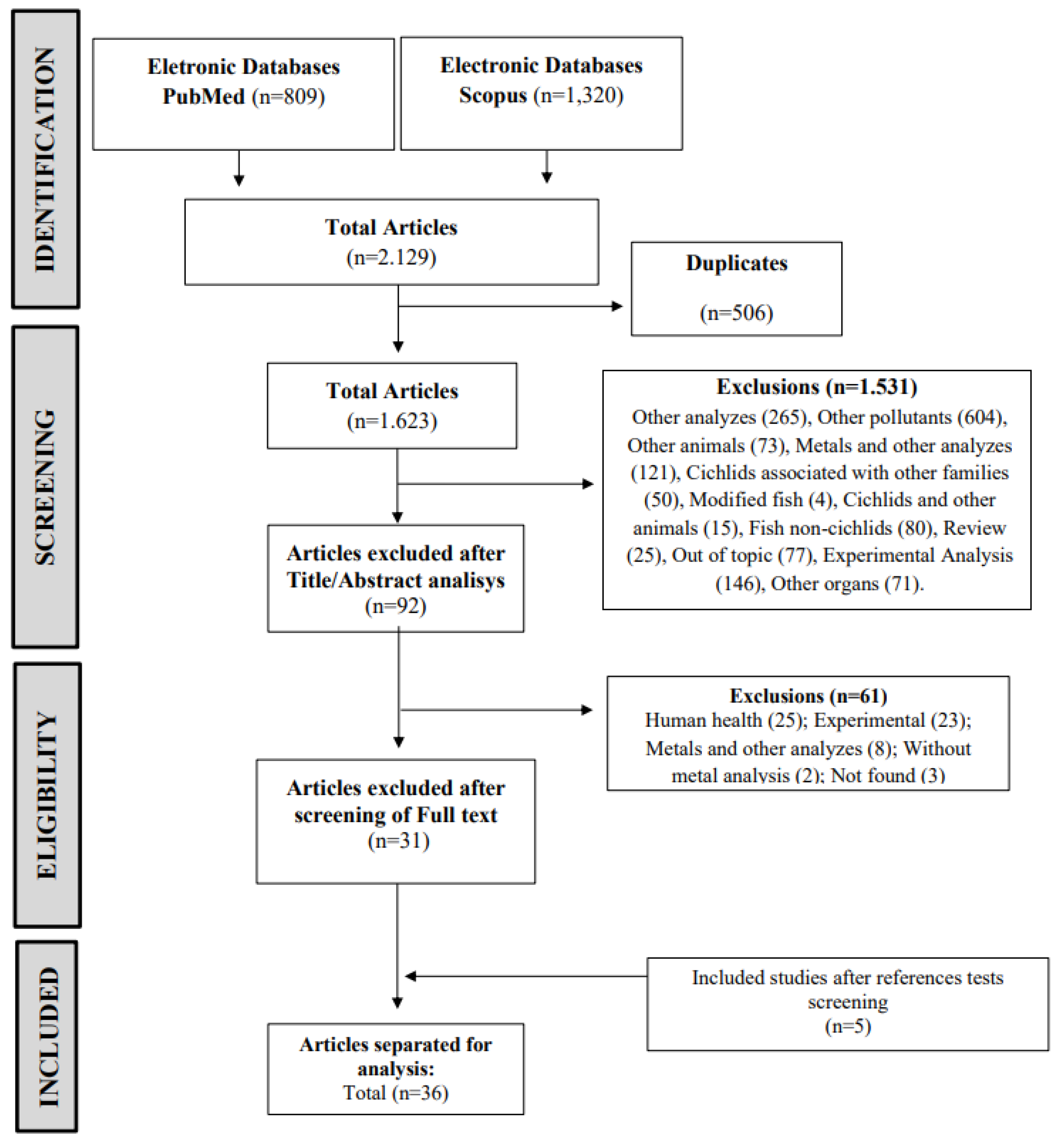
Figure 2.
Number of studies that found heavy metals in liver and gills of cichlid fish collected in the environment. Legend; Cd: cadmium, Cu: copper, Pb: lead, Zn: zinc, Mn: manganese. Fe: iron, Cr: chromium, Hg: mercury, Ni: nickel, Co: cobalt: Ag: silver, As: arsenic, Sr: strontium, Se: selenium, Ba: barium, Mo: molybdenum.
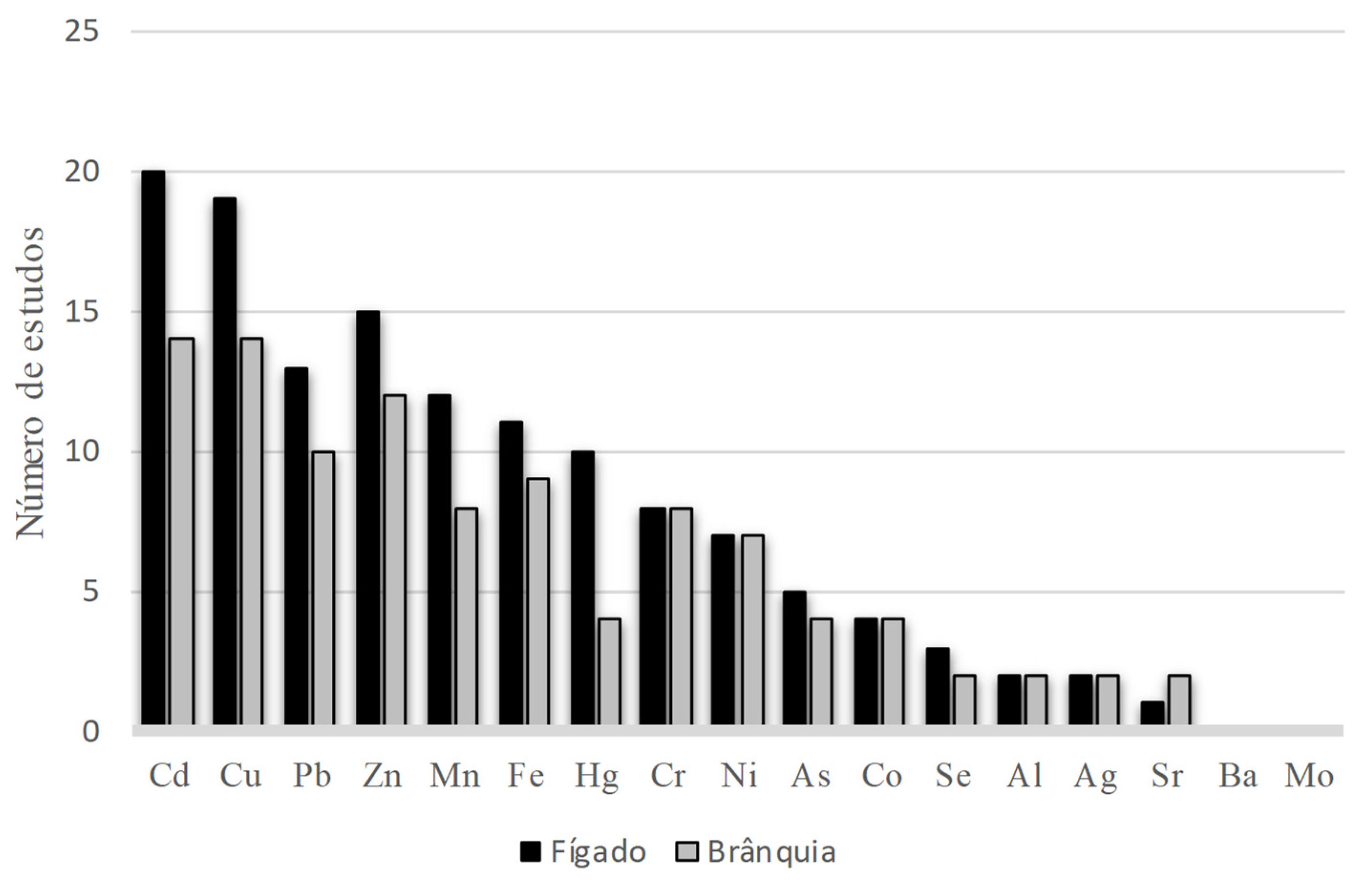
Figure 3.
Representation of the main effects of heavy metals on gills and liver of cichlid fish in the environment. Schematic diagram of the most common gill and hepatic lesions. Legend: SOD: Superoxide Desmutase, CAT: Catalase, GST: Glutathione S-Transferase, GPx: Glutathione Peroxidase, LPO: lipid peroxidation, PC: carbonyl protein, DNA: deoxyribonucleic acid; A) Cell rupture (arrow), macrovesicular steatis (asterisk), microvesicular steatosis (arrow head), congestion (star); B) Proliferation of mucous cells (circle), lamelar deformation (arrow), aneurysm and lamelar fusion (asterisk), lamelar destruction (arrow head), filamental fusion and lamelar hyperplasia (rectangle). Source: Authors.
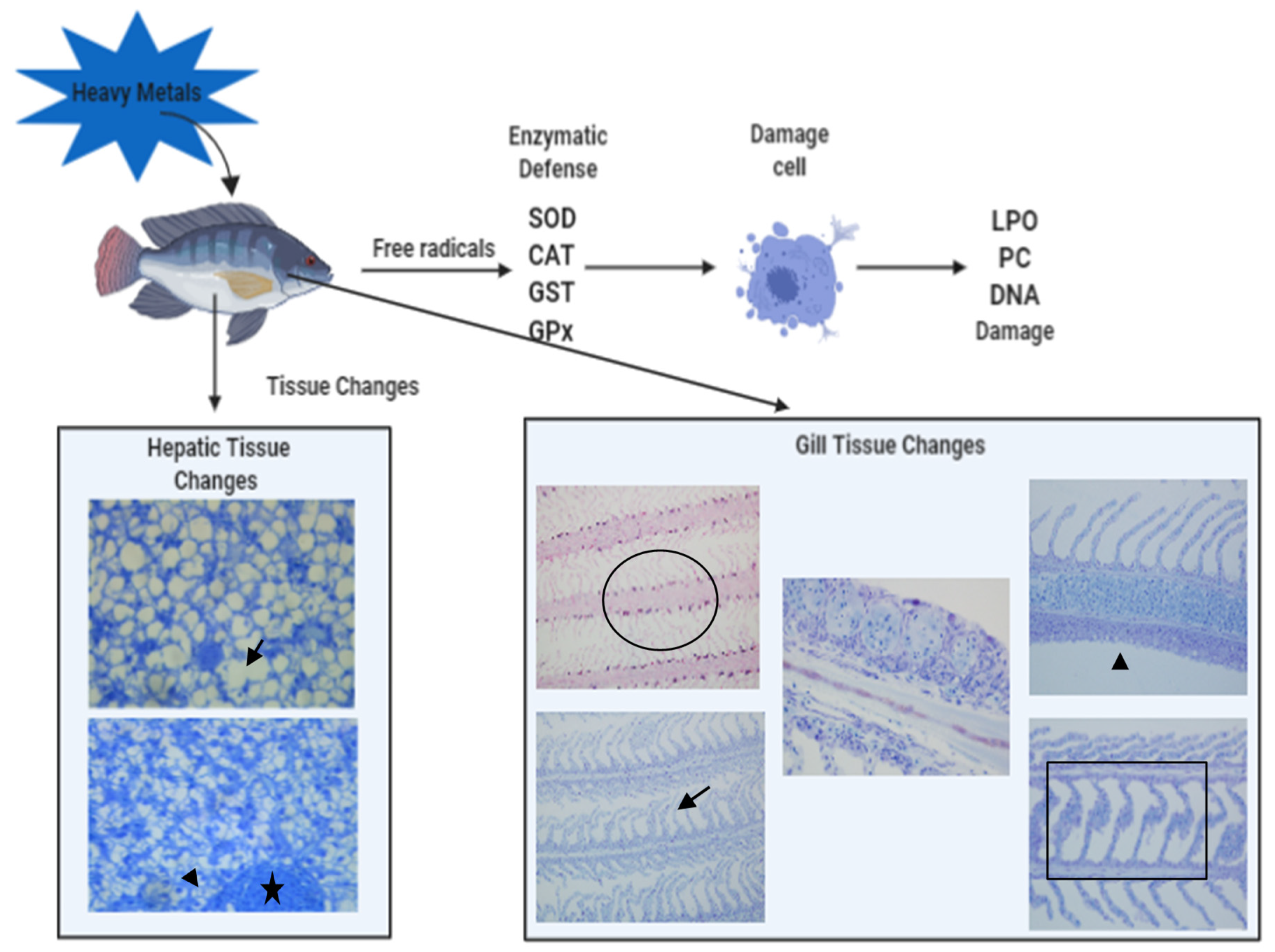
Figure 4.
Results of bias analysis (SYRCLE) of studies evaluating the effects of heavy metals on liver and gills of cichlid fish exposed to contamination in the environment. The evaluations (Q1-Q7) were scored with “yes”(+) indicating low risk of bias, “no” (-) indicating high risk of bias or “unclear” (?) indicating that the item was not reported, resulting in an unknown risk of bias. Legend; Q1: Was the allocation sequence generated and applied appropriately?; Q2: Were the groups similar at baseline or were they adjusted for confounding factors in the analysis?; Q3: Were the animals randomly selected to evaluate the results?; Q4: Was the results evaluator blind?; Q5: Were the incomplete data of the results treated properly?; Q6: Are the study reports free of reports of selective results?; Q7: Was the study apparently free of other problems that could result in a high risk of bias?
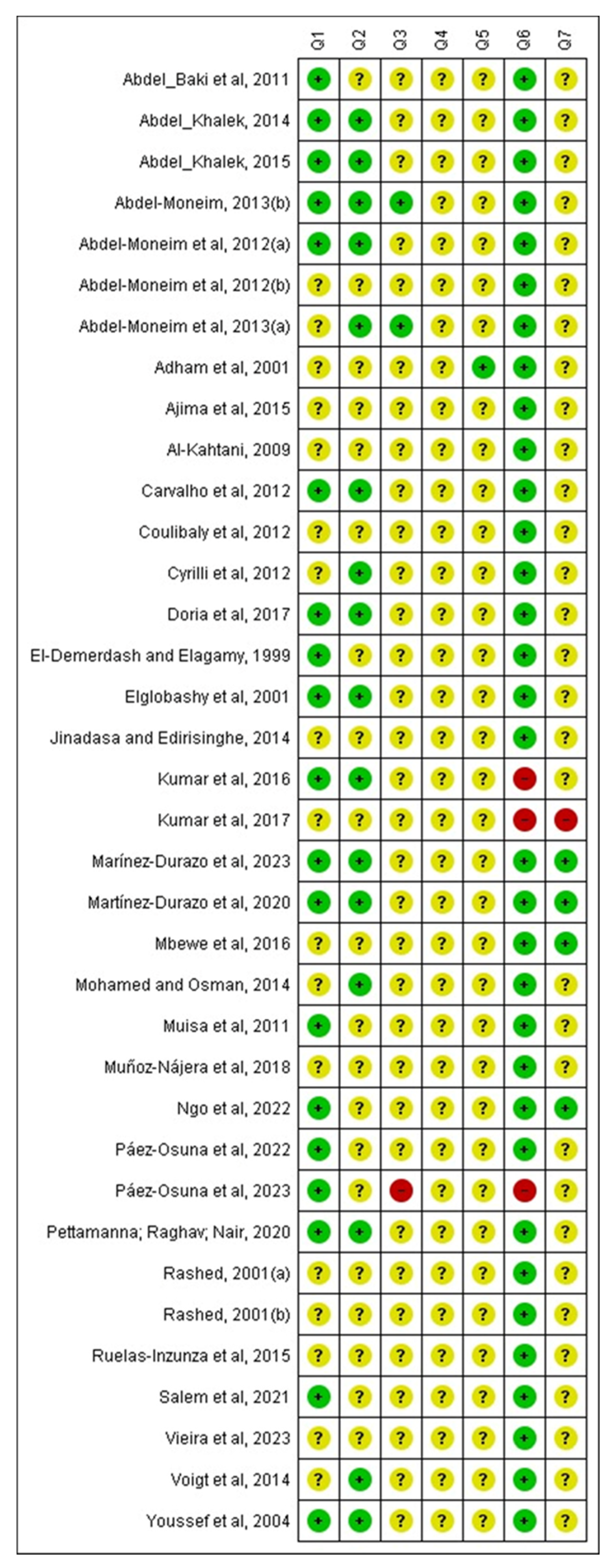
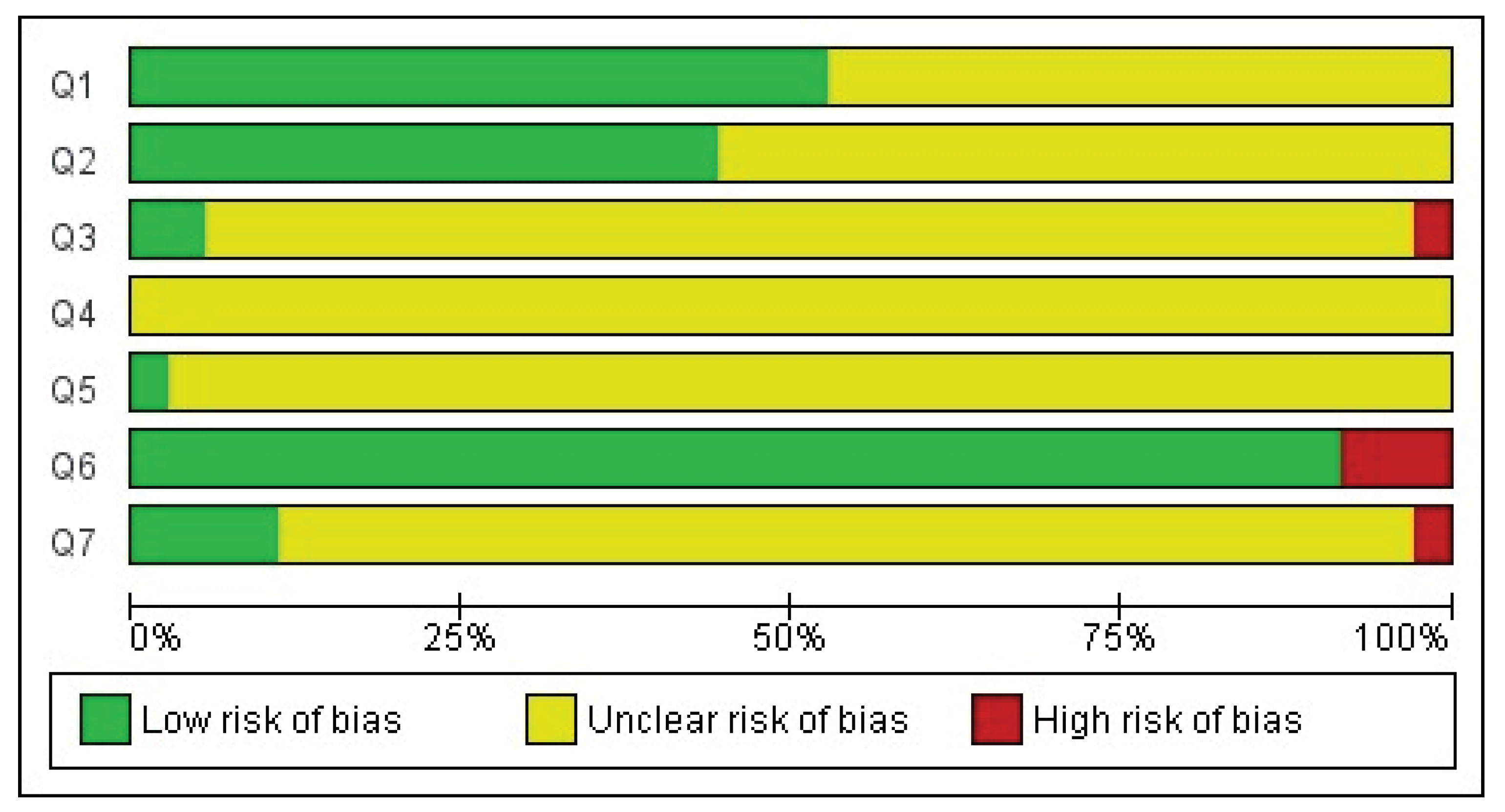

Table 1.
Características dos estudos que analisaram os efeitos da exposição à metais pesados em fígado e brânquias de ciclídeos coletados no ambiente.
Table 1.
Características dos estudos que analisaram os efeitos da exposição à metais pesados em fígado e brânquias de ciclídeos coletados no ambiente.
| References | Country | Species | Weight (g)/ Size (cm) | Number of animals | Groups of animals | Collection Period/ Station | |Analysis | Metals | Tissues |
|---|---|---|---|---|---|---|---|---|---|
|
El-Demerdash and Elagamy, 1999 [30] |
Egypt |
Tilapia nilótica (Oreochromis nilótica) |
200-250/ 15-20 | 20 | Test + Control | Yes/ ? | Metals in tissues and water, oxidative status, biochemical markers | Cd, Hg | Liver, Brain |
|
Rashed, 2001(a) [31] |
Egypt |
Tilapia nilótica (Oreochromis nilótica) |
440-1020/ ? | 50 | Tests (animal ages) | ? | Metals in tissues, water, sediment and aquatic plant | Co, Cr, Cu, Fe, Mn, Ni, Sr, Zn | Liver, Gills, Muscle, Stomach, Intestine, Vertebral Column, Scales |
|
Rashed, 2001(b) [18] |
Egypt |
Tilapia nilótica (Oreochromis nilótica) |
440-1020/ ? | 50 | Tests (animal ages) + Control | ? | Metals in tissues, water, sediment and aquatic plant | Pb, Cd | Liver, Gills, Muscle, Stomach, Intestine, Vertebral Column, Scales |
|
Elghobashy et al., 2001 [32] |
Egypt | Oreochromis niloticus | 150 ± 5/ ? | 30 | Tests (collection sites) + Control | Yes/ Summer | Metals in tissues, metals and physical-chemical parameters in water, oxidative status, biochemical markers | Fe, Cu, Zn, Pb, Cd | Liver, Gills, Kidney, Muscle |
|
Adham et al., 2001 [33] |
Egypt | Oreochromis niloticus | 150 ± 16,0/ 14,00 ± 1,4 | 200 | Tests (collection sites) + Control | Yes/ Fall-Winter, Spring-Summer | Metals and physical-chemical parameters in water, histopathology, biochemical markers | Pb, Cu, Mn, Zn, Fe, Ni, Hg | Liver, Gills, Blood |
|
Youssef et al., 2004 [34] |
Egypt | Oreochromis spp/ Sarotherodon galilaues | O. niloticus: 60-431/ 14,5-27 S. galilaeus: 3,6-37,5/ 8,0-11,5O. oreus: 21-151,8/ 10.5-20 | ? | Tests (animal species) | ? | Metals in tissues | Cu, Zn, Fe, Mn, Cd | Liver, Gills, Kidney, Muscle, Brain, Gonad |
|
Al-Kahtani, 2009 [35] |
Arabia | Oreochromis niloticus | 40-60/ 12-19 | ? | Tests (collection stations) | Yes/ Fall, Winter, Spring, Summer | Metals in tissues, metals and physical-chemical parameters in water | Fe, Zn, Cu, Pb, Mn, Cd | Liver, Muscle |
|
Abdel-Baki et al., 2011 [36] |
Saudi Arabia |
Tilapia nilótica (Oreochromis nilótica) |
?/ ? | 10 | Test | Yes/ Summer | Metals in tissues and water | Pb, Cd, Hg, Cu, Cr | Liver, Kidney, Intestine, Gills, Muscle |
|
Muisa et al., 2011 [37] |
Zimbabwe | Oreochromis niloticus | ?/ ? | ? | Tests (collection periods and sites) | Yes/ 2 periods | Metals in tissues, water and sediment | Al | Liver, Gills, Muscle, Kidney |
|
Abdel-Moneim et al., 2012(a) [38] |
Egypt | Oreochromis niloticus | 85-150/ 18-25 | 108 | Tests (collection sites) + Control | Yes/ ? | Metals in water, histopathology, oxidative status | Cd, Cu, Fe, Mn, Pb, Zn, Cr, Hg, Co, Ni | Gills |
|
Abdel-Moneim et al., 2012(b) [39] |
Saudi Arabia | Oreochromis niloticus | 90-130/ 18-22 | 120 | Tests (collection sites) | Yes/ ? | Metals and physical-chemical parameters in water, histopathology | Fe, Mn, Zn, Cu, Ni, Cd, Pb, Cr, Co, Hg | Liver, Gills |
|
Cyrille et al., 2012 [40] |
Ivory Coast | Sarotherodon melanotheron | Juvenile: 15,1-15,8/ ? Adult: 19,5-25,0/ ? | 15 | Tests (juvenile and adult animals) | Yes/ Dry, Rainy, Swelling | Metals in tissues, water physical-chemical parameters | Cd | Liver, Gills, Muscle |
|
Carvalho et al., 2012 [41] |
Brazil | Oreochromis niloticus | 90-200/ 15-20 | 32 | Tests (collection stations) + Control | Yes/ Fall, Winter, Spring, Summer | Metals and physical-chemical parameters in water, oxidative status, biochemical markers | Cr, Cd, Cu, Zn, Mn, Fe | Liver, Gills, Muscle |
|
Coulibaly et al., 2012 [42] |
Ivory Coast | Sarotherodon melanotheron | ?/ ? | 60 | Tests (collection stations) | Yes/ Dry, Rainy, Swelling | Metals in tissues, water and sediment | Cd, Cu, Pb, Hg, Zn | Liver, Kidney, Muscle, Brain |
|
Abdel-Moneim et al., 2013a [43] |
Egypt | Oreochromis niloticus | 125-210/ 20-26 | 120 | Test + Control | Yes/ ? | Metals in tissues and water, oxidative status, histopathology | Cd, Cu, Fe, Pb, Zn, Mn | Liver |
|
Abdel-Moneim, 2013b [44] |
Saudi Arabia | Oreochromis niloticus | 75-152/ 15-21 | ? | Tests (collection sites) + Control | Yes/ ? | Metals in water, histopathology | Ni, Fe, Zn, Co, Ba, Pb, Cu, Cd | Liver |
|
Jinadasa and Edirisinghe, 2014 [45] |
Sri Lanka | Tilapia sp | 216.6 ± 162,1/ 21,1 ± 4,0 | 80 | Tests (collection sites) | ? | Metals in tissues | Cd, Pb, Hg | Liver, Gills, Muscle |
|
Mohamed and Osman, 2014 [46] |
Sudan | Oreochromis niloticus | ?/ ~13 | ? | Tests (collection sites) | ? | Metals in tissues and water | Cd, Cr, Ni, Pb, Cu, Zn, Fe, Sr | Muscle, Gills |
|
Voigt et al., 2014 [47] |
Brazil | Geophagus brasiliensis | Male: 59,5-278,0/ 145-250 Female: 64,6-145,4/ 150-190 | Male:38 Female:17 | Tests (gender, weight and size of animals) | Yes/ Summer | Metals in tissues, water and sediment | Cu, Mn, Zn, Fe, Co, Cd, Cr, Ag, Ni | Liver, Gills, Muscle |
|
Abdel-Khalek, 2014 [48] |
Egypt | Oreochromis niloticus | 160-180/ 18,5-24,8 | 36 | Tests (collection sites) + Contol | Yes/ Summer | Metals in tissues and water, histopathology | Cu, Zn, Pb, Fe, Mn, Cd | Liver, Gills, Kidney, Muscle |
|
Abdel-Khalek, 2015 [49] |
Egypt | Oreochromis niloticus | 160-180/ 18,5-24,8 | 36 | Tests (collection sites) + Control | Yes/ Summer | Metals and physical-chemical parameters in water, oxidative status | Cu, Zn, Pb, Fe, Mn, Cd | Liver, Gills, Blood |
|
Ajima et al., 2015 [50] |
Nigeria |
Pelmatochromis guentheri/ Pelmatochromis pulcher |
35 ± 0,05/ ? | 18 | Tests (animal species) | ? | Metals in tissues, water and sediment | Pb, Zn, Cd, Cu, Fe | Gills, Muscle, Intestine |
|
Ruelas-Inzunza et al., 2015 [51] |
Mexico | Oreochromis aureus | Rainy station: 506,8 ± 100,5/ 30,9±2,4 Dry station: 563,4 ± 93,9/ 31,0±2,8 | 80 | Tests (collection stations) | Yes/ Dry, Rainy |
Metals in tissues | Hg | Liver, Muscle, Kidney |
|
Kumar et al., 2016 [52] |
India | Oreochromis mossambicus | ? | 54 | Tests (collection sites) | Yes/ ? | Metals in muscle, histopathology, oxidative status, biochemical markers | Cr, Mn, Co, Ni, Cu, Zn, As, Se, Cd, Hg, Pb | Liver, Gills, Kidney, Muscle, Brain, Gonad |
|
Mbewe et al., 2016 [53] |
Zambia |
Tilapia andersonii/ T. rendalli/ Oreochromis niloticus |
T.rendalli: 170-250/ ? O.niloticus: 150-300/ ?T.andersonii: 390/ ? | 3/ 4/ 3 | Tests (animal species and collection sites) | Yes/ ? | Metals in tissues and sediment | Cu, Cd, Pb, Mn, Cr, Ni, Fe | Liver, Gonads, Muscle, Intestines, Head. |
|
Doria et al., 2017 [54] |
Brazil | Geophagus brasiliensis | 148,8 ± 8,89/ 19,97 ± 2,87 | 55 | Tests (collection sites) | Yes/ Summer | Metals in tissues, histopathology, oxidative status, biochemical markers | Cu, Mn, Zn, Co, Cd, Cr, Ag, Pb, Ni, Al, As | Liver, Gills, Blood, Muscle, Brain |
|
Kumar et al., 2017 [55] |
India | Oreochromis mossambicus | ?/ ? | ? | Tests (collection sites) | Yes/ ? | Metals in tissues, water and sediment, oxidative status, biochemical markers | Cr, Mn, Co, Ni, Cu, Zn, As, Se, Cd, Hg, Pb | Liver, Gills, Kidney, Muscle, Brain, Gonad |
|
Muñoz-Nájera et al., 2018 [56] |
Mexico | Oreochromis niloticus | ?/ ? | 150 | Tests (collection periods) | Yes/ 5 periods | Metals in tissues, metals and physical-chemical parameters in water, biochemical markers | Cd, Cr, Cu, Pb | Liver, Muscle |
|
Pettamanna; Raghav; Nair, 2020 [57] |
Índia | Etroplus suratensis | 65,7-83,1/11-14,5 | 15 | Test + Control | Yes/Winter | Metals and physical-chemical parameters in water, histopathology, biochemical markers | Cu, Fe | Liver |
|
Martínez-Durazo et al., 2020 [58] |
Mexico |
Tilapia nilótica (Oreochromis niloticus) Largemouth bass (Micropterus salmoides) |
461,8-619,6/26,98-34,51 45,1-1.182,0/30,17-40,10 |
21-30 21-30 |
Tests (animal species) | Yes/Dry and Rainy | Metals in tissues. | Cr, Cu, Fe, Mn, Ni,Zn | Liver, gills, stomach, gonads and muscle |
|
Salem et al., 2021 [59] |
Egypt |
Tilapia nilótica (Oreochromis nilótica) |
80±5/17±2 | 16 males | Tests (collection sites) | Yesyear all | Metals in tissues, metals and physical-chemical in water, quantitative, biochemical markers Real-Time PCR. | Cd, Fe, Mn, Co e Pb | Liver |
|
Ngo et al., 2022 [1] |
Vietname |
common Carp (Cyprinus carpio), silver Carp (Hypothalmic molitrix), Tilapia nilótica (Oreochromis niloticus) |
? | 240/240/240 | Tests (animal species) | Yes/four seasons | Metals in tissues and wate, oxidative status, biochemical markers. | Zn, Cu, Pb, Cd | Livers, kidneys, gills |
|
Páez-Osuna et al., 2022 [60] |
Mexico | Oreochromis aureus | 200-840/22.2-41.3 | 15 | Tests (collection periods) | Yes/spring | Metals in tissues, sediments, metals and physical-chemical parameters in water and risk assessment. | As, Cd, Cu, Hg, Se, Zn | Liver, muscle, and guts |
|
Vieira et al., 2023 [61] |
Brazil |
Tucunare (Cichla sp). Piraíba (Braquiplatystoma filamentosum) Jaraqui (Semaprochilodus sp) |
? | 10/10/10 | Tests (animal species) | ? | Metals in tissues, oxidative status, biochemical markers and bioinformatic. | Hg | Liver, muscle |
|
Martínez-Durazo et al., 2023 [6] |
Mexico |
Tilapia nilótica (Oreochromis niloticus) Largemouth bass (Micropterus salmoides |
? | 20/20/20 | Tests (animal species) + control | Yes/Dry and Rainy | Metals in tissues, histopathology. | Cr, Cu, Fe, Mn Ni, Zn, As, Hg, Se. | Liver, gills, muscle, gonads |
|
Páez-Osuna, et al., 2023 [62] |
Mexico |
Carpa mon (Cyprinus carpio) Tilápia azul (Oreochromis aureus) Robalo (Micropterus salmoides) |
C.carpio 36,5-47/625/1.725 O. aureus 21-34/165-670 M. salmoides 24-38/170/740 |
7/22/16 | Tests (animal species) | ? | Metals in tissues, risk assessment. | As, Cd, Cu, Zn | Liver, gills, muscle, guts |
Legenda: Ag: Silver; Al: aluminum; As: Arsênico; Ba: Barium; Ni Cd: Cadmium; Co: Cobalt; Cr: Chrome; Cu: Copper; Se: Selenium; Fe: Iron; Hg: Mercury; Mn: Manganese; Mo: Molybdenum; Nickel; Pb: Lead; Se: Selênio; Sr: Strontium; Zn: Zinc; ?: uninformed.
Table 2.
Main liver and gill alterations found in cichlid fish after exposure to heavy metals in the environment.
Table 2.
Main liver and gill alterations found in cichlid fish after exposure to heavy metals in the environment.
| Organ | Analyses | Effects |
|---|---|---|
| Liver | Histopathologies | Cell lesions and degenerations Circulatory changes Cell proliferative changes Ultramorphological changes |
| Oxidative stress | SOD: ↑, GPx: ↑, CAT: ↑, MDA:↑, GSH: ↑, GST: ↑, LPO: ↑, PC: ↑ SOD: ↓, GPx: ↓, CAT: ↓ | |
| Bioaccumulation | Cd, Cu, Pb, Zn, Mn, Fe, Cr, Hg, Ni, Co, Ag, As, Se, Ba, Mo, Al | |
| Gill | Histopathologies | Cell proliferative changes Cell lesions and degenerations Circulatory changes Ultramorphological changes |
| Oxidative stress | SOD: ↑, GPx: ↑, CAT: ↑, MDA:↑, GSH: ↑, GST: ↑, LPO: ↑SOD: ↓, GPx: ↓, GSH: ↓, GR: ↓ |
|
| Bioaccumulation | Cd, Cu, Pb, Fe, Zn, Mn, Cr, Ni, Co, Ag, Hg, Al, Sr, As, Se, Ba, Mo |
MDA: Malondialdeide; SOD: Superoxide dismutase; CAT: Catalase; GSH: Glutathione; GST: Glutathione-S-Transferase; GPx: Glutathione Peroxidase; G6PD: Glucose-6-Phosphate dehydrogenase; GR: Glutathione reductase; LPO: Lipid peroxidation; PCO: Carbonyl protein; MT: Metalotionein; AChe: Acetylcholinesterase; ALPmt: Mitochondrial alkaline phosphatase; ALPmc: Microsomal alkaline phosphatase; GSTmt: Mitochondrial Glutathione-S-Transferase; GSTmc: Glutathione-S-Microsomal Transferase; * Higher bioaccumulation in the dry season; ** Higher bioaccumulation in the rainy season; No seasonal difference in bioaccumulation; ↑ increase; ↓ decrease.
Disclaimer/Publisher’s Note: The statements, opinions and data contained in all publications are solely those of the individual author(s) and contributor(s) and not of MDPI and/or the editor(s). MDPI and/or the editor(s) disclaim responsibility for any injury to people or property resulting from any ideas, methods, instructions or products referred to in the content. |
© 2024 by the authors. Licensee MDPI, Basel, Switzerland. This article is an open access article distributed under the terms and conditions of the Creative Commons Attribution (CC BY) license (http://creativecommons.org/licenses/by/4.0/).
Copyright: This open access article is published under a Creative Commons CC BY 4.0 license, which permit the free download, distribution, and reuse, provided that the author and preprint are cited in any reuse.


