
Submitted:
18 March 2024
Posted:
19 March 2024
You are already at the latest version
Abstract
Tuberculosis (TB) remains a global health threat, necessitating innovative strategies for control and prevention. This comprehensive review explores the Mycobacterium tuberculosis Lysine Exporter (LysE) gene, unveiling its multifaceted roles and potential uses in controlling and preventing tuberculosis (TB). As a pivotal player in eliminating excess L-lysine and L-arginine, LysE contributes to the survival and virulence of M. tuberculosis. This review synthesizes findings from different electronic databases and includes 13 studies focused on the LysE of M. tuberculosis. The research unveils that LysE can be a potential drug target, a diagnostic marker for TB, and a promising candidate for vaccine development. The absence of LysE in the widely used BCG vaccine underscores its uniqueness and positions it as a novel area for TB prevention. In conclusion, this review underscores the significance of LysE in TB pathogenesis and its potential as a drug target, diagnostic marker, and vaccine candidate. The multifaceted nature of LysE positions it at the forefront of innovative approaches to combat TB, calling for sustained research efforts to harness its full potential in the global fight against this infectious disease.
Keywords:
Lysine exporter
; mycobacterium tuberculosis
; LysE
; transmembrane protein
; lysine
; arginine
1. Introduction
Tuberculosis (TB) stands as a formidable and pervasive health threat, ranking as the 13th leading cause of death worldwide [1]. Before the emergence of the COVID-19 pandemic, tuberculosis (TB) stood as the leading cause of mortality attributable to a single infectious agent, surpassing even the impact of HIV/AIDS [2]. In 2022, it caused 1.3 million deaths globally, with the WHO South-East Asian Region seeing a surge in new cases [3]. Developing countries, including high TB-burden nations like India, face challenges such as inadequate diagnosis, treatment non-adherence, increasing drug-resistant cases, and low awareness, leading to a rapid rise in TB [4]. Tackling these issues is vital to curb TB’s impact and strengthen global health resilience.
In countries with a high burden of TB, such as India, there has been a concerning surge in the incidence of multidrug-resistant TB (MDR-TB) and extensively drug-resistant TB (XDR-TB), indicating a lack of effective control measures [5]. In these regions, the diagnosis of MDR-TB, XDR-TB, and other forms of drug-resistant TB, as well as TB associated with HIV, can pose challenges due to their complexity and the associated high costs [4]. In the present scenario, the Bacillus Calmette-Guérin (BCG) vaccine is the primary and sole tuberculosis (TB) vaccination for safeguarding newborns and children against TB meningitis. However, its effectiveness in reducing the transmission of pulmonary TB is somewhat constrained [6]. Recognizing this limitation, there is a critical need to explore Mycobacterium tuberculosis (M.tb) proteins absent in BCG that possess the potential to induce specific humoral, cell-mediated, and innate responses in the host [7].
The survival and virulence of M.tb are likely influenced by various ion channels and transporter proteins that the bacterium possesses [8]. These transporter proteins play a crucial role in the physiological processes of M.tb, contributing to its ability to survive and cause disease [8]. In mycobacteria, L-Lysine exporters (LysE), a transmembrane protein, play a pivotal role in eliminating surplus L-lysine and L-arginine resulting from metabolic activities within the cytosol. An extensive literature search on the M.tb L-lysine exporter (LysE) gene revealed that it removes excess metabolically produced basic amino acids such as L-lysine and L-arginine and their toxic analogues from the cytosol. The accumulation of these toxic analogues can lead to bacterial growth retardation, which reveals that LysE is an essential component for bacterial growth. The absence of these exporters results in elevated intracellular levels of L-lysine, impeding the growth of the bacteria [9].
LysE, an exporter of L-Lysine, has emerged as a promising candidate in various studies, highlighting its potential applications as a drug target, vaccine candidate, and prospective diagnostic marker [9,10,11,12]. Its absence in the widely used BCG vaccine defines its uniqueness, and hence, LysE can be a suitable new vaccine candidate [7,11,12]. Therefore, comprehensive knowledge about this exporter is required for its consideration as a potential drug target for inhibition, diagnostic marker and probable vaccine candidate. Much research has been conducted on Lysine and arginine exporters of Eshcherichia.coli (ArgO) and Corynebacterium glutamicum (LysE); however, the LysE of mycobacterium tuberculosis, which is responsible for bacterial growth and able to stimulate innate and adaptive immunity, is still a novel area for research. This review underscores the significance and functional role of LysE in Mycobacterium tuberculosis. It explores it as a potential target for drug inhibition, a diagnostic marker, and a promising candidate for vaccine development.
2. Methodology
2.1. Inclusion and Exclusion Criteria
We searched various databases, including PubMed, Scopus, Web of Science, CENTRAL, and Google Scholar, using specific keywords related to the Lysine Exporter gene of Mycobacterium tuberculosis. Inclusion criteria involved selecting articles from full journal publications. We excluded articles containing information about the Lysine-Exporter gene from other bacterial species like C. glutamicum, E. coli, H. pylori, etc. However, articles that discussed LysE of Mycobacterium tuberculosis along with LysE of other bacterial species were included. The data extraction, however, focused solely on the LysE of Mycobacterium tuberculosis. We included the studies of the last 25 years. No language restrictions were applied. We performed the search between September and October 2023 and presented search strategies for PubMed, Scopus, CENTRAL and Web of Science here.
2.2. Search Strategy
Pubmed
(((((((“lysine export*”[Title/Abstract]) OR (“lysine permea*”[Title/Abstract])) OR (LysE[Title/Abstract])) OR (“amino acid transport*”[Title/Abstract])) OR (“lysine efflux”[Title/Abstract])) OR (“lysine transport*”[Title/Abstract])) OR (Rv1986[Title/Abstract])) OR (lysine[MeSH Terms])
(((tubercul*[Title/Abstract]) OR (TB[Title/Abstract])) OR (mycobacteri*[Title/Abstract])) OR (“mycobacterium tuberculosis”[MeSH Terms])
(#1) AND (#2)
Scopus:
TITLE-ABS-KEY(“lysine export*”) OR TITLE-ABS-KEY(“lysine permea*”) OR TITLE-ABS-KEY(“lysine efflux”) OR TITLE-ABS-KEY(“lysine transport*”) OR TITLE-ABS-KEY (LysE) OR TITLE-ABS-KEY(Rv1986) OR TITLE-ABS-KEY(“amino acid transport*”) AND TITLE-ABS-KEY(tubercul*) OR TITLE-ABS-KEY(mycobacteri*) OR TITLE-ABS-KEY(“mycobacterium tuberculosis”) OR TITLE-ABS-KEY(TB)
Web of Science:
((((((KP=(“lysine export*”)) OR KP=(“lysine permea*”)) OR KP=(“lysine efflux”)) OR KP=(“lysine transport*”)) OR KP=(LysE)) OR KP=(Rv1986)) OR KP=(“amino acid transport*”)
((KP=(tubercul*)) OR KP=(mycobacteri*)) OR KP=(“mycobacterium tuberculosis”)
CENTRAL
Search Name: LysE_mycobacterium tuberculosis
Comment:
ID Search Hits
#1 “Lysine export*” 0
#2 “lysine permea*” 0
#3 “lysine transport*” 1
#4 “lysine efflux” 1
#5 LysE 112
#6 “amino acid transporter” 44
#7 MeSH descriptor: [Lysine] explode all trees and with qualifier(s): [biosynthesis - BI] 1
#8 #1 OR #2 OR #3 OR #4 OR #5 OR #6 OR #7 159
#9 tubercul* 9065
#10 mycobacteri* 2495
#11 TB 7621
#12 “mycobacterium tuberculosis” 1073
#13 MeSH descriptor: [Mycobacterium tuberculosis] explode all trees 415
#14 #9 OR #10 OR #11 OR #12 OR #13 14759
#15 #8 AND #14 0
3. Result
We initially identified 531 potential articles across various electronic databases, with the majority from PubMed (n=258), Scopus (n=271), and Web of Science (n=2), along with two articles from other sources. Following the removal of duplicates, a total of 438 articles were examined. Through initial screening based on title and abstract, 414 articles were excluded. Subsequently, the full texts of 24 articles were independently assessed, leading to the exclusion of 11 articles that lacked specific details on mycobacterial LysE or focused on details of lysine exporters of other bacterial species. Ultimately, 13 articles were identified for potential inclusion. We have presented a PRISMA flow diagram detailing the selection of articles in Figure 1.
3.1. Details of Included Studies
A total of 13 studies were included that describe the role of LysE in the bacterial system. Out of 13, the majority of the research on LysE was performed in China [7,10,13,14] and the United States of America [9,15,16,17], followed by Germany [18], Mexico [19], South Africa [12], United Kingdom [11] and in India [20]. We included the research of the last 25 years. In this section below, we have highlighted the primary research work of all included studies.
In 2021, Dubey et al. [20] focused on LysO, which mediates the export of L-lysine and L-thialysine in E.coli. They studied the hidden aspect of LysO topology and the mechanism it uses to facilitate export. In C. glutamicum and E. coli, the export of L-lysine is facilitated by two proteins, LysE and LysO, respectively. In Mycobacterium tuberculosis, L-Lysine export mediates by LysE, a transmembrane protein. Amino acid exporters are required to arbitrate resistance to toxic analogues of amino acids, which promote translational errors resulting in aberrant protein synthesis.
Georgieva et al. 2020 [15] purified and characterised the LysE gene biophysically. The function of LysE membrane exporters in bacteria is to remove the excess of metabolically produced basic amino acids such as L-lysine and L-arginine from the cytosol. LysE deficiency causes increased cellular levels of L-lysine, which inhibits bacterial growth. Consequently, the LysE proteins found in bacterial pathogens emerge as promising drug targets for inhibition. This underscores the importance of understanding these proteins comprehensively, encompassing their structures and the relationships between structure and function.
In another study, Georgieva et al. 2020 [9] focussed on the Mtb-encoded L-lysine exporter (LysE), which eliminates the excess of metabolically produced basic amino acids from the bacterial cytosol. LysE serves as a target for inhibiting bacterial growth because its absence results in the accumulation of toxic levels of L-lysine, suppressing bacterial growth. The most studied H37Rv strain of Mtb encodes two LysEs, but one of them, the Rv1986 protein, is considered the primary exporter. During the early stages of Mtb infection, Rv1986 expression is increased under hypoxia conditions. Thus, the LysE protein can be a probable vaccine candidate. This study developed custom protocols for cloning, expression, purification, and initial characterization of LysE. Protein is purified in lipodiscs made of native E. coli membranes and detergent.
Hasenoehrl et al., 2019 [16] explained that due to threonine auxotrophy in M.tb, LysE functions as a metabolic relief valve to remove surplus cytoplasmic lysine. This exporter also has a function in non-auxotrophic settings, as indicated by the Mtb lysE mutant’s delayed escape from the lag phase and sluggish development. During infection of resting and active macrophages, Rv1986 is generated and in a lysosomal exposure model, and it is also involved in human latent tuberculosis infection. The exporter has been linked to protective immunity in humans and is a significant target of IL-2-secreting memory T cells. Amino acids, particularly arginine, have been shown to influence human immunometabolism and T-cell responses. These findings highlight the significance of LysE-mediated export.
Chen et al. (2018) [7] conducted a comprehensive analysis across 12 M. Bovis BCG strains and 200 M. TB strains. They proposed the presence of proteins Rv1986, an amino acid transporter, and an integral membrane sulfoglycolipid transporter Rv3823c which is responsible for mycolic acid transport, in most epidemic strains but absent in BCG. These proteins exhibited a higher number of T cell and B cell epitopes. Notably, Rv1986 is found in virulent M. Bovis but not in the BCG strain. The researchers expressed recombinant M. tb proteins Rv1986 and Rv3823c in E. coli, testing their ability to stimulate innate and adaptive immune cell propagation and cytokine release. The results indicated that Rv1986 and Rv3823c have the potential to enhance protective immunity through the activation of T cells, plasma cells, and inflammatory cytokines. This suggests their possible roles in future vaccine development. Additionally, Rv1986 was identified as an immunodominant target for memory T cells, highlighting its diagnostic potential.
A study on Region of Deletion 2 (RD2) of M.Tb by Jiazhen Chen et al. 2018 [10] stated that RD2 is responsible for bacterial virulence and showed immunodominancy in infected cattle. Diagnostic ability in humans is an unexplored area. Antigens that showed immunogenicity in cattle are Rv1983, Rv1986, Rv1987, and Rv1989c. Various antigens from RD2 were assessed for diagnosis in 233 TB patients using a whole blood IFN-γ release assay. The active TB group exhibited significantly elevated IFN-γ release responses to Rv1986-P9, P15, P16, Rv1988-P4, P11, and Rv1987-P11 compared to the control groups (p<0.05). These findings indicate that the six epitopes originating from the RD2 region of M. tuberculosis hold promise as potential diagnostic markers for tuberculosis.
Schneefeld et al., 2017 [18] demonstrated that the Rv1985c protein binds to its own and the promoter region of Rv1986. In M. tuberculosis (Mt), Rv1985c and Rv1986 share identical genomic organization with lysG and lysE in C. glutamicum, including a promoter region where both genes overlap. The generation of a precisely defined Rv1985c deletion mutant in M. tuberculosis, coupled with gene expression analysis comparing the wild-type strain and the mutant, demonstrated that the Rv1985c protein activates the transcription of Rv1986 in a lysine-dependent manner. Consequently, it serves as an autoregulatory mechanism for its own expression. Furthermore, whole transcriptome expression analysis revealed that, in addition to lysE(Mt), the Rv1985c protein regulated three other genes, ppsB, ppsC, and ppsD, all of which are involved in cell wall metabolism. The author proposed renaming the Rv1985c protein to LysG(Mt) and naming the Rv1986 protein LysE(Mt) in mycobacterium tuberculosis.
One more study by Jiang et al. in 2017 [13] studied RD2 proteins of mycobacteria. The researchers illustrated that genes within the RD2 antigens exhibit higher variability than other mycobacterial regions, particularly in the T-cell epitopic region. This implies their potential involvement in genetic variation to evade host immunity. The study specifically involved amplifying and comparing the sequences of five genes within the RD2 region, which encompasses both T and B cell epitopic regions. The results indicate that proteins Rv1980c, Rv1985, and Rv1986 in the RD2 region undergo antigenic variation in response to host immune pressure, suggesting their potential role in ongoing immune evasion.
In 2015, Tsu and Saier [14] explored the LysE superfamily of transmembrane transport proteins, which facilitate the export of amino acids, lipids, and heavy metal ions. This newly identified superfamily comprises three families: L-lysine and L-arginine exporters (LysE), homoserine/threonine resistance proteins (RhtB), and cadmium ion resistance proteins (CadD). While LysE and RhtB proteins are involved in amino acid export, CadD proteins play a more distant role in cadmium (Cd2+) efflux. Members of the LysE Superfamily contribute to ionic homeostasis, protection against cytoplasmic heavy metals/metabolites, cell envelope assembly, and transmembrane electron flow. As a result, they impact the physiology and pathogenesis of various microbes, serving as potential targets for drug action. However, many family members remain poorly understood in terms of their functional and physiological roles.
Torres et al. [19] performed a randomized controlled trial including patients with latent tuberculosis infection (LTBI). Patients were randomized into two arms, immediate or delayed isoniazid treatment arms. The research pinpointed the primary inducers of IFN production in individuals with latent tuberculosis infection (LTBI) before isoniazid therapy, identifying Rv0849, Rv1737, and the RD2-encoded Rv1986 (absent in many commonly used BCG strains). Earlier investigations have demonstrated that peptides from Rv1986 exhibit potent stimulation of peripheral cells in both LTBI and active TB patients, inducing notably higher levels of IL2 compared to IFN-γ.
Gideon et al. in 2010 [12] identified the upregulation of two regions of difference (RD) 11 (Rv2658C and Rv2659c) and one RD2 (Rv1986), absent in widely used BCG strains. Exploring gene response patterns during prolonged hypoxia could unveil novel antigens for potential use in vaccines or diagnostics for tuberculosis. The Rv1986 gene exhibited significant elevation among tuberculosis patients during low oxygen levels. In TB patients, analysis of human immune responses to this protein revealed its strong recognition by cells producing interleukin-2 (IL-2), crucial for long-term immunological memory. These findings established Rv1986 as an immunodominant target for memory T cells. Further examination of BCG strains with or without Rv1986 is essential, holding promise for potentially enhancing the effectiveness of existing or new tuberculosis vaccines.
Cockle et al. in 2002 [11] directed their efforts towards pinpointing more precise reagents that could effectively differentiate between vaccination and infection. Their goal also involved the identification of subunit vaccine candidates to enhance the efficacy of tuberculosis vaccines. This study involved applying comparative genomics and identifying M. Bovis and M. tuberculosis antigens detection in the BCG vaccine. This study carefully selected 13 open reading frames (ORFs) from the RD1, RD2, and RD14 regions of M. tuberculosis. It was investigated in cattle infected with M. Bovis and in BCG-vaccinated and unvaccinated control cattle. This examination revealed eight particularly immunogenic antigens (Rv1983, Rv1986, Rv3872, Rv3873, Rv3878, Rv3879c, Rv1979c, and Rv1769). The potential of these antigens as diagnostic markers or subunit vaccines merits further exploration and study.
In Corynebacterium glutamicum, Vrljic et al. 1999 [17] explained that the LysE carrier protein exhibits remarkable functionality in exporting L-lysine. The investigation delves into the membrane topology of LysE, a protein composed of 236 amino acyl residues, by analysing PhoA- and LacZ-fusions. This study explains that LysE is a family of transmembrane proteins found in other bacterial species, such as E. coli, B. subtilis, M. tuberculosis and H. pylori. L-lysine export is required in environments with low concentrations of lysine-containing peptides. When the export carrier gene lysE is deleted, exceptionally high cytoplasmic concentrations of L-lysine (>1M) accumulate, leading to bacteriostasis.
We have shared Table 1, summarizing the last ten years of LysE research. It highlights LysE’s potential as a vaccine candidate, drug target, and diagnostic marker. This calls for more research on LysE to provide a quick overview of the work done in this field.
4. Discussion
Several amino acid biosynthesis pathways crucial for the survival and pathogenesis of Mycobacterium tuberculosis were investigated. These pathways play a key role in the replication machinery and cell wall synthesis of the bacterium. Importantly, many of these pathways or their significant enzyme homologs are absent in humans and animals [21]. One such pathway is the aspartate pathway, which synthesises essential amino acids such as lysine, isoleucine, threonine and methionine [16].
Lysine, an essential amino acid, is produced in mycobacteria via the aspartate pathway using Meso-diaminopimelate (DAP) as a direct precursor [22]. Meso-diaminopimelate (DAP) is a primary constituent of bacterial cell wall peptidoglycan) [22]. Excess lysine inside the bacterial cell leads to toxic effects on the cell resulting in growth retardation. Transmembrane proteins like L-lysine exporters (LysE) are vital in bacteria to remove excess L-lysine and L-arginine produced during metabolic processes from the cytosol. If these exporters are absent, cellular levels of L-lysine increase, impeding bacterial growth [9]. These lysine exporters are situated on the inner membrane of bacterial cells, facilitating the efficient removal of surplus lysine from the cytosol.
L-lysine exporter (LysE) manifests as a small transmembrane protein with a molecular weight ranging from 20 to 25 kDa [23]. Functioning as a dimer, this protein exhibits a structural arrangement characterized by five to six transmembrane hydrophobic spanning helices [17]. The specific configuration of LysE underscores its role as a functional unit with distinct characteristics crucial for its cellular activities [24,25]. LysE was discovered primarily in C. glutamicum and later in M. tuberculosis, B. subtilis, Escherichia coli, and Helicobacter pylori [17]. It was the first amino acid exporter to be molecularly recognized [24]. The LysE gene has sequence similarities to E. coli’s arginine exporter and Corynebacterium glutamicum’s lysine exporter, ArgO, and LysE, respectively. It has been reported that LysG stimulates the transcription of lysE in the presence of lysine or histidine [18]. Ionic homeostasis, transmembrane electron transport, protection from high cytoplasmic heavy metal/metabolite concentrations, and cell envelope assembly are all functions of the members of the LysE Superfamily [14]. LysE is observed to facilitate the unidirectional removal of L-lysine, along with other vital amino acids like L-arginine, through efflux. It is recognized as the exclusive pathway for the excretion of L-lysine. The proton motive force (H+ antiport) is thought to be the energy source [26].
LysE becomes a crucial target for bacterial inhibition due to its essential role in preventing the accumulation of toxic analogues or toxic levels of L-lysine. A deficiency in LysE results in the build-up of these harmful substances, inhibiting bacterial growth [9]. As such, targeting LysE presents a promising strategy to impede bacterial proliferation and ensure the effective control of bacterial populations. A study reported [20] that L-canavanine (CAN) and L-thialysine (Thl) are the toxic analogues of lysine and arginine, respectively. Incorporation of these toxic analogues during protein synthesis leads to aberrant protein synthesis and ultimately leads to toxicity or growth inhibition [20]. LysE, identified as the Rv1986 gene in Mycobacterium tuberculosis, stimulates the proliferation of both innate and adaptive immune cells, promoting the release of cytokines. According to the cytokine secretion and T cell population data, it can potentially boost protective immunity via T cells, plasma cells, and inflammatory cytokines [7,19]. LysE belongs to the region of deletion 2 (RD2) antigens responsible for bacterial virulence and growth, which was lost between 1927 to 1931 when vaccinologists reported the further attenuation of the BCG vaccine [13,27].
L-lysine exporter (LysE) emerges as a multifaceted candidate in several studies, being recognized not only as a potential vaccine candidate but also as a promising drug target and a prospective diagnostic marker [9,10,11,12]. Its absence in the widely used BCG vaccine defines its uniqueness, and hence, LysE can be a suitable new vaccine candidate [7,11,12]. These findings highlight the versatile applications of LysE in the realms of vaccine development, therapeutic intervention, and diagnostic advancements, showcasing its significance across various biomedical contexts. To prevent the further development of multidrug-resistant strain, inhibition or blocking such efflux pumps may be considered an effective therapeutic strategy.
4.1. Strength and Limitations
This review is the first comprehensive study of LysE in Mycobacterium tuberculosis in the last 25 years. It gathers all existing research, setting a strong base for future studies. Despite a small pool of studies, it covers all relevant findings, giving a deep insight into LysE. It also pinpoints areas needing more exploration, guiding future research to fully understand LysE’s role in tuberculosis. Although faced with challenges like limited access to full-text articles, the review effectively uses available resources, like abstracts, to provide a thorough overview of LysE research.
The lack of research on LysE in Mycobacterium tuberculosis poses a challenge to thoroughly analyzing certain aspects, potentially resulting in gaps in comprehension and interpretation. Relying solely on abstracts when full-text access is unavailable may limit the completeness and precision of data extraction, as abstracts might not capture the intricacies and discoveries of the original studies. Furthermore, including only accessible studies could unintentionally introduce bias by overlooking pertinent research published in journals or repositories with limited access.
5. Conclusion
Ending tuberculosis by 2030 is not just a health challenge but also a developmental opportunity. The World Health Organization’s End TB Strategy, adopted in 2014, aims to eliminate global TB in line with newly adopted Sustainable Development Goals. Challenges include drug-resistant TB (MDR and XDR-TB), non-adherence to treatment, and inappropriate diagnosis. The standard BCG vaccine is not effective for adults, highlighting the urgent need for a new drug target and a better vaccination. This review underscores the significance of LysE in TB pathogenesis and its potential as a drug target, diagnostic marker, and vaccine candidate. The multifaceted nature of LysE positions it at the forefront of innovative approaches to combat TB, calling for sustained research efforts to harness its full potential in the global fight against this infectious disease.
Author Contributions
Conceptualization: SU; methodology: SU, AD and ZSQ; validation: SU, AD and SK; resources: VA and ZSQ; supervision: VA and SK; writing-original draft preparation: SU and AD: writing-review and editing: VA, SK and ZSQ. All authors have read and agreed to the published version of the manuscript.
Funding
This work received no funding
Acknowledgements
We acknowledge the Research and Development Department of Datta Meghe Institute of Higher Education and Research for their administrative and financial support.
Conflicts of Interests
The authors declared no competing interest.
References
- World Health Organization. GLOBAL TUBERCULOSIS REPORT 2021. geneva; 2021.
- World Health Organization. Global tuberculosis report 2022 [Internet]. 2022. Available from: https://iris.who.int/bitstream/handle/10665/363752/9789240061729-eng.pdf?sequence=1.
- World Health Organization. Tuberculosis: Key Facts [Internet]. 2023. Available from: https://www.who.int/news-room/fact-sheets/detail/tuberculosis.
- WHO key facts. Tuberculosis: Key facts 2021 [Internet]. 2021. Available from: https://www.who.int/news-room/fact-sheets/detail/tuberculosis#:~:text=In%202020%2C%20an%20estimated%2010,all%20countries%20and%20age%20groups.
- WHO. Tuberculosis: Fact sheets [Internet]. 2021. Available from: https://www.who.int/news-room/fact-sheets/detail/tuberculosis.
- Pereira SM, Dantas OMS, Ximenes R, Barreto ML. Vacina BCG contra tuberculose: efeito protetor e políticas de vacinação. Rev Saúde Pública. 2007, 41 (suppl 1), 59–66. [Google Scholar] [CrossRef]
- Chen F, Shi T, Zhu Q, Xiao L. The Role of Mycobacterium tuberculosis Rv1986 and Rv3823c in Stimulating Humoral and Cell-Mediated Immune Responses. 2018, 1, 6.
- Dwivedi, M.; Mukhopadhyay, S.; Yadav, S.; Dubey, K.D. A multidrug efflux protein in Mycobacterium tuberculosis; tap as a potential drug target for drug repurposing. Comput. Biol. Med. 2022, 146, 105607. [Google Scholar] [CrossRef] [PubMed]
- Georgieva, E.R.; Fanouraki, C.; Borbat, P.P. Expression, purification and initial characterization of LysE membrane exporter from Mycobacterium tuberculosis: Towards comprehensive functional and structural study. FASEB J. 2020, 34, 1–1. [Google Scholar] [CrossRef]
- Chen, J.; Ruan, Q.; Shen, Y.; Wang, S.; Shao, L.; Zhang, W. Assessing and screening for T-cell epitopes from Mycobacterium tuberculosis RD2 proteins for the diagnosis of active tuberculosis. Braz. J. Infect. Dis. 2018, 22, 462–471. [Google Scholar] [CrossRef] [PubMed]
- Cockle, P.J.; Gordon, S.V.; Lalvani, A.; Buddle, B.M.; Hewinson, R.G.; Vordermeier, H.M. Identification of Novel Mycobacterium tuberculosis Antigens with Potential as Diagnostic Reagents or Subunit Vaccine Candidates by Comparative Genomics. Infect. Immun. 2002, 70, 6996–7003. [Google Scholar] [CrossRef] [PubMed]
- Gideon, H.P.; Wilkinson, K.A.; Rustad, T.R.; Oni, T.; Guio, H.; Kozak, R.A.; Sherman, D.R.; Meintjes, G.; Behr, M.A.; Vordermeier, H.M.; et al. Hypoxia Induces an Immunodominant Target of Tuberculosis Specific T Cells Absent from Common BCG Vaccines. PLOS Pathog. 2010, 6, e1001237. [Google Scholar] [CrossRef] [PubMed]
- Jiang, Y.; Liu, H.; Wang, X.; Xiao, S.; Li, M.; Li, G.; Zhao, L.; Zhao, X.; Dou, X.; Wan, K. Genetic diversity of immune-related antigens in Region of Difference 2 of Mycobacterium tuberculosis strains. Tuberculosis 2017, 104, 1–7. [Google Scholar] [CrossRef]
- Tsu, B.V.; Saier, M.H. The LysE Superfamily of Transport Proteins Involved in Cell Physiology and Pathogenesis. PLOS ONE 2015, 10, e0137184–e0137184. [Google Scholar] [CrossRef]
- Georgieva, E.R.; Fanouraki, C.; Borbat, P.P. Purification and Biophysical Characterization Of LysE Membrane Exporter From Mycobacterium tuberculosis in Lipodiscs Made of Native E. coli Membranes and Detergent. Biophys. J. 2020, 118, 503a–504a. [Google Scholar] [CrossRef]
- Hasenoehrl, E.J.; Sajorda, D.R.; Berney-Meyer, L.; Johnson, S.; Tufariello, J.M.; Fuhrer, T.; Cook, G.M.; Jacobs, W.R., Jr.; Berney, M. Derailing the aspartate pathway of Mycobacterium tuberculosis to eradicate persistent infection. Nat. Commun. 2019, 10, 4215. [Google Scholar] [CrossRef]
- Vrljic, M.; Garg, J.; Bellmann, A.; Wachi, S.; Freudl, R.; Malecki, M.J.; Sahm, H.; Kozina, V.J.; Eggeling, L.; Saier, M.H.; et al. The LysE superfamily: topology of the lysine exporter LysE of Corynebacterium glutamicum, a paradyme for a novel superfamily of transmembrane solute translocators. . 1999, 1, 327–336. [Google Scholar]
- Schneefeld, M.; Busche, T.; Geffers, R.; Kalinowski, J.; Bange, F.-C. The transcriptional regulator LysG (Rv1985c) of Mycobacterium tuberculosis activates lysE (Rv1986) in a lysine-dependent manner. PLOS ONE 2017, 12, e0186505–e0186505. [Google Scholar] [CrossRef]
- Torres, M.; García-García, L.; Cruz-Hervert, P.; Guio, H.; Carranza, C.; Ferreyra-Reyes, L.; Canizales, S.; Molina, S.; Ferreira-Guerrero, E.; Téllez, N.; et al. Effect of isoniazid on antigen-specific interferon-γ secretion in latent tuberculosis. Eur. Respir. J. 2014, 45, 473–482. [Google Scholar] [CrossRef] [PubMed]
- Dubey, S.; Majumder, P.; Penmatsa, A.; Sardesai, A.A. Topological analyses of the L-lysine exporter LysO reveal a critical role for a conserved pair of intramembrane solvent-exposed acidic residues. J. Biol. Chem. 2021, 297, 101168. [Google Scholar] [CrossRef] [PubMed]
- Yelamanchi, S.D.; Surolia, A. Targeting amino acid metabolism of Mycobacterium tuberculosis for developing inhibitors to curtail its survival. IUBMB Life 2021, 73, 643–658. [Google Scholar] [CrossRef] [PubMed]
- Pavelka, M.S.; Jacobs, W.R. Biosynthesis of diaminopimelate, the precursor of lysine and a component of peptidoglycan, is an essential function of Mycobacterium smegmatis. J. Bacteriol. 1996, 178, 6496–6507. [Google Scholar] [CrossRef] [PubMed]
- Sahm, H.; Eggeling, L. New ubiquitous translocators: amino acid export by Corynebacterium glutamicum and Escherichia coli. Arch. Microbiol. 2003, 180, 155–160. [Google Scholar] [CrossRef]
- Eggeling L, Sahm H. The cell wall barrier of Corynebacterium glutamicum and amino acid efflux. J Biosci Bioeng. 2001 Jan;92(3):201–13.
- Krämer AB, R. Bacterial amino acid transport proteins: occurrence, functions, and significance for biotechnological applications. Appl Microbiol Biotechnol. 2002, 58, 265–274. [Google Scholar] [CrossRef] [PubMed]
- Saier, Jr MH. Families of transmembrane transporters selective for amino acids and their derivatives The information presented in this review was initially prepared for presentation at the FASEB meeting on amino acid transport held in Copper Mountain, Colorado, June 26–July 1, 1999 and was updated in January 2000 following the meeting of the Transport Nomenclature Panel of the International Union of Biochemistry and Molecular Biology (IUBMB) in Geneva, November 28–30, 1999. The system of classification described in this review reflects the recommendations of that panel. Microbiology. 2000, 146, 1775–1795. [Google Scholar]
- Kozak, R.A.; Alexander, D.C.; Liao, R.; Sherman, D.R.; Behr, M.A. Region of Difference 2 Contributes to Virulence ofMycobacterium tuberculosis. Infect. Immun. 2011, 79, 59–66. [Google Scholar] [CrossRef] [PubMed]
Figure 1.
PRISMA Flow Diagram.
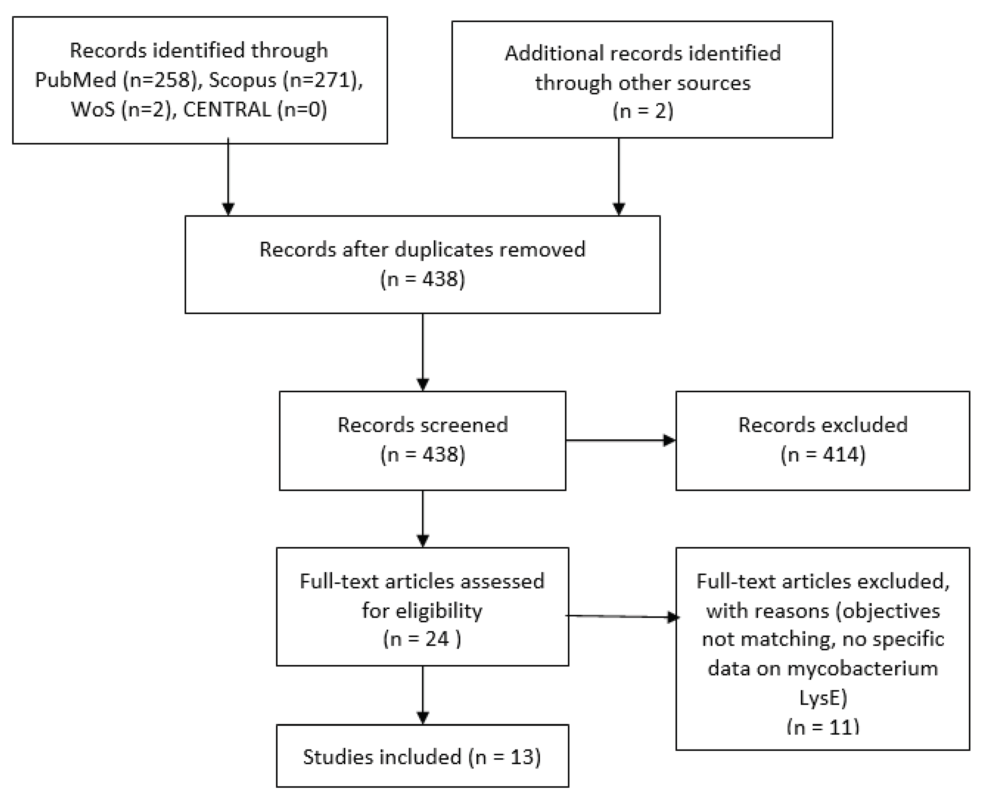

Table 1.
Research on the multifaceted role of LysE.
| S.No | Study ID | Findings of the study | Conclusion |
|---|---|---|---|
| 1. | Georgieva et al. 2020 [15] United States of America (USA) |
LysE deficiency causes increased cellular levels of L-lysine, which inhibits bacterial growth. Hence, LysE emerges as potential targets for drug inhibition, requiring a thorough examination of their structures and the relationships between structure and function. The study involved protein purification in Lipodisc made up of native E.coli membrane and detergents. |
This study claimed to be the first study on highly pure LysE conducted in a controlled environment. It sets the groundwork for more in-depth investigations into how these proteins function. |
| 2. | Georgieva et al. 2020 [9] United States of America (USA) |
The Rv1986 (LysE) protein of M.tb is the primary lysine exporter. Its expression is increased during the early stages of M.tb infection under hypoxia conditions. Hence, LysE can be a probable vaccine candidate. There is a need to have detailed knowledge about the less studied M.tb membrane transport system. |
This study represented the initial production of highly pure LysE, providing a platform for in-depth investigations into the functional mechanisms of this protein. |
| 3. | Hasenoehrl et al., 2019 [16] United States of America (USA) |
In M.tb, lysine permease LysE, encoded by Rv1986, catalyses lysine export. A deficiency of LysE contributes to bacteria’s poor growth. LysE is a primary target for memory T cells secreting IL-2 and is believed to contribute to human protective immunity. Resting macrophage and lysosomal exposure models revealed that LysE is involved in human latent TB infection. The absence of LysE in eukaryotic cells makes it a probable target for drug discovery. |
These results demonstrate that the aspartate pathway depends on a combination of metabolic control mechanisms in M.tb. This pathway is essential for persistence and a promising target for developing anti-tuberculosis drugs. |
| 4. | Chen et al. 2018 [7] China |
Currently available BCG vaccine is less effective in M.tb prevention and transmission. Therefore, it is crucial to investigate M. tb proteins absent in BCG that can induce specific humoral, cellular, and innate responses in the host. Rv1986 (LysE) shows diagnostic promise and is present in virulent M. bovis but not in the BCG strain. Studies highlight its role as an immunodominant target for memory T cells, containing numerous T and B cell epitopes. |
The findings indicated that Rv1986 is a highly immunogenic antigen capable of eliciting both humoral and cell-mediated immune responses. |
| 5. | Jiazhen et al., 2018 [10] China |
In infected cattle, Rv1983, Rv1986, Rv1987, and Rv1989c within RD2 were identified as immunodominant. This study assessed their diagnostic potential in humans. Out of 87 screened peptides from RD2 proteins, only ten were recognized by over 10% of active TB patients. The active TB group exhibited significantly higher IFN-γ responses to Rv1986 compared to the control groups (p < 0.05). |
This study concluded that six epitopes from RD2, including Rv1986, have potential diagnostic value in active TB. |
| 6. | Jiang et al., 2017 [13] China |
Region of Deletion 2 (RD2) plays a role in mycobacterial virulence, and its deletion from M.tb leads to a decrease in bacterial growth. The amplification and comparison of the five genes in RD2 indicate the presence of T and B cell epitopic regions. The study proposes that Rv1980c, Rv1985, and Rv1986 RD2 proteins belong to RD2. These proteins exhibit antigenic variation in response to host immune pressure, suggesting their potential involvement in ongoing immune evasion. |
The data confirmed that RD2 regions, aiming to evade host immunity, exhibit unexpected variability, particularly in immune-related antigens and T-cell epitope regions. |
| 7. | Schneefeld er al., 2017 [18] Germany |
The study showed that the Rv1985c (LysG) protein binds to its own gene and its promoter region of Rv1986 (LysE). Additionally, the Rv1985c protein induces the transcription of Rv1986 in a lysine-dependent manner and autonomously regulates the expression of its own gene. While the function of the Rv1986 protein remains elusive, studies have demonstrated its regulation and recognition by memory T cells in human TB. |
This study delineated the regulatory network of Rv1985c in M.tb. Given the resemblance to an orthologous gene pair in C. glutamicum, the study proposed renaming Rv1985c to lysG(Mt) and Rv1986 to lysE(Mt). |
| 8. | Tsu and Saier, 2015 [14] China |
The LysE superfamily, comprising transmembrane transport proteins vital for exporting amino acids, lipids, and heavy metal ions, plays pivotal roles in maintaining ionic balance, cell envelope assembly, and shielding against excessive cytoplasmic heavy metal/metabolite concentrations. | Consequently, they influence the physiology and pathogenesis of microbes and present potential drug targets. |
Disclaimer/Publisher’s Note: The statements, opinions and data contained in all publications are solely those of the individual author(s) and contributor(s) and not of MDPI and/or the editor(s). MDPI and/or the editor(s) disclaim responsibility for any injury to people or property resulting from any ideas, methods, instructions or products referred to in the content. |
© 2024 by the authors. Licensee MDPI, Basel, Switzerland. This article is an open access article distributed under the terms and conditions of the Creative Commons Attribution (CC BY) license (http://creativecommons.org/licenses/by/4.0/).
Copyright: This open access article is published under a Creative Commons CC BY 4.0 license, which permit the free download, distribution, and reuse, provided that the author and preprint are cited in any reuse.


