
Submitted:
04 March 2024
Posted:
05 March 2024
You are already at the latest version
Abstract
Among cephalopod discards, ink has revealed several beneficial properties regarding human health, environmental preservation and food preservation. In this study, different concentrations of an aqueous extract of cuttlefish (Sepia spp.) ink (CI) were added, respectively, to the packing medium employed during golden seabream (Sparus aurata) canning. Quality parameters of the resulting canned fish were determined and compared to the counterpart control and raw samples. As a result, an important effect of the CI concentration added to the packing medium was proved. The presence in the packing medium of a relatively low CI concentration led to lower (p<0.05) lipid oxidation development (fluorescent compound formation), lower (p<0.05) changes of colour parameters (L* and a* values) and lower (p<0.05) trimethylamine values in canned fish. Additionally, the two lowest CI concentrations tested led to higher average values of C22:6ω3 and ω3/ω6 ratios. Remarkably, higher average values of free fatty acids, polyene index and C20:5ω3 were detected in all kinds of CI-treated fish when compared to control canned samples. In agreement with environmental sustainability and circular economy requirements, the study provides a first approach to a novel and beneficial use of the present marine discard for the quality enhancement of canned fish.
Keywords:
cuttlefish
; ink
; golden seabream
; canning
; oxidation
; hydrolysis
; phospholipids
; ω3 fatty acids
; colour
; trimethylamine
1. Introduction
As a result of processing, the fishing and aquaculture industries generate a wide range of discards and wastes [1,2]. Such substrates are nowadays recognised as rich in healthy and nutritional constituents but constitute one of the most important environmental problems of coastline areas [3,4]. Among cephalopod discards, ink sacs are commonly removed manually and thrown without appropriate management. However, recent studies on this kind of discard have revealed several beneficial properties regarding human health [5,6,7], cosmetic purposes [8], and environmental preservation [9]. Regarding the preservation of seafood, previous studies have shown relevant antimicrobial [10] and antioxidant [11,12,13] properties of cephalopod ink extracts when applied to different kinds of seafood substrates.
The preservative behaviour of cephalopod ink has been indicated to reside in melanin and melanin-free fractions [14] by donating hydrogen atoms to any radical present in the reacting medium and thus preventing the formation of free radical formation [15]. The analysis of the melanin-free ink obtained from splendid squid (Loligo formosana) indicated that the highest antioxidant behaviour was present in fractions with molecular weight lower than 3 kDa, whose activity was not modified after a heat treatment (30 min at 90 ºC) [16]. Based on a chromatographic analysis, Senan [17] proved that the active principle in the methanolic extract obtained from cuttlefish (Sepia pharaonis) ink was a peptide molecule, which showed to be stable during the thermal treatment (30 min at 40-100 ºC). Recently, Xie et al. [18] indicated that water-extracted cuttlefish (Sepia sculenta) melanin contained a characteristic indole structure with irregular spherical structures and provided a protective effect on kojic and ascorbic acids.
Canning process can preserve marine species by the combination of sealing in a hermetic container and heating to destroy spoilage and pathogenic microorganisms and inactivate enzymes [19,20]. Provided duration and temperature during the heating and cooling cycle of the thermal process are properly carried out, canned seafood would be preserved for a long time. However, long-term high temperature may cause a decrease of the quality and the nutritional value (i.e., destruction of vitamins, protein components and unsaturated fatty acids, FAs) and a loss of the sensory quality (i.e., softer texture, separation of jelly and fat, discolouration, and undesirable taste) [21,22]. Therefore, the search for optimised canning conditions [23] and the employment of advanced preserving technologies [24] has been found necessary.
The present study focused on the use of cuttlefish (Sepia spp.) ink (CI). The basic objective was to analyse the preservative properties of an aqueous CI extract during the canning process of golden seabream (Sparus aurata). For it, different concentrations of CI were added, respectively, to the packing medium. Quality indices regarding lipid damage (lipid oxidation and hydrolysis development and changes in the FA profile), colour changes (L*, a*, and b* parameters) and trimethylamine (TMA) formation were measured in raw and canned samples. The effect of the CI concentration employed was analysed.
2. Materials and Methods
2.1. Initial ink and preparation of the ink extract
Commercial ink from cuttlefish (Sepia spp.) was provided by Sepink (Vilagarcía de Arousa, Pontevedra, Spain). The ink exhibited the following proximate composition (g·100 g-1): 78.4 (moisture), 6.4 (protein), 0.1 (lipids), 9.0 (ash), and 5.6 (total fibre).
To prepare the ink extract, a mixture of CI (40 g) and distilled water (400 mL) was stirred (30 s), sonicated (30 s), and centrifuged (3,500×g for 30 min at 4 °C). Then, the resulting supernatant was taken. The extraction process was repeated three more times. Finally, all supernatants were pooled together and made up to 1 L with distilled water.
2.2. Raw fish and fish canning
Farmed golden seabream (S. aurata) (44 specimens) (ca. 500 g each) were obtained in the fresh state in a local market and transported on ice to the laboratory. Then, four individuals were selected and considered as initial fish. For it, fish individuals were beheaded, eviscerated and filleted. After discarding the dark muscle, the resulting white muscle was minced and analysed independently in each of the fish individuals (n = 4).
On the same day, the remaining fish were divided into four batches (ten individuals per batch) that were considered independently during the current study (n = 4). In all batches, individuals were beheaded, eviscerated and filleted. Then, 45-g portions of fish fillets were placed in small flat rectangular cans (105 × 60 × 25 mm; 150 mL). Two cans were prepared from each individual fish. In each batch, 0, 10, 25, 40, and 80 mL of the above-mentioned CI extract were added, respectively, to the cans as packing media. Cans were filled by the addition of distilled water (95, 85, 70, 55, and 15 mL, respectively). As a result, CTR (canned control), and samples corresponding to CI-1, CI-2, CI-3 and CI-4 packing conditions were prepared, respectively.
The CI concentrations tested in this study were based on several preliminary tests. Thus, CI-4 condition corresponds to the highest concentration that can be employed without modifying the sensory descriptors of canned golden seabream (i.e., flesh colour, odour or flavour). In order to analyse the effect of the CI extract content, three conditions including lower CI concentrations (i.e., CI-1, CI-2, and CI-3 packing conditions) were also checked.
The cans were vacuum–sealed (SOMME 222, Ezquerra, San Adrián, Navarra, Spain) and sterilised (115 ºC, 45 min; Fo = 7 min) in a steam retort (Presoclave II 75L, JP Selecta, Barcelona, Spain). When the heating time was accomplished, the steam was cut off, the remaining steam was flushed away by the use of air, and water at reduced pressure was employed for cooling the cans.
The cans were opened after 3 months of storage at room temperature (20 ºC). For it, the liquid part was carefully drained off gravimetrically and filtered through a filter paper. After discarding the dark muscle, the white muscle of the canned fish was wrapped in filter paper and used for analysis. Each physico-chemical analysis was carried out in triplicate in each single sample.
In agreement with common practice employed in canneries, a 3-month storage was carried out. Canning manufacturers indicate that a minimum of a 2-3-month storage is required to optimise the sensory acceptability of commercial canned fish [25].
All chemical reagents and solvents employed were of reagent grade (Merck, Darmstadt, Germany).
2.3. Determination of lipid content and composition
The lipid fraction was obtained by extraction of the golden seabream muscle by applying the Bligh and Dyer [26] method, which employs a chloroform-methanol (1:1) mixture. Quantification was carried out according to Herbes and Allen [27]. Lipid content was calculated as g·kg-1 fish muscle.
The free fatty acid (FFA) content was determined on the lipid extract of the fish muscle by the Lowry and Tinsley [28] method, which is based on a complex formation with cupric acetate-pyridine followed by spectrophotometric (715 nm) assessment (Beckman Coulter DU 640 spectrophotometer, Beckman Coulter Inc., Brea, CA, USA). For quantitative purposes, oleic acid was employed as standard. Results were calculated as g FFAs·kg-1 lipids.
The phospholipid (PL) content was determined by measuring the organic phosphorus in the lipid extract in agreement with the Raheja et al. [29] procedure. This method implies a complex formation of organic phosphorus with ammonium molybdate. For quantitative purposes, 1,2-dipalmitoyl-rac-glycero-3-phosphocholine was employed as standard. Results were calculated as g PLs·kg-1 lipids.
Lipid extracts were converted into FA methyl esters (FAMEs) by employing acetyl chloride in methanol and then analysed using a Perkin-Elmer 8700 gas chromatograph (Madrid, Spain) equipped with a fused silica capillary column SP-2330 (0.25 mm i.d. x 30 m, 0.20 μm film, Supelco Inc., Bellefonte, PA, USA) [30]. Peaks corresponding to FAMEs were identified by comparing their retention times with those of standard mixtures (Qualmix Fish and Supelco 37 Component FAME Mix, Supelco Inc.). Peak areas were automatically integrated; C19:0 FA was used as the internal standard for quantitative purposes.
Content of each FA was calculated as g·100 g−1 total FAs. Such values were employed in order to obtain the content on FA groups, i.e., saturated (STFAs), monounsaturated (MUFAs), polyunsaturated (PUFAs) FAs, ω3 FAs, and ω6 FAs. Additionally, the ω3/ω6 ratio and the polyene index (PI), considered as the C20:5ω3 + C22:6ω3/C16:0 concentration ratio, were calculated.
2.4. Assessment of lipid oxidation development
The peroxide value (PV) was determined spectrophotometrically (520 nm) (Beckman Coulter, DU 640; London, UK) on the lipid extract by peroxide reduction with ferric thiocyanate [31]. Results were calculated as meq. active oxygen·kg-1 lipids.
The thiobarbituric acid index (TBA-i) was determined according to Vyncke [32]. For it, the content of the thiobarbituric acid reactive substances (TBARSs) was spectrophotometrically measured at 532 nm and calculated from a standard curve using 1,1,3,3-tetraethoxy-propane. Results were calculated as mg malondialdehyde·kg-1 muscle.
The determination of the interaction compound formation produced by reaction of oxidised lipids and protein-type molecules was carried out by fluorescence spectroscopy (Fluorimeter LS 45; Perkin Elmer España; Tres Cantos, Madrid, Spain). Fluorescent compound formation was measured at 393/463 and 327/415 nm in agreement with previous research [33]. The relative fluorescence (RF) was calculated according to the formula: RF = F/Fst, where F is the fluorescence measured at each excitation/emission wavelength pair and Fst is the fluorescence intensity of a quinine sulphate solution (1 µg·mL-1 in 0.05 M H2SO4) at the corresponding wavelength pair. The formation of fluorescent compounds was measured in the aqueous phase obtained from the lipid extraction [26] of the fish muscle.
2.5. Determination of muscle colour changes and TMA value
Colour parameters (L*, a* and b*) were determined in the surface of the raw and canned fish muscle. For it, instrumental colour analysis (CIE 1976 Lab), performed with a tristimulus Hunter Labscan 2.0/45 colorimeter, was carried out. For each sample analysis, colour scores were averaged over four determinations, which were taken by rotating the measuring head 90° between triplicate measurements per position.
The TMA content was determined using the picrate colorimetric method, as previously described by Tozawa et al. [34]. This method involves the preparation of a 5% trichloroacetic acid extract of the fish muscle (10 g/25 mL). Results were calculated as mg TMA-N·kg-1 muscle.
2.6. Statistical analysis
As previously expressed, four replicates (n = 4) were considered in this study. Data obtained were evaluated by analysis of variance (ANOVA) to explore differences resulting from the effect of the sterilisation step and the CI extract concentration added to the packing medium. For carrying out the comparison of average values, the least-squares difference (LSD) procedure was developed. A confidence interval at the 95% level (p < 0.05) was considered in order to establish significant differences among batches in all instances. For it, the PASW Statistics 18 software for Windows (SPSS Inc., Chicago, IL, USA) was employed.
3. Results and discussion
3.1. Determination of lipid oxidation evolution
The presence of peroxides (primary oxidation compounds) showed to be low (i.e., < 2.07) in all kinds of samples included in the current study (Table 1). In most cases, differences among samples were not significant (p > 0.05). However, a higher (p < 0.05) peroxide content in canned samples corresponding to the CI-1 condition than in the canned control was observed. No effect (p > 0.05) of the sterilisation step (comparison between the initial fish and the canned control fish) could be inferred.
As in the case of the peroxide determination, levels detected for the TBARSs were low, all values being below the 0.14 score (Table 1). No effect (p > 0.05) of the presence of the CI extract in the packing medium could be outlined on the TBARS formation. No effect (p > 0.05) was also inferred as a result of the sterilisation step.
Interaction compound formation is depicted in Figure 1. Comparison between the initial fish and fish corresponding to the canned control condition showed a remarkable (p < 0.05) increase as a result of the canning process. Among canned samples, the lowest average values were obtained in fish corresponding to the CI-1 and CI-2 packing conditions. An inhibitory effect (p < 0.05) on the fluorescent compound formation was proved by employing the CI-2 packing condition. Contrary, the CI-4 condition led to an increased formation (p < 0.05) of such kind of compounds when compared to the canned control samples.
During seafood thermal processing in general, levels detected for the different primary and secondary lipid oxidation compounds can be considered the result of several effects [21,22]. One side, the heating step (i.e., sterilisation in the present case) would increase the formation of such lipid oxidation compounds. On the other side, the heat treatment itself would facilitate the breakdown of peroxides and TBARSs, so that a content decrease of such molecules could be produced. Meantime, the heating process would facilitate the interaction between primary and secondary lipid oxidation compounds (i.e., electrophilic molecules) and protein-type compounds (i.e., nucleophilic compounds) present in the fish muscle. As a result of this interaction, an increase of the fluorescent compound formation would be produced [33,35,36]. Finally, and regarding the current study, the presence of the CI extract in the packing medium may interfere on the development of the lipid oxidation by avoiding the formation of oxidation compounds or preserving them from subsequent breakdown or interaction.
According to the low presence of primary and secondary lipid oxidation compounds in the present canned samples, no effect of the CI extract could be inferred on the content of such deteriorative compounds. However, an inhibitory effect on the formation of fluorescent compounds was observed if the CI-2 condition is considered. Contrary, a prooxidant effect was obtained if the CI-4 condition is taken into account. Therefore, a selective effect of the CI concentration added to the packing medium could be concluded.
Previous research has proved that the antioxidant activity of an antioxidant compound may depend on different kinds of factors, such as the lipid composition, antioxidant concentration, temperature, oxygen pressure, and the presence of other antioxidants and food components, e.g., proteins and water [37,38]. According to their molecular structure, antioxidants can exhibit substantial differences in effectiveness when used with different types of oils or fat-containing foods, and when used under different handling and processing conditions. A remarkable antioxidant effect was proved for ascorbate in steam- and microwave-cooked fish [39]; however, above critical values and in agreement with the present research, ascorbate showed pro-oxidant properties. Additionally, Medina et al. [40] proved a remarkable effect of polyphenol concentration on the antioxidant capacity during the thermal treatment of tuna (Thunnus alalunga).
No previous results are available, to the best of our knowledge, regarding the effect on canned seafood quality of ink extracts obtained from cephalopod species. However, an aqueous extract of cuttlefish (S. officinalis) ink led to an inhibitory effect on the formation of conjugated dienes and trienes and fluorescent compounds during a heating treatment of fish in a model system [12]. Additionally, a preservative effect of cephalopod ink has already been proved on non-thermally treated seafood. Thus, the presence of melanin-free ink obtained from splendid squid (L. formosana) led to a remarkable decrease of primary and secondary lipid oxidation development in chilled (15 days in ice) mackerel (Rastrelliger kanagurta) muscle [16]. Vate et al. [11] indicated a reduction of the peroxide and TBARS formation in refrigerated (20 days at 4 ºC) surimi gel sardine (Sardinella albella) by addition of melanin-free ink obtained from splendid squid (L. formosana); additionally, a content reduction of nonanal and 2-decenal was reported. An inhibitory effect on peroxide content was detected by Essid et al. [41] in smoked sardine (S. aurita) during cold storage (35 days at 4 ºC) by previous soaking in cuttlefish (Sepia officinalis) ink solution; furthermore, an extension of the shelf-life time was observed.
Previous studies have addressed the preservative effect resulting from the addition of antioxidant-enriched oils to the packing medium during seafood canning. Thus, the presence in the packing medium of polyphenol compounds included in extra-virgin olive oil (EVOO) led to a lower oxidation development in canned tuna (T. alalunga) [40] and in different canned fish (tuna, sardine, mackerel, anchovy) [42]. The employment of sunflower oil in the packing medium led to a lower TBARS content in canned yellowfin tuna (Thunnus albacares) [43] and to a lower fluorescent compound formation in canned sprat (Clupeonella cultriventris) [35]; in both studies, such lower values were explained by the presence of antioxidant compounds in the oil employed as packing medium.
The addition of plant extracts to the packing medium has led to a lower lipid oxidation development in canned fish. This is the case of rosemary extract and tomato juice with canned bonito (Sarda sarda) [44] and grape seed (Vitis vinifera) with canned sardine (Sardina pilchardus) [45]. Similarly, the presence of macroalgae extracts in the packing medium of canned fish has shown preservative effects. Thus, a decrease of the TBARS level was observed in canned Atlantic salmon (Salmo salar) by addition of algae (Durvillaea antarctica, Ulva lactuca, or Pyropia columbina) extracts [46] and a decrease of the fluorescent compound formation was proved in canned Atlantic mackerel (Scomber scombrus) by the presence of Bifurcaria bifurcata extracts [47] and in canned Chub mackerel (Scomber colias) by including algae (Fucus spiralis or U. lactuca) aqueous extracts [30]. Regarding the employment of waste substrates produced during seafood processing, a lower fluorescent compound formation was detected in canned horse mackerel (Trachurus trachurus) by including an aqueous extract of octopus (Octopus vulgaris) cooking liquor in the packing medium [36].
3.2. Determination of FFA and PL content
Comparison between initial fish and canned fish corresponding to the control condition indicated a marked increase (p < 0.05) of the FFA value as a result of the sterilisation step (Table 2). All CI-treated fish showed higher average values of FFAs than the control canned fish. However, differences were not found significant (p > 0.05).
In the current study, the FFA content can be influenced by different factors. First, the thermal process would lead to the hydrolysis of higher-molecular-weight lipid classes such as triacylglycerols (TAGs) and PLs [21,48]. Then, FFAs are likely to be oxidised or broken-down during the thermal process as providing a greater accessibility to oxygen and other pro-oxidant molecules when compared to TAG and PL classes [49,50]; consequently, this effect would produce a decrease of the FFA content. Current results have shown that the first effect was more important than the second one. The fact that all CI-treated fish showed higher average FFA levels than the canned control condition indicates that some preservative effect is produced on the FFA compounds by the presence of the CI extract. A preservative effect on FFAs was also inferred by Barbosa et al. [30] during Atlantic Chub mackerel (S. colias) canning by the presence of an aqueous extract of F. spiralis in the packing medium. The same authors [47] proved a higher FFA retention in canned Atlantic mackerel (S. scombrus) by the presence of B. bifurcata extracts in the packing medium.
No previous information is available regarding the effect of CI on the lipid hydrolysis development in processed seafood. The presence of an aqueous extract of cuttlefish (S. officinalis) ink in a fish-heated model system did not provide a definite trend on the formation of FFAs [12]. The addition of natural preservative compounds to the packing medium has led to contradictory conclusions regarding the FFA value. Thus, a lower FFA content was detected in canned fish (tuna, sardine, mackerel, and anchovy) by employing EVOO [42] and in canned Chub mackerel (S. colias) by including an aqueous extract of octopus (O. vulgaris) cooking liquor [51]. However, no effect was proved in canned Chub mackerel (S. colias) by addition of U. lactuca extracts [30].
Regarding the PL fraction in the current study, the content revealed a marked decrease (p < 0.05) as a result of the sterilisation treatment (comparison between initial fish and control canned fish) (Table 2). However, all CI-treated fish showed higher average values than the control canned condition. Remarkably, canned muscle corresponding to the CI-1 condition did not show a significant loss (p > 0.05) of the PL content when compared to the initial fish.
PLs have attracted a great attention as showing a high bioavailability and preserving effect as delivery systems on several diseases [52,53]. Based on the needs and requirements expressed by food and pharmaceutical industries, great efforts are being addressed to the retention of the PL fraction in seafood as presenting high PUFA levels [54,55].
Previous research regarding the effect of antioxidant compounds included in the packing system on the PL content in canned fish is scarce. The addition of an aqueous extract of F. spiralis to the packing medium of brine-canned Chub mackerel (S. colias) led to a preserving effect on the PL composition [30]. Similarly, Méndez et al. [36] proved a retention of the PL content in brine-canned horse mackerel (T. trachurus) by addition to the packing medium of an aqueous extract of octopus (O. vulgaris) cooking liquor.
3.3. Analysis of the FA profile
The FA profile of the initial fish indicated the following composition of individual FAs (g·100 g-1 total FAs): 1.34±0.07 (C14:0), 0.25±0.02 (C15:0), 16.54±0.45 (C16:0), 2.30±0.06 (C16:1ω7), 0.35±0.02 (C17:0), 4.73±0.25 (C18:0), 23.68±1.18 (C18:1ω9), 3.10±0.26 (C18:1ω7), 26.88±0.31 (C18:2ω6), 0.95±0.07 (C20:1ω9), 1.00±0.07 (C20:2ω6), 1.20±0.09 (C20:4ω6), 0.28±0.03 (C22:1ω9), 2.27±0.15 (C20:5ω3), 0.27±0.02 (C22:4ω6), 0.50±0.04 (C24:1ω9), 2.39±0.08 (C22:5ω3), and 11.96±0.74 (C22:6ω3). Expressed in terms of FA groups, the following composition was obtained for the initial fish (g·100 g-1 total FAs): 23.20±0.64 (STFAs), 30.82±1.56 (MUFAs), 45.98±0.93 (PUFAs), 16.62±0.94 (ω3 FAs), 29.36±0.21 (ω6 FAs). Additionally, the following FA ratio values were obtained: 0.57±0.03 (ω3/ω6 ratio) and 0.86±0.03 (PI).
Seafood consumption deserves currently a great interest according to their beneficial effects on human health. On the basis of recent studies [56,57], the total ω3 FA value is considered a highly valuable index. Thus, epidemiological and clinical studies have associated the eicosapentaenoic acid (EPA) consumption to an inhibitory effect on the development of inflammatory, coronary, and circulatory diseases [58]. Meantime, docosahexaenoic acid (DHA) has been associated with a positive effect on several health concerns such as foetal development, prevention of neurodegenerative diseases, and appropriate functioning of the nervous system and visual organs in the foetus [59]. Additionally, the ω3/ω6 ratio of foods included in the human diet has attracted a great interest [60,61]. Thus, a diet including values of 1:5 or higher for this ratio have been recommended in order to reduce the development of certain health concerns [62].
In the present study, FA analysis was carried out on control and CI-treated canned samples (data not shown). According to the above-mentioned nutritional and healthy properties, quality changes related to FA values will be focused on the ω3 FA fraction (namely, EPA, DHA, and total ω3 FA values) and to FA ratios (ω3/ω6 and PI).
Comparison of samples corresponding to the initial fish and the canned control condition revealed a remarkable loss (p < 0.05) of EPA and DHA values (Figure 2 and Table 3, respectively) as a result of the sterilisation step. Regarding the CI treatment, all CI-treated fish showed higher average EPA values than the canned control; notably, a significant loss could not be inferred (p > 0.05) in treated samples when compared to the initial fish. For the DHA value, samples corresponding to the CI-1 and CI-2 conditions showed higher average values than canned control fish; contrary, samples corresponding to CI-3 and CI-4 conditions revealed lower average values. This dependency on the CI concentration employed is in agreement with the results previously shown regarding the fluorescent compound formation (Figure 1). Total ω3 FA content indicated the same profile than the DHA value (Table 3). Thus, all canned samples (control and CI-treated) underwent a content decrease (p < 0.05) in comparison to the initial fish and no significant effect (p > 0.05) was concluded as a result of the CI addition to the packing medium. Notably, samples corresponding to CI-1 and CI-2 conditions showed higher average values of total ω3 FAs than the counterpart canned samples.
The analysis of the ω3/ω6 ratio indicated a marked decrease (p < 0.05) as a result of the sterilisation step (Table 3). Therefore, it could be inferred that ω3 FAs have been more susceptible to heat treatment than those belonging to the ω6 series. Comparison among canned samples (CI-treated and control) did not provide a significant effect (p > 0.05) of the presence of the CI extract in the packing medium. However, and in agreement with current results on the FR (Figure 1) and DHA content (Table 3), CI-1 and CI-2 conditions led to the highest average values of the ω3/ω6 ratio.
The assessment of the PI is considered a valuable and practical tool for measuring the possible loss of the PUFA content during marine species canning or processing in general [30,36,63]. In the present study, this quality parameter showed an average decrease as a result of the thermal treatment (Figure 3). Notably, canned fish corresponding to the CI-1 condition did not reflect a significant loss (p > 0.05) when compared to the initial fish. Compared to the control canned condition, all CI-treated samples provided higher average values for this index; however, differences were not found significant (p > 0.05). A preservative effect of the cuttlefish (S. officinalis) ink on PUFA compounds (i.e., higher PI) was already proved by Trigo et al. [12] during a heating treatment of fish in a model system.
Previous research related to the addition of antioxidant compounds to the packing medium has shown a preservative effect on the PUFA content. Thus, Ortiz et al. [47] showed a PI retention of canned Atlantic salmon (S. salar) muscle when packed in a medium including an ulte (basal part of alga D. antarctica) extract; however, no differences were obtained in such study when other algae (cochayuyo, frond of D. antarctica; U. lactuca; P. columbina) extracts were included in the packing system. Higher PI scores were observed in canned Atlantic mackerel (S. scombrus) by addition of B. bifurcata extracts [47] and in canned Chub mackerel (S. colias) by addition of F. spiralis or U. lactuca extracts [30]. Recently, a higher PI in canned Chub mackerel (S. colias) was proved by including an aqueous extract of octopus (O. vulgaris) cooking liquor in the packing medium [51].
3.4. Determination of colour changes in canned fish muscle
A remarkable increase (p < 0.05) of the L* value was detected as a result of the thermal treatment (Table 4). This increase was partially avoided in all cases by the presence of the CI extract in the packing medium. Thus, all CI-treated fish revealed lower (p < 0.05) L* values than the control canned fish. Additionally, canned fish corresponding to the CI-1 condition did not provide differences (p > 0.05) with the initial fish.
A general average decrease of the a* value was observed by comparison of the initial fish and all kinds of canned samples (Table 4). This decrease was found significant (p < 0.05) in the case of the canned control samples, but not (p > 0.05) in CI-treated fish. No effect (p > 0.05) of the CI concentration added to the packing medium could be inferred on this colour parameter.
Comparison between the initial fish and all kinds of canned samples indicated a relevant increase of the average b* value as a result of the canning process (Table 4). The presence of the CI extract in the packing medium led to lower average b* values than the canned control fish; however, differences were not found significant (p > 0.05). The lowest average value was observed for canned fish corresponding to the CI-1 condition.
According to the great effect on the appearance and acceptability of all kinds of seafood, the assessment of colour changes during processing has attracted a great attention [64]. The current evolution of colour parameters during the canning process is in agreement with previous studies regarding heating treatment of seafood. In general, the canning process has led to increased L* and b* values and to a* value decreases [43,65,66].
No previous research has focused on the effect of cephalopod ink on colour changes in canned seafood. However, the addition of preservative compounds to the packing medium has proved valuable effects. Thus, the employment of sunflower oil as packing medium in canned yellowfin tuna (T. albacares) led to lower L* and b* values when compared to coconut and ground nut oils [43]; this difference was justified on the basis of the presence of preservative compounds in sunflower oil. The addition of an aqueous extract of B. bifurcata to the packing medium of canned Atlantic mackerel (S. scombrus) led to decreased L* and b* values [47]. Recently, Méndez et al. [36] obtained lower L* and b* values in canned horse mackerel (T. trachurus) by addition of an aqueous extract of octopus (O. vulgaris) cooking liquor to the packing medium.
3.5. Determination of TMA content
A relevant (p < 0.05) TMA content increase was detected in all kinds of canned samples when compared to the initial fish (Table 2). Lower average values were found in CI-treated fish when compared to the counterpart canned control. Notably, the lowest average values were observed in fish samples corresponding to the CI-2 condition, which were significantly lower (p < 0.05) than those corresponding to the canned control samples. As for the lipid quality retention (FR, Figure 1; DHA and ω3/ω6 ratio values, Table 3), a selective effect of the CI concentration could be inferred.
TMA is recognised as a remarkable deteriorative compound whose value can reflect the quality degradation of seafood subjected to different kinds of technological procedures [67]. On the basis that TMA formation by microbial activity is not likely to be produced during the current study, formation of TMA should be originated from the thermal breakdown of protein-like compounds [20,24]. As in the current study, previous research accounts for a relevant TMA formation as a result of the sterilisation step in canned seafood [43,68,69].
In agreement with the results depicted in Table 2, an inhibitory effect on the TMA content has been observed for samples corresponding to the CI-2 batch. Unfortunately, no previous studies are available, to the best of our knowledge, regarding the effect of cephalopod ink extracts on the TMA value of canned seafood. However, an inhibitory effect on quality loss has already been proved on non-thermally treated seafood. Thus, Karim et al. [10] demonstrated that a previous squid melanin-free ink soaking of squid (Loligo duvaucelli) led to an increased shelf-life time and a decrease of the TMA and total volatile base (TVB) values during the refrigerated (4 ºC for 215 days) storage. Sadok et al. [70] proved that a previous cuttlefish (S. officinalis) ink soaking led to an inhibitory effect on the formation of off-odour compounds (TMA and TVB values) in cold-stored (0 ºC for 12 days and –2 ºC for 23 days) peeled shrimp (Penaeus kerathurus). Recently, an inhibitory effect on the formation of TVB and TMA was detected by Essid et al. [41] in smoked sardine (S. aurita) during cold storage (35 days at 4 ºC) by previous soaking in cuttlefish (S. officinalis) ink solution.
Previous research has also addressed the effect resulting from the addition of preservative compounds to the packing medium on the TMA value of canned seafood. As in the present results, a lower TVB value was observed in canned bonito (S. sarda) by including rosemary extract and tomato juice in the packing medium [44]. An inhibitory effect on TMA formation was detected in canned Chub mackerel (S. colias) by addition of an aqueous F. spiralis extract to the packing medium [69]. Contrary, no effect on the TVB and TMA values was detected in Atlantic mackerel (S. scombrus) [47] and Chub mackerel (S. colias) [30] by the presence of aqueous extracts of several macroalgae.
4. Conclusions
A preservative effect of the CI extract on the canned fish quality was concluded. However, an important effect of the CI concentration added to the packing medium was proved. According to the results obtained for lipid oxidation development (fluorescent compound formation), colour changes (L* and a* parameters) and TMA formation, a preservative effect (p < 0.05) of the CI extract was detected in the present study when employing CI concentrations corresponding to CI-2 condition. Additionally, higher average values of DHA and ω3/ω6 ratio were detected in canned fish corresponding to CI-1 and CI-2 conditions. Remarkably, higher average values of FFAs and PUFAs (i.e., PI and EPA content) were detected in all kinds of CI-treated fish when compared to control canned samples.
The present research provides a first approach to a novel and beneficial use of a cephalopod discard (i.e., cuttlefish ink) for the quality enhancement of canned fish. This research is in agreement with nowadays requirements for the search of new and natural sources of preservative compounds and also agrees with general commitments for environmental sustainability and circular economy. On the basis of the notable effect of the CI concentration employed, further research is considered necessary in order to optimise the CI extract to be employed.
Author Contributions
Conceptualization, B.M. and S.P.A.; methodology, B.M. and M.T.; data curation, M.T. and S.P.A.; writing—original draft preparation, S.P.A.; writing—review and editing, B.M. and S.P.A. All authors have read and agreed to the published version of the manuscript.
Funding
This research received no external funding.
Institutional Review Board Statement
Not applicable.
Informed Consent Statement
Not applicable.
Data Availability Statement
All data are comprised within the article.
Acknowledgments
The cuttlefish ink was kindly provided by Sepink (Vilagarcía de Arousa, Pontevedra, Spain).
Conflicts of Interest
The authors declare no conflicts of interest.
References
- Rustad, T.; Storro, I.; Slizyte, R. Possibilities for the utilisation of marine by-products. Int. J. Food Sci. Technol. 2011, 46, 2001–2014. [Google Scholar] [CrossRef]
- Olsen, R.L.; Toppe, J.; Karunasagar, I. Challenges and realistic opportunities in the use of by-products from processing of fish and shellfish. Trends Food Sci. Technol. 2014, 36, 144–152. [Google Scholar] [CrossRef]
- Atef, M.; Ojagh, M. Health benefits and food applications of bioactive compounds from fish byproducts: A review. J. Funct. Foods 2017, 35, 673–681. [Google Scholar] [CrossRef]
- Vázquez, J.A.; Meduiña, A.; Durán, A.I.; Nogueira, M.; Fernández-Compás, A.; Pérez-Martín, R.I.; Rodríguez-Amado, I. Production of valuable compounds and bioactive metabolites from by-products of fish discards using chemical processing, enzymatic hydrolysis, and bacterial fermentation. Mar. Drugs 2019, 17, 139. [Google Scholar] [CrossRef] [PubMed]
- Fahmy, S.R.; Ali, E.M.; Ahmed, N.S. Therapeutic effect of Sepia ink extract against invasive pulmonary aspergillosis in mice. J. Basic App. Zool. 2014, 67, 196–204. [Google Scholar] [CrossRef]
- Solano, F. Melanin and melanin-related polymers as materials with biomedical and biotechnological applications—Cuttlefish ink and mussel foot proteins as inspired biomolecules. Int. J. Mol. Sci. 2017, 18, 1561. [Google Scholar] [CrossRef]
- Momenzadeh, N.; Hajian, S.; Shabankare, A.; Ghavimi, R.; Kabiri-Samani, S.; Kabiri, H.; Hesami-Zadeh, K.; Shabankareh, A.N.T.; Nazaraghay, R.; Nabipour, I.; Mohammadi, M. Photothermic therapy with cuttlefish ink-based nanoparticles in combination with anti-OX40 mAb achieve remission of triple-negative breast cancer. Int. Immunopharm. 2023, 115, 109622. [Google Scholar] [CrossRef] [PubMed]
- Neifar, A.; Abdelmalek, I.B.; Bouajila, G.; Kolsi, R.; Bradai, M.N.; Abdelmouleh, A.; Gargouri, A.; Ayed, N. Purification and incorporation of the black ink of cuttlefish Sepia officinalis in eye cosmetic products. Color. Technol. 2013, 129, 150–154. [Google Scholar] [CrossRef]
- Wang, H.; Xu, T.; Zheng, B.; Cao, M.; Gao, F.; Zhou, G.; Ma, C.; Dang, J.; Yao, W.; Wu, K.; Liu, T.; Yuan, Y.; Fu, Q.; Wang, N. Cuttlefish ink loaded polyamidoxime adsorbent with excellent photothermal conversion and antibacterial activity for highly efficient uranium capture from natural seawater. J. Hazard. Mat. 2022, 433, 128789. [Google Scholar] [CrossRef]
- Karim, N.U.; Sadzali, N.L.; Hassan, M. Effects of squid ink as edible coating on squid sp. (Loligo duvauceli) spoilage during chilled storage. Int. Food Res. J. 2016, 23, 1895–1901. [Google Scholar]
- Vate, N.K.; Benjakul, S.; Agustini, T.W. Application of melanin-free ink as a new antioxidative gel enhancer in sardine surimi gel. J. Sci. Food Agric. 2015, 95, 2201–2207. [Google Scholar] [CrossRef] [PubMed]
- Trigo, M.; Paz, D.; Bote, A.; Aubourg, S.P. Antioxidant activity of an aqueous extract of cuttlefish (Sepia officinalis) ink during fish muscle heating. Antioxidants 2023, 12, 1996. [Google Scholar] [CrossRef] [PubMed]
- Liang, Y.; Han, Y.; Dan, J.; Li, R.; Sun, H.; Wang, J.; Zhang, W. A high-efficient and stable artificial superoxide dismutase based on functionalized melanin nanoparticles from cuttlefish ink for food preservation. Food Res. Int. 2023, 163, 112211. [Google Scholar] [CrossRef] [PubMed]
- Derby, C.D. Cephalopod Ink: Production, chemistry, functions and applications. Mar. Drugs 2014, 12, 2700–2730. [Google Scholar] [CrossRef]
- Chen, S.; Xue, C.; Li, Y.; Gao, Z.; Ma, Q. Studies on the free radical scavenging activities of melanin from squid ink. China J. Mar. Drugs, 2007, 26, 24–27. [Google Scholar]
- Vate, N.K.; Benjakul, S. Antioxidative activity of melanin-free ink from splendid squid (Loligo formosana). Int. Aquat. Res. 2013, 5, 9. [Google Scholar] [CrossRef]
- Senan, V.P. Antibacterial activity of methanolic extract of the ink of cuttlefish, Sepia pharaonis, against pathogenic bacterial strains. Int. J. Pharm. Sci. Res. 2015, 6, 1705–1710. [Google Scholar]
- Xie, J.; Li, H.; Che, H.; Dong, X.; Yang, X.; Xie, W. Extraction, physicochemical characterisation, and bioactive properties of ink melanin from cuttlefish (Sepia esculenta). Int. J. Food Sci. Technol. 2021, 56, 3627–3640. [Google Scholar] [CrossRef]
- Horner, W. Canning fish and fish products. In Fish Processing Technology, 2nd edition; Hall, G., Ed.; Blackie Academic and Professional, Chapman and Hall: London, UK, 1997; pp. 119–159. [Google Scholar]
- Veiga, A.; Martínez, E.; Ojea, G.; Caride, A. Principles of thermal processing in canned seafood. In Quality Parameters in Canned Seafoods; Cabado, A.G., Vieites, J.M., Eds.; Nova Science Publishers, Inc.: New York, USA, 2008; pp. 83–103. [Google Scholar]
- Lukoshkina, M.; Odoeva, G. Kinetics of chemical reactions for prediction of quality of canned fish during storage. App. Biochem. Microb. 2003, 39, 321–327. [Google Scholar] [CrossRef]
- Ling, B.; Tang, J.; Kong, F.; Mitcham, E.J.; Wang, S. Kinetics of food quality changes during thermal processing: A review. Food Bioprocess Technol. 2015, 8, 343–358. [Google Scholar] [CrossRef]
- Pitarch, J.L.; Vilas, C.; de Prada, C.; Palacín, C.G.; Alonso, A.A. Optimal operation of thermal processing of canned tuna under product variability. J. Food Eng. 2021, 304, 110594. [Google Scholar] [CrossRef]
- Tokur, B.; Korkmaz, K. Novel thermal sterilization technologies in seafood processing. In Innovative Technologies in Seafood Processing; Özoğul, Y., Ed.; CRC Press, Taylor and Francis Group: Boca Raton, FL, USA, 2020; pp. 303–322. [Google Scholar]
- Ruiz-Roso, B.; Cuesta, I.; Pérez, M.; Borrego, E.; Pérez-Olleros, L.; Varela, G. Lipid composition and palatability of canned sardines. Influence of the canning process and storage in olive oil for five years. J. Sci. Food Agric. 1998, 77, 244–250. [Google Scholar] [CrossRef]
- Bligh, E.; Dyer, W. A rapid method of total extraction and purification. Can. J. Biochem. Physiol. 1959, 37, 911–917. [Google Scholar] [CrossRef] [PubMed]
- Herbes, S.E.; Allen, C.P. Lipid quantification of freshwater invertebrates: Method modification for microquantitation. Can. J. Fish. Aquat. Sci. 1983, 40, 1315–1317. [Google Scholar] [CrossRef]
- Lowry, R.; Tinsley, I. Rapid colorimetric determination of free fatty acids. J. Am. Oil Chem. Soc. 1976, 53, 470–472. [Google Scholar] [CrossRef]
- Raheja, R.; Kaur, C.; Singh, A.; Bhatia, A. New colorimetric method for the quantitative determination of phospholipids without acid digestion. J. Lipid Res. 1973, 14, 695–697. [Google Scholar] [CrossRef] [PubMed]
- Barbosa, R.G.; Trigo, M.; Campos, C.A.; Aubourg, S.P. Preservative effect of algae extracts on lipid composition and rancidity development in brine-canned Atlantic Chub mackerel (Scomber colias). Eur. J. Lipid Sci.Technol. 2019, 1900129. [Google Scholar] [CrossRef]
- Chapman, R.; McKay, J. The estimation of peroxides in fats and oils by the ferric thiocyanate method. J. Am. Oil Chem. Soc. 1949, 26, 360–363. [Google Scholar] [CrossRef]
- Vyncke, W. Direct determination of the thiobarbituric acid value in trichloracetic acid extracts of fish as a measure of oxidative rancidity. Fette, Seifen, Anstrichm. 1970, 72, 1084–1087. [Google Scholar] [CrossRef]
- Aubourg, S.P.; Medina, I.; Pérez-Martín, R.I. A comparison between conventional and fluorescence detection methods of cooking-induced damage to tuna fish lipids. Z. Lebensm. Unters. Forsch. 1995, 200, 252–255. [Google Scholar] [CrossRef]
- Tozawa, H.; Erokibara, K.; Amano, K. Proposed modification of Dyer’s method for trimethylamine determination in codfish. In Fish Inspection and Quality Control; Kreuzer, R., Ed.; Fishing News Books Ltd.: London, UK, 1971; pp. 187–190. [Google Scholar]
- Naseri, M.; Rezaei, M. Lipid changes during long-term storage of canned sprat. J. Aquat. Food Prod. Technol. 2012, 21, 48–58. [Google Scholar] [CrossRef]
- Méndez, L.; Trigo, M.; Zhang, B.; Aubourg, S.P. Antioxidant effect of octopus by-products in canned horse mackerel (Trachurus trachurus) previously subjected to different frozen storage times. Antioxidants 2022, 11, 2091. [Google Scholar] [CrossRef] [PubMed]
- Nawar, W.W. Lipids. In Food Chemistry, 3rd edition; Fennema, O., Ed.; Marcel Dekker: New York, NY, USA, 1996; pp. 225–314. [Google Scholar]
- Pokorný, J. Introduction. In Antioxidants in Foods. Practical Applications; Pokorný, J., Yanishlieva, N., Gordon, M., Eds.; CRC Press: Boca Raton, USA, 2001; pp. 1–3. [Google Scholar]
- Ramanathan, L.; Das, N. Studies on the control of lipid oxidation in ground fish by some polyphenolic natural products. J. Agric. Food Chem. 1992, 40, 1–17. [Google Scholar] [CrossRef]
- Medina, I.; Satué, M.T.; German, J.B.; Frankel, E. Comparison of natural polyphenols antioxidant from extra virgin olive oil with synthetic antioxidants in tuna lipids during thermal oxidation. J. Agric. Food Chem. 1999, 47, 4873–4879. [Google Scholar] [CrossRef] [PubMed]
- Essid, I.; Aroussia, H.; Soufi, E.; Bouriga, N.; Gharbi, S.; Bellagha, S. Improving quality of smoked sardine fillets by soaking in cuttlefish ink. Food Sci. Technol, Campinas, 2022, 42, e65020. [Google Scholar] [CrossRef]
- Caponio, F.; Summo, C.; Pasqualone, A.; Gomes, T. Fatty acid composition and degradation level of the oils used in canned fish as a function of the different types of fish. J. Food Comp. Anal. 2011, 24, 1117–1122. [Google Scholar] [CrossRef]
- Mohan, C.O.; Remya, S.; Murthy, L.N.; Ravishankar, C.N.; Kumar, K.A. Effect of filling medium on cooking time and quality of canned yellowfin tuna (Thunnus albacares). Food Cont. 2015, 50, 320–327. [Google Scholar] [CrossRef]
- Kınay, A.G.; Duyar, H.A. Rosemary (Rosmarinus officinalis) as a preservative agent in canned bonito (Sarda sarda). Mar. Sci. Tech. Bull. 2021, 10, 406–415. [Google Scholar] [CrossRef]
- Bouriga, N.; Bahri, W.R.; Mili, S.; Massoudi, S.; Quignard, J.P.; Trabelsi, M. Variations in nutritional quality and fatty acids composition of sardine (Sardina pilchardus) during canning process in grape seed and olive oils. J. Food Sci. Technol. 2022, 59, 4844–4852. [Google Scholar] [CrossRef]
- Ortiz, J.A.; Vivanco, J.P.; Aubourg, S.P. Lipid and sensory quality of canned Atlantic salmon (Salmo salar): Effect of the use of different seaweed extracts as covering liquids. Eur. J. Lipid Sci. Technol. 2014, 116, 596–605. [Google Scholar] [CrossRef]
- Barbosa, R.G.; Trigo, M.; Fett, R.; Aubourg, S.P. Impact of a packing medium with alga Bifurcaria bifurcata extract on canned Atlantic mackerel (Scomber scombrus) quality. J. Sci. Food Agric. 2018, 98, 3462–3467. [Google Scholar] [CrossRef] [PubMed]
- Medina, I.; Sacchi, R.; Aubourg, S.P. A 13C-NMR study of lipid alterations during fish canning: Effect of filling medium. J. Sci. Food Agric. 1995, 69, 445–450. [Google Scholar] [CrossRef]
- Labuza, T. Kinetics of lipid oxidation in foods. CRC Crit. Rev. Food Technol. 1971, 2, 355–405. [Google Scholar] [CrossRef]
- Miyashita, K.; Tagagi, T. Study on the oxidative rate and prooxidant activity of free fatty acids. J. Am. Oil Chem. Soc. 1986, 63, 1380–1384. [Google Scholar] [CrossRef]
- Malga, J.M.; Trigo, M.; Martínez, B.; Aubourg, S.P. Preservative effect on canned mackerel (Scomber colias) lipids by addition of octopus (Octopus vulgaris) cooking liquor in the packaging medium. Molecules 2022, 27, 739. [Google Scholar] [CrossRef] [PubMed]
- Küllenberg, D.; Taylor, L.A.; Schneider, M.; Massing, U. Health effects of dietary phospholipids. Lipids Health Dis. 2012, 11, 3. [Google Scholar] [CrossRef] [PubMed]
- Li, J.; Wang, X.; Zhang, T.; Huang, Z.; Luo, X.; Deng, Y. A review on phospholipids and their main applications in drug delivery systems. Asian J. Pharm. Sci. 2015, 10, 81–98. [Google Scholar] [CrossRef]
- Aubourg, S.P.; Medina, I.; Pérez-Martín, R. Polyunsaturated fatty acids in tuna phospholipids: Distribution in the sn-2 location and changes during cooking. J. Agric. Food Chem. 1996, 44, 585–589. [Google Scholar] [CrossRef]
- Takahashi, K.; Inoue, Y. Marine by-product phospholipids as booster of medicinal compounds. Adv. Food Nutr. Res. 2012, 65, 31–46. [Google Scholar] [CrossRef]
- Magalhães, J.P.; Müller, M.; Rainger, G.; Steegenga, W. Fish oil supplements, longevity and aging. Aging 2016, 8, 1578–1582. [Google Scholar] [CrossRef]
- Devassy, J.G.; Leng, S.; Gabbs, M.; Monirujjaman, M.; Aukema, H.M. Omega-3 polyunsaturated fatty acids and oxylipins in neuroinflammation and management of Alzheimer disease. Adv. Nutr. 2016, 7, 905–916. [Google Scholar] [CrossRef]
- Swanson, S.; Block, R.; Mousa, S. Omega-3 fatty acids EPA and DHA: Health benefits throughout life. Adv. Nutr. 2012, 3, 1–7. [Google Scholar] [CrossRef]
- Ofosu, F.K.; Daliri, E.B.M.; Lee, B.H.; Yu, X. Current trends and future perspectives on omega-3 fatty acids. Res. J. Biol. 2017, 5, 11–20. [Google Scholar]
- Uauy, R.; Valenzuela, A. Marine oils: The health benefits of n-3 fatty acids. Nutrition 2000, 16, 680–684. [Google Scholar] [CrossRef] [PubMed]
- Komprda, T. Eicosapentaenoic and docosahexaenoic acids as inflammation-modulating and lipid homeostasis influencing nutraceuticals: A review. J. Funct. Foods 2012, 4, 25–38. [Google Scholar] [CrossRef]
- Simopoulos, A.P. The importance of the ratio of omega-6/omega-3 essential fatty acids. Biomed. Pharm. 2002, 56, 365–379. [Google Scholar] [CrossRef] [PubMed]
- Rustad, T. Lipid oxidation. In Handbook of Seafood and Seafood Products Analysis; Nollet, L.M., Toldrá, F., Eds.; CRC Press, Francis and Taylor Group: Boca Raton, FL, USA, 2010; pp. 87–95. [Google Scholar]
- Schubring, R. Quality Assessment of Fish and Fishery Products by Color Measurement. In Handbook of Seafood and Seafood Products Analysis; Nollet, L.M., Toldrá, F., Eds.; CRC Press, Francis and Taylor Group: Boca Raton, FL, USA, 2010; pp. 395–424. [Google Scholar]
- Mohan, C.O.; Remya, S.; Ravishankar, C.N.; Vijayan, P.K.; Srinivasa Gopal, T.K. Effect of filling ingredient on the quality of canned yellowfin tuna (Thunnus albacares). Int. J. Food Sci. Technol. 2014, 49, 1557–1564. [Google Scholar] [CrossRef]
- Gómez-Limia, L.; Carballo, J.; Rodríguez-González, M.; Martínez, S. Impact of the filling medium on the colour and sensory characteristics of canned European eels (Anguilla anguilla L.). Foods 2022, 11, 1115. [Google Scholar] [CrossRef] [PubMed]
- Özoğul, Y. Methods for freshness quality and deterioration. In Handbook of Seafood and Seafood Products Analysis; Nollet, L.M., Toldrá, F., Eds.; CRC Press, Francis and Taylor Group: Boca Raton, FL, USA, 2010; pp. 189–214. [Google Scholar]
- Selmi, S.; Monser, L.; Sadok, S. The influence of local canning process and storage on pelagic fish from Tunisia: Fatty acids profile and quality indicators. J. Food Proc. Preserv. 2008, 32, 443–457. [Google Scholar] [CrossRef]
- Aubourg, S.P.; Trigo, M.; Martínez, B.; Rodríguez, A. Effect of prior chilling period and alga-extract packaging on the quality of a canned underutilised fish species. Foods 2020, 9, 1333. [Google Scholar] [CrossRef]
- Sadok, S.; Abdelmoulah, A.; El Abed, A. Combined effect of sepia soaking and temperature on the shelf life of peeled shrimp Penaeus kerathurus. Food Chem. 2004, 88, 115–122. [Google Scholar] [CrossRef]
Figure 1.
Determination of the fluorescence ratio in initial and canned fish including different concentrations of cuttlefish ink (CI) in the packing medium. Average values of four replicates (n = 4); standard deviations are indicated by bars. Different letters (a, b, c, d) denote significant differences (p < 0.05). Abbreviations of sample names as expressed in Table 1.
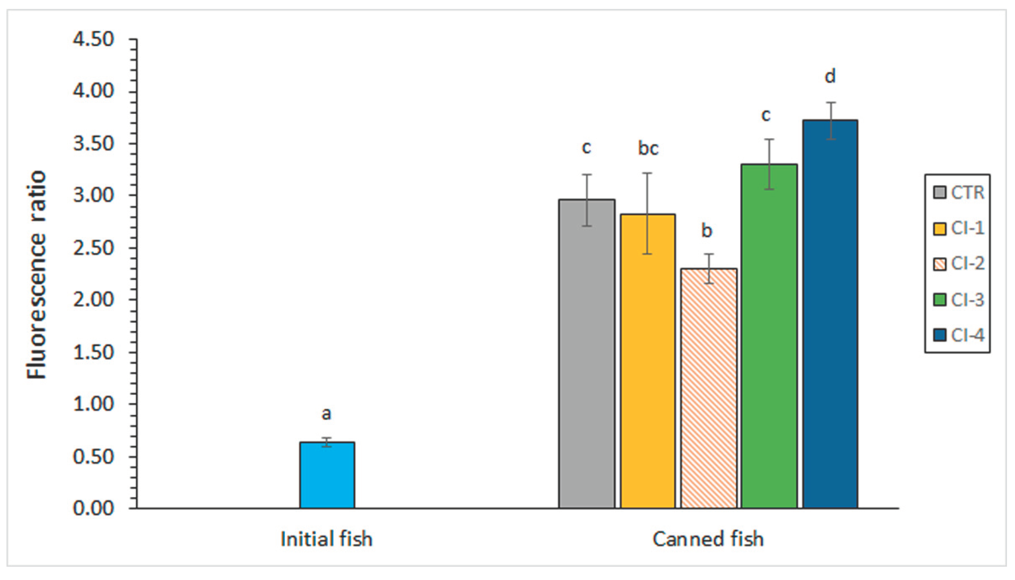
Figure 2.
Determination of the eicosapentaenoic acid (EPA) content (g·100 g-1 total fatty acids) in initial and canned fish including different concentrations of cuttlefish ink (CI) in the packing medium. Average values of four replicates (n = 4); standard deviations are indicated by bars. Different letters (a, b) denote significant differences (p < 0.05). Abbreviations of sample names as expressed in Table 1.
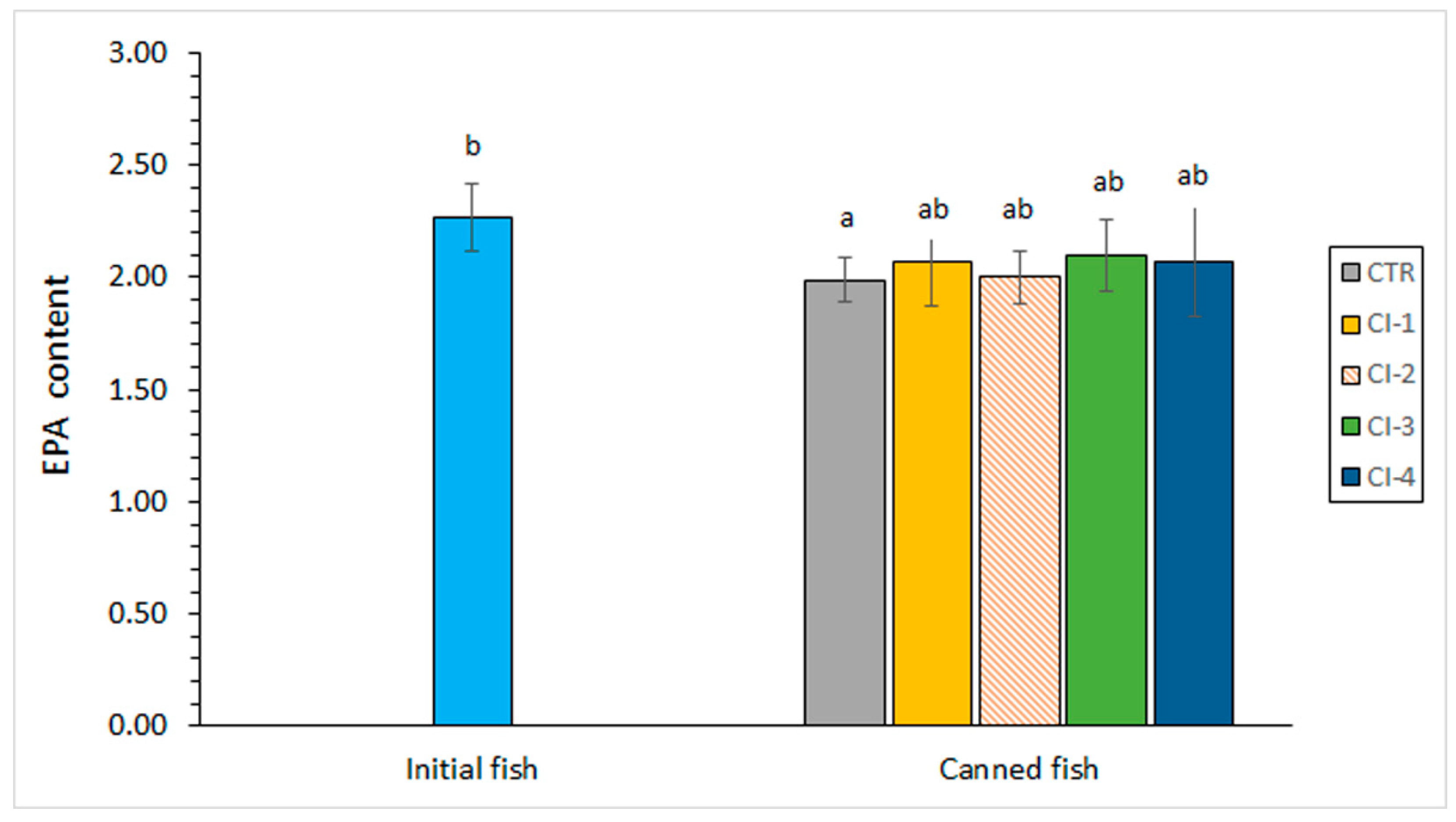
Figure 3.
Determination of the polyene index in initial and canned fish including different concentrations of cuttlefish ink (CI) in the packing medium. Average values of four replicates (n = 4); standard deviations are indicated by bars. Different letters (a, b) denote significant differences (p < 0.05). Abbreviations of sample names as expressed in Table 1.
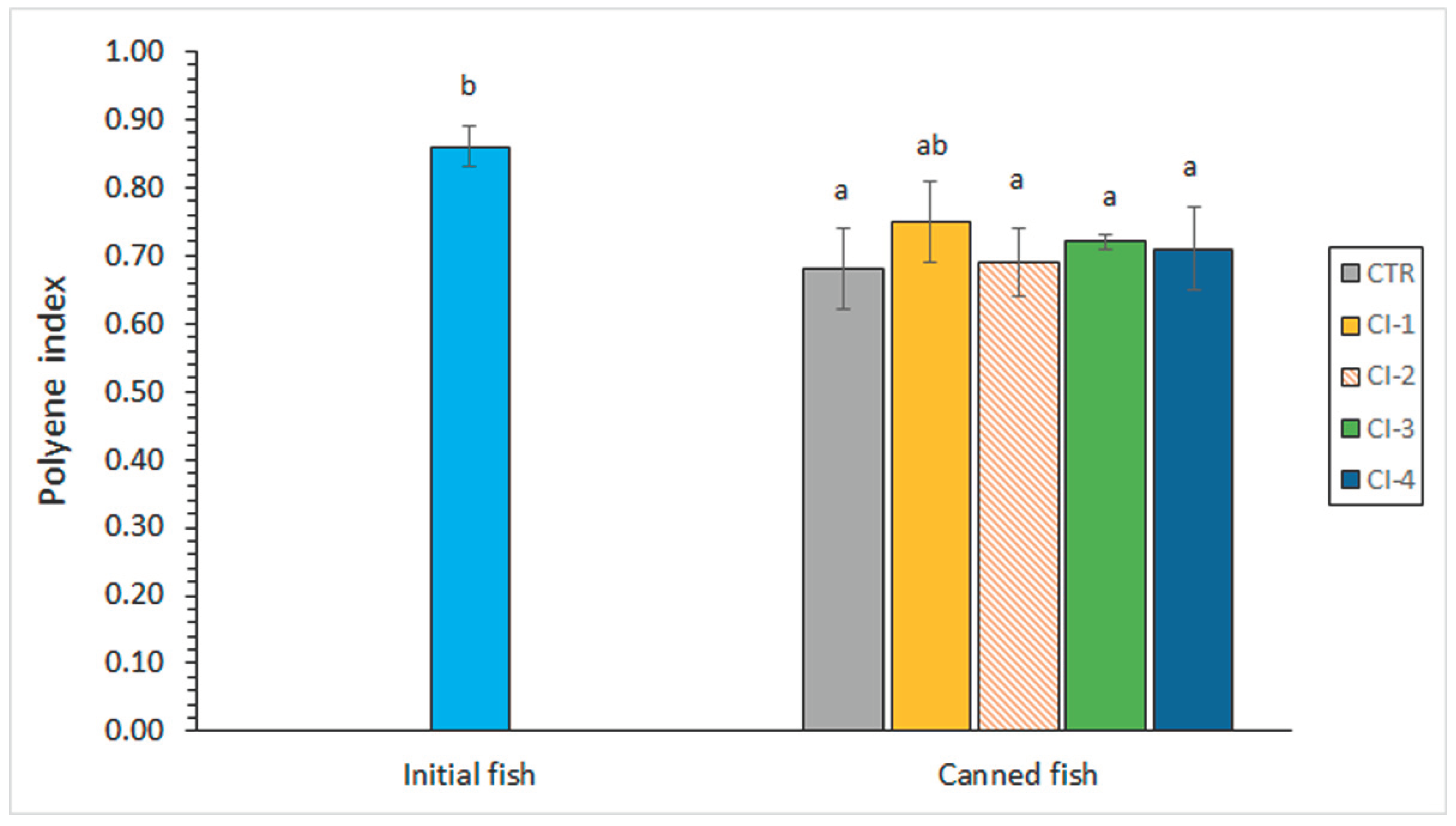

Table 1.
Determination* of primary and secondary lipid oxidation in initial and canned fish including different concentrations of cuttlefish ink (CI) in the packing medium**.
Table 1.
Determination* of primary and secondary lipid oxidation in initial and canned fish including different concentrations of cuttlefish ink (CI) in the packing medium**.
| Initial or canned sample | Lipid oxidation index | |
|---|---|---|
| Peroxide value (meq. active oxygen·kg-1 lipids) | Thiobarbituric acid index (mg malondialdehyde·kg-1 muscle) | |
| Initial fish | 1.65 ab (0.34) |
0.08 a (0.02) |
| CTR | 1.10 a (0.18) |
0.07 a (0.03) |
| CI-1 | 2.06 b (0.53) |
0.08 a (0.03) |
| CI-2 | 1.37 ab (0.63) |
0.09 a (0.03) |
| CI-3 | 1.53 ab (0.19) |
0.10 a (0.02) |
| CI-4 | 1.98 ab (0.85) |
0.13 a (0.03) |
* Average values of four replicates (n = 4); standard deviations are indicated in brackets. In each column, different letters (a, b) denote significant differences (p < 0.05). ** Abbreviations: CTR (canned control), CI-1, CI-2, CI-3, and CI-4 (canned samples including increasing concentrations of the CI extract in the packing medium, according to the Material and Methods section).
Table 2.
Determination* of free fatty acid (FFA), phospholipid (PL), and trimethylamine (TMA) values in initial and canned fish including different concentrations of cuttlefish ink (CI) in the packing medium**.
Table 2.
Determination* of free fatty acid (FFA), phospholipid (PL), and trimethylamine (TMA) values in initial and canned fish including different concentrations of cuttlefish ink (CI) in the packing medium**.
| Initial or canned sample | Chemical determination | ||
|---|---|---|---|
| FFA (g·kg-1 lipids) |
PL (g·kg-1 lipids) |
TMA (mg TMA-N·kg-1 muscle) |
|
| Initial fish | 0.49 a (0.05) |
39.69 b (3.84) |
2.71 a (0.33) |
| CTR | 1.58 b (0.31) |
29.30 a (3.74) |
5.25 c (0.94) |
| CI-1 | 1.90 b (0.26) |
35.01 ab (3.39) |
3.92 bc (0.41) |
| CI-2 | 1.87 b (0.19) |
27.01 a (4.51) |
3.90 b (0.30) |
| CI-3 | 1.96 b (0.60) |
28.75 a (3.46) |
4.51 bc (0.74) |
| CI-4 | 1.97 b (0.27) |
29.42 a (3.14) |
3.93 bc (0.42) |
* Average values of four replicates (n = 4); standard deviations are indicated in brackets. In each column, different letters (a, b, c) denote significant differences (p < 0.05). ** Abbreviations of sample names as expressed in Table 1.
Table 3.
Determination* of docosahexaenoic acid (DHA) and total ω3 fatty acid (FA) values (g·100 g-1 total FAs) and total ω3/total ω6 ratio in initial and canned fish including different concentrations of cuttlefish ink (CI) in the packing medium**.
Table 3.
Determination* of docosahexaenoic acid (DHA) and total ω3 fatty acid (FA) values (g·100 g-1 total FAs) and total ω3/total ω6 ratio in initial and canned fish including different concentrations of cuttlefish ink (CI) in the packing medium**.
| Initial or canned sample | FA parameter | ||
|---|---|---|---|
| DHA | Total ω3 | ω3/ω6 ratio | |
| Initial fish | 11.96 b (0.74) |
16.62 b (0.94) |
0.57 b (0.03) |
| CTR | 8.74 a (1.14) |
12.88 a (1.26) |
0.42 a (0.05) |
| CI-1 | 9.70 a (0.95) |
13.89 a (1.23) |
0.46 a (0.04) |
| CI-2 | 9.12 a (0.34) |
13.58 a (0.19) |
0.47 a (0.01) |
| CI-3 | 8.41 a (1.13) |
12.50 a (1.15) |
0.42 a (0.05) |
| CI-4 | 8.66 a (0.95) |
12.90 a (1.30) |
0.42 a (0.05) |
* Average values of four replicates (n = 4); standard deviations are indicated in brackets. In each column, different letters (a, b) denote significant differences (p < 0.05). ** Abbreviations of sample names as expressed in Table 1.
Table 4.
Determination* of colour changes in initial and canned fish including different concentrations of cuttlefish ink (CI) in the packing medium**.
Table 4.
Determination* of colour changes in initial and canned fish including different concentrations of cuttlefish ink (CI) in the packing medium**.
| Initial or canned sample | Colour parameter | ||
|---|---|---|---|
| L* | a* | b* | |
| Initial fish | 46.35 a (0.96) |
2.21 b (0.76) |
-3.76 a (0.24) |
| CTR | 81.40 c (2.03) |
-1.07 a (0.51) |
8.98 b (0.68) |
| CI-1 | 55.54 ab (8.39) |
2.18 b (0.67) |
6.81 b (1.79) |
| CI-2 | 63.48 b(6.09) | 1.38 b (0.83) |
7.31 b (1.06) |
| CI-3 | 62.46 b (4.58) |
1.53 b (0.36) |
8.66 b (1.95) |
| CI-4 | 60.39 b (5.50) |
1.91 b (0.36) |
7.39 b (0.99) |
* Average values of four replicates (n = 4); standard deviations are indicated in brackets. In each column, different letters (a, b, c) denote significant differences (p < 0.05). ** Abbreviations of sample names as expressed in Table 1.
Disclaimer/Publisher’s Note: The statements, opinions and data contained in all publications are solely those of the individual author(s) and contributor(s) and not of MDPI and/or the editor(s). MDPI and/or the editor(s) disclaim responsibility for any injury to people or property resulting from any ideas, methods, instructions or products referred to in the content. |
© 2024 by the authors. Licensee MDPI, Basel, Switzerland. This article is an open access article distributed under the terms and conditions of the Creative Commons Attribution (CC BY) license (http://creativecommons.org/licenses/by/4.0/).
Copyright: This open access article is published under a Creative Commons CC BY 4.0 license, which permit the free download, distribution, and reuse, provided that the author and preprint are cited in any reuse.


