Submitted:
28 February 2024
Posted:
28 February 2024
You are already at the latest version
Abstract
Study design: Technical note.
Objectives: To present a new technique of treatment for a patient with thoracolumbar focal kyphosis due to osteoporotic verte-bral fracture (OVFs).
Background : OVFs are common among the elderly population. Following a severe wedged fracture, patients often experience low back pain and disruptions in activities of daily living. Reconstruction surgeries, such as corpectomy, are among the treatment options for this condition. However, corpectomy requires longer surgical times and involves a significant amount of blood loss.
Materials and Methods : An 80-year-old woman presented to our hospital with severe low back pain and gait disturbance. She had experienced an L2 OVF six years prior. Although she had no neurological deficit, she could not walk more than 200m due to pain. Preoperative spinal radiographs revealed severe sagittal malalignment, with the L2 vertebra collapsed and focal kyphosis of L1-3 measuring 34 degrees.
Results: The patient underwent anterior and posterior surgery in the right decubitus position using a C-arm free technique. The surgical time was 133 minutes with an estimated blood loss of 100 ml. Hyperlordotic cages were inserted in the upper and lower disc space via a lateral approach, while percutaneous pedicle screws (PPS) were inserted from a posterior approach. These procedures were performed simultaneously under navigation guidance only. Post-operative images showed excellent spinal alignment, with local kyphosis reduced from 34 to 14 degrees, and no major or severe complications occurred. At one-year follow-up, the patient was able to walk smoothly, with her visual analogue scale for low back pain improving from 73mm to 13mm, and her Oswestry Disability Index improving from 53.3% to 13.3%.
Conclusions/Level of Evidence : The simultaneous use of hyperlordotic cages and PPS is an effective and innovative navigation technique that yields promising outcomes for thoracolumbar focal kyphosis resulting from OVFs. This approach not only reduces surgical time but also minimizes intraoperative blood loss. Level V.
Keywords:
Osteoporotic vertebral fractures
; thoracolumbar focal kyphosis
; C-arm free
; novel technique
; oblique lumbar interbody fusion
1. Introduction
Osteoporotic vertebral fractures (OVFs) are very common fractures in the elderly population [1]. After a severe wedged fracture, patients may experience low back pain, pulmonary dysfunction [2], and disturbances in activities of daily living [3]. Usually, conservative treatments for OVFs such as pain management [4], physiotherapy [5], and orthosis [6] are effective for most cases.
Severe OVFs occur when the fractured vertebral body height collapses to less than one-third of its original height [7]. These patients may experience intractable back pain, focal kyphosis deformities, progressive neurologic impairment, additional complicated morbidities, and even a heightened mortality risk [8]. According to the literature, conservative treatment for such patients is ineffective [9].
A wide variety of techniques and approaches have been described for the treatment of these fractures. The surgical goals include correction of deformity, restoration of collapsed vertebrae, maintenance of sagittal balance, and achieving ideal bone fusion with stable internal fixation. Among these, reconstruction surgeries such as corpectomy, pedicle subtraction osteotomy (PSO), and vertebral column resection (VCR) are considered for addressing focal kyphosis [10]. However, these procedures are often associated with significant challenges, including extensive surgical time, considerable blood loss, and the risk of long-term complications like proximal junctional kyphosis or structural failures [11].
Given the complexities and ongoing debates surrounding the optimal surgical intervention and the selection of fusion techniques and instrumentation [12], this discourse introduces a novel surgical technique. This new approach is tailored for patients experiencing thoracolumbar focal kyphosis as a result of severe OVF, a promising improvement over traditional methods.
2. Materials and Methods
This research was approved by the ethics committee of our institution (No. 472), and informed consent from the patient undergoing surgery was duly obtained.
2.1. Patient History
An 80-year-old woman presented to our hospital with severe low back pain and gait disturbance. She had sustained an L2 severe OVF six years prior to her visit. The patient had undergone long-term conservative treatment for pain management before coming to our hospital, which was ineffective for her condition.
2.2. Physical Examination
During the examination, she had no neurological deficit. However, due to severe back pain, she was unable to walk more than 200 meters. Her visual analog scale (VAS) for low back pain scored 73mm, and her Oswestry Disability Index (ODI) was 53.3%.
2.3. Preoperative Imaging
Preoperative spinal radiographs revealed severe sagittal malalignment. The L2 vertebra had collapsed, and the focal kyphosis between L1 and L3 was noted 34 degrees. Bone mineral density was 0.623 g/cm², and the T-score for the lumbar vertebrae was 3.5 SD (Figure 1).
2.4. Surgery
The procedure utilized a simultaneous anterior and posterior approach, performed in a single position using the right lateral decubitus position, without reliance on a C-arm, thereby minimizing radiation exposure. Continuous neuromonitoring was initiated at the beginning of the procedure to ensure the patient’s neurological safety throughout (Figure 3A).
Following sterilization and draping, a reference frame was carefully inserted into the left sacroiliac joint. Subsequently, a CT scan was obtained using the O-arm to provide detailed imaging guidance (Figure 3B). A meticulous 4 cm lateral skin incision was created, guided by a navigational probe. Through this incision, the external, internal, and transverse abdominal muscles were sequentially dissected, providing access to the surgical site.
After registering each surgical instrument, the L1/2 and L2/3 disc spaces were accurately exposed with the aid of a navigation probe. A self-retaining retractor with illumination was then placed to maintain clear visibility and access to the operative field. Discectomy at the L1/2 level was meticulously performed using a knife and navigated shavers (Figure 4A,B), followed by complete disc removal with a navigated Cobb elevator (Figure 4C,D).
Subsequently, the disc space was carefully expanded using a navigated spreader and trial (Figure 5). This precise preparation allowed for the insertion of a hyperlordotic cage (CLYDESDALE™ PTC SPINAL SYSTEM), angled at 12 degrees, under the guidance of navigation to ensure optimal placement (Figure 6). Following this, the same meticulous steps were repeated for the insertion of the second cage.
During the insertion of the cages, a second surgeon simultaneously inserted percutaneous pedicle screws from a posterior approach (Figure 7). A critical technique involved inserting the cranial screws before the cages to maintain the accuracy of the navigation system. Following the successful placement of six screws across L1, L2, and L3, rods were inserted. Compression forces were then applied through the screws to enhance lordosis, ensuring optimal spinal alignment and curvature correction (Figure 8).
The steps of this novel technique are illustrated in Figure 9 to provide a clearer understanding of the procedure.
2.5. Postoperative Imagings
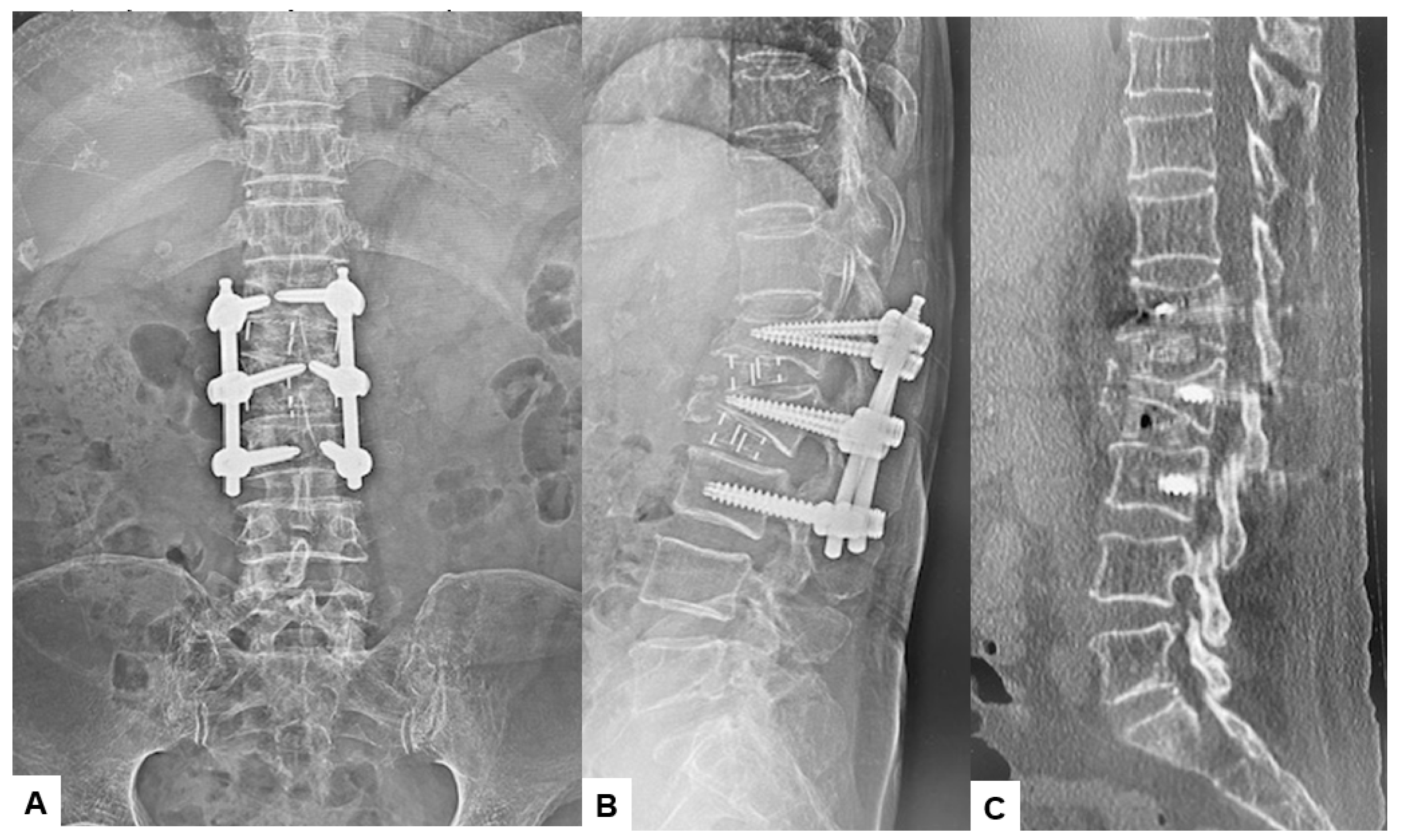
Postoperative radiographs and CT scans confirmed the successful realignment of the spine. The focal kyphosis angle between L1 and L3 was corrected from 34 degrees to 14 degrees. CT showed that, hyperlordotic cages and screws position were precisely inserted (Figure 10).
3. Results
The patient was treated successfully through surgery, with a total surgery time of 133 minutes and an estimated blood loss of 100 ml. After one-year follow-up, the patient is walking smoothly without any gait disturbance. The VAS for low back pain and ODI were improved from 73mm to 13mm, 53.3% to 13.3%, respectively. Furthermore, no major or severe complications were reported.
4. Discussion
Osteoporosis is recognized as a significant global health concern. One of the most prevalent complications associated with this condition is OVFs [13], which can lead to chronic pain, disability, spinal malalignment such as focal kyphosis, and consequently, functional decline and diminished quality of life for elderly patients. OVFs are predominantly located in the thoracolumbar region [14]. While usually OVFs result in mild symptoms and can be treated conservatively, approximately 30 to 40% of these fractures present with severe symptoms [15], necessitating surgical intervention. Indications for surgery include instability at the fracture site, posttraumatic kyphosis, intractable pain unresponsive to conservative treatments, neurological deficits, and significant spinal canal stenosis [16].
Thoracolumbar focal kyphosis is a common cause of sagittal spinal malalignment. The importance of sagittal plane balance in the spine cannot be overemphasized, as it is associated with good long-term functional outcomes [17]. Compression of the fractured vertebral body and exacerbation of the kyphotic deformity result in forward movement of the body’s center of gravity and an increase in the sagittal vertical axis [18]. When evaluating kyphotic deformities, it is important to distinguish between global and focal types. Mild global kyphosis deformities may be managed conservatively or sometimes may require isolated posterior column osteotomies [18]. In contrast, patients with fixed focal kyphotic deformities typically present with severe pain, early fatigue, forward leaning, and difficulty maintaining horizontal gaze [19], and may require more invasive methods depending on the desired correction.
In our patient, percutaneous balloon kyphoplasty (BKP) and percutaneous vertebroplasty (PVP) are not applicable due to the nature of the fracture. The fracture is old and consolidated, meaning that the bone has healed and solidified over time, making these procedures ineffective for addressing the condition [20]. Regarding osteotomy techniques for correcting kyphotic deformities, several methods have been reported in the literature [21], including Pontes osteotomy, Smith-Peterson osteotomy, pedicle subtraction osteotomy (PSO), and combined anterior-posterior correction [22]. While these techniques can achieve the desired correction, they also pose significant risks and serious complications, such as dural tears, nerve root injuries, and spinal cord injury, which can be disastrous [23]. Additionally, complications such as fixation failure, kyphosis recurrence, prolonged surgical time, and significant blood loss are associated with these techniques. It’s important to consider that the majority of these patients are elderly and have multiple comorbidities, so minimizing risks is paramount [24].
Combined anterior corpectomy and posterior fixation procedures have been observed to be associated with higher complication rates, regardless of whether the procedures are performed in one or two stages. These complications include severe blood loss, prolonged surgical time, nerve root and spinal cord injuries, and wound infections [25]. Additionally, in corpectomy procedures, non-union rates and cage subsidence are high [26]. Furthermore, cage placement can be challenging, especially when using wide footprint cages, which may result in cumbersome insertion processes [27,28]. One study demonstrated that the mean surgical time for minimally invasive corpectomy and posterior fixation was 275 minutes, which was significantly less than conventional open surgery [20]. However, in our study, the surgical time was less than half of that reported by that study, totaling only 133 minute.
It has been reported that correcting kyphotic deformities completely with a posterior-only approach, without anterior support, can be challenging [29]. Böhm et al. also noted that using combined surgery in patients with focal kyphosis could facilitate fusion development and alignment more effectively [30]. However, in our case, we chose not to pursue these techniques due to our patient’s advanced age and multiple comorbidities. Instead, we aimed to provide her with the best possible treatment while minimizing potential risks.
This is the first study to reveal details about focal kyphosis correction and improvement in sagittal balance following the minimally invasive procedure using double hyperlordotic cages with C-arm free technique. In our patient, surgery was performed in the lateral decubitus position to avoid the need for repositioning and redraping between anterior and posterior procedures. This approach helped to reduce operative time, risks of contamination, and the inconvenience of re-registration for navigation purposes. Hiyama et al. reported an additional average repositioning time of 34 minutes between lateral decubitus and prone positions [31]. Although percutaneous posterior pedicle screw fixation in the lateral position using C-arm fluoroscopy can be technically challenging, navigation technology makes it feasible and accurate [32]. Additionally, the lateral position is generally better tolerated by patients compared to prone surgery and avoids potential concerns associated with prone positioning, such as postoperative vision loss, cardiovascular complications, hypovolemia, reduced pulmonary compliance, and cardiac arrest [33].
Extended surgical times in elderly patients with multiple serious comorbidities can pose significant hazards. At times, procedures may need to be staged, leading to prolonged hospitalization and delayed mobilization, which carries its own set of risks [34]. In our technique, the surgical time was 133 minutes and blood loss was 100 ml, significantly less than the mean reported for other procedures. For example, Suk et al. compared anterior-posterior surgery versus closing wedge osteotomy for kyphotic osteoporotic vertebral fractures (OVFs) and reported mean blood loss of 2892 mL for the former and 1930 mL for the latter [35]. Postoperatively, our patient experienced notable improvements, with the VAS for low back pain decreasing from 73mm to 13mm and the ODI improving from 53.3% to 13.3%.
Placing the cage in the correct position typically necessitates the use of fluoroscopy. However, in our technique, the double hyperlordotic cages are navigated, allowing for three-dimensional visualization in all planes on the navigation monitor. This approach significantly reduces radiation exposure to the surgeon and operating room staff in terms of fluoroscopy time and exposure [36].
Our new technique does have several disadvantages. Further studies with a larger population and longer follow-up duration are needed to accurately evaluate the outcomes of this technique. Secondly, there is a risk of intraoperative CT-based surgical errors, particularly involving the misplacement of spinal implants due to inadvertent movement of the reference frame. Lastly, due to the simultaneous nature of the technique, it requires the involvement of two surgeons to perform the procedure.
5. Conclusion
The simultaneous double hyperlordotic cages with PPS is a useful technique, which reduce surgical time and intraoperative blood loss. Additionally, acceptable correction can be achieved with this minimally invasive technique. This novel navigation technique yields promising outcomes for patients with thoracolumbar focal kyphosis due to severe osteoporotic vertebral fracture.
Author Contributions
M.T.: conceptualisation, writing—original draft preparation; A.K.A.: writing—review and editing; S.J.E.: writing—review and editing; S.A: data collection; T.K.: data collection; T.T.: data collection.; K.U.: data collection, K.S.: data collection, All authors have read and agreed to the published version of the manuscript.
Funding
This study received funding from Japan Organization of Occupational Health and Safety.
Institutional Review Board Statement
This study was conducted in accordance with the guidelines of the Declaration of Helsinki and approved by the institutional review boards at Okayama Rosai Hospital (approval No. 472, Jan 10,, 2024).
Informed Consent Statement
Informed consent was obtained from all subjects involved in the study.
Data Availability Statement
The data presented in this study are available in the article.
Acknowledgments
This study was supported by Okayama Spine Group.
Conflicts of Interest
The authors declare no conflict of interest. The funders had no role in the design of the study; in the collection, analyses, or interpretation of data; in the writing of the manuscript; or in the decision to publish the results.
References
- Reginster JY, Burlet N. Osteoporosis: A still increasing prevalence. Bone 2006; 38:S4-S9. [CrossRef]
- Park JH, Lee SM, Shim SW, Baek SN, Choi YS. The Influence of Restrictive Pulmonary Dysfunction on Osteoporotic Thoracic Vertebral Fractures. Asian Spine J. 2021 Oct;15(5):659-663. [CrossRef]
- Jang HD, Kim EH, Lee JC, Choi SW, Kim HS, Cha JS, Shin BJ. Management of Osteoporotic Vertebral Fracture: Review Update 2022. Asian Spine J. 2022 Dec;16(6):934-946. PMC9827207. Ensrud KE, Schousboe JT. Clinical practice: vertebral fractures. N Engl J Med 2011;364:1634-42. [CrossRef]
- Rajasekaran S, Kanna RM, Schnake KJ, et al. Osteoporotic thoracolumbar fractures-how are they different?: classification and treatment algorithm. J Orthop Trauma 2017;31 Suppl 4:S49-56. [CrossRef]
- Sánchez-Pinto-Pinto B, Romero-Morales C, López-López D, de-Labra C, García-Pérez-de-Sevilla G. Efficacy of Bracing on Thoracic Kyphotic Angle and Functionality in Women with Osteoporosis: A Systematic Review. Medicina (Kaunas). 2022 May 24;58(6):693. [CrossRef]
- Kutsal FY, Ergin Ergani GO. Vertebral compression fractures: still an unpredictable aspect of osteoporosis. Turk J Med Sci. 2021; 51(2): 393–9. [CrossRef]
- Che H, Breuil V, Cortet B, et al. Vertebral fractures cascade: potential causes and risk factors. Osteoporosis Int. 2019; 30(3): 555–63. [CrossRef]
- Alpantaki K, Dohm M, Korovessis P, Hadjipavlou AG. Surgical options for osteoporotic vertebral compression fractures complicated with spinal deformity and neurologic deficit. Injury. 2018; 49(2): 261–71. [CrossRef]
- Cao Z, Wang G, Hui W, Liu B, Liu Z, Sun J. Percutaneous kyphoplasty for osteoporotic vertebral compression fractures improves spino-pelvic alignment and global sagittal balance maximally in the thoracolumbar region. PLoS One. 2020 Jan 30;15(1):e0228341. [CrossRef]
- Tanaka M, Singh M, Fujiwara Y, Uotani K, Oda Y, Arataki S, Yamauchi T, Takigawa T, Ito Y. Comparison of Navigated Expandable Vertebral Cage with Conventional Expandable Vertebral Cage for Minimally Invasive Lumbar/Thoracolumbar Corpectomy. Medicina (Kaunas). 2022 Mar 1;58(3):364. [CrossRef]
- Wen Z, Mo X, Zhao S, et al. Comparison of percutaneous kyphoplasty and pedicle screw fixation for treatment of thoracolumbar severe osteoporotic vertebral compression fracture with kyphosis. World Neurosurg. 2021; 152: e589–96. [CrossRef]
- Johnell O, Kanis JA. An estimate of the worldwide prevalence and disability associated with osteoporotic fractures. Osteoporos Int 2006;17:1726–33. [CrossRef]
- Goldstein CL, Chutkan NB, Choma TJ, Orr RD. Management of the elderly with vertebral compression fractures. Neurosurgery. 2015;77(4):S33–S45. [CrossRef]
- Park HY, Ahn JH, Ha KY, et al. Clinical and radiologic features of osteoporotic spine fracture with delayed neurologic compromises. World Neurosurg. 2018;120:e1295–e1300. [CrossRef]
- Spiegl U, Jarvers JS, Heyde CE, Josten C. Osteoporotic vertebral body fractures of the thoracolumbar spine: indications and techniques of a 3608-stabilization. Eur J Trauma Emerg Surg. 2017;43(1):27–33. [CrossRef]
- Katsumi K, Hirano T, Watanabe K, et al.. Surgical treatment for osteoporotic thoracolumbar vertebral collapse using vertebroplasty with posterior spinal fusion: a prospective multicenter study. Int Orthop. 2016;40(11):2309–2315. [CrossRef]
- Goldstein CL, Chutkan NB, Choma TJ, et al. Management of the elderly with vertebral compression fractures. Neurosurgery. 2015;77(Suppl 4):S33-45. [CrossRef]
- Diebo BG, Henry J, Lafage V, Berjano P. Sagittal deformities of the spine:Factors influencing the outcomes and complications. Eur Spine J. 2015;24:S3–15. [CrossRef]
- Terai, H., Takahashi, S., Yasuda, H., Konishi, S., Maeno, T., Kono, H., Matsumura, A., Namikawa, T., Kato, M., Hoshino, M., Tamai, K., Toyoda, H., Suzuki, A., & Nakamura, H. (2021). Differences in surgical outcome after anterior corpectomy and reconstruction with an expandable cage with rectangular footplates between thoracolumbar and lumbar osteoporotic vertebral fracture. *North American Spine Society Journal (NASSJ)*, *6*, 100071. [CrossRef]
- Diebo B, Liu S, Lafage V, et al. Osteotomies in the treatment of spinal deformities: indications, classification, and surgical planning. Eur J Orthop Surg Traumatol 2014;24Suppl 1:S11–20. [CrossRef]
- Been HD, Poolman RW, Ubags LH. Clinical outcome and radiographic results after surgical treatment of post-traumatic thoracolumbar kyphosis following simple type A fractures. Eur Spine J. 2004;13:101–107. [CrossRef]
- Kamerlink JR, Errico T, Xavier S, et al. Major intraoperative neurologic monitoring deficits in consecutive pediatric and adult spinal deformity patients at one institution. Spine. 2010;35:240–245. [CrossRef]
- Zeng Y, Chen Z, Sun C, et al. Posterior surgical correction of posttraumatic kyphosis of the thoracolumbar segment. J Spinal Disord Tech. 2013;26:37–41. [CrossRef]
- McAfee PC. Complications of anterior approaches to the thoracolumbar spine. Emphasis on Kaneda instrumentation. Clin Orthop Relat Res. 1994;306:110–119.
- K Uchida, S Kobayashi, H Nakajima, Y Kokubo, T Yayama, RSato, G Timbihurira, H Baba. Anterior expandable strut cage replacement for osteoporotic thoracolumbar vertebral collapse.J Neurosurg Spine, 4 (2006), 454-462. [CrossRef]
- JP Grant, TR Oxland, MF Dvorak.Mapping the structural properties of the lumbosacral vertebral endplates. Spine (Phila Pa 1976), 26, (2001), 889-896. [CrossRef]
- M Pekmezci, E McDonald, A Kennedy, R Dedini, T McClellan, CAmes, V Deviren. Can a novel rectangular footplate provide higher resistance to subsidence than circular footplates. An ex vivo biomechanical study. Spine (Phila Pa 1976), 37 (2012), E1177-E1181,. [CrossRef]
- Chokshi JJ, Shah M. Outcomes of including fracture level in short-segment fixation for thoracolumbar fracture-dislocation. Asian Spine J. 2019;13:56–60. [CrossRef]
- Dobran M, Nasi D, Brunozzi D, et al. Treatment of unstable thoracolumbar junction fractures: short-segment pedicle fixation with inclusion of the fracture level versus long-segment instrumentation. Acta Neurochir (Wien) 2016;158:1883–9. [CrossRef]
- Hiyama A., Sakai D., Sato M., Watanabe M. The analysis of percutaneous pedicle screw technique with guide wire-less in lateral decubitus position following extreme lateral interbody fusion. J. Orthop. Surg. Res. 2019;14:304. [CrossRef]
- Yamauchi T, Jaiswal A, Tanaka M, Fujiwara Y, Oda Y, Arataki S, Misawa H. Minimally Invasive L5 Corpectomy with Navigated Expandable Vertebral Cage: A Technical Note. Brain Sci. 2021 Sep 19;11(9):1241. [CrossRef]
- Kwee M.M., Ho Y.H., Rozen W.M. The prone position during surgery and its complications: A systematic review and evidence-based guidelines. Int. Surg. 2015;100:292–303. [CrossRef]
- Robertson P.A., Rawlinson H.J., Hadlow A.T. Radiologic stability of titanium mesh cages for anterior spinal reconstruction following thoracolumbar corpectomy. J. Spinal Disord. Tech. 2004;17:44–52. [CrossRef]
- Suk, S.I.; Kim, J.H.; Lee, S.M.; Chung, E.R.; Lee, J.H. Anterior-posterior surgery versus posterior closing wedge osteotomy in posttraumatic kyphosis with neurologic compromised osteoporotic fracture. Spine 2003, 28, 2170–2175. [CrossRef]
- Tanaka M, Fujiwara Y, Uotani K, Ayhan S, Yamauchi T, Sonawane S, Nakanishi K. Minimally invasive thoracolumbar corpectomy with navigated expandable vertebral cage: A technical note, Interdiscip Neurosurg, (2021), 24. [CrossRef]
Figure 1.
Preoperative spinal radiograms, A: Postero-anterior radiogram, B: Lateral spinal radiogram, C: Antero-posterior lumbar radiogram, D: Lateral lumbar radiogram.
Figure 1.
Preoperative spinal radiograms, A: Postero-anterior radiogram, B: Lateral spinal radiogram, C: Antero-posterior lumbar radiogram, D: Lateral lumbar radiogram.
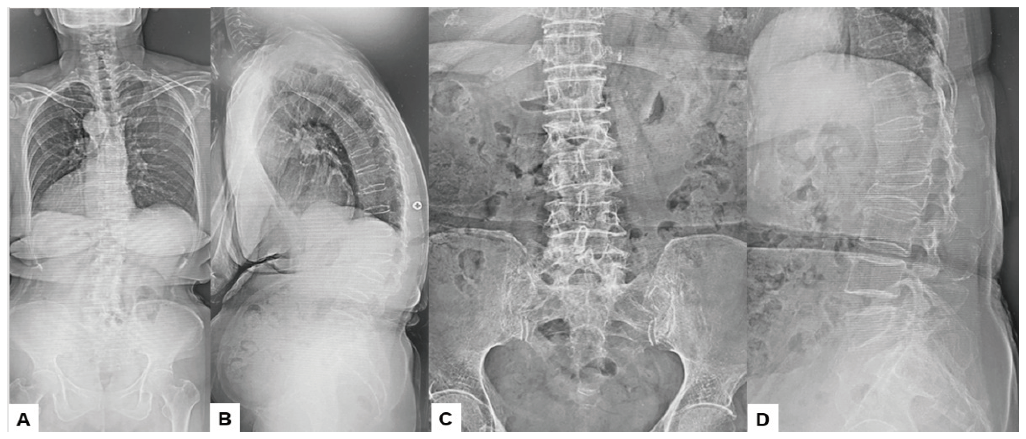
Figure 2.
Preoperative CT and MR imaging, A: Mid-sagittal reconstruction CT, B: 3D reconstruction CT, C: T2 weighted mid-sagittal MR imaging.
Figure 2.
Preoperative CT and MR imaging, A: Mid-sagittal reconstruction CT, B: 3D reconstruction CT, C: T2 weighted mid-sagittal MR imaging.
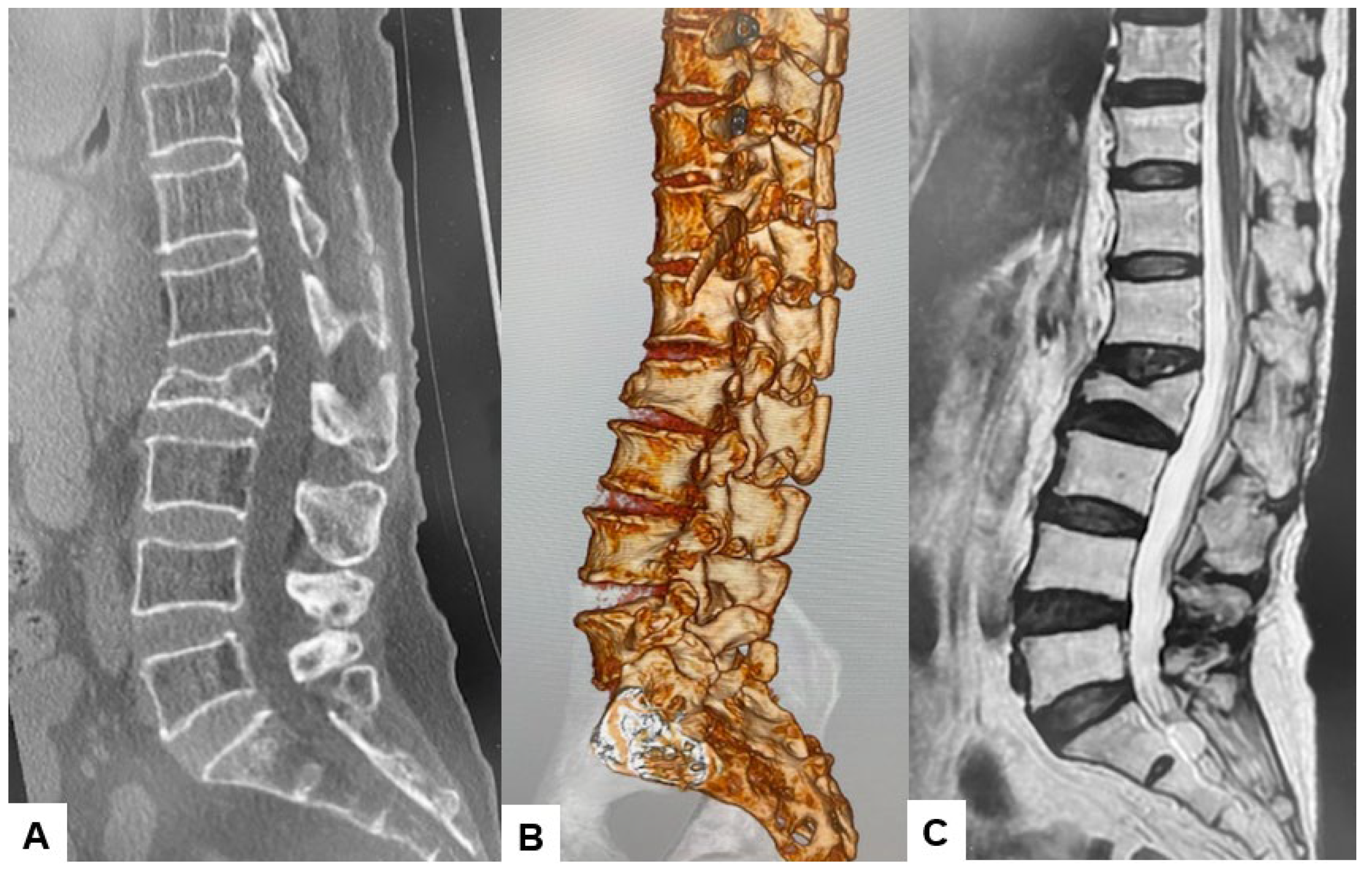
Figure 3.
O-arm and neuromonitouring, A: O-arm, B: Neuromonitouring.
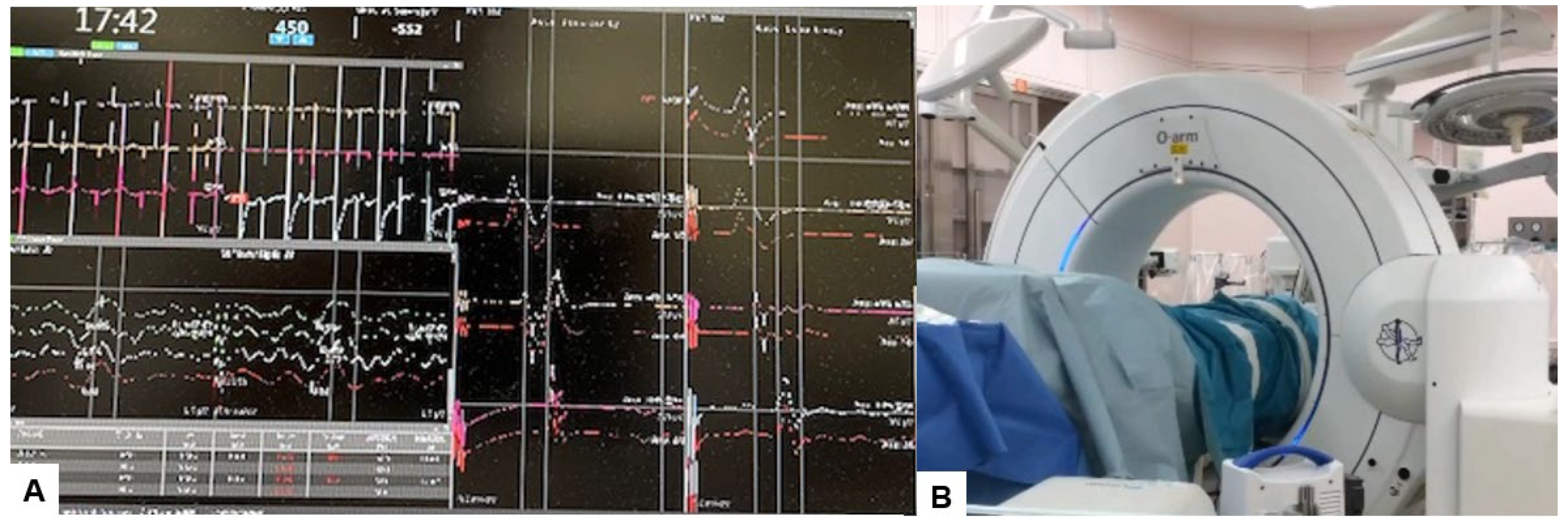
Figure 4.
The navigated shaver and Cobb elevetor. A: Coronal view of the navigated shaver, B: Axial view of the navigated shaver, C: Coronal view of the navigated Cobb elevetor, D: Axial view of the navigated Cobb elevetor.
Figure 4.
The navigated shaver and Cobb elevetor. A: Coronal view of the navigated shaver, B: Axial view of the navigated shaver, C: Coronal view of the navigated Cobb elevetor, D: Axial view of the navigated Cobb elevetor.
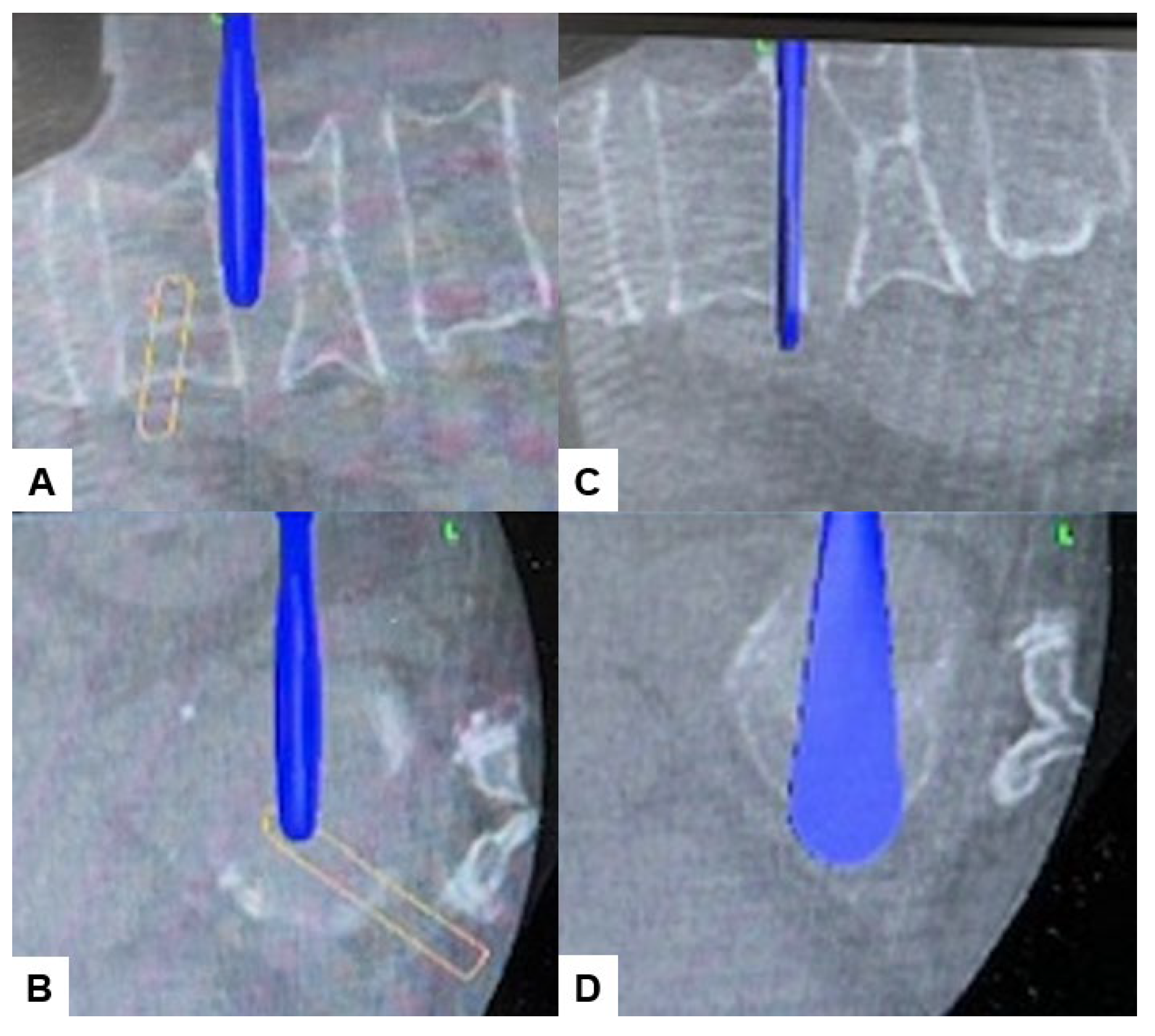
Figure 5.
The navigated spredor, A: Coronal view, B: Axial view, C: sagittal view, D: Intraoperative image.
Figure 5.
The navigated spredor, A: Coronal view, B: Axial view, C: sagittal view, D: Intraoperative image.
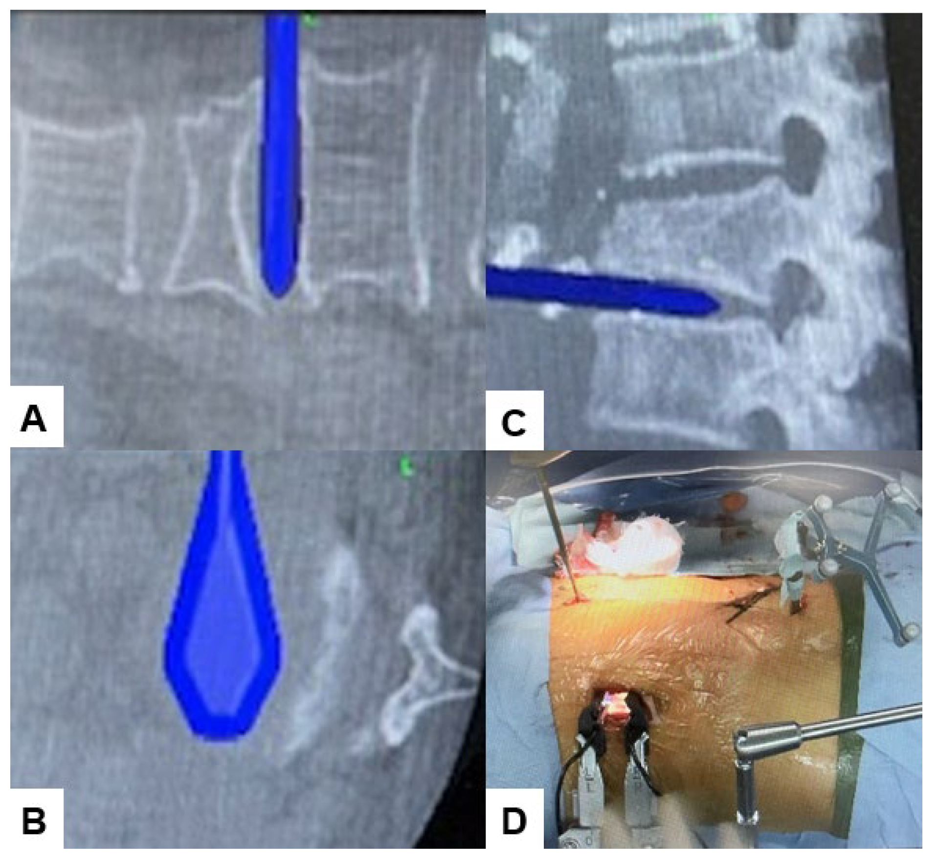
Figure 6.
The navigated cage, A: Coronal view, B: Axial view, C: sagittal view, D: 3D view.
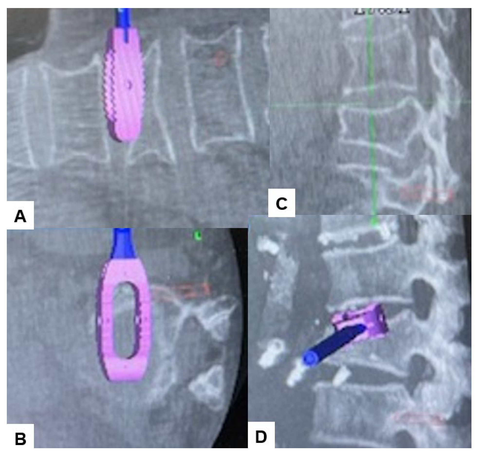
Figure 7.
Percutaneous pedicle screw insertion, A: Sagittal view, B: Axial view, C: 3D view, D: Coronal view.
Figure 7.
Percutaneous pedicle screw insertion, A: Sagittal view, B: Axial view, C: 3D view, D: Coronal view.
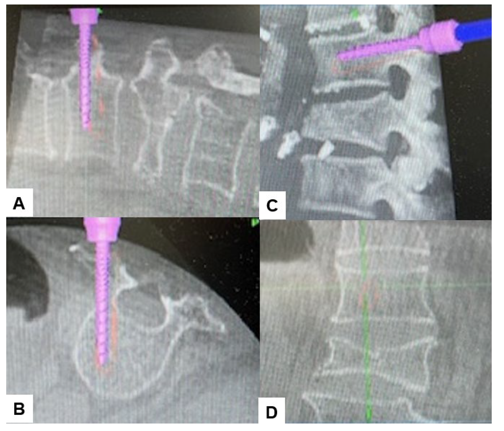
Figure 8.
Intraoperative images.
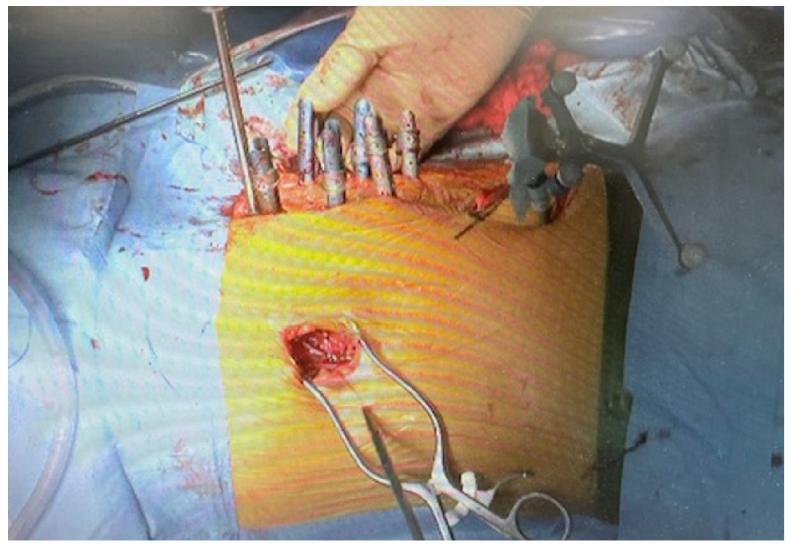
Figure 9.
The steps of dual cage technique, A, Insertion of cranial PPS, B, Simultaneous insertion of a cranial hyperlordotic cage and PPS, C, Insertion of a caudal hyperlordotic cage and rods and compression.
Figure 9.
The steps of dual cage technique, A, Insertion of cranial PPS, B, Simultaneous insertion of a cranial hyperlordotic cage and PPS, C, Insertion of a caudal hyperlordotic cage and rods and compression.
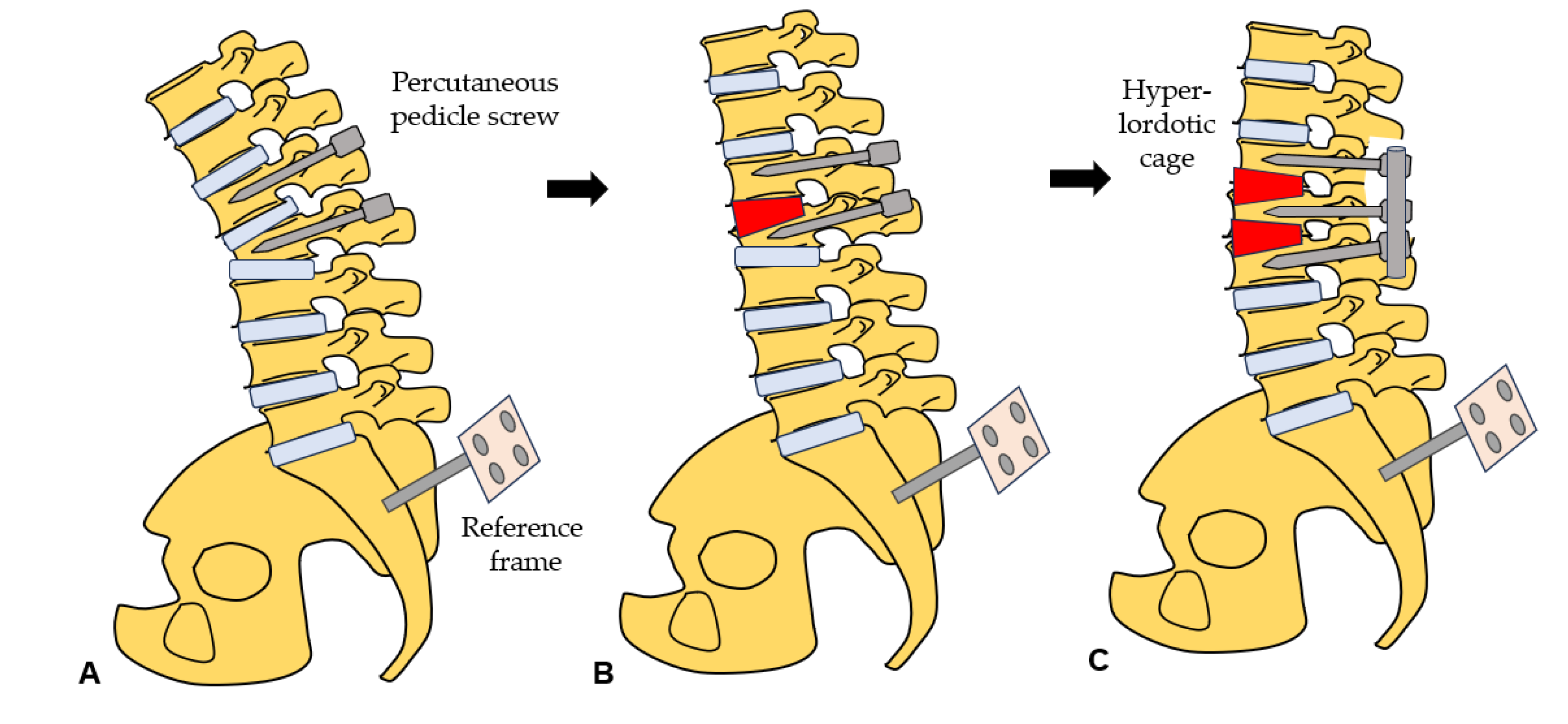
Figure 10.
Postoperative radiograms and CT, A: Antero-posterior lumbar radiogram, B: Lateral lumbar ragiogram, C: Mid-sagittal reconstraction CT.
Figure 10.
Postoperative radiograms and CT, A: Antero-posterior lumbar radiogram, B: Lateral lumbar ragiogram, C: Mid-sagittal reconstraction CT.
Disclaimer/Publisher’s Note: The statements, opinions and data contained in all publications are solely those of the individual author(s) and contributor(s) and not of MDPI and/or the editor(s). MDPI and/or the editor(s) disclaim responsibility for any injury to people or property resulting from any ideas, methods, instructions or products referred to in the content. |
© 2024 by the authors. Licensee MDPI, Basel, Switzerland. This article is an open access article distributed under the terms and conditions of the Creative Commons Attribution (CC BY) license (http://creativecommons.org/licenses/by/4.0/).
Copyright: This open access article is published under a Creative Commons CC BY 4.0 license, which permit the free download, distribution, and reuse, provided that the author and preprint are cited in any reuse.


