
Submitted:
26 February 2024
Posted:
26 February 2024
You are already at the latest version
Abstract
An observational cohort study of patients diagnosed with endometrial cancer (EC) stage IA G1, or atypical endometrial hyperplasia (AHE), undergoing organ-preserving treatment, was conducted. Objective of the study: to determine CDO1, PITX2, and CDH13 gene methylation levels in early endometrial cancer and atypical hyperplasia specimens obtained before organ-preserving treatment in the patients with adequate response and with insufficient response to hormonal treatment. Materials and methods: 41 endometrial specimens obtained during diagnostic uterine curettage in women with EC (n=28) and AEH (n=13), willing to preserve reproductive function, were studied; 18 specimens of endometrial cancer IA stage G1 from peri- and early postmenopausal women (comparison group) were included in the study. The control group included 18 endometrial specimens from healthy women obtained by diagnostic curettage for missed abortion and/or intrauterine adhesions. Methylation levels were analyzed using the modified MS-HRM method. Results: All 13 women with AEH had complete response (CR) to medical treatment. In the group undergoing organ-preserving treatment for uterine cancer IA stage G1 (n=28), 14 patients had complete response (EC CR group) and 14 had not (non-CR). It was found that all groups had statistically significant differences in CDO1 gene methylation levels with the control group (p<0.001) except for the EC CR group (p=0.21). The p-value of difference between EC CR and EC non-CR groups was <0.001. The differences in PITX2 gene methylation levels between the control and study groups were also significantly different (p<0.001), except for AEH group (p=0.21). For the difference between EC CR and EC non-CR groups, the p-value was 0.43. For CDH13 gene methylation levels, statistically significant differences were found between the control and EC non-CR groups (p<0.001), and the control and EC comparison groups (p=0.005). When comparing the EC CR group with EC non-CR group, the p-value for this gene was <0.001. Simultaneous assessment of CDO1 and CDH13 genes methylation allowed to accurately distinguish between EC CR and EC non-CR groups (AUC=0.96). Conclusion: Assessment of CDO1 and CDH13 gene methylation in endometrial specimens from patients with endometrial cancer (IA stage G1), planned for medical treatment, can predict the treatment outcome.
Keywords:
methylation
; atypical endometrial hyperplasia
; endometrial cancer
; organ-preserving treatment
; MS-HRM
; CDO1
; CDH13
1. Introduction
Endometrial cancer in young women of reproductive age is rare: only 4% of patients are under 40 years of age [1]. The standard treatment of endometrial cancer is hysterectomy and bilateral salpingo-oophorectomy with or without lymphadenectomy. However, for patients who wish to preserve their reproductive function, conservative treatment of EC, based on progestins, is possible. FIGO guidelines suggest that conservative treatment can be considered in patients with a histologically confirmed well differentiated endometrioid adenocarcinoma G1, or atypical hyperplasia (AEH) [2]. Patients undergoing conservative treatment may experience unfavorable outcomes such as absence of treatment effect, disease recurrence, and disease progression [3]. Expression of progesterone receptors has been considered as one of the predictors of complete response, but in a study by Yamazawa et al. only 50% effectiveness with a specificity of 100% was observed for this treatment option [4]. Analysis of the expression of mismatch repair (MMR) proteins showed even lower effectiveness (22%) [5]. Today, there are no reliable prognostic markers for use in clinical practice [6]. Methylation changes of some genes are associated with poor prognosis of cancer treatment. The study by Hirano et al. on genome-wide DNA methylation profiles, showed that more aggressive endometrial tumors in young women had a specific methylation profile [7]. Thus, it can be assumed that the outcomes of treatment with progestins may be associated with the methylation of certain genes. In this study, we analyzed the methylation levels of three tumor suppressor genes, CDO1, PITX2, and CDH13, in endometrial specimens obtained before organ-preserving treatment. Cysteine dioxygenase (CDO) is an enzyme that is involved in the biodegradation of toxic cysteine. CDO1 gene methylation has been found in several types of cancers. [8]. An increased CDO1 gene methylation is associated with poor treatment outcomes. The CDH13 gene is a tumor suppressor gene belonging to the cadherin gene superfamily. Its expression is low or absent in various tumors. The level of the gene’s methylation is associated with the depth of muscle infiltration in endometrial cancer [9]. PITX2 is a transcription factor that plays an important role in embryo development. PITX2 methylation has shown a strong correlation with breast cancer recurrence [10].
The aim of this study was to identify differences between groups with complete response and non-complete response to hormonal treatment based on the CDO1, PITX2 and CDH13 gene methylation levels in endometrial specimens obtained before treatment.
2. Results
All 13 women with atypical endometrial hyperplasia had a complete response to the treatment, and the disease did not progress (AEH group). Among the women with uterine cancer, 14 patients had complete response (EC CR group) and 14 patients had non-complete response (EC non-CR group): partial response (n=3); stable disease (n=2); the disease recurrence within one year after successful 6-month course of treatment (n=5); or the disease progression (n=4). The age and BMI of the patients are presented in Table 2. Patients either with stable disease or with the disease progression underwent hysterectomy. The rest underwent repeated courses of therapy. Four of them had atrophy of all endometrial glands after two additional treatment courses, and subsequently started the IVF treatment. The outcomes are presented in Table 3.
The methylation of the CDO1, PITX2, and CDH13 genes was carried out in the AEH and EC groups, in the control group (healthy women), and the comparison group (women with uterine cancer stage IA in premenopause and early postmenopause).
Figure 1.
Methylation level of CDO1 gene in the studied groups. **- p<0.001.
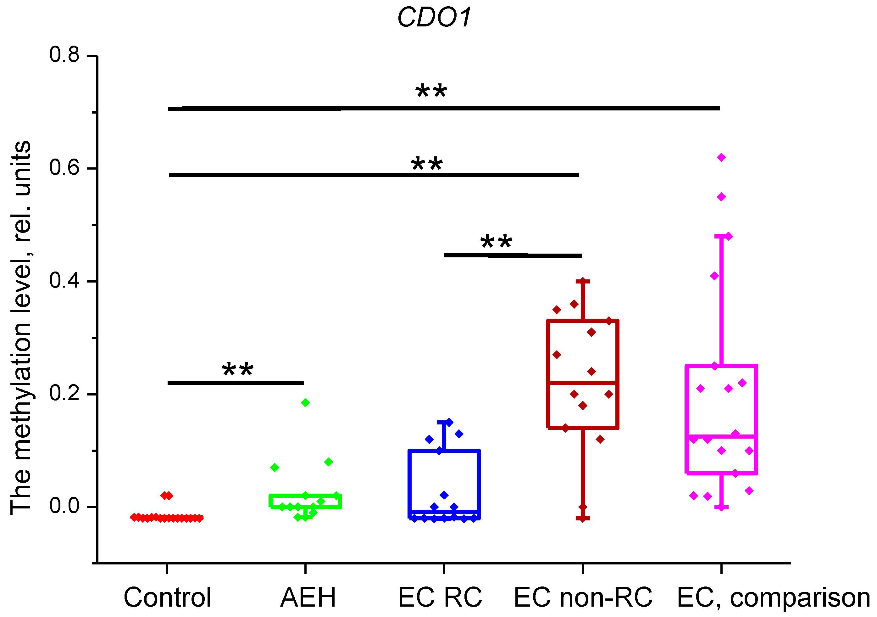
It was found that all groups, CDO1 methylation levels were significantly different from the control group (p<0.001), except for the EC CR group (p=0.21). The p-value of difference between EC CR and EC non-CR groups was <0.001.
Figure 2.
Methylation level of PITX2 gene in the studied groups. **- p<0.001.
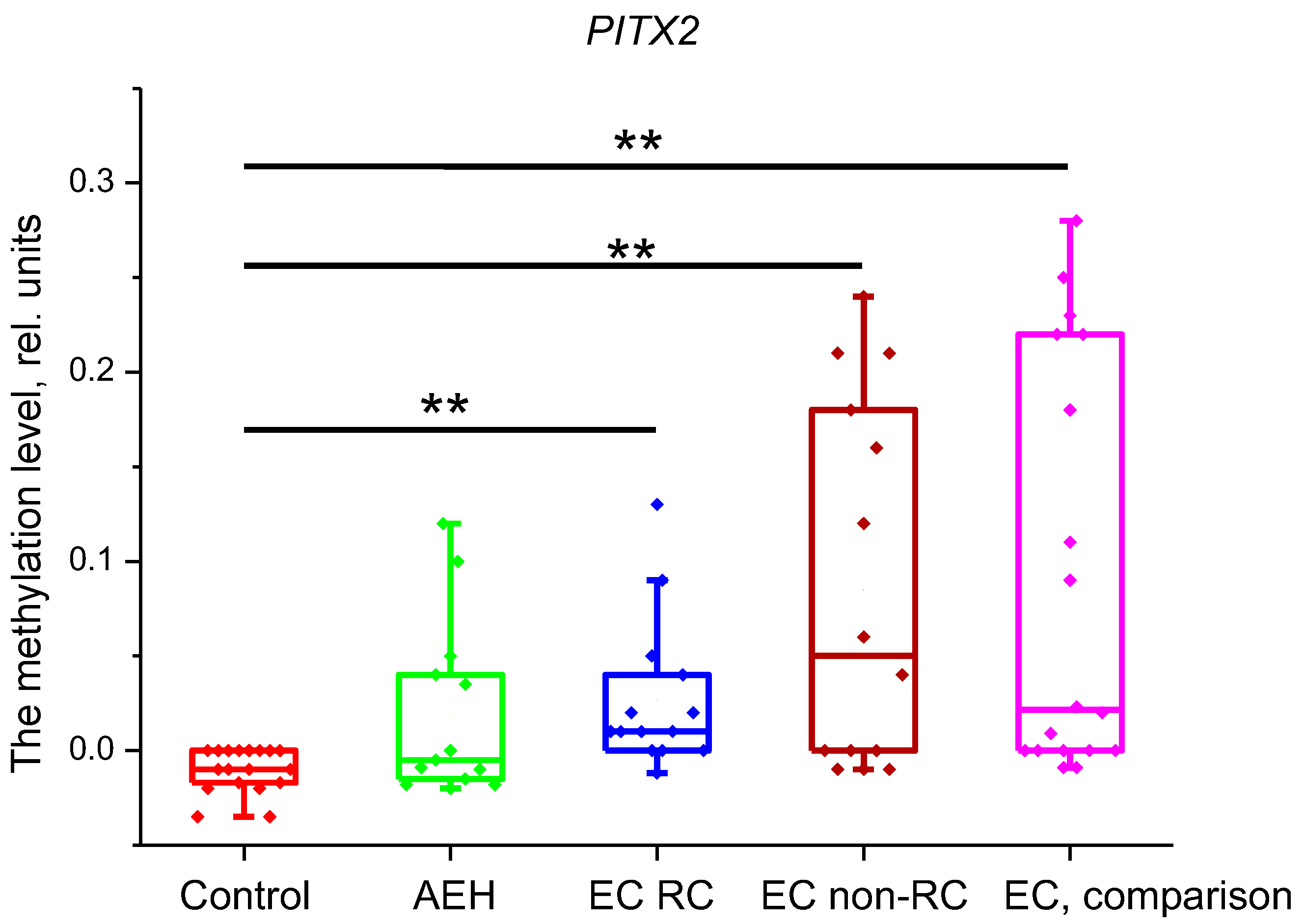
Assessment of PITX2 methylation level showed that all groups had statistically significant differences with the control group (p<0.001) except for AEH group (p=0.21). For the difference between EC CR and EC non-CR groups, the p-value was 0.43.
Figure 3.
Methylation level of CDH13 gene in the studied groups. **- p<0.05, **- p<0.001.
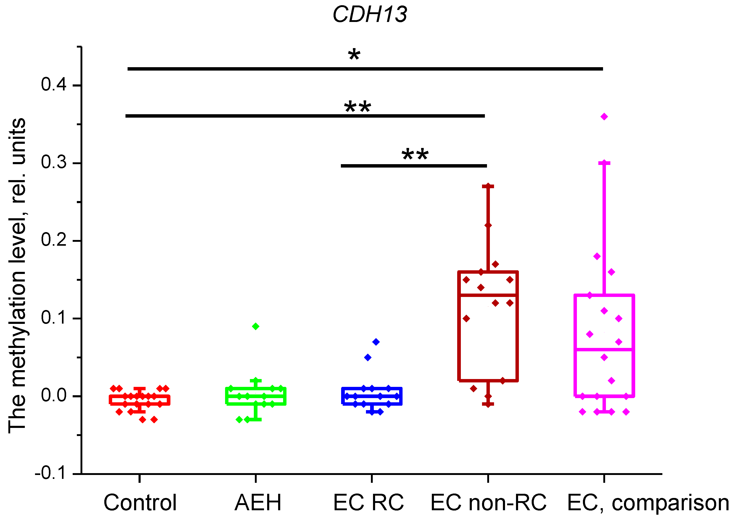
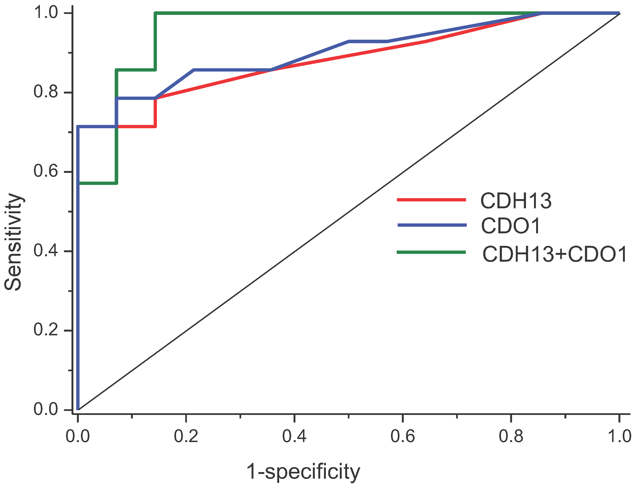
CDH13 gene methylation levels were significantly different between the control and EC non-CR groups (p<0.001), and the control and EC comparison groups (p=0.005). When comparing the EC CR group with EC non-CR group, the p-value for this gene was <0.001. ROC analysis showed high diagnostic value for CDH13 gene, with AUC=0.88(0.75-1), and for CDO1 gene, with AUC=0.9(0.89-1). Logistic regression for both genes showed AUC=0.96(0.89-1). ROC curves are presented in Figure 3.
Figure 3.
ROC curves for prognostic markers of EC treatment outcomes.
3. Discussion
Treatment of early EC, attempting to preserve fertility has become possible, since this disease is most likely to have a favorable prognosis. However, some patients experience recurrence or progression of the disease. In the latter case, urgent radical surgery is necessary due to the increased risk to the patient's life. Several prognostic molecular markers for treatment outcomes have been proposed. The most studied are progesterone receptors; however, the related findings are contradictory. Ki67, Nrf2, SPAG9, and MMR proteins were also considered as prognostic markers, but they did not show high diagnostic value [6]. In this study, we determined CDO1, PITX2, and CDH13 genes methylation levels in EC and AEH specimens of patients before conservative therapy, as well as in the endometrium of healthy women and in the EC specimens obtained after hysterectomy (a comparison group). This group served as an additional control for the obtained results.
All genes were found to have increased methylation levels in EC. The analysis of the treatment outcomes for endometrial cancer IA stage G1 showed that the methylation level of CDO1 and CDH13 genes may help to predict a positive treatment outcome (complete response) with high accuracy (AUC=0.96). We could not assess statistical differences in methylation levels between various subgroups with non-complete response, because we could not find a sufficient number of participants within the four years study period; this is a certain limitation of our study. In the comparison group the values of methylation levels of all studied genes were intermediate between the corresponding levels in EC CR and non-CR groups; this additionally confirms the validity of the obtained results.
In AEH, only CDO1 gene showed increased methylation level. We were not able to assess the methylation levels in patients with AEH non-responsive to treatment, since in our study all patients had a complete response; probably, the methylation level would be higher in patients with subsequent negative treatment outcomes.
4. Materials and Methods
An observational cohort study of patients diagnosed with atypical endometrial hyperplasia (AEH) and endometrial cancer (EC) IA stage G1, subjected to conservative treatment, was conducted. Patients were followed from 2019 to 2023. The study included 41 patients: patients with AEH (n=13) and patients with EC (n=28). Inclusion criteria were: no signs of invasion into the muscular layer according to contrast-enhanced magnetic resonance imaging (MRI) or ultrasound examination, no signs of metastatic lesions in lymph nodes or ovaries. The patients were consulted by a reproductologist before therapy. In addition, each patient was informed that the recommended type of treatment was not standard for endometrial cancer and gave their consent. The patients diagnosed with AEH were treated with levonorgestrel-releasing intrauterine device (LNG-IUD); the patients diagnosed with endometrial cancer IA stage G1 were treated with LNG-IUD and goserelin 3.6 mg per month. The initial treatment period was 6 months. According to the treatment outcomes, patients were divided into two groups: a group with complete response (CR) and a group with non-complete response (non-CR). The latter group was subdivided into four subgroups: a group with partial response to treatment, a group with stable disease, a group with the disease recurrence within a year after successful 6-month course of treatment, and a group with the disease progression under treatment. The study also included the patients in peri- and early postmenopause with confirmed uterine cancer IA stage G1, who underwent radical treatment by hysterectomy with adnexa (comparison group, n=18). The control group included endometrial samples from healthy women after diagnostic curettage for missed abortion and/or intrauterine adhesions (n=18).
The response of AEH and EC to hormonal treatment was assessed pathomorphologically. The presence of hormonal atrophy of all endometrial glands and a decidual reaction of the stroma, and/or the presence of typical glandular hyperplasia complexes was considered as complete response in both conditions. For endometrial cancer, partial response was stated by the presence of atypical endometrial complexes. EC was considered stable in case of residual carcinoma complexes of the same degree. A decrease in the degree of cancer differentiation was considered as the sign of EC progression.
The methylation study was carried out in the cytology laboratory of the Federal State Budgetary Institution “The Research Center for Obstetrics, Gynecology and Perinatology named after Academician V.I. Kulakov” of the Ministry of Health of the Russian Federation. The methylation level was assessed using a modified MS-HRM method described in the article by Krasnyi A.M. et al. [11]. The primers for analysis are presented in Table 1.

Table 1.
Primers for MS–HRM.
| Gene | Forward Primer | Reverse Primer |
| CDO1 | GGGAGGATGAATTTTATAGATTTG | TAAACTTCCATAATAACCTACACCT |
| PITX2 | GTAGGAAGGAAATTAGAATTAAAT | AAAACTTACTACTAACTACCTCTTTTC |
| CDH13 | GGGGTTTTTTTGTTTTTAGATT | CTTATCCACCCACTTACAAACTAC |
Amplification of fragments of CpG islands of the studied genes was performed according to the following protocol: 95 °С — 5 min; (95 °С — 15 sс, 60 °С — 30 s, 72 °С — 45 s) ×30; (95 °С — 15 s, 50 °С — 30 s, 72 °С — 45 s) ×25.
Statistical analysis was carried out in IBM SPSS Statistics 20. The significance of differences between the studied groups was assessed using the Mann–Whitney U test. The data are presented as median values, 1 and 3 quartiles (M (Q1;Q3)). We applied logistic regression and receiver operating characteristic (ROC) curve analysis, separately, to assess diagnostic value of the studied parameters. The difference was significant at p < 0.05. The figures were plotted using the OriginPro 8.5.
5. Conclusions
Thus, we have shown that assessment of the CDO1 and CDH13 genes methylation levels in endometrial specimens from patients with endometrial cancer (IA stage G1) can predict the treatment outcome. The DNA methylation changes in AEH in the absence of a complete response to treatment could not be assessed and require additional study. Also, in case of repeated treatment courses, it is important to evaluate methylation changes in dynamics; this may allow to decide on their feasibility for the disease follow-up. In addition, our study indicates the potential value of genome-wide methylation assessment for selecting an optimal panel of prognostic markers for treatment outcomes.
Author Contributions
Conceptualization, methodology, and formal analysis, Aleksey M. Krasnyi; investigation, Lyubov T. Gadzhieva, Diana N. Kokoeva, Mark G. Kosenko; writing—original draft preparation, Aleksey M. Krasnyi; writing—review and editing, Ekaterina L. Yarotskaya, Stanislav V. Pavlovich, Levon A. Ashrafyan; supervision, Gennadiy T. Sukhikh.
Institutional Review Board Statement
The study was conducted in accordance with the Declaration of Helsinki, and approved by the Ethics Committee of National Medical Research Center for Obstetrics, Gynecology and Perinatology of Ministry of Healthcare of Russian Federation (Protocol №11, 19.12.2019).
Informed Consent Statement
Informed consent was obtained from all subjects involved in the study.
Data Availability Statement
The data that support the findings of this study are available from the corresponding author, upon request.
Conflicts of Interest
The authors have no conflict of interest to declare that are relevant to the content of this article.
References
- Lee, N.K.; Cheung, M.K.; Shin, J.Y.; Husain, A.; Teng, N.N.; Berek, J.S.M.; Kapp, D.S.; Osann, K.; Chan, J.K. Prognostic Factors for Uterine Cancer in Reproductive-Aged Women. Obstet. Gynecol. 2007, 109, 655–662. [Google Scholar] [CrossRef] [PubMed]
- Colombo, N.; Creutzberg, C.; Amant, F.; Bosse, T.; González-Martín, A.; Ledermann, J.; Marth, C.; Nout, R.; Querleu, D.; Mirza, M.R.; et al. ESMO-ESGO-ESTRO Consensus Conference on Endometrial Cancer: Diagnosis, treatment and follow-up. Ann. Oncol. Erratum in Ann Oncol. 2015; Erratum in Ann Oncol. 2017, 28, 167–168. 2016, 27, 16–41. [Google Scholar] [CrossRef] [PubMed]
- Gallos, I.D.; Yap, J.; Rajkhowa, M.; Luesley, D.M.; Coomarasamy, A.; Gupta, J.K. Regression, relapse, and live birth rates with fertility-sparing therapy for endometrial cancer and atypical complex endometrial hyperplasia: a systematic review and metaanalysis. Am. J. Obstet. Gynecol. 2012, 207, 1–12. [Google Scholar] [CrossRef] [PubMed]
- Yamazawa, K.; Hirai, M.; Fujito, A.; Nishi, H.; Terauchi, F.; Ishikura, H.; Shozu, M.; Isaka, K. Fertility-preserving treatment with progestin, and pathological criteria to predict responses, in young women with endometrial cancer. Hum. Reprod. 2007, 7, 1953–1958. [Google Scholar] [CrossRef] [PubMed]
- Raffone, A.; Catena, U.; Travaglino, A.; Masciullo, V.; Spadola, S.; Della Corte, L.; Piermattei, A.; Insabato, L.; Zannoni, G.F.; Scambia, G.; et al. Mismatch repair-deficiency specifically predicts recurrence of atypical endometrial hyperplasia and early endometrial carcinoma after conservative treatment: A multi-center study. Gynecol. Oncol. 2021, 161, 795–801. [Google Scholar] [CrossRef] [PubMed]
- Giampaolino, P.; Cafasso, V.; Boccia, D.; Ascione, M.; Mercorio, A.; Viciglione, F.; Palumbo, M.; Serafino, P.; Buonfantino, C.; De Angelis, M.C.; et al. Fertility-Sparing Approach in Patients with Endometrioid Endometrial Cancer Grade 2 Stage IA (FIGO): A Qualitative Systematic Review. BioMed Res. Int. 2022, 27. [Google Scholar] [CrossRef] [PubMed]
- Hirano, T.; Arai, E.; Fujimoto, M.; Nakayama, Y.; Tian, Y.; Ito, N.; Makabe, T.; Yamagami, W.; Susumu, N.; Aoki, D.; et al. Prognostication of early-onset endometrioid endometrial cancer based on genome-wide DNA methylation profiles. J. Gynecol. Oncol.. 2022, 33, e74. [Google Scholar] [CrossRef] [PubMed]
- Brait, M.; Ling, S.; Nagpal, J.K.; Chang, X.; Park, H.L.; Lee, J.; Okamura, J.; Yamashita, K.; Sidransky, D.; Kim, M.S. Cysteine Dioxygenase 1 Is a Tumor Suppressor Gene Silenced by Promoter Methylation in Multiple Human Cancers. PLOS ONE 2012, 7, e44951. [Google Scholar] [CrossRef] [PubMed]
- Sheng, Y.; Wang, H.; Liu, D.; Zhang, C.; Deng, Y.; Yang, F.; Zhang, T.; Zhang, C. Methylation of tumor suppressor gene CDH13 and SHP1 promoters and their epigenetic regulation by the UHRF1/PRMT5 complex in endometrial carcinoma. Gynecol. Oncol. 2016, 140, 145–151. [Google Scholar] [CrossRef] [PubMed]
- Maier, S.; Nimmrich, I.; Koenig, T.; Eppenberger-Castori, S.; Bohlmann, I.; Paradiso, A.; Spyratos, F.; Thomssen, C.; Mueller, V.; Nährig, J.; et al. DNA-methylation of the homeodomain transcription factor PITX2 reliably predicts risk of distant disease recurrence in tamoxifen-treated, node-negative breast cancer patients – Technical and clinical validation in a multi-centre setting in collaboration with the European Organisation for Research and Treatment of Cancer (EORTC) PathoBiology group. Eur. J. Cancer 2007, 43, 1679–1686. [Google Scholar] [CrossRef] [PubMed]
- Krasnyi, A.M.; Sadekova, A.A.; Kometova, V.V.; Rodionov, V.V.; Yarotskaya, E.L.; Sukhikh, G.T. Methylation Profile of Small Breast Cancer Tumors Evaluated by Modified MS–HRM. Int. J. Mol. Sci. 2023, 24, 12660. [Google Scholar] [CrossRef] [PubMed]
Table 2.
Age and BMI of patients by groups.
| Clinical data | Control | AEH | EC CR | EC non-CR | EC comparison group | p* |
| Age | 30(33;34.4) | 37(34;42) | 36(34.5;41) | 34(29.5;34.5) | 57.5(42;62.7) | 0.01 |
| BMI | 32(30.4;32.9) | 25(24;30) | 31.5(30;32.5) | 32(30;33.5) | 25.5(22.5;33.7) | 0.54 |
*p-value is presented for EC groups with complete response and non-complete response.
Table 3.
Reproductive outcomes by the disease.
| Disease/reproductive outcome | Childbirth | Missed abortion | Pregnancy not considered | |
| AEH (n=13) | 3 | 2 | 8 | |
| EC CR (n=14) | 4 | 5 | 5 | |
| EC non-CR (n=14) | No effect after 6 months of treatment; or disease progression; or disease recurrence with partial response to additional treatment courses (n=10) | - | - | - |
| Disease recurrence or partial response to the initial treatment course: additional treatment courses resulted in complete response (n=4) | 0 | 4 | 0 | |
Disclaimer/Publisher’s Note: The statements, opinions and data contained in all publications are solely those of the individual author(s) and contributor(s) and not of MDPI and/or the editor(s). MDPI and/or the editor(s) disclaim responsibility for any injury to people or property resulting from any ideas, methods, instructions or products referred to in the content. |
© 2024 by the authors. Licensee MDPI, Basel, Switzerland. This article is an open access article distributed under the terms and conditions of the Creative Commons Attribution (CC BY) license (http://creativecommons.org/licenses/by/4.0/).
Copyright: This open access article is published under a Creative Commons CC BY 4.0 license, which permit the free download, distribution, and reuse, provided that the author and preprint are cited in any reuse.


