Submitted:
04 February 2024
Posted:
05 February 2024
You are already at the latest version
Abstract
Inflammation is a vital aspect of the body's immune response, appearing either as an acute reaction that swiftly develops (e.g., after injury or during an infection) or as a chronic condition that persists over a prolonged period, often linked with autoimmune disorders or cancer. This study introduces potential TNF-alpha inhibitors investigated by Molecular Docking investigation. They are Dactolisib with a binding energy of -10.1 kcal/mol, Nilotinib with -9.6 kcal/mol, Eltrombopag with -9.7 kcal/mol, Radotinib with -9.6 kcal/mol, and Nilotinib again with -9.7 kcal/mol. Additionally, Hypericin exhibited a notable binding energy of -9.9 kcal/mol, suggesting its potential as a natural TNF-alpha inhibitor. While these computational results are preliminary, they provide a foundation for further biological studies to confirm their efficacy. The significance of these findings extends to the scientific community, offering potential applications in the field of inflammatory diseases.
Keywords:
autodock vina
; pyrx program
; TNF-alpha
; inflammation
; docking studies
1. Introduction
Inflammation constitutes a crucial component of the body's immune reaction. It may manifest as an acute response, rapidly emerging (such as following trauma or during an infection), or it can persist over an extended period (chronic) in association with conditions like autoimmune diseases or cancer [1,2,3]. Inflammation indices are released into the bloodstream as inflammation indices in response to significant inflammation in a particular organ or tissue, resulting from injury, ischemia, or infection. Assessing these indicators proves valuable in identifying the presence of diseases involving inflammation and tracking their progression, particularly post-initiation of therapy[4,5]. Moreover, these markers can aid in assessing the risk of developing specific pathologies[4].
Typically, tests for inflammatory markers are prescribed alongside other diagnostic examinations when a doctor suspects an acute or chronic disease linked to tissue or cell damage in the patient[4,5]. Inflammation often leads to the production of inflammatory cytokines, vital proteins within the immune system that play a pivotal role in the inflammatory response. These proteins are essential for activating and coordinating immune cells involved in defending the body against injury and infection. Nevertheless, an excessive amount of inflammatory cytokines can be detrimental, emphasizing the significance of maintaining a proper balance in their production and activation[6,7].
There are also anti-inflammatory cytokines. These cytokines play a role in balancing the immune system and modulating the inflammatory response. While inflammatory cytokines promote inflammation to defend the body from injury and infection, anti-inflammatory cytokines act to limit and control this process, contributing to the resolution of inflammation [8,9].
Some notable anti-inflammatory cytokines include interleukin (IL-10). These play a key role in maintaining homeostasis and preventing an excessive inflammatory response that could damage tissues. The balance between inflammatory and anti-inflammatory cytokines is critical for adequate regulation of the immune response[6,7,8,9,10].
The current study employed molecular docking studies [11,12] to investigate various drugs and natural substances in their interaction with TNF-alpha.
The objective was to understand which compounds exhibit a strong binding affinity within the ligand binding site of this cytokine.
Tumor necrosis factor (TNF), a vital cytokine produced by white blood cells, plays a crucial role in regulating the immune response, particularly in the context of tumor-related pathologies. TNF functions by promoting inflammation, facilitating the production of cells involved in the inflammatory response, and aiding in cellular healing[13,14]. Often denoted as TNF-alpha, it holds significance in inflammatory bowel diseases (IBD), including Crohn's disease and ulcerative colitis[15].
2. Material and Methods
-Crystal Structure of TNF-alpha was taken from Protein Data Bank (PDB Code:2AZ5).
3. Results and Discussion
Inflammation plays a pivotal role in the body's immune response, presenting as either an acute reaction that rapidly arises (e.g., post-trauma or during an infection) or as a chronic state that persists over an extended duration, commonly associated with conditions such as autoimmune diseases or cancer [1,2,3].
The present investigation utilized molecular docking studies [11,12] to explore the interactions of various drugs and natural substances with TNF-alpha. The primary goal was to discern which compounds demonstrate a robust binding affinity within the ligand binding site of this cytokine.
Tumor necrosis factor (TNF), an essential cytokine produced by white blood cells, plays a pivotal role in regulating the immune response, particularly in the context of tumor-related pathologies. TNF operates by promoting inflammation, facilitating the production of cells involved in the inflammatory response, and aiding in cellular healing[13,14]. Commonly referred to as TNF-alpha, it carries significance in inflammatory bowel diseases (IBD), including Crohn's disease and ulcerative colitis[15].Developing effective TNF-alpha inhibitors is a promising avenue for intervention in various health conditions and underscores the importance of targeted therapies in the field of medicine. Molecular docking methods [16,17,18] are useful for this goal.
Indeed, the present work presents for the first time, some potential drugs TNF-alpha inhibitors as Dactolisib with binding energy of -10.1 kcal/mol, Nilotinib, with binding energy of -9.6 kcal/mol, Eltrombopag with binding energy of -9.7 kcal/mol, Radotinib with binding energy of -9.6 kcal/mol and Nilotinib with binding energy of -9.7. Additonally Hypericin with binding energy of -9.9 kcal/mol could be considered natural TNF-alpha inhibitor. These computational results are preliminary and necessary for further biological studies to confirm the results of these data are very important for the scientific community, potentially applicable in the field of infiammatory malatties.
Molecular docking methods [16,17,18] are proved valuable in the pursuit of identifying TNF-alpha inhibitors. In this study, novel potential drugs with inhibitory effects on TNF-alpha are introduced for the first time. Dactolisib exhibited a promising binding energy of -10.1 kcal/mol, followed by Nilotinib at -9.6 kcal/mol, Eltrombopag at -9.7 kcal/mol, Radotinib at -9.6 kcal/mol, and Nilotinib again at -9.7 kcal/mol. Additionally, Hypericin demonstrated a noteworthy binding energy of -9.9 kcal/mol, positioning it as a potential natural TNF-alpha inhibitor.
These computational findings, though preliminary, lay the foundation for further biological studies that are crucial to confirming their efficacy. The implications of these results extend to the scientific community, holding potential applications in the realm of inflammatory diseases.
Figure 1.
displays the docking outcomes of TNF-alpha Crystal Structure of TNF-alpha in conjunction with crystal ligand 307(6,7-DIMETHYL-3-[(METHYL{2-[METHYL({1-[3-(TRIFLUOROMETHYL)PHENYL]-1H-INDOL-3-YL}METHYL)AMINO]ETHYL}AMINO)METHYL]-4H-CHROMEN-4-ONE) with binding energy of -8.8 kcal/mol within the Ligand Binding Site, as analyzed by Autodock Vina with pyrx program. On the left side, 2D diagrams illustrate the residue interactions between the protein and 307. Meanwhile, the right side exhibits the Ligand Binding Site of the protein, highlighting the specific location of 307.
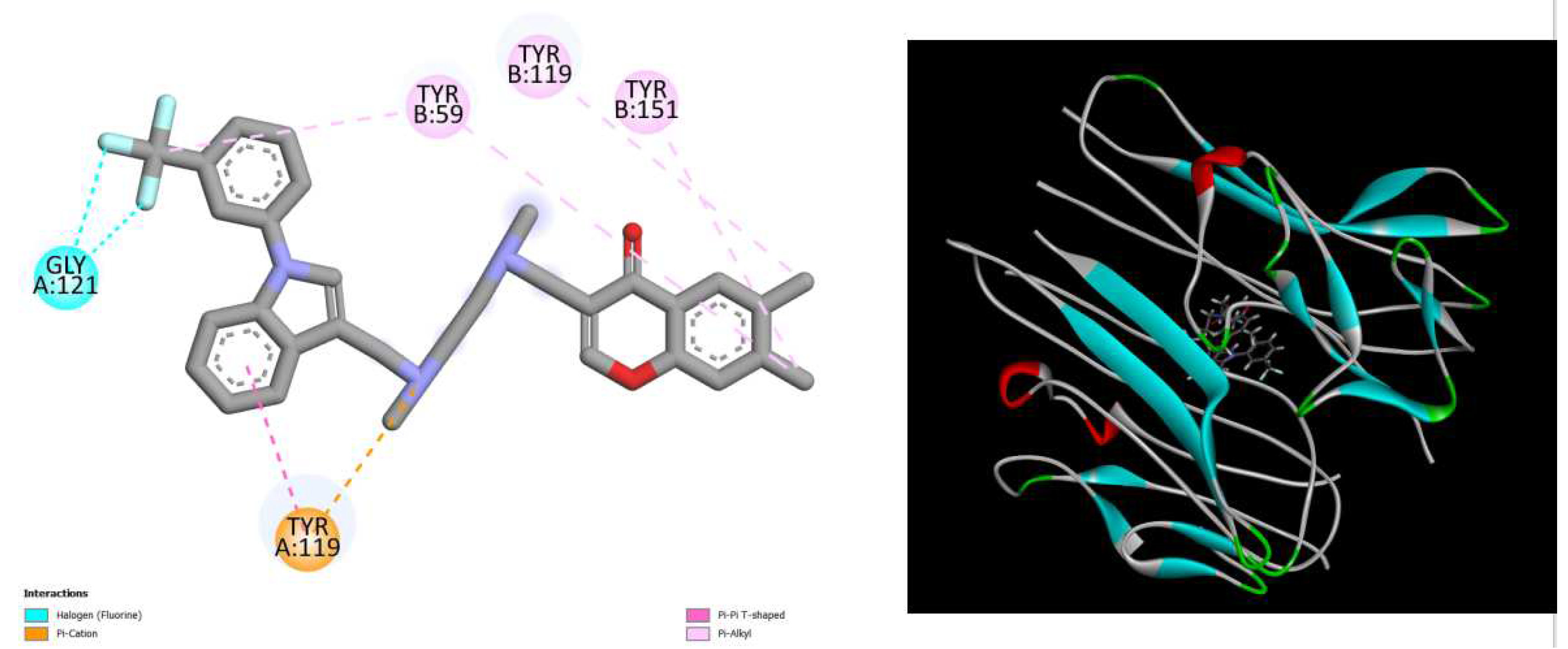
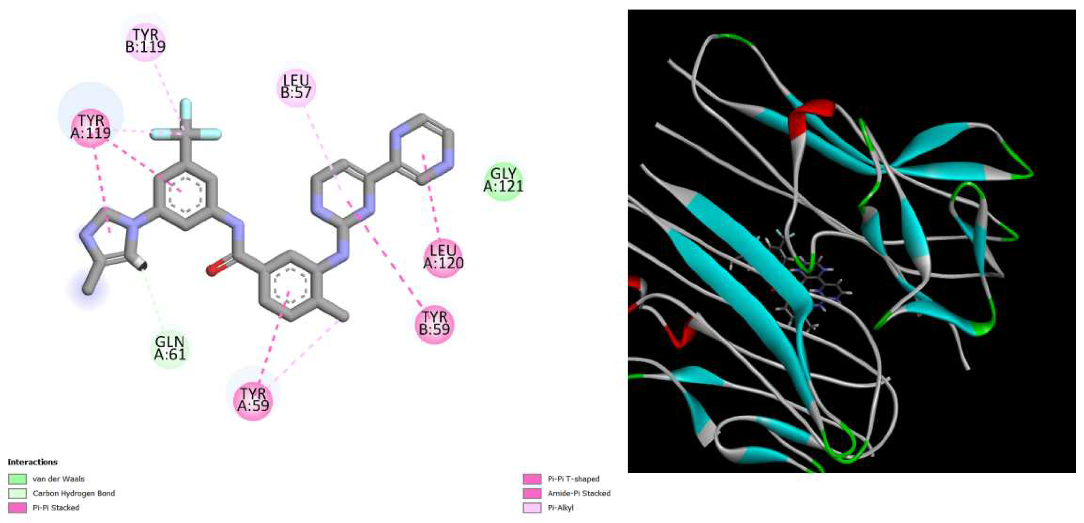
Figure 2.
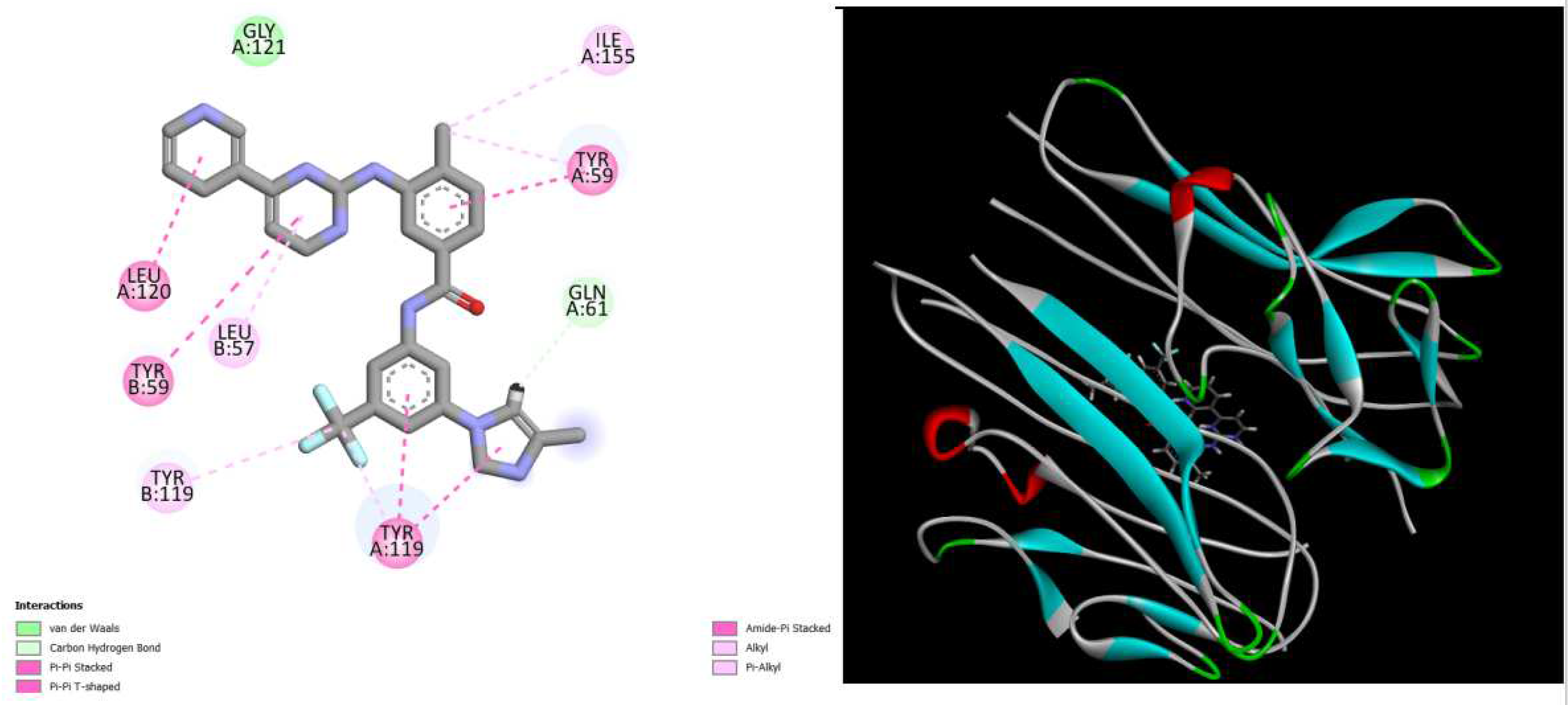
displays the docking outcomes of TNF-alpha Crystal Structure of TNF-alpha in conjunction with docked dactolisib with binding energy of -10.1 kcal/mol within the Ligand Binding Site, as analyzed by Autodock Vina with pyrx program. On the left side, 2D diagrams illustrate the residue interactions between the protein and dactolisib. Meanwhile, the right side exhibits the Ligand Binding Site of the protein, highlighting the specific location of dactolisib.
Figure 2.
displays the docking outcomes of TNF-alpha Crystal Structure of TNF-alpha in conjunction with docked dactolisib with binding energy of -10.1 kcal/mol within the Ligand Binding Site, as analyzed by Autodock Vina with pyrx program. On the left side, 2D diagrams illustrate the residue interactions between the protein and dactolisib. Meanwhile, the right side exhibits the Ligand Binding Site of the protein, highlighting the specific location of dactolisib.
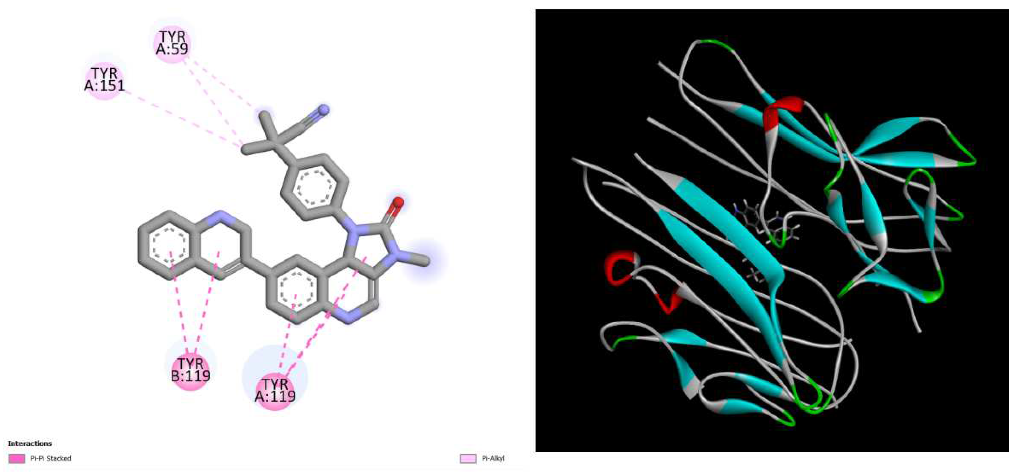
Figure 3.
displays the docking outcomes of TNF-alpha Crystal Structure of TNF-alpha in conjunction with docked Eltrombopag with binding energy of -9.7 kcal/mol within the Ligand Binding Site, as analyzed by Autodock Vina with pyrx program. On the left side, 2D diagrams illustrate the residue interactions between the protein and Eltrombopag. Meanwhile, the right side exhibits the Ligand Binding Site of the protein, highlighting the specific location of Eltrombopag.
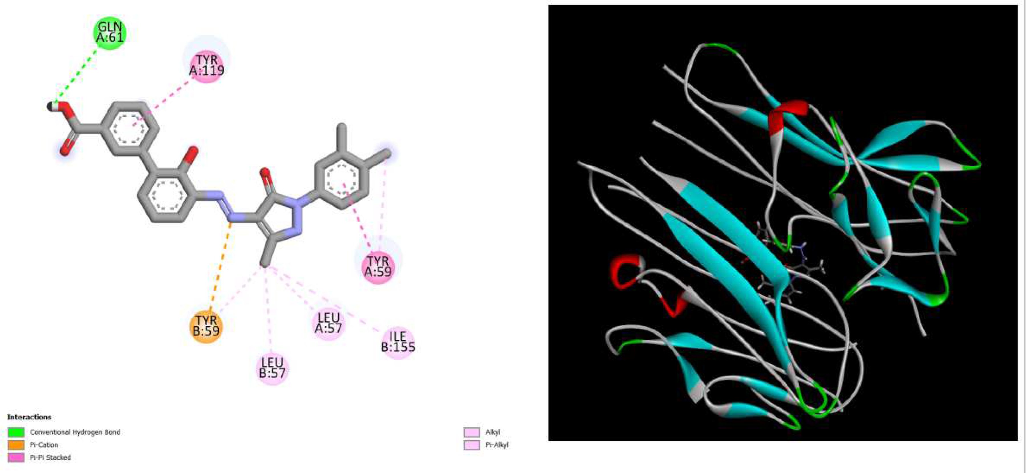
Figure 4.
displays the docking outcomes of TNF-alpha Crystal Structure of TNF-alpha in conjunction with docked Radotinib with binding energy of -9.6 kcal/mol within the Ligand Binding Site, as analyzed by Autodock Vina with pyrx program. On the left side, 2D diagrams illustrate the residue interactions between the protein and dRadotinib.
Meanwhile, the right side exhibits the Ligand Binding Site of the protein, highlighting the specific location of Radotinib.
Figure 5.
displays the docking outcomes of TNF-alpha Crystal Structure of TNF-alpha in conjunction with docked hypericin with binding energy of -9.9 kcal/mol within the Ligand Binding Site, as analyzed by Autodock Vina with pyrx program. On the left side, 2D diagrams illustrate the residue interactions between the protein and hypericin Meanwhile, the right side exhibits the Ligand Binding Site of the protein, highlighting the specific location of hypericin.
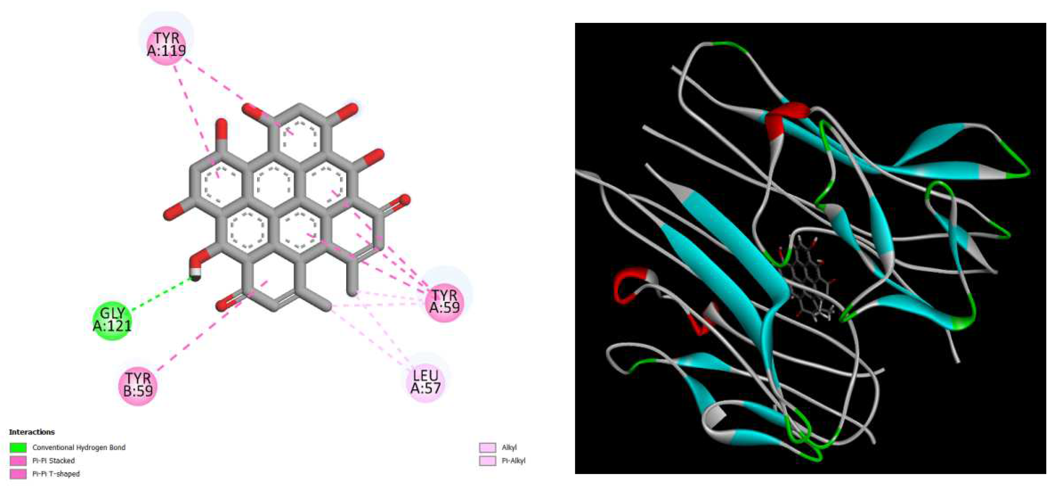
Figure 6.
displays the docking outcomes of TNF-alpha Crystal Structure of TNF-alpha in conjunction with docked nilotinib with binding energy of -9.6 kcal/mol within the Ligand Binding Site, as analyzed by Autodock Vina with pyrx program. On the left side, 2D diagrams illustrate the residue interactions between the protein and nilotinib. Meanwhile, the right side exhibits the Ligand Binding Site of the protein, highlighting the specific location of nilotinib.
4. Conclusion
This study utilized molecular docking methods to explore potential TNF-alpha inhibitors among various drugs and natural substances. Tumor necrosis factor-alpha (TNF-alpha), a crucial cytokine in immune regulation and inflammation, is implicated in various health conditions, particularly inflammatory bowel diseases (IBD) and tumor-related pathologies. The presented findings introduce novel potential inhibitors, including Dactolisib, Nilotinib, Eltrombopag, Radotinib, and Hypericin, each demonstrating promising binding affinities within TNF-alpha's ligand binding site. While these computational results are preliminary, they lay the groundwork for further biological studies essential to confirming the efficacy of these potential inhibitors. Developing effective TNF-alpha inhibitors holds promise for targeted therapeutic interventions in inflammatory diseases.
Conflicts of Interest
The authors declare no conflicts of interest.
References
- van Loo, G.; Bertrand, M.J. Death by TNF: A road to inflammation. Nature Reviews Immunology 2023, 23, 289–303. [Google Scholar] [CrossRef] [PubMed]
- Zhao, Y.; Simon, M.; Seluanov, A.; Gorbunova, V. DNA damage and repair in age-related inflammation. Nature Reviews Immunology 2023, 23, 75–89. [Google Scholar] [CrossRef] [PubMed]
- Marchi, S.; Guilbaud, E.; Tait, S.W.; Yamazaki, T.; Galluzzi, L. Mitochondrial control of inflammation. Nature Reviews Immunology 2023, 23, 159–173. [Google Scholar] [CrossRef] [PubMed]
- Zhao, Y.; Simon, M.; Seluanov, A.; Gorbunova, V. DNA damage and repair in age-related inflammation. Nature Reviews Immunology 2023, 23, 75–89. [Google Scholar] [CrossRef] [PubMed]
- Lauritano, D.; Mastrangelo, F.; D’Ovidio, C.; Ronconi, G.; Caraffa, A.; Gallenga, C.E.; Conti, P. Activation of Mast Cells by Neuropeptides: The Role of Pro-Inflammatory and Anti-Inflammatory Cytokines. International Journal of Molecular Sciences 2023, 24, 4811. [Google Scholar] [CrossRef]
- Gupta, K.K.; Khan, M.A.; Singh, S.K. Constitutive inflammatory cytokine storm: a major threat to human health. Journal of Interferon & Cytokine Research 2020, 40, 19–23. [Google Scholar]
- Vaillant, A.A.J.; Qurie, A. Interleukin. In StatPearls [Internet]; StatPearls Publishing, 2022. [Google Scholar]
- Leppkes, M.; Neurath, M.F. Cytokines in inflammatory bowel diseases–update 2020. Pharmacological Research 2020, 158, 104835. [Google Scholar]
- Stromsnes, K.; Correas, A.G.; Lehmann, J.; Gambini, J.; Olaso-Gonzalez, G. Anti-inflammatory properties of diet: Role in healthy aging. Biomedicines 2021, 9, 922. [Google Scholar] [CrossRef] [PubMed]
- Steen, E.H.; Wang, X.; Balaji, S.; Butte, M.J.; Bollyky, P.L.; Keswani, S.G. The role of the anti-inflammatory cytokine interleukin-10 in tissue fibrosis. Advances in wound care 2020, 9, 184–198. [Google Scholar] [PubMed]
- Stanzione, F.; Giangreco, I.; Cole, J.C. Use of molecular docking computational tools in drug discovery. Progress in Medicinal Chemistry 2021, 60, 273–343. [Google Scholar] [PubMed]
- Menchaca, T.M.; Juárez-Portilla, C.; Zepeda, R.C. Past, present, and future of molecular docking. In Drug Discovery and Development-New Advance; IntechOpen, 2020. [Google Scholar]
- Ulich, T.R. Tumor necrosis facto. In Cytokines of the Lun; CRC Press, 2022; pp. 307–332. [Google Scholar]
- Laha, D.; Grant, R.; Mishra, P.; Nilubol, N. The role of tumor necrosis factor in manipulating the immunological response of tumor microenvironment. Frontiers in immunology 2021, 12, 656908. [Google Scholar] [PubMed]
- Jang, D.I.; Lee, A.H.; Shin, H.Y.; Song, H.R.; Park, J.H.; Kang, T.B.; Yang, S.H. The role of tumor necrosis factor alpha (TNF-α) in autoimmune disease and current TNF-α inhibitors in therapeutics. International journal of molecular sciences 2021, 22, 2719. [Google Scholar] [PubMed]
- Eberhardt, J.; Santos-Martins, D.; Tillack, A.F.; Forli, S. AutoDock Vina 1.2. 0: New docking methods, expanded force field, and python bindings. Journal of chemical information and modeling 2021, 61, 3891–3898. [Google Scholar] [PubMed]
- Tang, S.; Chen, R.; Lin, M.; Lin, Q.; Zhu, Y.; Ding, J.; Wu, J. Accelerating autodock vina with gpus. Molecules 2022, 27, 3041. [Google Scholar] [CrossRef] [PubMed]
- Pawar, R.P.; Rohane, S.H. Role of autodock vina in PyRx molecular docking. 2021. [Google Scholar]
Disclaimer/Publisher’s Note: The statements, opinions and data contained in all publications are solely those of the individual author(s) and contributor(s) and not of MDPI and/or the editor(s). MDPI and/or the editor(s) disclaim responsibility for any injury to people or property resulting from any ideas, methods, instructions or products referred to in the content. |
© 2024 by the authors. Licensee MDPI, Basel, Switzerland. This article is an open access article distributed under the terms and conditions of the Creative Commons Attribution (CC BY) license (http://creativecommons.org/licenses/by/4.0/).
Copyright: This open access article is published under a Creative Commons CC BY 4.0 license, which permit the free download, distribution, and reuse, provided that the author and preprint are cited in any reuse.


