Submitted:
27 May 2025
Posted:
28 May 2025
You are already at the latest version
Abstract
Per- and polyfluoroalkyl substances (PFAS), a large group of developing contaminants, have recently become the subject of increased concern due to their potentially hazardous effects. They are classified as poisonous substances that can be found in a variety of aquatic situations. The widespread usage of PFAS across numerous industries has resulted in a high environmental and biological accumulation of the substance. Identification and elimination of PFAS from the environment is crucial since they are tenacious and have the potential to cause cancer. Traditional methods of PFAS content assessment, while useful in some cases, are often inadequate for continuing environmental control and monitoring. Within academia, there is a noticeable desire for rapid, cost-effective, durable, and readily transportable techniques targeted at detecting PFAS compounds in field settings. As a result, environmental labs and other governmental and non-governmental bodies may start testing more often as mandated by legislation. PFAS-detecting sensors, which offer an innovative solution that can be applied in situ and is affordable and simple to use, have emerged as a promising method for assessment based on the existing research. In addition, it may give administrators and users of water worldwide useful information they can use. This article provides a thorough summary of recent developments, limitations, and performance considerations in PFAS detection sensors. The Surface-enhanced Raman scattering (SERS) method has also been covered because of its excellent sensitivity and specificity for detecting pollutants, making it a potential solution for sensing PFAS. Intelligent sensing systems for PFAS detection should benefit from this research.
Keywords:
per- and polyfluoroalkyl substances
; PFAS detection sensors
; surface-enhanced Raman scattering
; emerging contaminants
1. Introduction
Perfluorocarbons, fondly referred to as the elusive perfluorinated compounds (PFCs), engage in a captivating dance within the molecular realm, exuding a mysterious allure reminiscent of their counterparts, the PFASs. These molecular maestros, with shared traits and clandestine chemistry, weave a tale of intrigue in the scientific symphony [1,2]. Despite these similarities, PFCs exhibit fundamental differences from PFASs. Unlike PFAS compounds, PFCs consist solely of carbon and fluorine atoms, with the absence of oxygen, hydrogen, sulfur, and nitrogen atoms [3,4]. The inception of PFASs dates back to 1950, and they are entirely synthetic in nature [1,5,6,7]. These compounds consist of at least one perfluoroalkyl group (CnF2n+1) linked to a hydrophilic head group [8,9,10]. Approximately 4,700 distinct PFAS have already been registered in the global market, with nearly 4,000 of them identified in diverse matrices [11]. The significant chemical structures of PFAS are illustrated in Figure 1.
Across a historical expanse exceeding six and a half decades, the intricate craft of producing PFASs for commercial purposes has unfolded through the adept utilization of two principal methodologies: the rigorous Electrochemical Fluorination and the meticulous artistry of Telomerization. These stalwart methods have stood the test of time, contributing to the nuanced legacy of PFAS manufacturing [1,13,14]. Electrochemical fluorination was stopped in 2002 due to the large amount of branched-chain PFAS it produced. Within the scholarly realm, the venerable electrochemical telomerization, an alchemical symphony orchestrating the generation of fluorotelomer alcohols, emerges as the preeminent method in the illustrious craft of PFAS production. Its widespread adoption attests to its prominence, casting an academic spotlight on the nuanced processes underlying the intricate world of per- and polyfluoroalkyl substances. Linear fluorinated molecules with an even number of fluorine atoms are often the result of telomerization [15]. Coated with PFAS, nonstick cookware and fast-food packaging (including pizza boxes and popcorn bags) can withstand both water and oil [16]. Pharmaceuticals, personal care items, cosmetics, and medical equipment are just a few of the many industries that use PFAS [17,18]. Fluorotelomer alcohol molecules in commercial items, poor treatment of industrial wastewater, or precursor degradation discharge PFAS into the environment [19]. In daily life, water, air, food, and everyday items intertwine to introduce individuals to the enigmatic compounds of perfluorooctanoic acid (PFOA) and perfluorooctane sulfonic acid (PFOS) [20].
Reducing pollution from sources like PFAS necessitates effective waste management and recycling programs [21,22,23,24]. The release of substances containing PFAS into the environment can be avoided through the implementation of effective waste management practices [24]. Realizing that PFAS could be in recycling streams highlights the need to implement responsible waste recycling strategies to reduce environmental contamination [22,25,26,27,28,29,30]. Efficient wastewater cotreatment of Dissolved Organic Nitrogen (DON) and PFAS becomes a linchpin in preventing environmental contamination [31,32,33]. Responsible waste recycling strategies act as guardians, averting the inadvertent release of PFAS into recycling streams and reinforcing the commitment to effective waste management practices [33]. Comprehensive PFAS management strategies are necessary because effective sanitation and hygiene practices are critical for minimizing the spread of contaminants [34,35,36,37]. Artificial intelligence (AI) can also optimize sorting processes and identify possible sources of PFAS contamination, which can improve the efficiency of PFAS management systems [38,39,40,41,42]. AI-powered waste treatment and environmental monitoring systems help take preventative actions against PFAS exposure [38].
Research on the toxicological consequences of PFASs has shown that these chemicals can have a wide variety of negative effects, such as on the immune system, reproductive system (both during embryonic and postnatal development), liver health, developmental processes, and the endocrine system [43]. In a global regulation, Germany, the US, and Canada harmonize efforts to restrict PFOA and PFOS in drinking water. Their shared pursuit transcends limitations, aiming to diminish both the production and consumption of these compounds [44]. In the potable water systems, the United States Environmental Protection Agency (EPA) has issued a sovereign decree, limiting PFOA and PFOS to a mere 0.07 ng/L [45]. This regulatory vigilance ensures the sanctity of our life-sustaining elixirs. Consequently, health departments and regulatory organizations monitor PFAS traces [46]. The health concerns posed by PFOA and PFOS can be felt even at nanomolar amounts [47]. Therefore, sensitive detection techniques or equipment are required for these PFAS. Various precise and sensitive analytical procedures are available. Some of the problems with analytical procedures are the time and effort needed to prepare samples, the high expenses of operations, the necessity for trained individuals to evaluate and interpret the results, and the fact that they aren’t applicable in real-world scenarios [48]. Therefore, it is essential to create unique sample preparation and quick detection techniques.
Limitations in sensitivity and the need for labor-intensive processes are some of the problems with current technologies for PFAS detection. The improved sensitivity, selectivity, stability, adaptability, and non-destructive properties of SERS make it an attractive method for PFAS detection. By overcoming the drawbacks of current PFAS detection techniques, SERS has the potential to transform PFAS detection and make a substantial contribution to our knowledge of these persistent environmental pollutants. Future directions for research include creating new SERS substrates with improved PFAS sensitivity and selectivity, incorporating SERS sensors into field-based portable devices, increasing the range of PFAS compounds that can be detected with SERS, and using SERS for environmental monitoring and analysis in the real world [49].
2. Current PFAS Detection Methods
2.1. PFAS Sample Preparation and Extraction
Preparing, cleaning, and concentrating samples for PFAS detection can be somewhat challenging because to their complicated matrices and limited environmental presence. Figure 2 shows the whole procedure for PFAS extraction from solid and liquid sources using well-established methods such as ion pair extraction, dispersive liquid-liquid microextraction, liquid-liquid extraction (LLE), and alkaline digestion [50].
These methods are sometimes inefficient, lengthy, and necessitate the co-extraction of lipids, which makes chromatographic analysis more difficult. Additionally, they often require analysis to be performed off-site. Non-polar solvents are capable of extracting non-ionic chemicals from their solution matrices. It is commonly recommended to employ weak anion exchange (WAX) cartridges when dealing with shorter chain PFCs in the range of C4 to C6. Conversely, for longer-chain PFAS exhibiting substantial recoveries in water and soil samples, the use of C18 and hydrophilic-lipophilic-balanced (HLB) cartridges is advised. A study conducted by L. Wang et al. in 2010 found that PFAS levels in bivalves’ soft tissues may be evaluated using methyl-tert-butyl ether (MTBE) extraction followed by SPE purification on a WAX phase, despite the fact that this approach contains critical steps that might lead to analyte loss [52]. In an unconventional approach, acetonitrile, coupled with shaking or sonication, proves adept at extracting PFCs from diverse biological matrices—ranging from insect larvae to forage fish, crustaceans, and bivalves. The efficacy of oasis WAX cartridges in purging environmental contaminants, including short chain perfluorinated carboxylic acids (PFCAs), is underscored in two enlightening studies [53,54]. In 2017, a study was conducted on 12 perfluorinated compounds in the coastal area of the Shandong peninsula [55]. The adept researchers employed Oasis HLB (0.2 g, 6 mL) to discern the presence of PFAS [55]. In a pioneering stride beyond conventional SPE, in 2018, two researchers orchestrated a magnetic method of SPE, a bespoke choreography designed for the exquisite extraction ballet of PFCs [12]. This innovation gracefully sidesteps the customary filtering interlude, ushering in a streamlined era where the separation process unfurls with newfound ease and celerity. In 2019, another research suggested an electrochemical preconcentration approach [56]. In a mere 10-minute cadence, this methodology deftly extracted 10 distinct PFAS variants from ambient samples, showcasing concentrations that spanned the spectrum from 0.5 ng/L to 500 ng/L [56]. In a collaborative efforts of two researchers in 2018 unfolded a tale where PFCs were meticulously extracted from industrial effluent [57]. The method of choice, akin to a delicate brushstroke, involved the sophisticated artistry of hollow fiber microextraction techniques. A honeycomb-like cylindrical core, embraced by four intricately sealed hollow fibers, manifested as the elegant embodiment of experimental design [57]. In a quest to maximize the volumetric extraction potential nestled within the wall pores, a pioneering method emerged—hollow fiber-based liquid-pressured microextraction. This innovative approach delved into the intricate art of saturating the hollow fiber wall pores without deliberately infiltrating the lumen with solvent. The composition involved submerging the assembly of hollow fiber extraction instruments into the solvent extractant, revealing the efficacy of ultrasonic solid-liquid extraction in this scientific symphony [58]. The results showed that this process is a safe, quick, easy, and inexpensive way to extract PFAS from food matrices and popcorn bags, with recovery rates near to 100%. In 2021, a research suggested that ultrasonic solid-liquid extraction has proven to be a simple and rapid extraction process with high PFAS recoveries from paper packaging materials [59]. The advent of this novel method unfolds as a tapestry woven with threads of heightened sensitivity, abbreviated analytical timelines, and the graceful streamlining of extraction procedures. However, amid the triumphs of this approach, the traditional SPE methods cast shadows with potential drawbacks—whispers of sample contamination and the subtle departure of surface-active PFAA to the very vessels cradling the samples. In addition, SPE is complicated enough without having to deal with column obstruction.
2.2. Analysis of PFAS via Chromatography
Table 1 unveils a panoramic view of diverse techniques employed in the extraction and detection symphony, orchestrating the revelation of PFAS levels in environmental samples. The virtuosity of HPLC adorned with conductivity or fluorimetric detectors, alongside the prowess of LC-MS/MS, graces the scientific stage as commonplace methodologies. Gas Chromatography (GC) lends its precision to the measurement of volatile PFAS, while Ion Chromatography (IC), Nuclear Magnetic Resonance (NMR), and the symphony of Fourier Transform Infrared Spectroscopy (FTIR) emerge as less frequented methods [60,61]. Delving into the depths of radiochemical techniques unveils an ability to discern branched-chain PFAS [1]. Among these, LC-MS stands as a luminary, offering a promising pathway for evaluating PFAS in a spectrum of environmental matrices, showcasing detection limits that venture into the nanogram realm. The scientific community’s gaze has progressively shifted towards LC-MS, leveraging its capabilities for the identification of PFAS in sludge, water, and serum [62,63,64].
2.3. PFAS Detection by Sensors
Within the biosensing, a singular entity arises—an amalgamation of vital components meticulously choreographed to collect, with precision, both quantitative and analytical data. This architectural marvel features a biological receptor seamlessly interfacing with a transduction device, encapsulating the essence of a biosensor. Comprising fundamental elements, this symphony harmonizes a biological recognition entity, a transducer, and an intricately designed signal processing system, as elucidated in a scholarly exploration [75]. The discerning and specific nature of biosensors relies on both a biorecognition system and a transducer. Sensitive biosensors are crafted using phages, whole-cell organisms, antibodies, and aptamers [76]. In molecular mimicry, Molecularly Imprinted Polymers (MIPs) emerge as virtuosos, replicating interactions reminiscent of substrate-enzyme and antigen-antibody engagements. The evolution of intricate sensing apparatus owes its prowess to the synergy of biomimetic materials and electrochemical technologies, transcending the limitations of unmodified or catalyst-modified sensors and unfurling novel realms of application potential. Within this molecular ballet, MIPs engage with target molecules, orchestrating a transformative symphony that alters their mass, absorbance, and refractive index—a nuanced performance dictated by the intricate tapestry of structure and surface charge [77]. MIPs craft a polymeric symphony in electrochemical elegance, sculpting a matrix adorned with celestial voids and recognition sites. These cosmic formations, akin to celestial bodies, dance in perfect harmony with the target analyte on the electrode surface, an exquisite ballet illustrated in the captivating canvas of Figure 3 [78]. The utilization of MIPs enhances both the sensitivity and selectivity of sensors due to their expansive surface area. Table 2 unfolds as a curated panorama, offering a succinct overview of the myriad sensor systems meticulously crafted for the discernment of PFAS.
2.4. Nanomaterial Based Sensor
Nanomaterials’ strong electrical and magnetic conductivity, as well as their large surface area, make them useful for resolving lower detection limit problems in monitoring applications. The use of nanoparticles (NPs) offered a new strategy for creating inexpensive and portable PFAS detection devices [85]. Nanomaterials, including the luminescent brilliance of silver and gold nanoparticles, the metallic prowess of metal nanoparticles (MNPs), and the quantum wonders of quantum dots (QDs), assume a pivotal role in elevating the performance of biosensors to new heights. An uncomplicated sensor for detecting PFOA was developed. Early efforts to modify AuNPs involved utilizing thiol-terminated polystyrene. Upon introduction to the solution of AuNPs, the PFOA demonstrated a proclivity for aggregation, guided by the captivating dance of fluorine-fluorine interactions. The test was performed admirably, with a noticeable shift in color from red to purple corresponding to increasing PFOA concentrations [85]. A study documented the assessment of PFCs using a probe based on the Au-NPs [86]. This innovative approach employs a fusion of polyethylene glycol-terminated (PEG-thiols) and perfluoroalkyl-terminated (F-thiols) alkanethiol to intricately modify gold particles, sculpting them into a bespoke probe. In a mere half-hour temporal embrace, the ingeniously crafted probe unfurls its prowess, discerning the presence of PFCs within concentrations ranging from 0.1 to 1000 μg/L. This enhanced sensor manifests remarkable stability, boasting a detection limit finely tuned to 10 μg/L. Rigorous assessments conducted by the author within the realms of municipal and river water unveiled a sensor adorned with high selectivity specifically tailored for PFOA and PFOS [87].
In the eloquent narrative penned by a study, a groundbreaking chapter unfolded introducing upconversion nanoparticles (UCNPs) as the luminescent protagonists in the sensor saga, meticulously crafted for the detection of PFOS [88]. The genesis of these UCNPs took shape through the meticulous artistry of solvothermal synthesis. The synthesis and deposition of covalent organic framework (COF) on the surface of UCNPs occurred simultaneously. Increased selectivity and sensitivity for PFOS were noted in the synthesized UCNPs-COFs. In the unveiling of a sophisticated sensing platform, its application to scrutinize authentic water samples unfolds as a narrative of triumph, showcasing recoveries within the captivating range of 106% to 108% and an exquisite limit of detection at 0.000075 ng/L for perfluorooctane sulfonate (PFOS). Embarking on a quest for a streamlined PFOS detection sensor, a research introduces a tale interwoven with titanium dioxide nanotubes (TiO2-NTAs) [89]. In the sensor realm, a trio of protagonists—acrylamide, ethylene glycol dimethacrylate, and 2,2-azobis(2-methylpropionitrile)—takes center stage. Each assumes a distinctive role, serving as the functional monomers, cross-linker, and initiator agent in the alchemical ballet of polymerization imprinting. The sensor born from this symphony stands as a testament to the marriage of selectivity and sensitivity, unfurling its prowess in discerning perfluorooctane sulfonic acid (PFOS), boasting a discerning detection limit at 86 µg/L and a linear range spanning from 250 to 5001 µg/L. Results from using the technique on environmental samples were positive. In order to detect PFOA in environmental samples, another study created a QDs sensor [70]. Embarking on a scientific odyssey, CdTe@CdS-QDs materialized through the alchemical artistry of the sol-gel technique. The saga continued with the creation of silica films, meticulously imprinted at the molecular level onto CdTe@CdS-QDs. Aqueous ammonia, akin to a catalyst in this intricate ballet, presided over the proceedings. The narrative of creation unfolded with a polymerization technique, where 3-aminopropyltriethoxysilane donned the mantle of the functional monomer, and tetraethoxysilane stood as the cross-linker, weaving the intricate tapestry of molecular imprinting. The method demonstrated effectiveness, yielding a detection limit of 10.35 µg/L and a linear range spanning from 104 to 6211 µg/L [51]. Values of recoveries seen by the author varied between 91 and 107%, with a relative standard deviation of 5.6%. PFOA in water was detected using the designed sensor with high selectivity. Research by S. Chen and colleagues in 2015, a luminescent symphony unfolded as they meticulously crafted a luminophore imprinting polymer, adorning the ultrathin nanosheets of carbon nitride (utg-C3N4) through the ballet of an electropolymerization technique [81]. The resulting sensor, a testament to their precision, revealed a harmonious linearity extending across detection ranges from 20 to 40,000 ng/L. Finding a limit of detection of 10 ng/L was a significant achievement. Also, the sensor was used for the examination of water samples, with satisfactory recoveries (96%-103.8%) being attained. There was also significant concordance between HPLC-MS/MS and the findings.
In their scholarly endeavour to unveil the fingerprint of PFOS within biological samples, a study conceptualized a sensor ingeniously fashioned with the radiance of carbon quantum dots (CQD) [85]. The integration of CQD with a chitosan hydrogel was orchestrated through the methodical process of electrochemical polymerization, culminating in the formation of a durable covalent bond. The interaction between sulfonates from chitosan and QDS, and amino groups from PFOS through electrostatic repulsion, forms a compound. This novel approach demonstrates sensitivity in detecting various perfluorinated compounds, including PFOA, PFOS, PFBS, and perfluorooctanoic acid. The highest imprinting factor discovered by the author was 2.75. The concentrations that could be added up to 1 ng/L were only as low as 0.00002 ng/L. The designed sensor yielded satisfactory recoveries (81-98%). Using a water-soluble CdS-QDS, another study created a straightforward and quick fluorometric method for measuring PFOA [79]. The birth of radiant CdS-QDs, aglow with intense luminosity, unfolded through the transformative touch of 3-mercaptopropionic acid (MPA). In a dance of molecular interplay, the entrance of PFOA became the catalyst for an enchanting aggregation of MPA-CdS QDs, orchestrated by the alluring forces of fluorine-fluorine interaction. This mystical collaboration cast a spell, conjuring forth an augmented fluorescence emanating from the embrace of MPA-CdS QDs. The saga of detection unfolded with a determined linearity stretching from 207.03 µg/L to 16.56 mg/L, revealing its secrets with a calculated limit of detection at 124.2 µg/L.
2.5. PFAS Detection by Surface-Enhanced Raman Spectroscopy
In a recent scientific expedition, the canvas of detection unfolded with the mastery of SERS as the chosen medium for exploring the mysteries of the PFAS [90]. The mesmerizing SERS effect, harnessed through the localized surface plasmon resonance (LSPR) of 40 nm silver nanoparticles (AgNPs), became the enchanting brushstroke in this artistic endeavor. In a temporal whisper of fewer than 30 seconds, the SERS technique exhibited its prowess, unveiling the ability to detect PFAS concentrations as ethereal as 20 femtograms per liter. The allure of the SERS technique lies in its potential to transcend the boundaries of PFAS contamination studies, offering a tableau of quicker, more sensitive, and less labour-intensive analyses, all within the realm of optical sensitivity. Within the intricate dance of Raman spectroscopy, where an excitation photon intertwines with a phonon in the analyte, losing energy along the way, wavelengths shift, and vibrational frequencies of phonons emerge as beacons for molecular identification. Amplification becomes a necessity for low analyte concentrations due to the rarity of spontaneous Raman scattering. In this narrative, 40 nm AgNPs assumed a pivotal role, entering a water symphony at a harmonious ratio of 2:3, casting their essence through the medium of drop-casting onto aluminum substrates to orchestrate the optimization of SERS enhancement. The spectral odyssey unfolded within the melodic cadence of 200 to 1800 cm⁻¹, where the rhythmic resonance of the asymmetric stretching mode of the difluoromethylene (CF₂) group, the very essence of PFAS, played its enchanting tune. In this symphony of wavelengths, the molecular harmonies revealed the intricate secrets of PFAS, painting a mesmerizing portrait in the language of spectroscopy, revealing itself as a Raman feature peak around 1300 cm⁻¹. Concentration calibration curves, akin to musical scales, emerged, with the feature peak at 1300 cm⁻¹ serving as the guiding melody to discern the concentration of water samples—each note unravelling the secrets of PFOA and PFOS.
In another research, SERS methods were used to find persistent flame retardants (PFAS), a major environmental problem, in firefighting foams [49]. With a 50 ppb PFOA detection limit, they were able to successfully identify PFOS, PFOA, and 6:2FTS. Graphene oxide (GO) membrane and nanosphere lithography were used to create two SERS substrates that showed GO’s strong affinity for fluoro surfactants (FS). They created a loaded dye-FS precipitate ion pair on the substrates, dye-FS-Ag and dye-FS-GO, both of which successfully detected FS, using EV dye as a Raman probe. Dye-FS-Ag’s Raman spectra showed a noticeable signal amplification on the Ag surface. Controlled tests using the GO membrane showcased the augmentation of FS loading in the presence of a dye [49]. This experimental demonstration unfolded as a controlled symphony, revealing the dynamic interplay between the membrane and the dye, a testament to the orchestrated finesse in material interactions.
Another study created a method for detecting PFOA, a contaminant belonging to the perfluoroalkyl substances (PFAS) class, utilizing SERS [91]. The inherent fluorescence of PFOA makes conventional SERS detection difficult because it masks the Raman signal. To address this issue, researchers applied a longer Raman excitation wavelength of 633 nm, effectively suppressing fluorescence while maintaining adequate Raman intensity. The researchers used crystal violet (CV) as a Raman label to improve the Raman signal and eliminate fluorescence. CV and PFOA interact to form ion pairs, resulting in a synergistic increase in Raman activity. This combination technique produced an amazing limit of detection (LOD) for PFOA of 10.52 ppb, confirming the method’s exceptional sensitivity and promise for real-world environmental monitoring. This approach not only has high sensitivity but also allows for the detection of PFOA even in the presence of other chemicals. Achieving this specificity involves employing an indirect analysis approach, wherein the Raman signal of CV is heightened in the presence of PFOA, serving as an indicator for the existence of PFOA. The SERS technique’s adaptability is further demonstrated by its ability to detect another common PFAS molecule, perfluorooctanesulfonic acid (PFOS). The method’s expansive versatility in PFAS detection is vividly illustrated by its remarkable detection limit at the ultra-sensitive scale of 10-12 M. This numerical testament echoes the method’s prowess, resonating across a broad spectrum of potential applications for precise detection of PFAS [49].
3. Conclusions
In PFAS detection, post-sample preparation unveils a plethora of detection methods, each boasting advantages in terms of speed, simplicity, cost-effectiveness, and sensitivity when juxtaposed with traditional chromatography. The brisk pace of assays necessitates the evolution of innovative extraction procedures, heralding a paradigm shift in analytical methodologies. Analyzing PFAS in the intricate landscape of food encounters challenges rooted in the absence of standardized methods, beckoning the need for pioneering approaches. Continuous monitoring emerges as a beacon of efficacy, transcending the constraints of batch techniques and embracing the vast domains of both food and environmental samples. While the majority of approaches have concentrated on PFAS detection in water, their performance within complex matrices demands meticulous assessment and augmentation to enable direct detection. A harmonious convergence of disciplines becomes imperative to sculpt sensors of impeccable design, where nanoparticles play the role of virtuosos, orchestrating signal amplification to elevate sensitivity and selectivity. Despite the promising strides in sensor research, the crucible of real sample analysis and selectivity must occupy the forefront, sculpting flawless sensing systems attuned to the nuances of PFAS. Validation stands as the cornerstone in this odyssey, a pivotal step ensuring the accuracy of PFAS measurements and paving the way for widespread adoption. In the grand tapestry of innovation, these sensors and sensing systems emerge as catalysts, potentially revolutionizing the screening of real-world samples for PFAS by regulatory agencies and other entities. Their efficiency may outshine conventional analytical methods, ushering in a new era of precision and effectiveness in the quest for PFAS detection.
References
- R. C. Buck et al., “Perfluoroalkyl and polyfluoroalkyl substances in the environment: terminology, classification, and origins,” Integrated environmental assessment and management, vol. 7, no. 4, pp. 513–541, 2011.
- C. Zhang, K. Yan, C. Fu, H. Peng, C. J. Hawker, and A. K. Whittaker, “Biological utility of fluorinated compounds: from materials design to molecular imaging, therapeutics and environmental remediation,” Chemical Reviews, vol. 122, no. 1, pp. 167–208, 2021.
- M. Li et al., “Theoretical studies of perfluorochemicals (PFCs) adsorption mechanism on the carbonaceous surface,” Chemosphere, vol. 235, pp. 606–615, 2019.
- Z. Du et al., “Adsorption behavior and mechanism of perfluorinated compounds on various adsorbents—A review,” Journal of hazardous materials, vol. 274, pp. 443–454, 2014.
- J. Walkowiak-Kulikowska, “Poly/Perfluorinated Alkyl Substances (PFASs)–Synthetic Methods, Properties and Applications,” Perfluoroalkyl Substances: Synthesis, Applications, Challenges and Regulations, 2022.
- F. Dixit et al., “Removal of zwitterionic PFAS by MXenes: comparisons with anionic, nonionic, and PFAS-specific resins,” Environmental Science & Technology, vol. 56, no. 10, pp. 6212–6222, 2022.
- I. M. Militao, F. A. Roddick, R. Bergamasco, and L. Fan, “Removing PFAS from aquatic systems using natural and renewable material-based adsorbents: A review,” Journal of Environmental Chemical Engineering, vol. 9, no. 4, p. 105271, 2021.
- R. Mueller and V. Yingling, “History and use of per-and polyfluoroalkyl substances (PFAS),” Interstate Technology & Regulatory Council, 2017.
- B. Bokkers et al., “Per-and polyfluoroalkyl substances (PFASs) in food contact materials,” 2019.
- K. A. Barzen-Hanson, “Per-and Polyfluoroalkyl Substances (PFASs) and Aqueous Film-Forming Foam Impacted Sites: New PFAS Discovery and Sorption of Anionic, Zwitterionic, and Cationic PFASs,” 2017.
- F. Li et al., “Short-chain per-and polyfluoroalkyl substances in aquatic systems: Occurrence, impacts and treatment,” Chemical Engineering Journal, vol. 380, p. 122506, 2020.
- C. Fang, X. Zhang, Z. Dong, L. Wang, M. Megharaj, and R. Naidu, “Smartphone app-based/portable sensor for the detection of fluoro-surfactant PFOA,” Chemosphere, vol. 191, pp. 381–388, Jan. 2018. [CrossRef]
- J. Radjenovic, N. Duinslaeger, S. S. Avval, and B. P. Chaplin, “Facing the challenge of poly-and perfluoroalkyl substances in water: is electrochemical oxidation the answer?,” Environmental Science & Technology, vol. 54, no. 23, pp. 14815–14829, 2020.
- A. He et al., “Vital environmental sources for multitudinous fluorinated chemicals: new evidence from industrial byproducts in multienvironmental matrices in a fluorochemical manufactory,” Environmental Science & Technology, vol. 56, no. 23, pp. 16789–16800, 2022.
- H. Zhu and K. Kannan, “A pilot study of per-and polyfluoroalkyl substances in automotive lubricant oils from the United States,” Environmental Technology & Innovation, vol. 19, p. 100943, 2020.
- K. L. Vorst, N. Saab, P. Silva, G. Curtzwiler, and A. Steketee, “Risk assessment of per-and polyfluoroalkyl substances (PFAS) in food: Symposium proceedings,” Trends in Food Science & Technology, vol. 116, pp. 1203–1211, 2021.
- A. F. Zakaria, N. Yahaya, M. Raznisyafiq, S. H. Loh, and S. Kamaruzaman, “Recent advances in applications of hybrid natural polymers as adsorbent for perfluorinated compounds removal–review paper,” Journal of Polymer Research, vol. 29, no. 1, pp. 1–19, 2022.
- H. A. Langberg et al., “Paper product production identified as the main source of per-and polyfluoroalkyl substances (PFAS) in a Norwegian lake: Source and historic emission tracking,” Environmental Pollution, vol. 273, p. 116259, 2021.
- Y.-C. Lin, W. W.-P. Lai, H. Tung, and A. Y.-C. Lin, “Occurrence of pharmaceuticals, hormones, and perfluorinated compounds in groundwater in Taiwan,” Environmental Monitoring and Assessment, vol. 187, no. 5, pp. 1–19, 2015.
- E. Papadopoulou et al., “Sampling strategy for estimating human exposure pathways to consumer chemicals,” Emerging Contaminants, vol. 2, no. 1, pp. 26–36, 2016.
- Md. A. Ahmed, M. Hossain, and M. Islam, Prediction of Solid Waste Generation Rate and Determination of Future Waste Characteristics at South-western Region of Bangladesh Using Artificial Neural Network. KUET, Khulna, Bangladesh: WasteSafe 2017, KUET, Khulna, Bangladesh, 2017.
- T. Stoiber, S. Evans, and O. V. Naidenko, “Disposal of products and materials containing per-and polyfluoroalkyl substances (PFAS): A cyclical problem,” Chemosphere, vol. 260, p. 127659, 2020.
- M. A. Ahmed and S. D. Chakrabarti, “SCENARIO OF EXISTING SOLID WASTE MANAGEMENT PRACTICES AND INTEGRATED SOLID WASTE MANAGEMENT MODEL FOR DEVELOPING COUNTRY WITH REFERENCE TO JHENAIDAH MUNICIPALITY, BANGLADESH,” presented at the 4 th International Conference on Civil Engineering for Sustainable Development (ICCESD 2018), Khulna, Bangladesh: Department of Civil Engg., KUET, Feb. 2018.
- S. Garg et al., “A review on the sources, occurrence and health risks of per-/poly-fluoroalkyl substances (PFAS) arising from the manufacture and disposal of electric and electronic products,” Journal of Water Process Engineering, vol. 38, p. 101683, 2020.
- D. Page, J. Vanderzalm, A. Kumar, K. Y. Cheng, A. H. Kaksonen, and S. Simpson, “Risks of perfluoroalkyl and polyfluoroalkyl substances (PFAS) for sustainable water recycling via aquifers,” Water, vol. 11, no. 8, p. 1737, 2019.
- M. A. Ahmed and S. M. Moniruzzaman, “A STUDY ON PLASTIC WASTE RECYCLING PROCESS IN KHULNA CITY,” presented at the 4 th International Conference on Civil Engineering for Sustainable Development (ICCESD 2018), Khulna, Bangladesh: Department of Civil Engg., KUET, Feb. 2018.
- C. Berg et al., “Developing innovative treatment technologies for PFAS-containing wastes,” Journal of the Air & Waste Management Association, vol. 72, no. 6, pp. 540–555, 2022.
- M. A. Ahmed, P. Roy, M. H. Shah, D. P. Argha, D. Datta, and R. H. Riyad, “Recycling of cotton dust for organic farming is a pivotal replacement of chemical fertilizers by composting and its quality analysis,” ERT, vol. 4, no. 2, Art. no. 2, Jun. 2021. [CrossRef]
- P. Roy, Md. A. Ahmed, and Md. H. Shah, “Biogas generation from kitchen and vegetable waste in replacement of traditional method and its future forecasting by using ARIMA model,” Waste Dispos. Sustain. Energy, vol. 3, no. 2, pp. 165–175, Jun. 2021. [CrossRef]
- C. Zhang et al., “Turning Waste into Wealth: An Efficient Platform for Capturing, Recycling and Reusing Perfluorinated Compounds,” 2023.
- M. A. Ahmed, D. B. P. Argha, M. R. Rashid, and R. H. Riyad, “Forms, Importance and Sources of Dissolved Organic Nitrogen (DON) in the Environment: A Review,” SVU-International Journal of Engineering Sciences and Applications, vol. 5, no. 2, pp. 18–27, Dec. 2024. [CrossRef]
- M. N. Goukeh and N. Alamdari, “Removal of Contaminants in Stormwater via Subsurface-Flow Wetlands: A Review with Focus on Nutrients, Heavy Metals, and PFAS,” Journal of Environmental Engineering, vol. 150, no. 3, p. 03124001, 2024.
- K. Sivagami et al., “Electrochemical-based approaches for the treatment of forever chemicals: Removal of perfluoroalkyl and polyfluoroalkyl substances (PFAS) from wastewater,” Science of The Total Environment, vol. 861, p. 160440, 2023.
- J. Kearns, “The role of chemical exposures in reducing the effectiveness of water–sanitation–hygiene interventions in Bangladesh, Kenya, and Zimbabwe,” Wiley Interdisciplinary Reviews: Water, vol. 7, no. 5, p. e1478, 2020.
- P. Roy, M. A. Ahmed, and A. Kumer, “AN OVERVIEW OF HYGIENE PRACTICES AND HEALTH RISKS RELATED TO STREET FOODS AND DRINKING WATER FROM ROADSIDE RESTAURANTS OF KHULNA CITY OF BANGLADESH,” EJERE, vol. 3, no. 2, Art. no. 2, Dec. 2019, Accessed: Aug. 08, 2023. [Online]. Available online: https://dergipark.org.tr/en/pub/ejere/issue/49620/590483.
- M. Dettori, A. Arghittu, G. Deiana, P. Castiglia, and A. Azara, “The revised European Directive 2020/2184 on the quality of water intended for human consumption. A step forward in risk assessment, consumer safety and informative communication,” Environmental Research, vol. 209, p. 112773, 2022.
- R. del V. Patrocinio and W. H. Organization, “Keeping our water clean: the case of water contamination in the Veneto Region, Italy,” 2017.
- S. Modak, H. Mokarizadeh, E. Karbassiyazdi, A. Hosseinzadeh, and M. R. Esfahani, “The AI-assisted removal and sensor-based detection of contaminants in the aquatic environment,” in Artificial Intelligence and Data Science in Environmental Sensing, Elsevier, 2022, pp. 211–244.
- A. Hosseinzadeh, A. Altaee, X. Li, and J. L. Zhou, “Machine learning-based modeling and analysis of perfluoroalkyl and polyfluoroalkyl substances controlling systems in protecting water resources,” Current Opinion in Chemical Engineering, vol. 42, p. 100983, 2023.
- D. B. P. Argha and M. A. Ahmed, “A Machine Learning Approach to Understand the Impact of Temperature and Rainfall Change on Concrete Pavement Performance Based on LTPP Data,” SVU-International Journal of Engineering Sciences and Applications, vol. 5, no. 1, pp. 150–155, Jun. 2024. [CrossRef]
- J. Roostaei, S. Colley, R. Mulhern, A. A. May, and J. M. Gibson, “Predicting the risk of GenX contamination in private well water using a machine-learned Bayesian network model,” Journal of Hazardous Materials, vol. 411, p. 125075, 2021.
- Z. Lin and W.-C. Chou, “Machine learning and artificial intelligence in toxicological sciences,” Toxicological Sciences, vol. 189, no. 1, pp. 7–19, 2022.
- S. Rainieri, N. Conlledo, T. Langerholc, E. Madorran, M. Sala, and A. Barranco, “Toxic effects of perfluorinated compounds at human cellular level and on a model vertebrate,” Food and Chemical Toxicology, vol. 104, pp. 14–25, 2017.
- D. Longpré, L. Lorusso, C. Levicki, R. Carrier, and P. Cureton, “PFOS, PFOA, LC-PFCAS, and certain other PFAS: A focus on Canadian guidelines and guidance for contaminated sites management,” Environmental Technology & Innovation, vol. 18, p. 100752, 2020.
- US EPA, “Drinking water health advisory for perfluorooctane sulfonate (PFOS).” Office of Water (4304T), Health and Ecological Criteria, 2016.
- A. L. Hagstrom et al., “Yale School of Public Health Symposium: an overview of the challenges and opportunities associated with per-and polyfluoroalkyl substances (PFAS),” Science of the Total Environment, vol. 778, p. 146192, 2021.
- A. Nadal, I. Quesada, E. Tuduri, R. Nogueiras, and P. Alonso-Magdalena, “Endocrine-disrupting chemicals and the regulation of energy balance,” Nature Reviews Endocrinology, vol. 13, no. 9, pp. 536–546, 2017.
- M. Trojanowicz and M. Koc, “Recent developments in methods for analysis of perfluorinated persistent pollutants,” Microchimica Acta, vol. 180, no. 11, pp. 957–971, 2013.
- B. M.b. et al., “Detection of PFAS via surface-enhanced Raman scattering: Challenges and future perspectives,” Sustainable Chemistry for the Environment, vol. 3, p. 100031, Sep. 2023. [CrossRef]
- L. T. Miaz et al., “Temporal trends of suspect-and target-per/polyfluoroalkyl substances (PFAS), extractable organic fluorine (EOF) and total fluorine (TF) in pooled serum from first-time mothers in Uppsala, Sweden, 1996–2017,” Environmental Science: Processes & Impacts, vol. 22, no. 4, pp. 1071–1083, 2020.
- S. Ganesan, C. Chawengkijwanich, M. Gopalakrishnan, and D. Janjaroen, “Detection methods for sub-nanogram level of emerging pollutants—Per and polyfluoroalkyl substances,” Food and Chemical Toxicology, vol. 168, p. 113377, Oct. 2022. [CrossRef]
- L. Wang, H. Sun, L. Yang, C. He, W. Wu, and S. Sun, “Liquid chromatography/mass spectrometry analysis of perfluoroalkyl carboxylic acids and perfluorooctanesulfonate in bivalve shells: Extraction method optimization,” Journal of chromatography A, vol. 1217, no. 4, pp. 436–442, 2010.
- M. D. Malinsky, C. B. Jacoby, and W. K. Reagen, “Determination of perfluorinated compounds in fish fillet homogenates: method validation and application to fillet homogenates from the Mississippi River,” Analytica chimica acta, vol. 683, no. 2, pp. 248–257, 2011.
- J. Janda, K. Nödler, H.-J. Brauch, C. Zwiener, and F. T. Lange, “Robust trace analysis of polar (C2-C8) perfluorinated carboxylic acids by liquid chromatography-tandem mass spectrometry: method development and application to surface water, groundwater and drinking water,” Environmental Science and Pollution Research, vol. 26, no. 8, pp. 7326–7336, 2019.
- Y. Wan et al., “Perfluoroalkyl acids (PFAAs) in water and sediment from the coastal regions of Shandong peninsula, China,” Environmental monitoring and assessment, vol. 189, no. 3, pp. 1–14, 2017.
- Y. Cao et al., “1000-fold preconcentration of per-and polyfluorinated alkyl substances within 10 minutes via electrochemical aerosol formation,” Analytical chemistry, vol. 91, no. 22, pp. 14352–14358, 2019.
- S. X. L. Goh and H. K. Lee, “Automated bundled hollow fiber array-liquid-phase microextraction with liquid chromatography tandem mass spectrometric analysis of perfluorinated compounds in aqueous media,” Analytica chimica acta, vol. 1019, pp. 74–83, 2018.
- C. Moreta and M. T. Tena, “Determination of perfluorinated alkyl acids in corn, popcorn and popcorn bags before and after cooking by focused ultrasound solid–liquid extraction, liquid chromatography and quadrupole-time of flight mass spectrometry,” Journal of Chromatography A, vol. 1355, pp. 211–218, 2014.
- G. W. Curtzwiler, P. Silva, A. Hall, A. Ivey, and K. Vorst, “Significance of Perfluoroalkyl Substances (PFAS) in Food Packaging,” Integrated Environmental Assessment and Management, vol. 17, no. 1, pp. 7–12, 2021. [CrossRef]
- C. A. Moody, W. C. Kwan, J. W. Martin, D. C. Muir, and S. A. Mabury, “Determination of perfluorinated surfactants in surface water samples by two independent analytical techniques: liquid chromatography/tandem mass spectrometry and 19F NMR,” Analytical chemistry, vol. 73, no. 10, pp. 2200–2206, 2001.
- G. N. Hebert, M. A. Odom, S. C. Bowman, and S. H. Strauss, “Attenuated total reflectance FTIR detection and quantification of low concentrations of aqueous polyatomic anions,” Analytical chemistry, vol. 76, no. 3, pp. 781–787, 2004.
- S. Na, R. Hai, X. Wang, N. Li, and D. Chen, “Concentrations and seasonal variations of perfluorinated compounds in sludge from three wastewater treatment plants in China,” Analytical Letters, vol. 53, no. 15, pp. 2400–2412, 2020.
- M. Wu et al., “Analysis of perfluorinated compounds in human serum from the general population in Shanghai by liquid chromatography-tandem mass spectrometry (LC-MS/MS),” Chemosphere, vol. 168, pp. 100–105, 2017.
- J. Wu, M. Junaid, Z. Wang, W. Sun, and N. Xu, “Spatiotemporal distribution, sources and ecological risks of perfluorinated compounds (PFCs) in the Guanlan River from the rapidly urbanizing areas of Shenzhen, China,” Chemosphere, vol. 245, p. 125637, 2020.
- J. Wang, Y. Shi, and Y. Cai, “A highly selective dispersive liquid–liquid microextraction approach based on the unique fluorous affinity for the extraction and detection of per-and polyfluoroalkyl substances coupled with high performance liquid chromatography tandem–mass spectrometry,” Journal of Chromatography A, vol. 1544, pp. 1–7, 2018.
- Guohong Deng, Yu Zhang, Xuegang Luo, and Jiayi Yang, “Direct extraction of U(VI) from a simulated saline solution by alkali-activated collagen fiber,” Journal of Radioanalytical and Nuclear Chemistry, vol. 318, no. 2, pp. 1109–1118, 2018.
- N. Park, Y. Kho, and J. Kim, “Levels of Perfluorinated Compounds in Liquid Milk Products in Korea,” Journal of Food Hygiene and Safety, vol. 36, no. 4, pp. 310–315, 2021.
- T. E. Lockwood, M. Talebi, A. Minett, S. Mills, P. A. Doble, and D. P. Bishop, “Micro solid-phase extraction for the analysis of per-and polyfluoroalkyl substances in environmental waters,” Journal of Chromatography A, vol. 1604, p. 460495, 2019.
- M. Zhang et al., “Bioaccumulation and human exposure of perfluoroalkyl acids (PFAAs) in vegetables from the largest vegetable production base of China,” Environment international, vol. 135, p. 105347, 2020.
- L. Zheng et al., “Core-shell quantum dots coated with molecularly imprinted polymer for selective photoluminescence sensing of perfluorooctanoic acid,” Talanta, vol. 194, pp. 1–6, Mar. 2019. [CrossRef]
- L. Xiang et al., “Determination of trace perfluoroalkyl carboxylic acids in edible crop matrices: matrix effect and method development,” Journal of agricultural and food chemistry, vol. 65, no. 39, pp. 8763–8772, 2017.
- G. Riviere et al., “Food risk assessment for perfluoroalkyl acids and brominated flame retardants in the French population: results from the second French total diet study,” Science of the Total Environment, vol. 491, pp. 176–183, 2014.
- S. Dalahmeh, S. Tirgani, A. J. Komakech, C. B. Niwagaba, and L. Ahrens, “Per-and polyfluoroalkyl substances (PFASs) in water, soil and plants in wetlands and agricultural areas in Kampala, Uganda,” Science of the Total Environment, vol. 631, pp. 660–667, 2018.
- H. Chen et al., “Occurrence and inputs of perfluoroalkyl substances (PFASs) from rivers and drain outlets to the Bohai Sea, China,” Environmental Pollution, vol. 221, pp. 234–243, 2017.
- V. Naresh and N. Lee, “A review on biosensors and recent development of nanostructured materials-enabled biosensors,” Sensors, vol. 21, no. 4, p. 1109, 2021.
- S. Singh, V. Kumar, D. S. Dhanjal, S. Datta, R. Prasad, and J. Singh, “Biological Biosensors for Monitoring and Diagnosis,” in Microbial Biotechnology: Basic Research and Applications, J. Singh, A. Vyas, S. Wang, and R. Prasad, Eds., in Environmental and Microbial Biotechnology. Singapore: Springer, 2020, pp. 317–335. [CrossRef]
- A. Herrera-Chacon, X. Cetó, and M. del Valle, “Molecularly imprinted polymers—towards electrochemical sensors and electronic tongues,” Analytical and Bioanalytical Chemistry, vol. 413, no. 24, pp. 6117–6140, Apr. 2021. [CrossRef]
- N. Karimian, A. M. Stortini, L. M. Moretto, C. Costantino, S. Bogialli, and P. Ugo, “Electrochemosensor for Trace Analysis of Perfluorooctanesulfonate in Water Based on a Molecularly Imprinted Poly(o-phenylenediamine) Polymer,” ACS Sens., vol. 3, no. 7, pp. 1291–1298, Jul. 2018. [CrossRef]
- Q. Liu, A. Huang, N. Wang, G. Zheng, and L. Zhu, “Rapid fluorometric determination of perfluorooctanoic acid by its quenching effect on the fluorescence of quantum dots,” Journal of Luminescence, vol. 161, pp. 374–381, May 2015. [CrossRef]
- M. Takayose, K. Akamatsu, H. Nawafune, T. Murashima, and J. Matsui, “Colorimetric Detection of Perfluorooctanoic Acid (PFOA) Utilizing Polystyrene-Modified Gold Nanoparticles,” Analytical Letters, vol. 45, no. 18, pp. 2856–2864, Nov. 2012. [CrossRef]
- S. Chen, A. Li, L. Zhang, and J. Gong, “Molecularly imprinted ultrathin graphitic carbon nitride nanosheets–Based electrochemiluminescence sensing probe for sensitive detection of perfluorooctanoic acid,” Analytica Chimica Acta, vol. 896, pp. 68–77, Oct. 2015. [CrossRef]
- N. Cennamo et al., “A Molecularly Imprinted Polymer on a Plasmonic Plastic Optical Fiber to Detect Perfluorinated Compounds in Water,” Sensors, vol. 18, no. 6, Art. no. 6, Jun. 2018. [CrossRef]
- J. Zhang et al., “A rapid and high-throughput quantum dots bioassay for monitoring of perfluorooctane sulfonate in environmental water samples,” Environmental Pollution, vol. 159, no. 5, pp. 1348–1353, May 2011. [CrossRef]
- Y. H. Cheng et al., “Metal–Organic Framework-Based Microfluidic Impedance Sensor Platform for Ultrasensitive Detection of Perfluorooctanesulfonate,” ACS Appl. Mater. Interfaces, vol. 12, no. 9, pp. 10503–10514, Mar. 2020. [CrossRef]
- Z. Jiao, J. Li, L. Mo, J. Liang, and H. Fan, “A molecularly imprinted chitosan doped with carbon quantum dots for fluorometric determination of perfluorooctane sulfonate,” Microchim Acta, vol. 185, no. 10, p. 473, Sep. 2018. [CrossRef]
- S. Suarasan et al., “Doxorubicin-Incorporated Nanotherapeutic Delivery System Based on Gelatin-Coated Gold Nanoparticles: Formulation, Drug Release, and Multimodal Imaging of Cellular Internalization,” ACS Appl. Mater. Interfaces, vol. 8, no. 35, pp. 22900–22913, Sep. 2016. [CrossRef]
- A. Pistocchi and R. Loos, “A map of European emissions and concentrations of PFOS and PFOA,” Environmental Science & Technology, vol. 43, no. 24, pp. 9237–9244, 2009.
- J. Li et al., “Surfactant-Sensitized Covalent Organic Frameworks-Functionalized Lanthanide-Doped Nanocrystals: An Ultrasensitive Sensing Platform for Perfluorooctane Sulfonate,” ACS Omega, vol. 4, no. 14, pp. 15947–15955, Oct. 2019. [CrossRef]
- T. Tran.T et al., “Molecularly imprinted polymer modified TiO2 nanotube arrays for photoelectrochemical determination of perfluorooctane sulfonate (PFOS),” Sensors and Actuators B: Chemical, vol. 190, pp. 745–751, Jan. 2014. [CrossRef]
- T. Huang, A. McClelland, and T. H. Zeng, “Trace PFAS Detection in Water Sources Using Silver Nanoparticles for Surface-Enhanced Raman Spectroscopy (SERS),” in 2022 IEEE 22nd International Conference on Nanotechnology (NANO), Jul. 2022, pp. 342–345. [CrossRef]
- B. Keskin, A. Üzer, and R. Apak, “Colorimetric sensing of ammonium perchlorate using methylene Blue−Modified gold nanoparticles,” Talanta, vol. 206, p. 120240, Jan. 2020. [CrossRef]
Figure 1.
Chemical composition of perfluorinated [12].
Figure 1.
Chemical composition of perfluorinated [12].
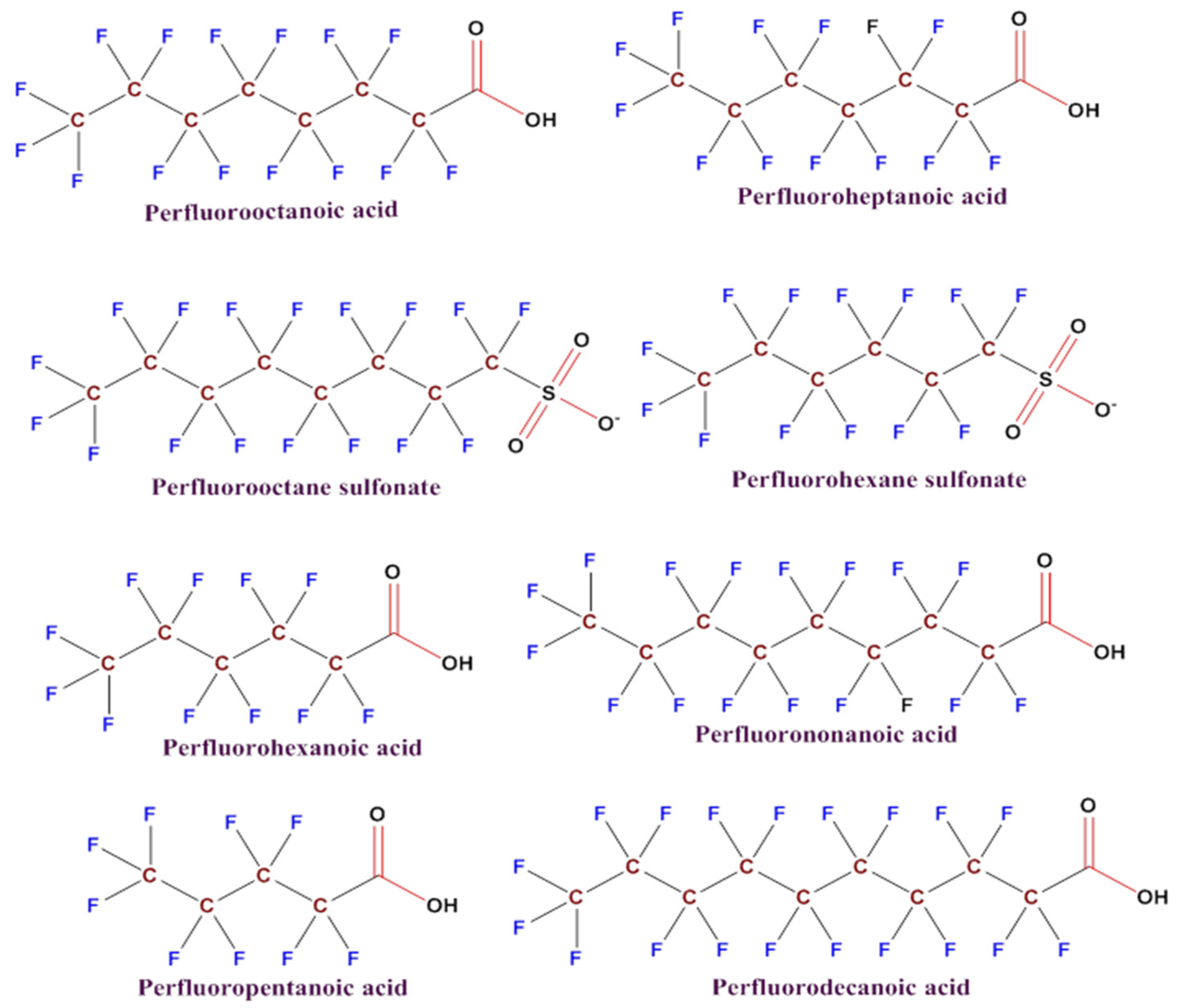
Figure 2.
a) An overview of the PFAS extraction process from solid or liquid samples. b) A range of detection techniques for finding PFAS in environmental samples [51]. Notes: LLE = Liquid-liquid extraction, SPE = Solid phase extraction, GC = Gac chromatography, HPLC = High-performance liquid chromatography, LC-MS/MS = Liquid chromatography with tandem mass spectrometry.
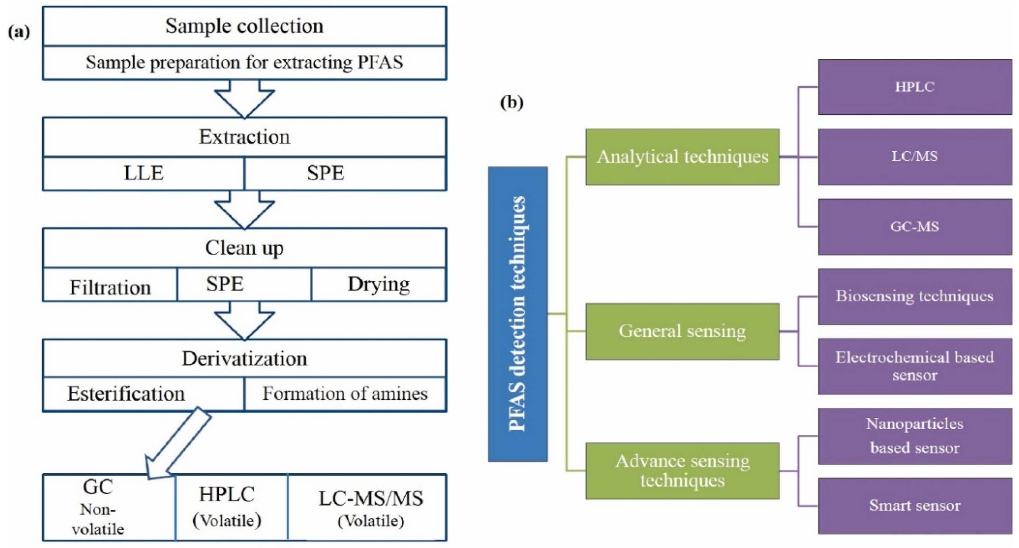
Figure 3.
The use of a gold electrode for molecular imprinting and detection of PFAS: a schematic depiction. Po-PD = poly (o-phenylenediamine), MIP = molecularly imprinted polymer, o-PD = ortho-phenylenediamine (o-phenylenediamine) [78].
Figure 3.
The use of a gold electrode for molecular imprinting and detection of PFAS: a schematic depiction. Po-PD = poly (o-phenylenediamine), MIP = molecularly imprinted polymer, o-PD = ortho-phenylenediamine (o-phenylenediamine) [78].
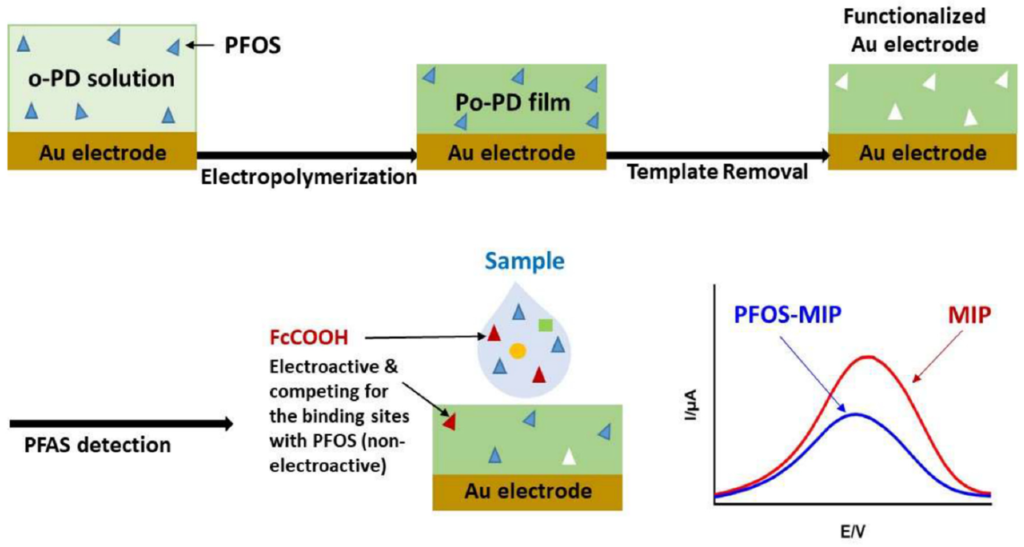

Table 1.
Several methods are currently accessible for extracting and detecting PFAS levels in environmental samples.
Table 1.
Several methods are currently accessible for extracting and detecting PFAS levels in environmental samples.
| Techniques | Samples Type | Limit of Detection (LOD) | Extraction Methods | Detected PFASs | References |
|---|---|---|---|---|---|
| LC-MS/MS | Water | 0.6–8.7 ng/L | LLE | PFOA, PFOS, PFHxA, PFODA, PFHpS, PFDS | [65] |
| HPLC-MS/MS | Water | 0.01-1.15 ng/L | SPE | PFOS, PFOA, PFNA, PFHpA, PFDA, PFHxS, | [66] |
| LC-MS/MS | Milk | 0.057 ng/L (PFOA), 0.021 ng/L (PFOS) | LLE | PFNA, PFDA, PFOA, PFHpA, PFBS, PFHxS, PFOS, PFUnA | [67] |
| LC-MS/MS | Liquid Sample | 0.29–6.6 ng/L | SPE | PFOS, PFOA, PFNA, PFDA, PFBA, PFUnA, | [68] |
| LC-MS/MS | Fruits and Vegetables | 0.07 ng/g (PFOS) | SPE | PFOS, PFOA | [69] |
| HPLC-MS/MS | Sediment | 1.5–10.9 ng/L | LLE | PFOS, PFDA, PFOA, PFDoA, PFHxS, PFNA | [70] |
| HPLC-MS/MS | Cabbage, lettuce, mustard leaf, | 0.017-0.180 ng/g | Ultrasonic extraction | PFOS, PFHxS | [71] |
| LC-MS/MS | Vegetables | 0.002–3.73 ng/g | LLE | PFOS, PFOA, PFDA, PFBA, PFBS | [72] |
| HPLC-MS/MS | Surface water | 50–1790 ng/L | SPE | FOSA, MeFOSA, EtFOSA, MeFOSE | [73] |
| HPLC-MS/MS | Different Water Samples | 0.05–0.22 ng/L | SPE | PFOS and PFOA | [74] |
Notes: PFOA = Perfluorooctanoic acid, PFOS = Perfluorooctane sulfonic acid, PFHxA = Perfluorohexanoic acid, PFODA = Perfluorooctadecanoic acid, PFHpS = Perfluoroheptane sulfonate, PFDS = Perfluorodecane sulfonate, PFNA = Perfluorononanoic acid, PFHpA = perfluoroheptanoic acid, PFDA = Perfluorodecanoic acid, PFHxS = Perfluorohexane sulfonate, PFBS = Perfluorobutane sulfonate, PFUnA = Perfluoroundecanoic acid, PFBA = Perfluorobutyrate, PFDoA = Perfluorododecanoic acid, FOSA = Perfluoroctano sulfonamid, MeFOSA = N-methyl perfluorooctane sulfonamide, EtFOSA = N-ethyl perfluorooctane sulfonamide, MeFOSE = N-Methyl perfluorooctane sulfonamido ethanol.
Table 2.
Some of the PFAS detection sensor systems that are now available.
| Detection System | Matrix | Limit of Detection (ng/L) | Detection Range (ng/L) | References |
|---|---|---|---|---|
| Spectrofluorometer | MPA-CdS QDs | 124200 | 207000–16563000 | [79] |
| Colorimetric detection | Gold nanoparticles | - | - | [80] |
| Electro chemiluminescence | Ultrathin nanosheets of carbon nitride | 10 | 20–4000 | [81] |
| Optical | Novel SPR | 210 | 0–200000 | [82] |
| Optical density | Bio-Gold Nanoparticles | 2.5 | 2.5–75 | [83] |
| Smartphone camera | Smart sensor | 0.5 | 10000–1000000 | [12] |
| Impedimetric | PFOS | 0.0005 | 0.00005–50000 | [84] |
Note: MPA = 3-mercaptopropionic acid, CdS = Cadmium sulfide, QDs = Quantum Dots.
Disclaimer/Publisher’s Note: The statements, opinions and data contained in all publications are solely those of the individual author(s) and contributor(s) and not of MDPI and/or the editor(s). MDPI and/or the editor(s) disclaim responsibility for any injury to people or property resulting from any ideas, methods, instructions or products referred to in the content. |
© 2025 by the authors. Licensee MDPI, Basel, Switzerland. This article is an open access article distributed under the terms and conditions of the Creative Commons Attribution (CC BY) license (https://creativecommons.org/licenses/by/4.0/).
Copyright: This open access article is published under a Creative Commons CC BY 4.0 license, which permit the free download, distribution, and reuse, provided that the author and preprint are cited in any reuse.


