Submitted:
10 June 2024
Posted:
12 June 2024
Read the latest preprint version here
Abstract
The purpose of the study is to analyse and to identify specific structural characteristics of melanocytic nevi. So, for better conclusions, using the optical microscope, could be possible a proper describtion related melanocytic nevi, reffering to youth pacients. In this research direction, good to mention that in a human individual life, play a significant role, different factors. That include in line, genetic, epigenetic, microbiomic, and proteomic factors. Future directions refers to preventive and prophylactic methods.
Keywords:
youth age
; patients
; epiderm
; melanocytic nevi
; analyse
Introduction
In order to define a disease, must have in attention a lot of differents factors such as historical points, or social and cultura.. Results of research studies, show us that some connective cells such as fibroblasts, lose their identity, in pathological conditions. [1] Another specific cells, namely melanocytes are known that having a specific structural point that is consider important in structural pathological description. [2] Refering to melanocytic nevi, in medical specific field of study and of research, various pigmented lesions of the epiderm, known as nevi, could be observe in different pars of the body. [3] For a proper diagnostic, an atypical nevus,can be biopsied.[4] In this direction, is important to practice a biopsy beside the extended clinical evaluation in melanocytic nevi. Researchers and specialists, usualy are looking also for changes that surrounding nevi . A great point in this field of research, could be possible the genetic susceptibility for morphological and functional alterations, in nevi. [5] A complete medical examination, play a great point for establishing the medical conduct, for a healthy status improving.[6,7] Structural analysis describe specific cells namely melanocytes as aggregated in ‘nests’, which conduct forming the nevus cells.[8] To the human persons with different age, this specific cells knowing as melanocytes could be found in various areas of the epiderm of the parts of the body. [9,10] Theoretical and practical studies, show that melanocytic nevi developing in utero present genetical differences from those that appear later.[11,12] In the present field, we can mention about various informations from scientific literature, referring to specific nevi. [13] Also from literature and from practicum actually are known different scientific informatiuons about extending melanocytic nevi, having specific scientific names.[14] Because are many cases in all of the world, diagnosed as melanocitic nevi, we can mention that currently, the proper treatment of epidermal nevi is challenging. [15,16,17,18]Congenital melanocytic nevi it is known as a subject of research that offer controversy. [19] Clinical monitoring in congenital melanocytic nevi is important for diagnosis and for possible medical treatment strategies applications. [20] A complete examination of the human body, during a medical examination, is important. [21,22] Best to mention that the nowadays higher incidence in melanoma is in acompaniament of the nevi existence of the body and of the increase exposure to the ultraviolet light. [23,24] Practical biopsy is important for diagnosis. [25]
Material and Methods
For the purpose of the study we can mention a little bit about classic laboratory method used and about the materials needed. For the fulfilment of permanent microscopic preparations was followed the steps of the classic method, using Hematoxylin Eosin staining. The samples used were from male and female youth patients, before mature age, from urban and rural residence. The operative pieces are intended to bring in the pathological anatomy service for macroscopic examination for diagnostic purposes. All pieces are examined by performing the optical microscope technique and analysis.
Results
Epiderm protect us during the life, from different factors. For a morphological analyse, structural and ultrastructural characteristics could be describes, using optical and electron microscope. Structural analyse of the epiderm, using colour laboratory techniques, is able to describe the specific layers with their characteristics. More than, using electron microscope, specific compounds as filaggrin which is knowing as an important epidermal protein and/or tight junction located in the granular layer of the epiderm, could be observed. For this purpose, transmission electron microscope examination, is consider one proper method for analyse. Scanning electron microscopy is also a modern method for analyse, which offer results that demonstrate abnormalities in the epiderm ultrastructure. (Figure 1) The human body is covers by skin and the epiderm contein differents types of glands, as sebaceous glands and sudoripar glands. In this study direction, it is known a typical physiopathlogic mechanism for the functionality of the body, including epidermal compounds and their body sorroundings. (photo 1)
Figure 1.
Methods for epiderm analyse.
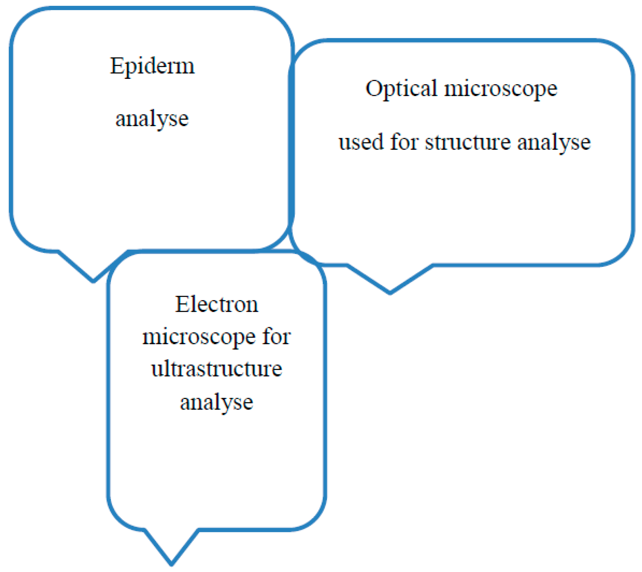
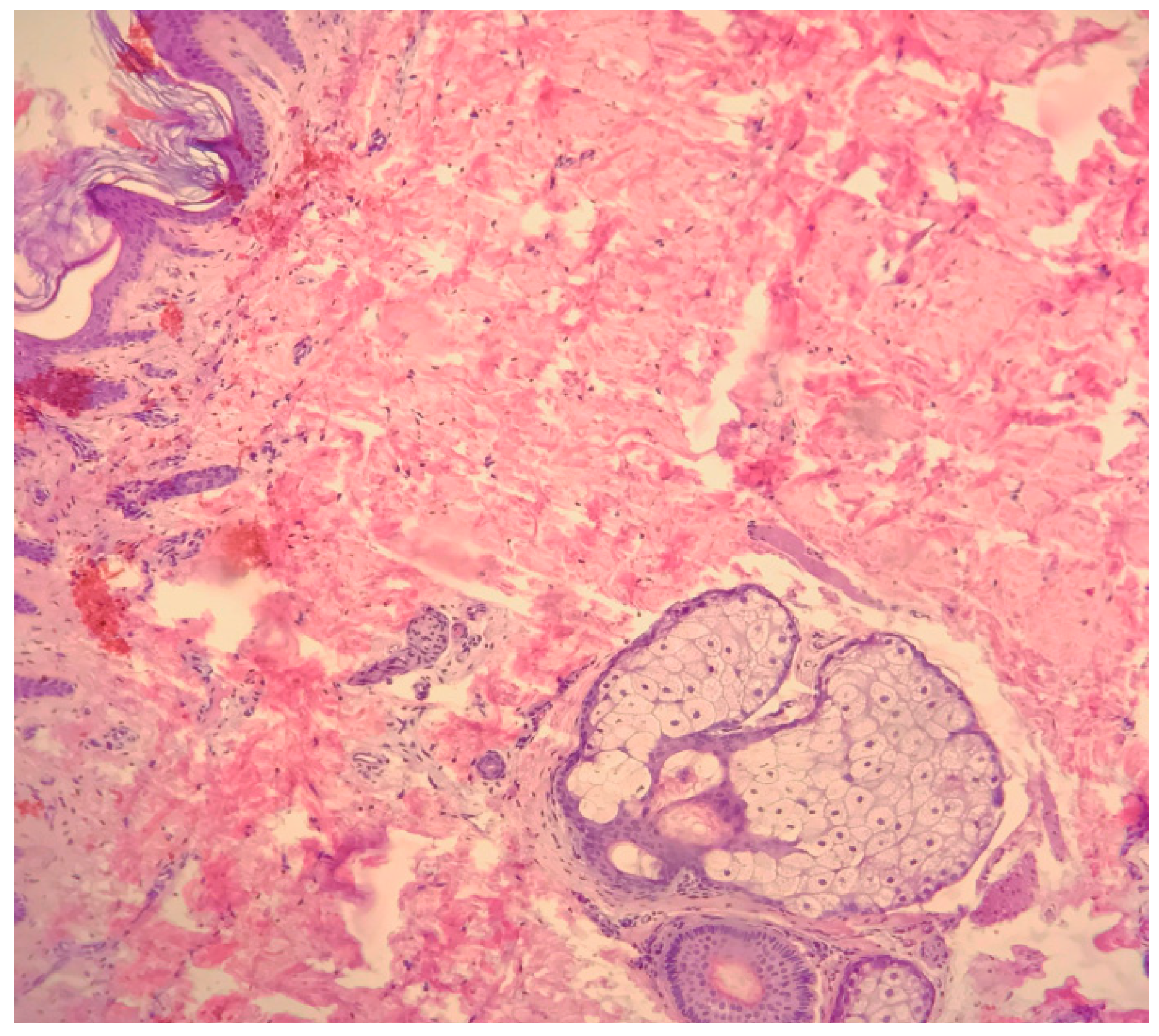
Photo 1.
Epiderm and annex glandsx10 Hematoxylin-Eosin staining.
Histopathological analyse describe various modifications to the melanocytic nevi aspect, located on various regions of the body. So we can mention asymmetry, irregular form, cytologic atypia, and mitotic activity. Medical specialists, describe and conclude that to benign melanocytic nevi, could be possible a describtion for atypical pathological characteristics of nevi and more important to mention characteristics when benign nevi are traumatized.(Figure 2) Dermoscopy play a role for a proper diagnostic important in practice to all ages, including, youth age and children.
Epiderm is a barrier, but is able for conducing to an illness status if include modifications in structural compounds.
Histopathological analyse describe the melanocytic nevi located on various regions of the body,with asymmetry, irregular form, cytologic atypia and mitotic activity. More than, medical specialists, describe and conclude related to the structural aspects in benign traumatized melanocytic nevi. In this field, dermoscopy play a role for a proper diagnostic.
Discussions
Great interest in knowing epidermal compounds. So, the epiderm, is composed of a number of specific lyers. Specific cells are known. One of the role of the epiderm is implication in differents injuries. Alterations in the compunds of the epiderm layers, contribute to the visual signs of pathologic conditions. One research direction, refer to the role of benign melanocytic lesions with alterations, which conduct to malignant cutanat melanoma. Related to melanocytic nevi, in some circumstances, could be possible that the prognosis be poor having in attention the healthy of the patients having comorbidities.
Conclusions
Prevention and educational methods, are important. More than early detection in melanocytic nevi is a great point in order to try to treat and to avoid maybe possible malignancy degeneration. Techniques for the laboratory diagnosis that are implications in monitoring previously pathological status, are implied and conduct to a proper quality of life in patients diagnosed with melanocytic nevi. .In this direction, implication of an interprofessional team strategies is one of the proper condition. Future trends, new laboratory techniques for diagnosis.
References
- Salzer M.C., Lafzi A., Berenguer-Llergo A., Youssif C., Castellanos A., Solanas G., Peixoto F.O., Stephan-Otto Attolini C., Prats N., Aguilera M., Martín-Caballero J., Heyn H., Benitah S.A. Identity noise and adipogenic traits characterize dermal fibroblast aging. Cell. 2018;175:1575–1590.e22. [CrossRef]
- Palicka GA, Rhodes AR. Acral melanocytic nevi: prevalence and distribution of gross morphologic features in white and black adults. Arch Dermatol. 2010 Oct;146(10):1085-94. [CrossRef]
- Wang M, Xu Y, Wang J, Cui L, Wang J, Hu XB, Jiang HQ, Hong ZJ, Yuan SM. Surgical Management of Plantar Melanoma: A Retrospective Study in One Center. J Foot Ankle Surg. 2018 Jul-Aug;57(4):689-693. [CrossRef]
- Richtig E. ASCO Congress 2018: melanoma treatment. Memo. 2018;11(4):261-265. [CrossRef]
- Wang B, Qu XL, Chen Y. Identification of the potential prognostic genes of human melanoma. J Cell Physiol. 2019 Jun;234(6):9810-9815. [CrossRef]
- Bristow I, Bower C. Melanoma of the Foot. Clin Podiatr Med Surg. 2016 Jul;33(3):409-22.
- Lallas A, Kyrgidis A, Koga H, Moscarella E, Tschandl P, Apalla Z, Di Stefani A, Ioannides D, Kittler H, Kobayashi K, Lazaridou E, Longo C, Phan A, Saida T, Tanaka M, Thomas L, Zalaudek I, Argenziano G. The BRAAFF checklist: a new dermoscopic algorithm for diagnosing acral melanoma. Br J Dermatol. 2015 Oct;173(4):1041-9. [CrossRef]
- Tronnier M. Melanotische Flecke und melanozytäre Nävi. In: Plewig G, Ruzicka T, Kaufmann R, Hertl M. Braun-Falcos Dermatol. Venerol. Allergol. Springer Berlin Heidelberg, 7. Auflage, 2016.
- Tolleson WH. Human Melanocyte Biology, Toxicology, and Pathology. J Environ Sci Health Part C 2005; 23: 105–61.
- Thomas AJ, Erickson CA. The making of a melanocyte: the specification of melanoblasts from the neural crest. Pigment Cell Melanoma Res 2008; 21: 598–610. [CrossRef]
- Colebatch AJ, Ferguson P, Newell F et al. Molecular genomic profiling of melanocytic nevi. J Invest Dermatol 2019; 139: 1762–8. [CrossRef]
- Kinsler VA, Thomas AC, Ishida M et al. Multiple congenital melanocytic nevi and neurocutaneous melanosis are caused by postzygotic mutations in codon 61 of NRAS. J Invest Dermatol 2013; 133: 2229–36. [CrossRef]
- Bandyopadhyay D. Halo nevus. Indian Pediatr 2014; 51: 850.
- Fernandez-Flores A. Eponyms, Morphology, and Pathogenesis of some less mentioned types of melanocytic nevi. Am J Dermatopathol 2012; 34: 607–18.
- Kim JJ (Department of Dermatology, Henry Ford Health System, Detroit, MI 48202, USA), Chang MW, Shwayder T. Topical tre-tinoin and 5-fluorouracil in the treatment of linear verrucous epi dermal nevus. J Am Acad Dermatol. 2000 Jul;43(1 Pt 1):129–132.
- Brown HM, Gorlin RJ. Oral mucosal involvement in nevus unius lateris (Icthyosis Hysterix). Arch Dermatol. 1960 Apr;81:509–515.
- Zvulunov A (Soroka Medical Center, Ben-Gurion University, Beer-Sheva Israel), Grunwald MH, Halvy S. Topical calcipotriol for treatment of infammatory linear verrucous epidermal nevus. Arch Dermatol. 1997 May;133(5):567–568.
- Boyce S (Washington Institute of Dermatologic Laser Surgery, Washington, DC 20037, USA), Alster T. CO2 laser treatment of epidermal nevi: Long-term success. Dermatol Surg. 2002 Jul;28(7):611–614.
- Arad Ehud, Zuker M. Ronald, The shifting paradigm in the management of giant congenital melanocytic nevi: review and clinical applications, Plast Reconstr Surg., 2014 Feb;133(2):367-376.
- Mologousis MA, Tsai SY, Tissera KA, Levin YS, Hawryluk EB., Updates in the Management of Congenital Melanocytic Nevi., Children (Basel). 2024 Jan 2;11(1):62. [CrossRef]
- Bristow I, Bower C. Melanoma of the Foot. Clin Podiatr Med Surg. 2016 Jul;33(3):409-22.
- Lallas A, Kyrgidis A, Koga H, Moscarella E, Tschandl P, Apalla Z, Di Stefani A, Ioannides D, Kittler H, Kobayashi K, Lazaridou E, Longo C, Phan A, Saida T, Tanaka M, Thomas L, Zalaudek I, Argenziano G. The BRAAFF checklist: a new dermoscopic algorithm for diagnosing acral melanoma. Br J Dermatol. 2015 Oct;173(4):1041-9. [CrossRef]
- Bartoš V, Kullová M. Malignant Melanomas of the Skin Arising on the Feet. Klin Onkol. 2018 Summer;31(4):289-292. [CrossRef]
- Palicka GA, Rhodes AR. Acral melanocytic nevi: prevalence and distribution of gross morphologic features in white and black adults. Arch Dermatol. 2010 Oct;146(10):1085-94. [CrossRef]
- Bristow IR, de Berker DA, Acland KM, Turner RJ, Bowling J. Clinical guidelines for the recognition of melanoma of the foot and nail unit. J Foot Ankle Res. 2010 Nov 01;3:25. [CrossRef]
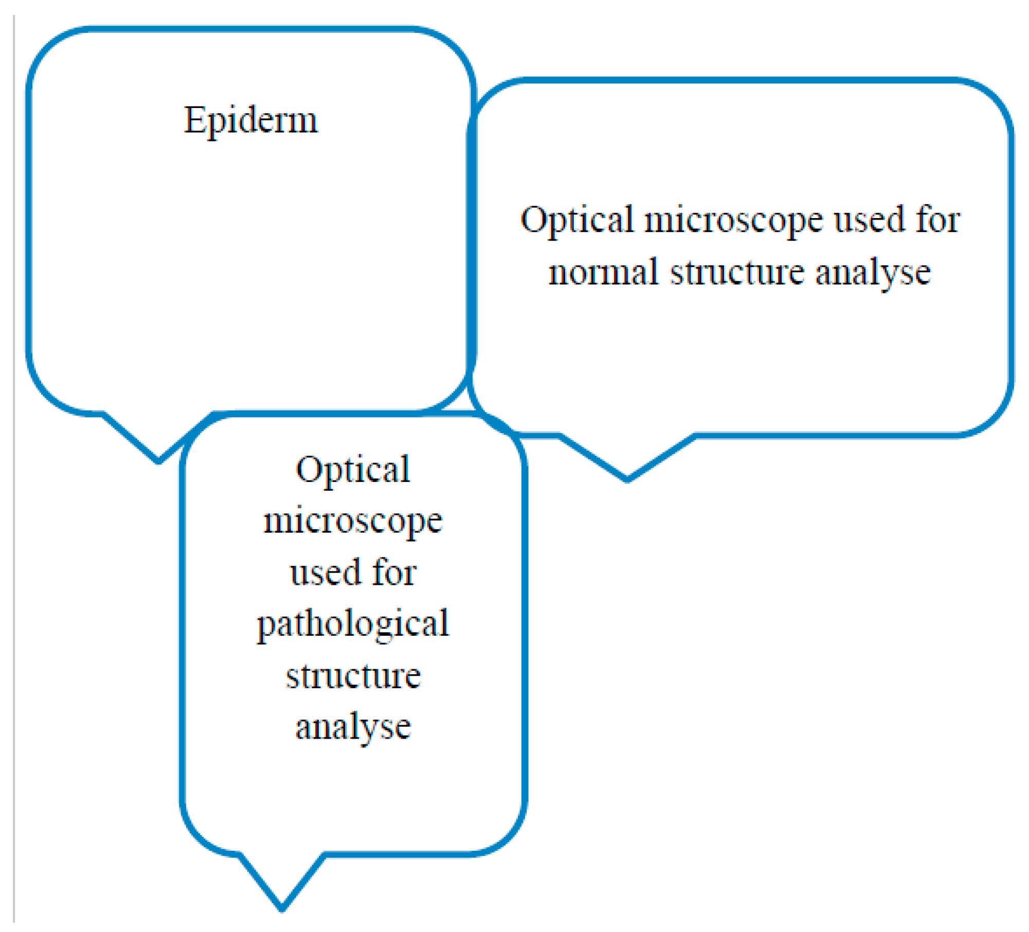
Figure 2.
Changes in epidermal compounds - from normal to pathological.
Disclaimer/Publisher’s Note: The statements, opinions and data contained in all publications are solely those of the individual author(s) and contributor(s) and not of MDPI and/or the editor(s). MDPI and/or the editor(s) disclaim responsibility for any injury to people or property resulting from any ideas, methods, instructions or products referred to in the content. |
© 2024 by the authors. Licensee MDPI, Basel, Switzerland. This article is an open access article distributed under the terms and conditions of the Creative Commons Attribution (CC BY) license (http://creativecommons.org/licenses/by/4.0/).
Copyright: This open access article is published under a Creative Commons CC BY 4.0 license, which permit the free download, distribution, and reuse, provided that the author and preprint are cited in any reuse.


