Submitted:
18 December 2023
Posted:
19 December 2023
You are already at the latest version
Preprints on COVID-19 and SARS-CoV-2
Abstract
Because the including of human anatomy forms the foundation for clinical medicine, it has contributed a pivotal role in medical education for many centuries. Thus with advancements in the science of modern, current day medicine, its continued place in the medical school curriculum deserves careful attention. In an attempt to provide guidance to decision-makers involved in clinical anatomy curriculum development at the medical school level, the Educational Affairs Committee of the American Association of Clinical Anatomists (AACA) developed this document, which defines the contours of a gross anatomy curriculum leading to the M.D. or D.O. degree. The main body of the document sets forth the anatomical concepts as well as the subject matter a student should master prior to graduation from medical school. Gross anatomy dissection is still a crucial topic in graduate and medical school. The nature and content of gross anatomy, which was historically regarded as a core course in the first year of medical education in MBBS, MD, DO, and some PhD and Graduate curricula as well as in other disciplines of selective graduate education and non-allopathic medical programs, have been significantly impacted by emerging challenges in the pedagogy of delivering medical education in the past ten years. A seemingly ever-increasing emphasis on additional modules in molecular biology, emerging pharmacotherapeutics and pathophysiology content have resulted in a condensation and apparent erosion of previously allotted classroom and laboratory hours in the gross anatomy curriculum. In addition, an imposition of financial constraints and a decreasing availability of sufficient cadaver donations to support gross anatomy dissection labs has also occurred in some locations. An innovative clinically focused cadaveric anatomy program is described which has improved the delivery and content retention of medical and graduate students and has become one of the most sought-after modules in the revised clinically oriented curriculum and has been deemed highly relevant to current medical education and graduate studies.
Keywords:
Anatomy
; cadaveric dissection
; prosection
; virtual dissection
; clinical integration
; COVID-19 pandemic
; fiberoptic examination
; surface anatomy.
1. OVERVIEW
Historically, the practice of anatomic cadaveric dissection has long been considered an essential component and necessary milestone in the medical education of aspiring physicians and biomedical scientists since the earliest recorded history of medical education. In ancient Egypt, cadaveric dissection originated as a religious ritual that was required as a rite of passage to the kingdom of the dead, where the procedure resembled more of a crude autopsy than an anatomical dissection as we might perform the process today. The earliest recorded medical school was founded in Salerno, Italy in 1235. In that program, cadaveric anatomy soon ascended to a prominent position in their medical curriculum. At Salerno, human dissection was performed as a sacramental procedure that illustrated the dissertations of revered ancient authors of the era. During the Renaissance, with the opening of the Anatomical Theatres in Padua (1490) and Bologna (1637), anatomy was considered an essential albeit artistic and spiritual exploration of the life, suffering, and death of the deceased, although most causes of death were then unknown. Anatomists began to dissect more extensively to investigate the structure of the body and produced texts that illustrated the images that were based on their dissections, such that pathological variations might be noted. The modern era of scientific aspects of human anatomy was highlighted by the publication of the main opera from Andrea Vesalius (von Wesel), who is considered prominent among the real fathers and ancestors of the scientific nature of modern anatomy as we know it today.
In the 17th century, the renowned Dr. Nicolaes Tulp, as a young, fledgling artist in his early 20s, painted the now famous rendition of cadaveric dissection. Renditions of his painting now adorn most modern anatomy departments. He was well known in Amsterdam as a practicing physician and surgeon. He was also a prominent civic leader and an anatomist who was appointed as Praelector in Anatomy. Among his duties and responsibilities were to regularize public dissections; he was also charged by the Surgeons' Guild to apprentice new surgeons. Nicolaes Tulp (9 October 1593 – 12 September 1674) was a Dutch surgeon and the mayor of Amsterdam. Dr. Tulp was well known for his upstanding moral character and as the subject of Rembrandt's famous painting The Anatomy Lesson of Dr. Nicolaes Tulp [1,2,3,4]. Dr Nicholaes Tulp has since earned centuries of acclaim in medical anatomy following his famous rendition of a cadaveric dissection since first portrayed in 1832 while he was still the emerging artist we now recognize as Rembrandt. Thankfully the teaching of anatomy has enjoyed many advances in pedagogy and process since that time, although the basics displayed by Dr Tulp remain important in today’s discussion [1,4]. The 18th to the 20th century saw a large expansion of medical schools throughout most countries of the world, each with their own model of inclusion of cadaveric dissection. Indications of religious overtones were sometimes notes, as cadaveric skeletal remains have recently been unearthed in Cambridge MA Holden Chapel and others, then the sites of early US medical schools. [5,6,7] Dissection instrumentation and decorum was similar to the surgeons tools of the day and has undergone vast improvements to the present day where it may resemble a modern surgical theatre with an expanded capacity of multiple cadavers simultaneously to accommodate the larger numbers of medical students now common to most Instructions.(refs) Moreover, since the outset of the recent Covid-19 pandemic, the incorporation of alternative strategies in the modernization and reorganization of anatomy curricula have placed increasing economic and didactic pressures on previously existing curricular models but have not outpaced the need for some back to the basics skills in dissection. Regional dissection is still what most surgeons and anatomists do best and requires more than textbook and virtual illustrations for the developing practitioner to excel in their vocation.
In the recent past decades, the delivery of the gross anatomy course in medical and graduate education used to occupy up to a full academic year of the curriculum. However, the entire course is now often reduced to a single semester. Moreover, the travel and public assembly restrictions forced by the pandemic required the adoption of off-campus modules for much of the traditional lecture content and limited the opportunities for the dissection element of the instruction. This mandated adjustment has included substantial reduction in both didactic lecture availability and laboratory dissections that were once a major core course and the major hallmark of a first-year medical student’s and some graduate student’s academic pursuits [9,10,11,12,13,14]. The results are that the role of gross anatomy as a core course in the medical curriculum has been forced to undergo much needed and welcome transition in scope and content in many institutions around the globe based on a variety of economic, epidemiological, and other factors [14].
With great advancements in educational technology and in the overall content and complexity and a drive toward a more clinically oriented focus of basic medical sciences in recent decades, the necessity for broad progressive updates and topic integration has now assumed an urgency in curriculum development. Because a fewer number of the generous clock hours previously allocated for gross anatomy instruction still remain available, the necessity for close review of the most essential topics, critical course content and compressed time for laboratory sessions are now imminent. It is noteworthy that the overall subject content, depth of instruction and student comprehension in the developing current curricular models has not diminished, while student expectations have likely increased. If anything, the overall content has not only maintained its overwhelming criticality in importance, but it has also increased in its complexity in its potential applications [15]. Therefore a driving need exists to condense such courses into shorter time frames and clock hours of participation to the extent possible. These benefits may be attained by making more efficient use of resources to include the incorporation of a stronger clinical focus, by integrating computer-assisted virtual applications, simulation exercises, 3-D modeling, problem-based learning (PBL) approaches, and by adding a presurgical approach to the laboratory modules by including surgical theatre demeanor and clinically relevant procedures [15,16]. The net result of the above is geared toward providing a stronger and more appropriate instructional program that is more closely related to the current time, period, and circumstances of contemporary interest. By offering a more relevant content and technologically-enhanced quality of medical education an institution may better meet the challenges and success on qualifying examinations the future may present to the new medical or bioscience graduate [17].
If the above objectives are achieved, including presenting the resulting course content in the most imaginative and cost- effective manner possible within the available time constraints, students should be able to complete their primary medical or graduate qualification and subsequent graduation from medical or graduate school within the current 4 or more years, depending on the nature of the program (i.e., MBBS, MD, PhD, or combined program). To accomplish these objectives, the didactic lectures are often oriented along organ system-based settings for each phase of the lectures, incorporated with a virtual anatomy system such as Anatomage® or similar virtual instructional assist and organized with a defined clinical focus and clear objectives and end points in mind [18,19,20]. Small group lecture discussions and demonstrations are encouraged to include topics in Pathophysiology, Medical Biophysics, Medical Epidemiology, and preclinical introduction to selected clinical procedures. In addition, a discussion and practical demonstration of meridians, acupuncture and acupressure points may be integrated where appropriate to further broaden the scope of the lesson [20].
The culmination of the dissection traditionally ends with each student being able to demonstrate the above procedures to a defined standard, including a complete functional surgical dissection often accomplished with an unembalmed cadaver, as the aforementioned cadaver can add additional realism and clinical focus to the process [18,19,20,21]. The anatomic content is reinforced and revisited in subsequent years in modules in Clinical Anatomy, Pathophysiology, Pathology, Forensic Medicine, and such other topics deemed relevant to review prior to the student’s formal entry to the clinical years.[18] With careful attention to planning, curriculum, and adherence to allocations of permissible timeframes, one can likely fulfill the needs of tomorrow’s medical practitioners, surgeons, physicians and biomedical scientists; with careful attention to content and need. To accomplish the academic challenges successfully will require the undeterred will and motivation of a dedicated faculty and staff, and availability of emerging educational technologies now available. With such resources in mind, one can help to attain the challenge that will enable the students of today and tomorrow to master the field of anatomy, physiology, and neurobiology like never before [22,23].
2. INTRODUCTION
Emerging Global Changes in Medical Education have increased the pressures to increase the focus, content, and efficiency of delivery in medical education in many subject areas to keep pace with advances in scientific discovery of new therapeutics and emerging diseases without losing sight of the many significant albeit often historical advances in clinical medicine which have transpired in recent decades [9,10,11,12,13]. While some have questioned the future directions of traditional education in Anatomy with suggestions that it is a declining field of interest for medical and graduate study, the importance as a core requirement in medical education remains as strong now as in the past generations [9].
Although many everchanging advances in technology have occurred and traditional approaches to pedagogy have presented new challenges, the core specialties including the specializations of surgery, radiology and others continue to remain heavily dependent upon a solid mastery of anatomy [11]. These pressures often occur with simultaneously shrinking budgets, limited faculty, and challenging resources, including decreases in available cadaver donations and changing perceptions of need in medical education across the globe, with severe limitations in some countries [12,15,24]. This has generated a shift towards a greater emphasis on molecular biological and pharmacotherapeutic aspects than on the more traditional physical aspects of conventional medical practice [10,10,11,12,13,18].
Emerging communicable infectious illnesses, including the recent coronavirus pandemic have now added an additional extraordinary burden to the educational process by forcing compromises in classroom and laboratory participation. These factors have further impacted negatively on cadaver availability due to sometimes unresolved specimen safety concerns, all without easy solutions especially when using fresh or unembalmed cadavers [14,21]. The greatest challenge however may be progressive schedule reallocations of laboratory hours complicated with ongoing decreases in available clock hours for some subjects that previously could be allotted to the cadaveric dissection experience [9,10,16]. Also, these limitations combined with shortages of experienced and knowledgeable faculty, as retiring faculty may not always be replaced with the same disciplines, talents, experience levels and specializations as in the past, or at the same rate as institutional needs become obligated to adjust to a changing classroom, a shrinking budget, and progressive changes in the laboratory and educational environment made available. Unlike a computer- based laboratory, cadaveric labs require adequate supervision and safety concerns, thereby placing further constraints on opportunities for individualized modes of instruction where unique dissecting skills and surgical techniques may be gained [13,14].
The role of biomedical physics and body mechanics applications to clinical medicine might also be mentioned here, as clinicians are often presented with a broad range of musculoskeletal, orthopedic and rheumatology issues [18,19,20]. Thus, a solid grounding in the biomedical physics of body mechanics and joint articulations and their many ramifications can be conveniently integrated into the anatomy curriculum both as practical and biomedical engineering objectives. Respiratory illness or vertebral injuries, for example may change one’s posture sufficiently as to alter a patient’s gait and posture and suggest a need for physical therapy or other aspects of physical medicine to resolve the issues, often without overdependence on opiates or other analgesic agents.
2.1. Determine the Objectives for a Successful Outcome in Advanced Level Cadaveric Dissection Clearly before you start the course.
Every successful anatomy experience should best start with an investigation of the desired objectives and desired outcomes, and supported with a well thought out lesson plan that will help to achieve the objectives deemed important for the lesson and dissection experience. In planning such an experience, consideration should be given to a) the level of prior education and experience of the intended audience; b) the need for any additional pre-dissection instruction before the actual dissection commences; c) the availability of suitable cadavers, dissection tables, instructional staff and availability of additional resources deemed necessary for a successful experience; and d) the group or class size undertaking the dissection laboratory must not exceed the capacity limitations of the dissecting theatre and available tables and instrumentation resources. Securing the availability of adequate resources typically requires significant preplanning, often commencing several weeks or months in advance of the planned laboratory experience, so as to insure access and availability of needed resources. Specific points one might entertain developing the plan are Included but are not limited to the items summarized below.
2.2. Determine what the critical endpoint in the Anatomy Course Duration and Content should be ? How long should it Last and determine what goals you wish to Accomplish.
Cadavers are a highly valuable but often scarce and expensive resource and must be utilized judiciously to obtain the best advantage and experience for the student. Historically traditional Gross Anatomy courses were once spaced over a 2 to 3 semester experience, staffed with dedicated faculty and assisted by surgical residents, fellows, and graduate assistants, themselves often still in training. This support staff could readily assist a faculty member in modelling and developing effective course delivery and provided the future physicians and surgeons with a comprehensive knowledge of anatomical features, clinical perspectives and provide a meaningful introductory presurgical experience that may carry over to the clinical environment [22]. Those assets Institutions were once accustomed to are now often pared down to a mere one semester block of instruction but with the same or greater projected endpoints in terms of expected academic content and stated course outcomes [10]. These adjustments have thereby necessitated shortcuts such as demonstrations, prosections, and abbreviated, condensed lab sessions compared to the more extensive and time-consuming dissections of yesteryear.
2.3. Instruction in Cadaveric Anatomy has Increased to Include a More Clinically Oriented Focus
The graduated physician is still expected to have mastered the same or greater grand level of knowledge of anatomy, physiology, and neurobiology as in the past. Moreover, one can be assured that the ‘board exams’ [i.e., the USMLE or equivalent comprehensive assessment] when taken, as well as the senior resident mentor, will expect no less than the same amount of the detailed knowledge-based information likely needed to navigate the most complex procedure one might encounter in the theatre [17]. In addition, one can also be assured that the ‘board exams’ will be certain to cover the same range of body parts and physiologic processes on those written examinations that one may have missed or inadvertently forgotten along the intervening years between the in-course experience and the ultimate qualifying examinations for entry into the advanced training years. In addition, as mentioned above, a looming shortage of qualified and experienced instructors in the field of human anatomy, physiology and neurobiology has been reported and is likely to continue to continue to be present in the future, as abundant opportunities for graduate training in this field appear to have been lacking [4,8]. Moreover, in recent years as the emphasis had been shifting toward cellular and molecular biology-oriented topics, and with what many may perceive as more exciting fields of study, particularly in the gross anatomy side of the profession. The cumulative impact of these changes in the delivery of anatomy education appears to have led to a lack of qualified instructors in some of the more basic and rudimentary elements of the curriculum [9,10,11,12,13,14].
2.4. Necessity has Often been Stated as the ‘Mother’ of Invention: Anatomy Budgets and resources may become constrained and require adjustments in pedagogy and course structure.
In most institutions, class size is often a deciding factor as to whether a particular module or class may be offered. As academic budgets and class times evolve and become readjusted, class size considerations can become a hot topic, as the larger the class size, the more cost effective it can become for the Institution to conduct the class. This is especially true for lecture type instruction where larger numbers of students may need the same required course. Small class sizes and laboratory expenses can become cost-prohibitive if the number of enrolled students per session is too small, and most institutions have well established lower limits that typically border around a certain single- or double- digit number that essentially revolves at least in part around cost recovery for offering elective and other course modules. For the most effective student- to-faculty ratio in medical instruction however, those lower numbers simply often do not work very well in many scenarios. Thus, anatomy lab as well as other specialized medical topics is one of those particular scenarios where a closer faculty to student ratio may be more effective but less economically feasible, thereby imposing limitations in the overall anatomy laboratory dissection experience. The importance of the class may be overshadowed by the cost to deliver it efficiently despite the fundamental nature and importance of the topic to the aspiring student.
2.5. Anatomy Essentials are Best Incorporated as a Foundation course early in the Medical Curriculum with continued emphasis throughout the duration of the foundation and clinical years.
Anatomy lecture and lab is traditionally a first-year course in medical education, where class lecture sizes are of necessity large, often 100 to 200 or more to a lecture session. For laboratory exercises however, small group instruction is much preferred to large group demonstration, such that everyone around the dissection table can more easily participate and more effectively visualize the details of the specimen and participate effectively in the exercise [18,19,20,21,22]. In such settings there can also be greater opportunity for effective student-faculty interaction, particularly in pointing out the fine detail of particular or individualized anatomic findings that may be present, and which are often unique to a particular cadaver. It is valuable to have an experienced mentor, preferably one with prior surgical or pathology experience, such as a retired or senior surgeon or pathologist who can readily identify and discuss specific pathologic teaching points and their likely sequela along the way during the dissection. As such, it presents a more effective teaching and learning situation, with freer uninhibited participation and with more student-instructor interchange resulting in less perceived student hesitation or perceived instructor intimidation. In smaller settings such as are described here there can be better and more effective attention paid to the uniqueness of each cadaver, and the more individualized teaching points that it may present.
It is also always useful to have both a gross anatomy reference. A pathology reference and a whiteboard readily available nearby during dissections, such that spontaneous discussions may ensue where students and the instructor assigned to the table may become engaged in creative discussions about the subtopic at hand. The ready availability of reference resources such as an anatomy or pathophysiology text close at hand may further encourage instructor-student shared exchanges and thus enhance the dissection experience. The greater the student involvement, the more likely the retention factor will be improved for later reference and correlation to clinical findings by the student in subsequent modules. The ready availability of appropriate supporting resources enables one to research topics of interest that may arise during a dissection procedure, rather than to delay such topics until after the moment has passed and instant interest has cooled. The whiteboard may be used to aid in the discussion, or perhaps for example to outline a surgical approach for a particular surgical procedure, then during the dissection, undertake such a procedure as part of the regional dissection, thereby adding a valuable clinical focus to the dissection. The uses for a readily available whiteboard adjacent to a dissection table are virtually unlimited and can add much to a dissection exercise.
2.6. Cadaveric dissection is an intense, focused experience; Recording Devices, Cell Phones and extraneous socialization are Usually Prohibited in Dissection Settings Unless Specifically Preapproved
Virtually every student attending today’s classroom arrives with a cell phone in hand. The use of cellphones is typically prohibited in a dissection laboratory however, with the exception of the camera use for limited, preapproved instructional or legitimate educational purposes only. In such instances, where one must be careful to not photograph the cadaver per se for identification purposes, and the instructor must remain vigilant to ensure that the safety and cadaver confidentiality remain intact. However, taking a picture of the whiteboard notes may be allowed under some circumstances, but should only be attempted with the prior express (preferably written) consent of the author or instructor whereby some forms of recording for later instructional purposes or topic review may be undertaken. It should be noted that some laboratories maintain surveillance cameras for safety concerns and security purposes, typically placed at a safe distance from the dissecting tables so as to be able to record human activity and student behavior only while not focusing on cadaver specifics per se [18,19,20,21].
2.7. A Clinically Oriented Focus Can Enhance the Significance of the Dissecting Experience
It is fundamentally logical and important to establish a sound clinical focus during anatomic instruction where it likely contributes to facilitating a greater content retention of the subject matter presented and discussed. It is usually beneficial to introduce some clinical procedures to the cadaver prior to the commencing the dissection per se, where such incorporation may offer the distinct advantage that it may provide an orientation to the subject, contribute to instructional realism, and ultimately can cause no harm to the student or the cadaver. It is essential that all fresh or unembalmed cadavers be safety tested and care taken to conduct the lab in a safe environment, such as state-of-the-art dissection laboratory facility equipped with adequate laminar- flow ventilation to minimize unwanted distraction during the often-condensed duration of the planned laboratory experience [21,22]. In addition, the laminar air flow minimizes the risks of inhalation and exposure to noxious fumes or unpleasant odors during the dissection exercise. Prior to the dissection, important sensitivity instruction should be given, including acceptable respect, clinical PPE attire including surgical scrubs and gowns, proper dissection instrumentation and surgical technics and attention to expected personal demeanor from all in attendance during the dissection. Clinically oriented procedures one might incorporate include such items including:
- Surgical attire and conduct in an anatomy theatre is typically similar to that of a surgical theatre, including surgical setup, instrument tray, instrumentation technic and supporting resources.
- Surgical draping of the cadaver, regionally specific to the anatomic dissection under discussion, followed by reassembly of the major components following the dissection.
- Demonstration of an endotrachoscopy, bronchoscopy or other instrument assisted visual examination procedure.
- Introduce and practice multiple suturing technics: These are easily performed on fresh unembalmed cadavers and can yield a great additional value-added dimension to the dissection; suturing human skin tends to generate a different sensation to the student than occurs with other materials.
- Time permitting, when the dissection progresses, remove the lungs intact and connect to a heart lung apparatus and demonstrate the physiologic elasticity of the respiratory system, and then demonstrate and examine the tissues for environmental injury such as smoking, drowning, forensic indications, etc. including examination of selected tissues under a dissecting microscope.
- Endoscopy of internal structures such as gastrointestinal, uterine, or other structures or and organs with an endoscope adds a dimension seldom available prior to a clinical setting and is an educationally rewarding experience likely to further reinforce long term recall to the student; Injection of a simple dye into cardiovascular and neural networks to better visualize such structures such as cardiac vessels and neural pathways that may be of interest. Simple dark colored food dyes are inexpensive any may be utilized as a substitute to more expensive tissue dyes for visualizing vasculature for classroom demonstrations should the tissue dyes be unavailable. As an added benefit, the dyes often reveal partial cardiovascular occlusions or other lesions that become excellent teaching points, and which otherwise may otherwise escape observation during the small or larger group dissections.
- Demonstration of surgical palpation, as the sense of touch in the surgeon’s hands and fingers become an important extension of his visibility, for anatomic features that the surgeon cannot see directly. The palpation of individual organs becomes an important part of the dissection, as much as how one holds the instruments, removes tissues, or obtains biopsy specimens is an important component of presurgical theatre. The opportunity to obtain biopsy specimens for integrated histologic evaluation adds an additional valued dimension to the dissection experience and may help the participating students to determine the likely cause of death of the cadaver.
- Numerous reports attest such as suturing, ultrasonography technique and other clinical procedures including fiberoptic examinations of internal organs are improved when first practiced on fresh cadavers over plastic models or live patients [15,16], and which when performed on fresh, unembalmed or lightly embalmed cadavers reflect observations more typical of those which may be observed in live specimens [19,20,21] Professor Smith, of Brighton and Sussex medical school once reported that when one presses a knife into human flesh, the sensation subtly and uniquely changes compared to that of other species or artificial tissues as the knife penetrates and travels through its incision [19]. With each cut of the knife, it facilitates the creation of a valuable learned sensation and teaching point in surgical technique. To slice into a human artery in contrast, one may feel a bit of spring back due to the elasticity of the structure, while dissection of veins or lymphatics tends to result in a more passive or flat surgical response and do not produce the same spring back sensation and nerves a response intermediate to the forgoing examples. Acquisition of such skills early in one’s medical career can bring greater satisfaction to the surgeon or emergency room physician throughout their subsequent career, and hopefully the early learned skill transfer may reduce the likelihood of a future medical or surgical mishap at an inopportune time where it could potentially result in a dire outcome [22,24].
- The dissecting laboratory should be equipped with adequate laminar flow air circulation, regardless of the type of cadaveric specimens used so as to minimize any potential untoward effects of formaldehyde or other noxious chemicals or odors [25,26]. This is especially important when students are expected to spend long hours over many days in the presence of the cadaver or cadaveric specimen. During the dissections, which typically last for several days only on each fresh cadaver, key surgical and medical procedures that are likely to be needed during forward clinical years are demonstrated on each cadaver, including insertion of chest tubes, spinal taps, Intubation, topical and cosmetic suturing, endoscopy another fiberoptic examinations, 3-D imaging, surgical techniques, point location and others [18,19,20,21].
2.8. The cadaver is the center of interest and attraction. High Quality Cadaver Availability is Important to the Instructional Success of the Dissection Experience
The availability of suitable cadavers is a highly valued and often scarce resource in medical education, and the instructor lesson plan should provide for and enable all parts of the cadaver to be utilized effectively [10]. To achieve this objective, the cadaver might best be sectioned especially if several dissections are ongoing at different tables simultaneously. In such settings, students may take turns to circulate in small groups to each table in order to maximize the overall dissection experience and thus take the best advantage of different anatomic regions of the cadaver. In addition, whenever possible both male and female cadavers should be made available, although historically the availability of male cadavers tend to outnumber the availability of female cadavers by a proportion of approximately two to one.
2.9. Cadavers deserve sensitivity, respect, and reverence; Sensitivity Training should Precede the Onset of Each Dissection Experience. Some institutions engage a chaplain to assist in this objective.
Prior to the dissection, important sensitivity instruction is preferably given, including requesting and ensuring acceptable respect for the cadaver is maintained throughout the totality of the dissection experience [18,19,20,21]. Cadavers are an often scarce and highly valued entity in an anatomy laboratory, and the experience should not be taken lightly by the participants. The instructor must foster and ensure a degree of solemness of the occasion and ensure the professional demeanor of the student participants is maintained throughout. Students who may inadvertently stray or distract from the professional atmosphere may impede the learning opportunity of the more serious students from the academic substance of the module and such excursions are best not to be tolerated by the faculty and staff.
2.10. Establish a Professionally oriented Presurgical Atmosphere to the Dissection Exercise, in Addition to Enhancing the Safety of the Participants; proper attention to surgical attire and demeanor helps to prepare the students for later clinical experiences.
The proper donning of clinical PPE attire including surgical scrubs and gowns often begins in the anatomy lab; proper dissection and surgical technics and attention to expected personal demeanor are emphasized during the dissection, where they add an additional dimension to the importance and significance of the occasion. During the dissections, which may typically last for several days to a week on each fresh cadaver, key elements of clinical examination, surgical and medical procedures that are likely to be needed during forward clinical years three and four are demonstrated on each cadaver, including insertion of chest tubes, spinal taps, Intubation, practical instruction of topical and cosmetic suturing, fiberoptic examination including endoscopy and others, 3-D imaging, multiple surgical techniques, surface anatomy markers, point location and others [18,19,20,21].
2.11. The Student Must Demonstrate the Essential and Proper Identification, Physiologic Function and Kinesthesia, Application of Major Muscle Groups and the Innervation to academic Standard.
The culmination of the dissection ends with each student being able to demonstrate the above procedures to standard and having completed a complete functional surgical dissection of the cadaver, having been informed via the expectations in a lesson plan in advance of the laboratory. The anatomic content of year one anatomy lab is discussed, reinforced and revisited in subsequent years in Clinical Anatomy, Pathophysiology, and Pathology modules with continued as needed access to the virtual anatomy program, prior to entry to the clinical years [27,28,29,30,31,32,33]. In a condensed timeframe, with limitations in resources it may seem to remain a challenge to adequately incorporate the quality and content of anatomic education fully needed and essential to retain anatomic proficiency and ensure competency in anatomy education. However, with careful attention to planning, one can fulfill the needs of tomorrow’s medical practitioners, surgeons, and physician biomedical scientists. In doing so, it will require careful attention to content and need, plus the will and undeterred motivation with the emerging educational technologies now available to attain the challenge will enable the students of today and tomorrow to master the field like never before.
2.12. The Dissection experience typically starts with a clinically oriented examination of the cadaver, noting any changes from a normal appearance or function, and may guide additional focus in the dissection experience; Incorporation of Surface Anatomy and Body Painting Modules is often Best Illustrated Prior to the Dissection Experience to assist in achieving this objective.
The early incorporation of surface anatomy modules into the laboratory is a highly useful technic which several authors have shown value in identifying key anatomic landmarks, and in determining a number of outward clinical features that are important in descriptive clinical anatomy [34,35,36,37,38]. Findings of surface anatomy instruction and demonstration, when applied to diagnostic criterion and to the art of physical diagnosis and clinical medicine via anatomically correct terminology may enhance the student’s long-term retention. The role of surface anatomy is often covered in Clinical Anatomy, covered in Year two of the USAT MBBS and MD curriculum. Living and Surface anatomy includes multimedia, body painting, and living anatomy which are useful for demonstration of body movements, body mechanics and thus have many applications to clinical and physical medicine. Indeed, the dynamics of conducting routine physical examinations are perhaps the most commonly used clinical applications of surface anatomy in clinical medicine in all specialties [10,39,40]. The study of Living and Surface anatomy is included in numerous anatomy programs, where it plays an increasingly important and well accepted role and contributes to a student’s understanding of physiologic aspects of musculoskeletal anatomic functions in addition to more subtle aspects of body contours and visualizations.
Body painting, including marks placed by indelible marking pens is an inexpensive and easily mastered approach to define variations in physical features and is well accepted by students as an effective instructional technique, and one in which students can often participate. An indelible line or mark is only ‘skin deep’ and can easily be superimposed when an error occurs, while a mark or premature cut made by a scalpel blade not so much, and when misplaced can complicate later steps and including deeper layered tissues in the dissection. Study of surface anatomy often incorporates body painting as a useful tool to enhance physiologic implications thus enhance a student’s understanding of the relationships between muscle groups, kinesiology and body movements and has proven beneficial adjunct to anatomy understanding and musculoskeletal physiology [34,35,36]. Peer physical examinations may sometimes be considered a useful tool in conducting living and surface anatomy sessions and assessment of clinical biometry, but many medical students may feel uncomfortable in such settings, thus the use of anatomic models or professional subjects more accustomed to such examinations is preferred, especially when sensitive body parts that may be at stake are under discussion and are often preferred in such sensitive settings. Addition of a course or module in clinical anatomy as the student enters year two of basic medical sciences can augment the introductory course content in anatomy and can enhance critical preclinical dimensions to the overall long-term retention factor for anatomy which will be very beneficial when the student enters the clinical years [41].
2.13. Safety and Cadaver Preparation are Important Considerations in Planning a Dissection Laboratory
The preparation of the cadaver becomes an important consideration for the type of laboratory one intends to conduct [25,26,29]. For dissections of shorter durations, of one week or less, unembalmed cadavers maintained in a cool, well-ventilated environment often work very well and in our experience are to be preferred. For dissections or prosections of up to one to 3 months duration, lightly embalmed cadavers in a cool, well-ventilated environments with intermittent cooling during times when the cadavers are not in use and where the formaldehyde is diluted to approximate “half strength” may be used. For traditional dissections of longer duration, embalmed cadavers usually via formalin remain the specimen of choice among most anatomists as they may be preserved for many months to years at conventional cool (~65- 68°F) room temperatures if kept with a moist formaldehyde cover. In our experience the fresh cadavers were most preferred by students however, as the absence of the formaldehyde not only minimizes any potential carcinogenic or other health related risk, and the absence of the formalin smell renders them more compatible with student acceptance both in and outside of the laboratory.
In addition, advantages recognized during the dissection of unembalmed cadavers enables the scheduling of longer durations of laboratory periods often extending of up to 8 hours in a single day. In contrast, with formaldehyde embalmed cadavers, labs are limited to approximately 2 hours, one or two times a week, thereby making them not only less cost effective but more drawn out and longer to accomplish with a corresponding loss of momentum in the longer scheme of events [25,29]. Recently developed computer assisted virtual learning with the Anatomage® and similar systems enables students to attain a greater depth of anatomy and neurobiology when combined with the small group short term dissection with greater retention than in the past, as they can return to the virtual system for refresher and self-study after the dissection without penalty as they prepare for the medical ‘board’ exams of their choice. In addition, the above circumvents the potential longer term health risks of formaldehyde exposure which have been proposed elsewhere [25,26].
2.14. The endpoint should enable the student to achieve clinical Competency in Anatomy
Competency in the teaching of anatomy requires considerably more than just structure identification that a student might gain from a textbook reading and/or review of anatomic diagrams or limited study with virtual anatomy resources [22,23]. While it has often been said that ‘a good picture is ‘worth a thousand words’ a good cadaver is more likely worth more along the value of a textbook or encyclopedia full of words. The more complex interrelationship with the innervation and interrelationship with adjacent structures in health and disease, identification of major vascular and neural networks, both virtually and cadaveric, and how specific therapeutic agents might impact on those actions can be demonstrated, could hardly be covered in a picture. An encyclopedia, probably more likely, but finally, demonstration to standard of the principles of the lesson in small group settings via cadaveric instruction helps to demonstrate the teaching points of the day, and the uniqueness of the individual cadaver, and correlate with the text and whiteboard references described above. An inquisitive instructor and with 6 or 8 enthusiastic students there is little that is likely to be omitted from the lessons of the day. The uniqueness of each individual cadaver will often add the subspecialty discussion to the process. Such as “what caused the left hip osteoporosis? “or “why did we find extra renal arteries only in this cadaver ?” “Is this what bone cancer looks like?’ “Is this a cyst or a tumor?’ there is no end to the questions that may surface during a small group dissection that may add greater dimension and instructional value to the exercise. It’s almost like it can be a preface to the first day in clinic [18,19,20,21]. Except maybe it is, and the patient is always obliged to be cooperative and respective of your professional demeanor in theatre.
2.15. New Technology Presents Opportunities for Change; judiciously incorporated, it can Fill both a Need and an Opportunity to Introduce Fresh New Ideas and discuss special features observed.
The opportunity to incorporate emerging developments in not only the pedagogy of teaching, but also in developments in teaching and learning techniques in anatomy education brings new challenges and opportunities to the anatomy lab. The advancements and technologies introduced in the last two centuries have been nothing short of remarkable [39]. Technological change is an ever-on-going process in all aspects of education, at all levels from primary to post-graduate, higher education instructional methodology. In your grandparent’s generation, the invention of computers may only have been a dream in someone’s imagination, while in the past four decades they have made major inroads if not superhighways and jet streams into every aspect of higher education. If we can accept the computer assisted virtual dissection of man and animals that is now possible with some of the current computerized programs as a new starting point in medical education, we are now off to a challenging opportunity to further enhance the process, content, and clinical relevance of medical and graduate education. In the last century, our professors would seldom allow a then new- fangled devise called a tape recorder in the lab or classroom; now multimedia is expected to be included in virtually all levels of education, where now virtually all lectures are recorded, some with video, and are designed to bring about a more comprehensive and greater retention of examined materials and concepts on virtually any topic. A student can truly reach for the stars or whatever horizon they may envision now in whatever discipline they may choose, as those once seemingly distant vistas are a lot closer due to innovations in educational technology and pedagogy than ever before: now those once distant realizations are closer than just the number of gigabytes and bandwidths needed to access and bring them to fruition.
3. RESULTS
3.1. Modernization of Anatomy and Neurobiology Instruction at the USAT Department of Anatomy
The Anatomy dissection program at this University developed progressively over a period of over 10 years to its present state. The program was developed under the oversight of the State-of-the-Art Miami Anatomic Research Center, who provided the extensive resources needed to develop the program.[42] From the outset, only unembalmed safety tested cadavers were utilized in anatomic dissections, and were maintained in a cooled and well- ventilated dissection parlor equipped with sanitized, stainless dissection tables when not in active dissection exercises The laboratory was fully equipped with presterilized, complete and presterilized instrument trays, protective gowns (PPE) and such other items as necessary, including a portable whiteboard dedicated to each active table in the room. Prior to the onset of each laboratory session, proper respect was offered for the cadaver. Prior to the dissection instruction, students received training on an Anatomage® table as part of their didactic anatomy lecture course, depicted in Figure 2. In Figure 1, a rendition of Rembrandt’s the Anatomy Lesson of Dr. Tulp, the renowned Dutch anatomist is depicted and contrasted with the current instructional dissection model at USAT, as depicted in Figure 2. Note the attentiveness to the subject detail in each figure, where a detailed dissection is shown in Figure 1 and in Figure 2, a unique pathologic anatomic finding has been identified such that all students may be called to observe and discuss the finding.
In Figure 2, When unique or remarkable findings such as depicted here are discovered during a dissection, the participants of all tables have the opportunity to join in the observation, dissection and discussions pertaining to the anatomic findings and treatment plans clinical outcomes.
In Figure 2, a typical group of USAT students is depicted undergoing a small group mentored cadaveric dissection, during which all participants cooperate and take an active part in the dissection process. Each dissection table can accommodate a maximum of 10 to 12 students, although for planning purposes most sessions limit each table to a maximum 8 students, while more are permitted to observe and participate during specific preplanned procedures as depicted in Figure 2. Among the procedures illustrated in the small group sessions include demonstration of a cardiac massage and dye enhanced visualization of the coronary arteries prior to further dissection of the heart, again with maximum student participation in process and discussions. It was not uncommon for the students to visualize and palpate an arterial occlusion with this technic. In additional modules, endoscopy or bronchoscopy is routinely performed during the first phase of the dissection process.
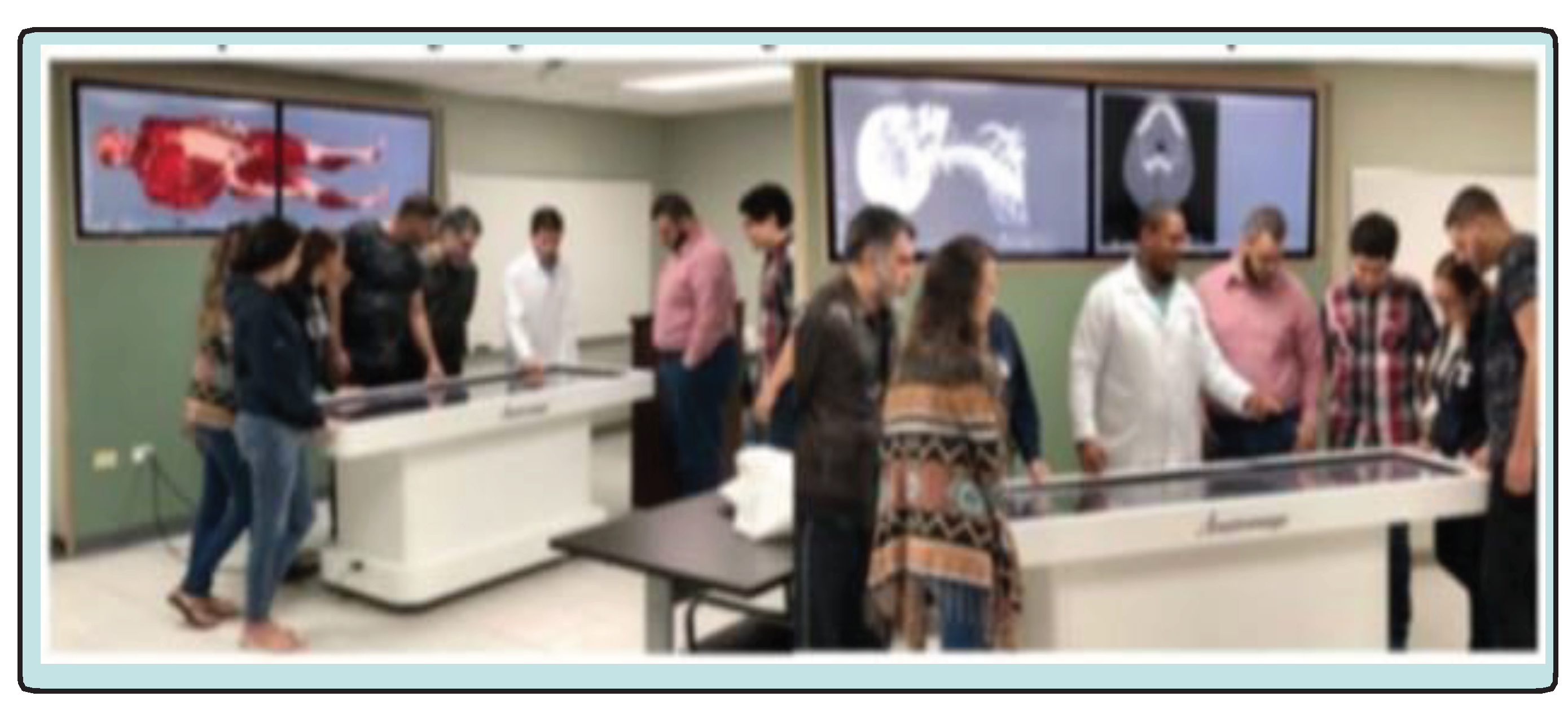
In Figure 3, a photoshoots of student participation on an Anatomage® virtual instruction module, where the images are also shown on the larger screen adjacent to the Anatomage® table. Both students and faculty can manipulate the images and have the opportunity to confirm the prerecorded identity of the structures depicted to provide immediate feedback and this further enhance the importance and retention of the content of the modules. In addition, students can return to the Anatomage® table for audit, follow-up, and reinforcement throughout their academic experiences during designated times through both basic science and clinical years.
Additional lab exercises include 3-D printing of body parts, endoscopy, Bronchoscopy, colonoscopy, wound closure and suturing techniques, and others, where a substantial volume of pathophysiology may be discovered during the course of a cadaveric examination and dissection. Overall, the mastery of staged anatomic instruction enables students to better appreciate and reinforce the clinical manifestations of the anatomy, physiology, and neurobiology with carryover to the clinical years and beyond. Anatomy teaching hours have become significantly compressed in recent years to yield room for additional topics in molecular biology and numerous other topics now included in medical basic sciences, many of which have a connection to basic functional anatomy. As a likely result of the innovative emphasis placed on this and other topics during medical basic sciences, student test scores on the basic sciences and clinical sciences pre-licensing examinations have trended upwards with virtually all applicants finding success in their pursuit of postgraduate residency training in the USA and other nations. As a consequence of the above cadaveric dissection program, the previously once somewhat unpopular course of the past has become one of the most popular course modules in the medical basic sciences, with many students requesting the opportunity to audit the module multiple times after their initial experience. Will they become future anatomists or surgeons? Only time may answer that question.
4. SUMMARY AND CONCLUSIONS
An innovative clinically focused cadaveric anatomy program is described which has improved the delivery and content retention of anatomy education for medical and graduate students and has become one of the most sought-after modules in the revised curriculum. Clearly, your grandfather’s anatomy lab has left the building and is not here anymore, but it hasn’t gone out entirely with the discarded debris, as the fundamental importance of anatomy dissection to medical education remains. Although grandad’s lab it is not likely to return any time soon in its earlier format, the fundamentals, importance and need for mastery of the instruction in gross anatomy remain solid and committed and haven’t changed over the intervening generations. The early process has provided the fundamental basis developed over hundreds of years, since Dr Nicholaes Tulp’s early demonstration of human dissection as artistically recorded in the ‘Anatomy Lesson of Dr. Tulp’ (ca 1632). Today’s anatomy dissection and modularized learning programs, including computer assisted virtual anatomy programs for human and animal anatomic study via a blended-SPOC (Small Platform Online Course) format now enable and facilitate anatomy instruction to be more meaningful and longer lasting than ever before.
The old adage has often been stated, “necessity is the mother of invention” and which rings truer today than ever before with increasing pressures to improve the efficiency and outcomes of laboratory exercises. With new and developing technology in the form of virtual anatomy, biophysics, fiberoptics, and electronic media applications. One can simulate neurophysiologic and biophysical phenomenon with predictive accuracy at the molecular level and apply fiberoptic visualizations and 3-D printing exercises not previously adaptable to the anatomy laboratory to supplement the age-old dissecting instruments of yesterday’s lab. This may include computer assisted photography, radiographic imaging, introduction to surgical techniques and procedures, all focused to better prepare the student of today for the clinical experience and demands of tomorrow. Neurophysiologic pathways can be modeled, and virtual dose related pharmacologic alterations predicted. In small group discussions, students can discover the uniqueness of each cadaver, relate them to live situations and examples, and engage in healthy and productive discussion with the participating faculty and in attendance, and with computer assisted library resources available as needed. The anatomy laboratory of the 21st century is an evolving program, which will continue to evolve and develop as technology advances and will continue to present an exciting and dynamic opportunity for tomorrow’s medical students to not only master the skills for today but to build on and to develop the skills for a stronger tomorrow. No single approach by itself is likely to be as effective as the combined approach using new and developing multimedia approaches. Thus, development of an innovative clinically focused cadaveric anatomy program is described which has improved the delivery and content retention of medical and graduate students and has become one of the most sought-after modules in the updated and revised clinically oriented curriculum.
Acknowledgements
The authors are grateful for the assistance of the many faculty and staff of the Miami Anatomic Research Institute for support and assistance in developing the above program for anatomy dissection for USAT medical students, many of whom entered the program with advanced certifications and degrees, seeking career advancement in the medical professions. Special thanks for the late Dr David Karam, whose dynamic lecture and brilliant surgical dissection talents were an inspiration to many of his students and to the writing of this manuscript.
Competing Interests
Authors have declared that no competing interests exist.
References
- Hunter M. Alder Hey report condemns doctors, management, and coroner. BMJ: British Medical Journal. 2001 Feb 2;322(7281):255. [CrossRef]
- Bergman EM, Prince KJ, Drukker J, van der Vleuten CP, Scherpbier AJ. How much anatomy is enough?. Anatomical sciences education. 2008 Jul;1(4):184-8. [CrossRef]
- Craig S, Tait N, Boers D, McAndrew D. Review of anatomy education in Australian and New Zealand medical schools. ANZ journal of surgery. 2010 Apr;80(4):212-6. [CrossRef]
- Older J. Anatomy: a must for teaching the next generation. The Surgeon. 2004 Apr 1;2(2):79-90. [CrossRef]
- Grauer, A. (1995). Bodies of Evidence: Reconstructing History through Skeletal Analysis. New York, NY: Wiley-Liss.
- Hodge, CJ. (2012). Non-bodies of knowledge: Anatomized remains from the Holden Chapel collection. Journal of Social Archaeology. J Social Archeology. Volume 13 (1): 1-27 . [CrossRef]
- Blakely RL, Harrington JM(1997b) Grave consequences: The opportunistic procurement of cadavers at the Medical College of Georgia. In: Blakely RL, Harrington JM (eds) Bones in the Basement: Postmortem Racism in Nineteenth-century Medical Training, Washington: Smithsonian Institution Press, pp. 162-183.
- The Anatomy Lesson of Dr. Nicolaes Tulp, by Rembrandt; 1632.
- Drake RL, McBride JM, Lackman N, Pawlina W. Medical Education in the anatomical sciences: The winds of change continue to blow. Anat ScI Educ. 2009;2(6):253-259. [CrossRef]
- Bowsher D. What should be taught in anatomy? Med Educ. 1976;10(2): 132-134. [CrossRef]
- Louw G, Eizenberg N, Carmichael SW. The place of anatomy in medical education: AMEE Guide no Med Teach. 2009;31(5): 373-386. [CrossRef]
- Ang E-T, Sugand K, Hartman M, Seow X-S, Bay B-H, Abrahams P. Singapore's anatomical future: Quo Vadis? Anat Sci Educ. 2012;5(4): 234-240. [CrossRef]
- Habicht JK, Kiessling C, Winklemann A. Bodies for Anatomy Education in Medical Schools: An Overview of the Sources of Cadavers Worldwide. Acad Med. 2018;93(9):1293- 1300. [CrossRef]
- Periya SN. Teaching Human Anatomy in the Medical Curriculum: A Trend Review. IJAR. 2017;5(4):445-448. [CrossRef]
- Smith C. Water, water everywhere: The pandemic has caused a shortage of cadavers. The Economist, Britain, May 8th 2021 edition. Available: https://www.economist. com/printedition/2021-05-08.
- Periya SN. Teaching anatomy in the medical curriculum: a trend review. Int J Adv Res. 2017;5(4): 445-448. [CrossRef]
- Shiffer CD, Boulet JR, Cover LJ, Pinsky WW. Advancing the Quality of Medical Education Worldwide: ECFMGs 2023 Medical School Accreditation Requirement. J Med Reg. 2019;105(4): 8-16. [CrossRef]
- Tulp OL, Ortiz-Bustillo M, Einstein GP, Sainvil F, Konyk CM, Branly RL. Innovation in Functional Anatomy Education: The Anatomy Lesson of Dr Tulp from the past to the Present. The FASEB Journal. 2018;33(S1): 505.4-505.4. [CrossRef]
- Tulp OL, Sainvil F, Einstein GP, Scrianka A, Branly R, Wu H, et al. Incorporation of virtual enhancement in anatomy, physiology and clinical pathology instruction in medicine and veterinary medicine basic sciences. FASEB J. 36(S1). [CrossRef]
- Tulp OL, Sainvil F, Wu H, Feleke Y, Einstein GP, Branly R. Modernization of Anatomy and Graduate Medical Education from the Past to the Present. The FASEB J. 2021;35(S1). [CrossRef]
- Leung KK, LU KS, Huang TS, Hsieh BS. Anatomy Instruction in medical schools: connecting the past and the future. Adv Health Sci Educ Theory Pract. 2006;11(2):209-215. [CrossRef]
- Anastakis DJ, Regehr G, Reznich RK. Assessment of technical skills transfer from the bench training model to the human model. Am J Surg. 1999;177(2):167-170. [CrossRef]
- Keim Janssen SA, VanderMeulen SP, Shostrom VK, Lomneth CS. Enhancement of anatomical learning and developing clinical competence of first-year medical and allied health profession students. Anat Sci Edu. 2014;7(3): 181-190. [CrossRef]
- Agu A. The Dissecting room experience: A factor in the choice of organ and whole body donation-A Nigerian survey.
- Kalanjati VP, Prasetiowati L, Alimsardjono H. The use of lower-formalin-containing embalming solution for anatomy cadaver preparation. Med J Indones. 2012;21(4): 203-207. [CrossRef]
- Whitehead MC, Savoia MC. Evaluation of methods to reduce formaldehyde levels of cadavers in the dissection laboratory. Clin Anat. 2008;21(1): 75-81. [CrossRef]
- Ocel J, Natt N, Tiegs R, Arora AS. Formal procedural skills training using a fresh frozen cadaver model: a pilot study. Clin Anat. 2006;19(2): 142-146. [CrossRef]
- Prasad N, Kumar S, Manjunath R, Bhadauria D, Kaul A, Sharma R. Real-time ultrasound guided percutaneous renal biopsy with needle guide by nephrologists decreases post-biopsy complications. Clin Kidney J. 2015;8(2): 151- 156. [CrossRef]
- Reed AB, Crafton C, Giglia JS, Hutto JD. Back To Basics: Use of fresh cadavers in vascular surgery training. Surgery. 2009;146(4):757-763. [CrossRef]
- Keim Janssen SA, VanderMeulen SP, Shostrom VK, Lomneth CS. Enhancement of anatomical learning and developing clinical competence of first-year medical and allied health profession students. Anat Sci Edu. 2014;7(3): 181-190. [CrossRef]
- Are C, Lomneth C, Stoddard H, Azarow K, Thompson JS. A preliminary review of a pilot curriculum to teach open surgical skills during general surgery residency with initial feedback. Am J Surg. 2012;204(1): 103-109. [CrossRef]
- Meek M, Meek J, Hollowoa B, Li R, Deloney L, Phelan, K. Lightly embalmed cadavers as a training tool for ultrasound-guided procedures commonly used in interventional radiology. Ac Rad. 2018;25(11): 1503-1509. [CrossRef]
- Hoyer R, Means R, Robertson J. Ultrasound-guided procedures in medical education: a fresh look at cadavers. Intern Emerg Med 2016;11(3): 431-436. [CrossRef]
- Asad MR, Nasir N. Role of Living and Surface Anatomy in Current Trends of Medical Education. Acad Radiol. 2018;25(11):1503-1509.
- Finn GM. Twelve tips for running a successful body painting teaching session in freshman gross anatomy. Med Teaching. 32:887-890. [CrossRef]
- Finn GM, McLachlan JC. A qualitative study on the student responses to body painting. Anat Sci Educ. 2010;3:33-38. [CrossRef]
- Nanjunundaiah K, Chowdapurkar S. Body painting: a tool which can be used to teach surface anatomy. J Clin Diagn Res. 2012;6(8): 1405-1408. [CrossRef]
- Collett T, Kirvell D, Nakorn A, McLachlan JC. The role of living models in the teaching of surface anatomy: some experiences from a UK medical school. Med Teach. 2009;31(3): e90-e96. [CrossRef]
- Bickley LS, Szilagyi PG, Hoffman RM, Soriano RP. In: Bate's Guide to Physical Examination and History taking. 12th edn. Lippincott Pubs. 2020.
- Hoyek N, Collet C, Di Rienzo F, De Almeida M, Guillot A. Effectiveness of three-dimensional digital animation in teaching human anatomy in an authentic classroom context. Anat Sci Educ. 2014;7(6): 430-437. [CrossRef]
- Hodge CJ. Non-bodies of knowledge: Anatomized remains from the Holden Chapel collection, Harvard University (2013). Journal of Social Archaeology.13(1). [CrossRef]
- Miami Anatomic Research Institute, 8850 NW 20th St, Doral, FL 33172, previously known as the Miami Anatomic Research Center.
Figure 1.
The Anatomy Lesson of Dr. Tulp, by Rembrandt (1632, Hague Museum).
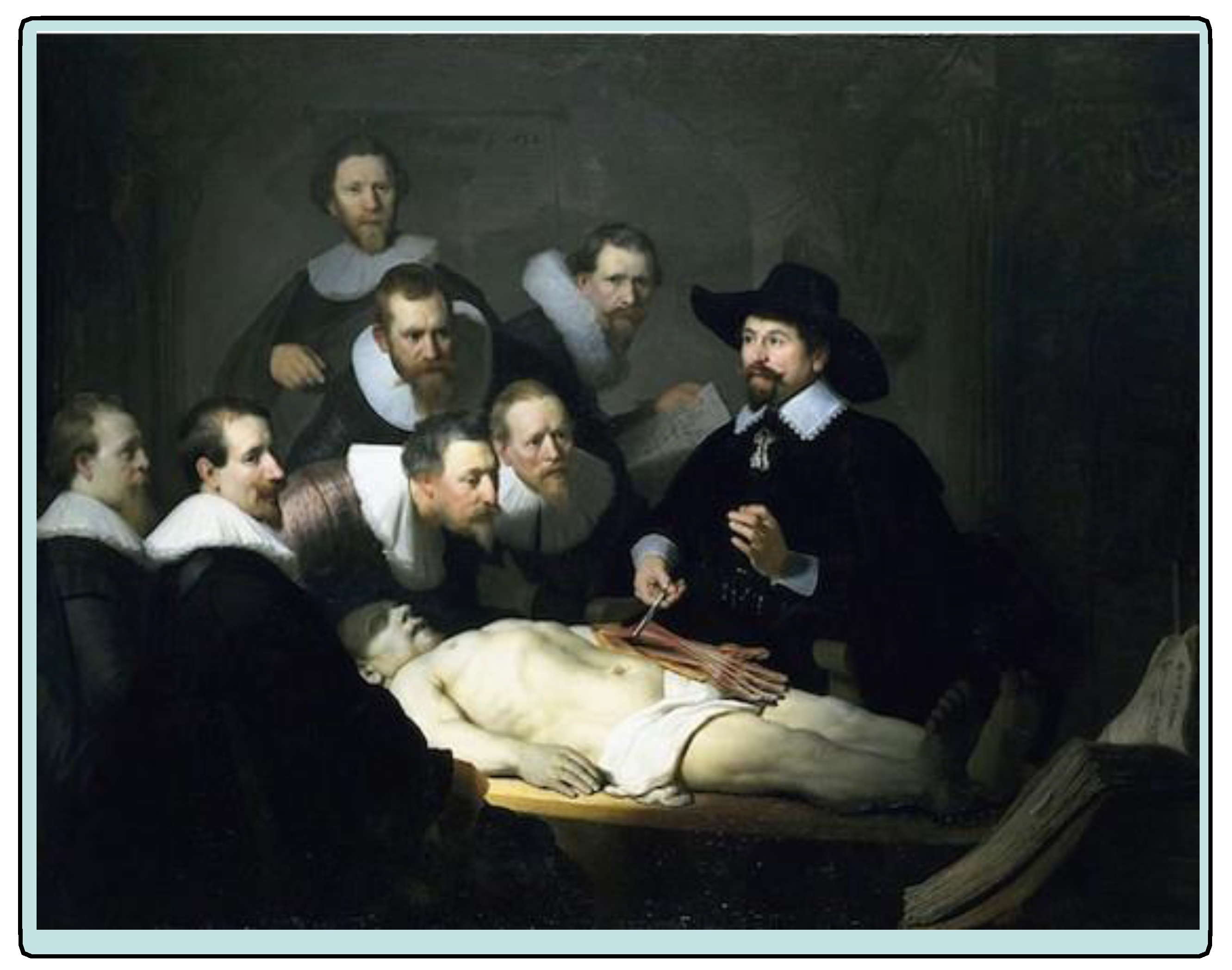
Figure 2.
The Anatomy Dissection Lesson at USAT Montserrat, 2018.
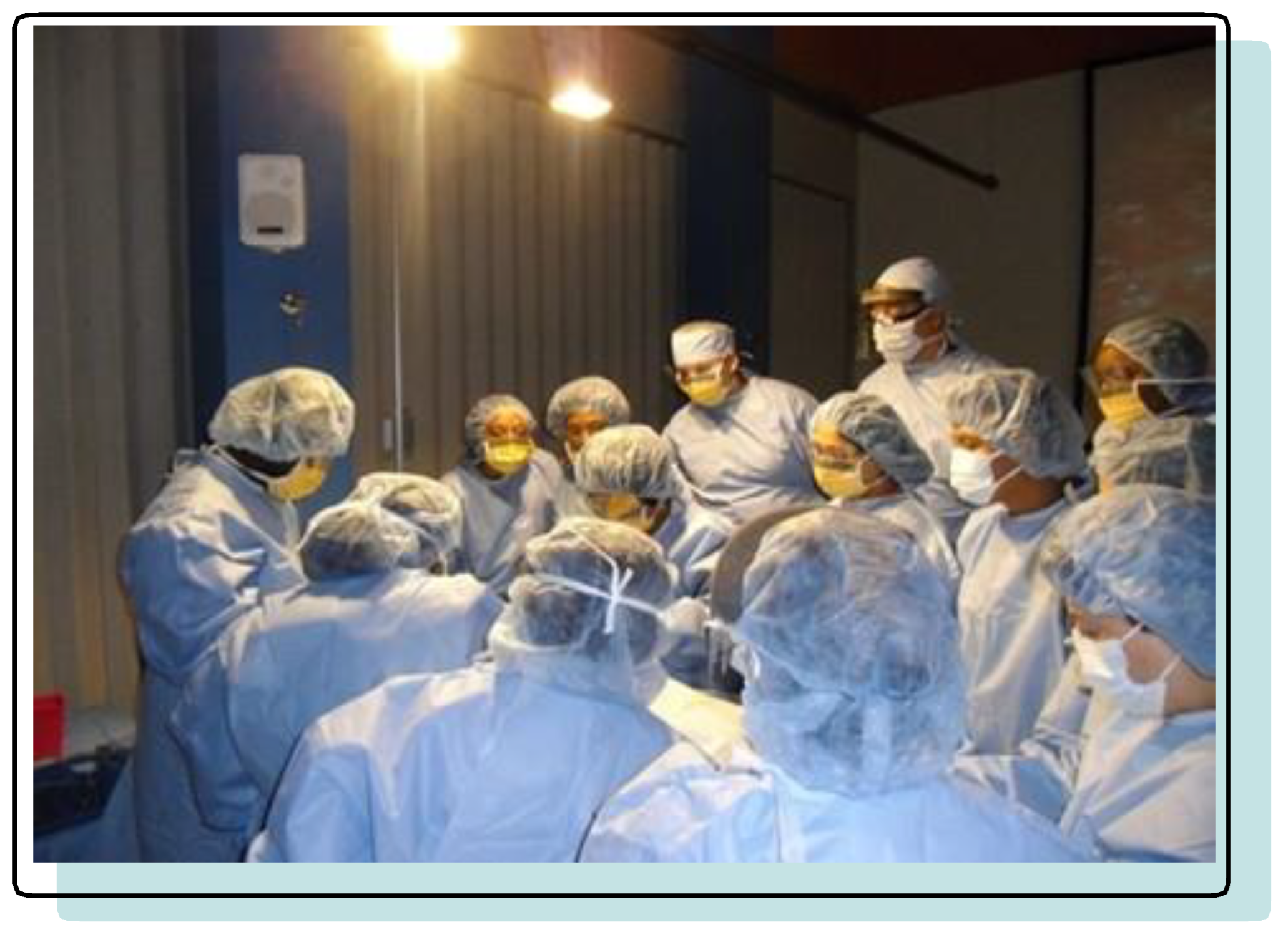
Figure 3.
Virtual pre-dissection classroom-based anatomy laboratory depicting musculoskeletal (L) and (R) cranial- cervical skeletal anatomy. Students can expose different anatomic structures and test themselves in a preprogrammed test module.
Figure 3.
Virtual pre-dissection classroom-based anatomy laboratory depicting musculoskeletal (L) and (R) cranial- cervical skeletal anatomy. Students can expose different anatomic structures and test themselves in a preprogrammed test module.
Disclaimer/Publisher’s Note: The statements, opinions and data contained in all publications are solely those of the individual author(s) and contributor(s) and not of MDPI and/or the editor(s). MDPI and/or the editor(s) disclaim responsibility for any injury to people or property resulting from any ideas, methods, instructions or products referred to in the content. |
© 2023 by the authors. Licensee MDPI, Basel, Switzerland. This article is an open access article distributed under the terms and conditions of the Creative Commons Attribution (CC BY) license (http://creativecommons.org/licenses/by/4.0/).
Copyright: This open access article is published under a Creative Commons CC BY 4.0 license, which permit the free download, distribution, and reuse, provided that the author and preprint are cited in any reuse.


