Submitted:
04 May 2025
Posted:
07 May 2025
Read the latest preprint version here
Abstract
The purpose of this article is to identify the results of structural analyze from liver samples. In the study were used both normaly and pathologicaly liver samples. Also good to mention that in this study were used liver samples, collected during necropsy, from healthy patients and from patients diagnosed with cirrhosis .This previously mentioned, are known as routinally found methods, in medical practice, used for hepatitis C diagnosis, which conduct to hepatic cirrhosis Folowing steps from the classic structural study, it is possible to analyse normal and alterate liver structure, on the colored preparates with specific stainings.
Keywords:
liver
; samples
; laboratory
; technique
; diagnosis
Introduction
Currently , HCV infection could be consider as one of bad results after percutaneous blood administration to ill patients. Also good to mention that HCV infection it is known as one of the most commonly through injection drug use. [1] The first and most important step in the care cascade is testing for HCV. Actually, is an enlarge number of expansion of populations eligible for testing for HCV prevention. [1,2,3]
It is know about care cascade, including multiple key points in diagnosis HCV infection. An HCV care cascade is consider as a model for identifying opportunities and barriers in order to improve laboratory tests, linkage to care, and proper treatment access. [4,5]
Relativelly recently, there have been increases in HCV detection among women of childbearing age. [6,7] Reinfection with HCV after curative therapy in illness status in different patients, is an important key point for medical team. [8,9,10] The proper laboratory techniques include immunoglobulin (Ig) G antibody enzyme immunoassays (anti-HCV) and nucleic acid tests (NAT), as modern methods, in conection with blood tests.[11] In case that need to distinguish between two directions such as true or false positivity of the anti-HCV antibody result, previously mentined tests, may be done with a second FDA-approved HCV antibody assay that is different from previously used for testing [11,12] Morphologically, HCV is an enveloped, positive-sense, single-stranded RNA virus of the Flavivirdae family.[13,14,15]
The great key point knowing as a start of the direct-acting antiviral (DAA) era was in 2011. The important role in this direction was the introduction of two NS3/4A protease inhibitors. Both previously mentioned were used in combination with interferon-based regimens for chronic HCV treatment to ill patients diagnosed.[16] Results of studies show that the HCV replication process is error prone. Finally results practically could be observe in variant viruses knowing as quasispecies.[17,18] Nowadays there are 7 genotypes of HCV. So there are known 6 major genotypes and the recent addition of genotype 7. This last 7 genotip has been found only in a few cases diagnosed to HCV positive patients. [19] Hepatitis C virus (HCV) infection is a great cause of various liver diseases as cirrhosis and hepatocellular carcinoma. Following promising news, significant scientific discovering things remain in attention for reducing morbidity and mortality, associated to HCV. [20,21] Understanding the properties of hepatitis C virus (HCV) viral RNA and proteins facilitates the development of diagnosis methods and also a proper treatment, including antivirals.[22,23,24] In addition we can mention that HCV genotyping assays approved for in vitro diagnostic use are commercially available[25,26] Cirrhosis, as a nowadays disease, is characterized by fibrosis and nodule formation of the liver. In the secondary plan, it is known as a chronic injury, which leads to alteration of the normal lobular organization of the liver. A complex of factors, such as life style, or environmentals, can injure the liver, and beside also including viral infections, toxins.
With each injury, the liver suffer alterations as fibrosis. Finally but after a long-standing injury, liver functionalteration, develop in time cirrhosis as a complex diseases. Ethiology of the chronic liver diseases usually progress unfortunately in cirrhosis, following pathological mechanisms.[27] Scientific knowledges referring to the severity of liver cirrhosis, as a disease with a bad prognostic on the public health, is still not well characterized. [28] Liver diseases are without doubts, the most common in the world. [29] Researchers must be carefully to ideea refering to demonstration that drug injury is present in liver structure injury that conduct to pathology. [30]
Material and Methods
In order to assist medical staff in understanding the concerns outlined, a series of digital images have been prepared. The operative pieces are intended to bring in the pathological anatomy service for macroscopic examination for diagnostic purposes. This are examined by performing the optical microscopic analysis. Using an optical microscope, could be possible liver structure analyze. We can observe hepatocytes and interlobular spaces and septa.[Figure 1] On a normal liver structure but using another specific stain known as Argentic impregnation Gomori.
The functional unit of the liver is the lobule with hexagonal form. Kienann space is specific for liver strucutre, including a portal triad (portal vein, hepatic artery, bile duct) sits at each corner of the hexagon. Mitochondri as points observing with lens x40, using Goldner Szekely stain. [Figure 2]
In liver cirrhosis, on samples could be observe tissue fibrosis and alterations in the normal liver architecture in abnormal structural nodules. [Figure 3]
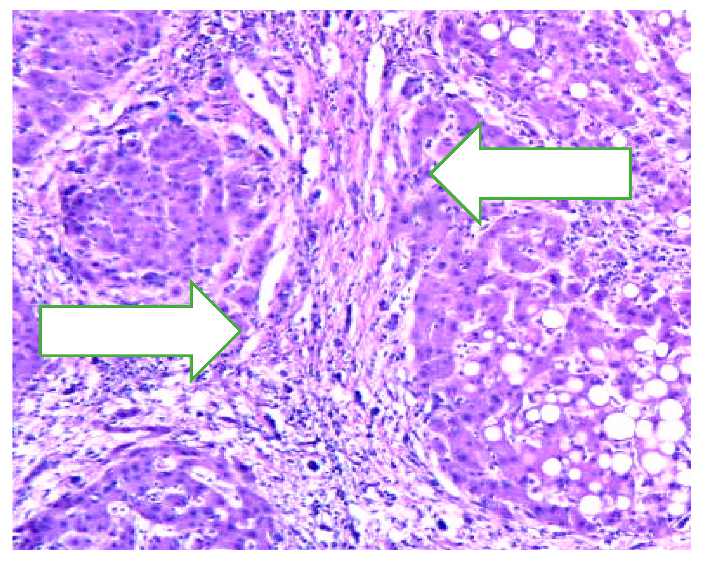
A similar coming image with specific structural changes in liver structure and with vascularized fibrotic septa in cirrhosis More than , we can mention in this disease, about portal hypertension. [Figure 4]
Prevention and treatment of liver cirrhosis are best done by an interdisciplinary medical team.
Animal models play a significant role in liver pathology diagnosis for example in rats with cirrhosis. Cirrhosis, as a nowadays disease, is characterized by fibrosis and neoformation noduls in the liver architecture. In addition, cirrhosis it is known as a chronic injury, which leads to alteration of the normal lobular organization of the liver. [29,30,31] A complex of factors, such as life style, or environmental factors, can affect the liver, for better or for worse. [32] [33] In addition to optimal management of cirrhosis complications per the recent EASL guidelines, patients with advanced liver disease also require support and management for substance/alcohol abuse, nutrition and frailty. [34,35]
4. Complications in Cirrhosis
Hepatic encephalopathy (HE) is an important major neuropsychiatric disorder in liver cirrhosis. There are known two types. So minimal hepatic encephalopathy (MHE), known as a cognitive deficit found in the ealier past time using specific psychological tests and Grade I HE. More than, also it is known about overt hepatic encephalopathy (OHE), with specific clinical symptoms.[36,37]
Ascites is knowing as a common complication in hepatic cirrhosis. Ascites is accompanied with portal hypertension. This previously mentioned complication in cirrhosis, it is known as a specific one, defined as an accumulation of a fluid quantity in the peritoneal cavity.[38]
Ascites infection is a possible accompaning event for ill persons diagnosed with cirrhosis. Infection in ascitis is often known as a bacterial peritonitis (SBP) somethimes with accompaning fungal infections.[39,40]
Variceal bleeding represent a relatively comon accompanied complication to patients diagnosed with cirrhosis. [41] In liver damage as cirrhosis, esophageal and gastric variceal bleeding together with rectal variceal bleeding.[42]
Hepatorenal syndrome and kidney injury represent a common complication to patients diagnosed with hepatic cirrhosis. [43]
Infections are also relatively commons in hepatic cirrhosis. In differents parts of the body could be find after signs and simpotoms, infections as tissues infections,bacteremia, pneumonia, urinary tract infections. [44,45] From years ago,till nowadays, infections caused by multidrug-resistant organisms (MDRO) play a significant role in liver damages such them from hepatic cirrhosis.[46]
5. Cellular and Molecular Key Points
Folowing embryogenesis stages, hepatic cells starting with progenitor cells namely hepatoblasts, during differentiation, become finally hepatocytes.
In the cellular and molecular mechanisms, signals play a great importance. So, hematopoietic cells, manage the hepatoblast proliferation prcess. In this direction, E-cadherin-mediated cell junction in hepatoblasts evolutive nechanism. Also are knowing other contributors in fibally hepatocytes evolution. It is useful to mention that the gradient of TGF-β secreted from the portal vein mesenchyme play also a great role in previously mentioned direction with finality hepatocytes. Cellular and molecular mechanism reffering to mechanistically, show a higher TGF-β signaling in portal vein in hepatic cells that drives cholangiocyte physiologically direction. Research studies, show the decreasing expression of CCAAT/ enhancer binding protein (C/EBP) α and which promoting expression of Hnf6 (aka Oc1) and Hnf1β. More that, also results of studies, show the transcription profile. In this direction it is good to mention a bit about cholangiocyte-specific gene transcription process. More exactly, by HNF6 and HNF1β, are implications in suppressing hepatocyte genes by decreasing C/EBPα levels.[47]
In the developmentally mechanism, the hepatocytes, and the cholangiocytes, emerge from the specific region in the definitive endoderm. In the initiations signals reffering to hepatic cells, include fibroblast growth factor (FGF), and also bone morphogenic proteins (BMPs). [47]
Studies also show as that non-alcoholic fatty liver disease (NAFLD) is a key point in liver diseases.In this direction, we can mention that the pathologically mechanisms, follow steps starting with steatosis having finally to cirrhosis Also good to mention that liver enzymes play also a role. In this mechanism, it is known about ALT:AST ratio. [48,49]
Discussions
Patient lifestyle changes, unfortunately cannot cure cirrhosis. Complications accompanying hepatic cirrhosis include, portal hypertension, edema in the abdomen and lower extremities, splenomegaly, infections, hepatic encephalopathy.
Behavioral modifications can prevent or at least delay disease progression and provide symptomatic relief.
Lifestyle changes, include factors, as eliminating ethanol consumption and dietary interventions as possible low-sodium diet, in order to reduce water retention. Regulate protein intake according to their doctor's directions and some medical recommandations, will be proper in the treatment of cirrhosis.
Cirrhosis secondary to HBV and HCV is one of the common risk factor for liver degeneration in cirrhosis.. Practically monitoring of cirrhotic patients is recommended, with at least six monthly screenings. Liver biopsy is the gold standard technique highly promising non-invasive methodology under development, that are used in diagnosis. Liver transplantation (LT) is also an effective therapeutic option for the management of cirrhosis end-stage. Relatively recently research investigations try to elucidate the signal transduction pathways that link hepatocytes alterations including cellular disfunctionality.
Conclusions
Research studies predict about not so a good prognosis of patients diagnosed with cirrhosis, knowing laboratory results and clinical points. Knowing the liver as a heterogeneous organ, t under a physiological control, the diagnostic in the ill patients, medical specialists could applay the proper treatment. Curently diagnostic methods and a proper control of liver functions conduct to a proper diagnostic. In idea that hepatic cirrhosis is hard or impossible to cure, we are waiting from future research dirrections and plans.
References
- AASLD-IDSA. HCV Guidance: Recommendations for Testing, Managing, and Treating Hepatitis C 2016. Available from: http://hcvguidelines.org/sites/default/files/HCV-Guidance_October_2016_a.pdf. Guidance produced by the American Association for the Study of Liver Disease and the Infectious Disease Society of America (AASLD-IDSA) covering recommendations for hepatitis C testing, clinical management and treatment.
- Recommendations for prevention and control of hepatitis C virus (HCV) infection and HCV-related chronic disease. Centers for Disease Control and Prevention. MMWR Recomm Rep. 1998, 47, 1–39, Recommendations from the Centers for the Disease Control and Prevention definng populations for risk-based testing of hepatitis C. [Google Scholar]
- Moyer, V.A. Screening for hepatitis C virus infection in adults: U.S. Preventive Services Task Force recommendation statement. Ann Intern Med. 2013, 159, 349–357, Hepatitis C testing recommendations for adults from the United States Preventive Services Task Force including risk-based testing and testing for adults born from 1945–1965. [Google Scholar] [CrossRef]
- Holmberg, S.D.; Spradling, P.R.; Moorman, A.C.; Denniston, M.M. Hepatitis C in the United States. N Engl J Med. 2013, 368, 1859–1861, Description of a care cascade integrating data from the Chronic Hepatitis Cohort Study and the National Health and Nutrition Examination Survey. [Google Scholar] [CrossRef]
- Viner, K.; Kuncio, D.; Newbern, E.C.; Johnson, C.C. The continuum of hepatitis C testing and care. Hepatology. 2015, 61, 783–789, Description of an HCV continuum of care in a US urban center highlighting important areas where patients become lost at each stage. [Google Scholar] [CrossRef]
- Koneru, A.; Nelson, N.; Hariri, S.; Canary, L.; Sanders, K.J.; Maxwell, J.F.; et al. Increased Hepatitis C Virus (HCV) Detection in Women of Childbearing Age and Potential Risk for Vertical Transmission - United States and Kentucky, 2011–2014. MMWR Morb Mortal Wkly Rep. 2016, 65, 705–710, Analysis finding that the proportion of infants born to HCV-infected mothers increased 68% nationally and 124% in Kentucky. [Google Scholar] [CrossRef]
- Kuncio, D.E.; Newbern, E.C.; Johnson, C.C.; Viner, K.M. Failure to Test and Identify Perinatally Infected Children Born to Hepatitis C Virus-Infected Women. Clin Infect Dis. 2016, 62, 980–985, Analysis of surveillance data from the Philadelphia Department of Public Health finding a significiant number of women giving birth in Philadelphia tested positive for HCV but that most of their children were not tested for HCV. [Google Scholar] [CrossRef]
- Lambers, F.A.; Prins, M.; Thomas, X.; Molenkamp, R.; Kwa, D.; Brinkman, K.; et al. Alarming incidence of hepatitis C virus re-infection after treatment of sexually acquired acute hepatitis C virus infection in HIV-infected MSM. AIDS. 2011, 25, F21–F27. [Google Scholar] [CrossRef]
- Martin, T.C.; Martin, N.K.; Hickman, M.; Vickerman, P.; Page, E.E.; Everett, R.; et al. Hepatitis C virus reinfection incidence and treatment outcome among HIV-positive MSM. AIDS (London, England) 2013, 27, 2551–2557. [Google Scholar] [CrossRef] [PubMed]
- Ingiliz, P.; Martin, T.C.; Rodger, A.; Stellbrink, H.J.; Mauss, S.; Boesecke, C.; et al. HCV reinfection incidence and spontaneous clearance rates in HIV-positive men who have sex with men in Western Europe. J Hepatol. 2016. [CrossRef]
- Testing for HCV infection: an update of guidance for clinicians and laboratorians. MMWR Morb Mortal Wkly Rep. 2013, 62, 362–365, Updated guidance for clinicians and laboratorians from the Centers for Disease Control and Prevention on diagnostic testing procedures for hepatitis C.
- Moyer, V.A. Screening for hepatitis C virus infection in adults: U.S. Preventive Services Task Force recommendation statement. Ann Intern Med. 2013, 159, 349–357, Hepatitis C testing recommendations for adults from the United States Preventive Services Task Force including risk-based testing and testing for adults born from 1945–1965. [Google Scholar] [CrossRef]
- Thomas, D.L.; Seeff, L.B. Natural history of hepatitis C. Clin Liver Dis. 2005, 9, 383–98. vi. [Google Scholar] [CrossRef]
- Mack, C.L.; Gonzalez-Peralta, R.P.; Gupta, N.; Leung, D.; Narkewicz, M.R.; Roberts, E.A.; et al. NASPGHAN practice guidelines: Diagnosis and management of hepatitis C infection in infants, children, and adolescents. J Pediatr Gastroenterol Nutr. 2012, 54, 838–855. [Google Scholar] [CrossRef]
- Thein, H.H.; Yi, Q.; Dore, G.J.; Krahn, M.D. Estimation of stage-specific fibrosis progression rates in chronic hepatitis C virus infection: a meta-analysis and meta-regression. Hepatology. 2008, 48, 418–431. [Google Scholar] [CrossRef]
- Conteduca, V.; Sansonno, D.; Russi, S.; Pavone, F.; Dammacco, F. Therapy of chronic hepatitis C virus infection in the era of direct-acting and host-targeting antiviral agents. J Infect. 2014, 68, 1–20. [Google Scholar] [CrossRef] [PubMed]
- Gomez, J.; Martell, M.; Quer, J.; Cabot, B.; Esteban, J.I. Hepatitis C viral quasispecies. J Viral Hepat. 1999, 6, 3–16. [Google Scholar] [CrossRef]
- Duffy, S.; Shackelton, L.A.; Holmes, E.C. Rates of evolutionary change in viruses: patterns and determinants. Nat Rev Genet. 2008, 9, 267–276. [Google Scholar] [CrossRef]
- Bukh, J. The history of hepatitis C virus (HCV): Basic research reveals unique features in phylogeny, evolution and the viral life cycle with new perspectives for epidemic control. J Hepatol. 2016, 65 (1 Suppl), S2–S21. [Google Scholar] [CrossRef]
- Ly, K.N.; Hughes, E.M.; Jiles, R.B.; Holmberg, S.D. Rising Mortality Associated With Hepatitis C Virus in the United States, 2003–2013. Clin Infect Dis. 2016, 62, 1287–1288, Describes rising HCV-associated mortality in the United States from 2003–2013. During that time period, HCV-associated deaths surpassed 60 other nationally natofiable infectious conditions combined. [Google Scholar] [CrossRef]
- Allison, R.D.; Tong, X.; Moorman, A.C.; Ly, K.N.; Rupp, L.; Xu, F.; et al. Increased incidence of cancer and cancer-related mortality among persons with chronic hepatitis C infection, 2006–2010. J Hepatol. 2015, 63, 822–828, Analysis of a cohort of HCV-infected persons that found the the incidence of liver cancer and many types of non-liver cancers were higher, and age at diagnosis and death younger, in patients with chronic HCV infection compared to the general population. [Google Scholar] [CrossRef]
- Alberti, A.; Chemello, L.; Benvegnù, L. Natural history of hepatitis C. J Hepatol. 1999, 31 (Suppl. 1), 17–24. [Google Scholar] [CrossRef]
- Hoofnagle, J.H. Course and outcome of hepatitis C. Hepatology. 2002, 36, S21–S29. [Google Scholar] [CrossRef]
- Chen, S.L.; Morgan, T.R. The natural history of hepatitis C virus (HCV) infection. Int J Med Sci. 2006, 3, 47–52. [Google Scholar] [CrossRef]
- Chevaliez, S.; Pawlotsky, J.M. Virology of hepatitis C virus infection. Best Pract Res Clin Gastroenterol. 2012, 26, 381–389. [Google Scholar] [CrossRef]
- Saludes, V.; González, V.; Planas, R.; Matas, L.; Ausina, V.; Martró, E. Tools for the diagnosis of hepatitis C virus infection and hepatic fibrosis staging. World J Gastroenterol. 2014, 20, 3431–3442. [Google Scholar] [CrossRef]
- Naveau, S.; Perlemuter, G.; Balian, A. [Epidemiology and natural history of cirrhosis]. Rev Prat. 2005, 55, 1527–32. [Google Scholar]
- Kleinbloesem, C.H.; van Harten, J.; Wilson, J.P.; Danhof, M.; van Brummelen, P.; Breimer, D.D. Nifedipine: kinetics and hemodynamic effects in patients with liver cirrhosis after intravenous and oral administration. Clin Pharmacol Ther. 1986, 40, 21–28. [Google Scholar] [CrossRef]
- Bircher J, Benhamou JP, McIntyre N, Rizzetto M, Rodes J, editors. Oxford Textbook of Clinical Hepatology. 2nd Edition Oxford University Press; 1999.
- Sherlock S, Dooley J, editors. Diseases of the Liver and Biliary System. 11th Edition Blackwell Science; Oxford, UK; Malden, MA: 2002.
- Schiff ER, Sorrell MF, Maddrey EC, editors. Schiff’s Diseases of the Liver. 9th Edition Lippincott, Williams & Wilkins; Philadelphia: 2003.
- Desmet, V.J.; Roskams, T. Cirrhosis reversal: a duel between dogma and myth. J Hepatol. 2004, 40, 860–867. [Google Scholar] [CrossRef]
- European Association for the Study of the Liver EASL Clinical Practice Guidelines for the management of patients with decompensated cirrhosis. Hepatol. 2018, 69, 406–460. [CrossRef]
- Wanless, I.R.; Nakashima, E.; Sherman, M. Regression of human cirrhosis. Morphologic features and the genesis of incomplete septal cirrhosis. Arch Pathol Lab Med. 2000, 124, 1599–1607. [Google Scholar] [CrossRef]
- Macdonald, S.; Jepsen, P.; Alrubaiy, L.; Watson, H.; Vilstrup, H.; Jalan, R. ,Quality of life measures predict mortality in patients with cirrhosis and severe ascites. Aliment Pharmacol Ther. 2019, 49, 321–330. [Google Scholar] [CrossRef]
- Häussinger, D.; Dhiman, R.K.; Felipo, V.; Görg, B.; Jalan, R.; Kircheis, G.; Merli, M.; Montagnese, S.; Romero-Gomez, M.; Schnitzler, A.; Taylor-Robinson, S.D.; Vilstrup, H. Hepatic encephalopathy. Nat Rev Dis Primers. 2022, 8, 43. [Google Scholar] [CrossRef] [PubMed]
- Ridola, L.; Faccioli, J.; Nardelli, S.; Gioia, S.; Riggio, O. Hepatic Encephalopathy: Diagnosis and Management. J Transl Int Med. 2020, 8, 210–219. [Google Scholar] [CrossRef] [PubMed]
- European Association for the Study of the Liver. EASL clinical practice guidelines on the management of ascites, spontaneous bacterial peritonitis, and hepatorenal syndrome in cirrhosis. J Hepatol. 2010, 53, 397–417. [Google Scholar] [CrossRef]
- Gravito-Soares, M.; Gravito-Soares, E.; Lopes, S.; Ribeiro, G.; Figueiredo, P. Spontaneous fungal peritonitis: a rare but severe complication of liver cirrhosis. Eur J Gastroenterol Hepatol. 2017, 29, 1010–1016. [Google Scholar] [CrossRef] [PubMed]
- Fiore, M.; Chiodini, P.; Pota, V.; Sansone, P.; Passavanti, M.B.; Leone, S.; Aurilio, C.; Pace, M.C. Risk of spontaneous fungal peritonitis in hospitalized cirrhotic patients with ascites: a systematic review of observational studies and meta-analysis. Minerva Anestesiol. 2017, 83, 1309–1316. [Google Scholar] [CrossRef]
- Alqahtani, S.A.; Jang, S. Pathophysiology and Management of Variceal Bleeding. Drugs 2021, 81, 647–667. [Google Scholar] [CrossRef]
- Baiges, A.; Hernández-Gea, V. Management of Liver Decompensation in Advanced Chronic Liver Disease: Ascites, Hyponatremia, and Gastroesophageal Variceal Bleeding. Clin Drug Investig. 2022, 42, 25–31. [Google Scholar] [CrossRef]
- Molleston, J.P.; Bennett, W.E., Jr. Mortality, Risk Factors and Disparities Associated with Esophageal Variceal Bleeding in Children’s Hospitals in the US. J Pediatr. 2021, 232, 176–182. [Google Scholar] [CrossRef]
- Van der Merwe, S.; Chokshi, S.; Bernsmeier, C.; Albillos, A. The multifactorial mechanisms of bacterial infection in decompensated cirrhosis. J Hepatol. 2021, 75 (Suppl. 1), S82–S100. [Google Scholar] [CrossRef]
- Choudry, N.; Sasso, R.; Rockey, D.C. Infection in Hospitalized Cirrhosis Patients: Changing Epidemiology and Clinical Features. Am J Med Sci. 2022, 363, 114–121. [Google Scholar] [CrossRef] [PubMed]
- Fernández, J.; Piano, S.; Bartoletti, M.; Wey, E.Q. Management of bacterial and fungal infections in cirrhosis: The MDRO challenge. J Hepatol. 2021, 75 (Suppl 1), S101–S117. [Google Scholar] [CrossRef] [PubMed]
- Trefts Elijah, GannonMaureen, Wasserman David H. , The liver, Published in final edited form as: Curr Biol. 2017, 27, R1147–R1151.
- Su, W.; Mao, Z.; Liu, Y.; Zhang, X.; Zhang, W.; Gustafsson, J.A.; Guan, Y. Role of HSD17B13 in the liver physiology and pathophysiology. Mol Cell Endocrinol. 2019, 489, 119–125. [Google Scholar] [CrossRef] [PubMed]
- Adams, L.A.; Angulo, P.; Lindor, K.D. Nonalcoholic fatty liver disease. CMAJ. 2005, 172, 899–905. [Google Scholar] [CrossRef]
- Malinchoc, M.; Kamath, P.S.; Gordon, F.D.; Peine, C.J.; Rank, J.; ter Borg, P.C. A model to predict poor survival in patients undergoing transjugular intrahepatic portosystemic shunts. Hepatology. 2000, 31, 864–871. [Google Scholar] [CrossRef]
- Godfrey, E.L.; Kueht, M.L.; Rana, A.; Awad, S. MELD-Na (the new MELD) and peri-operative outcomes in emergency surgery. Am J Surg. 2018, 216, 407–413. [Google Scholar] [CrossRef]
Figure 1.
Normal liver x10. Argentic impregnation Gomori.
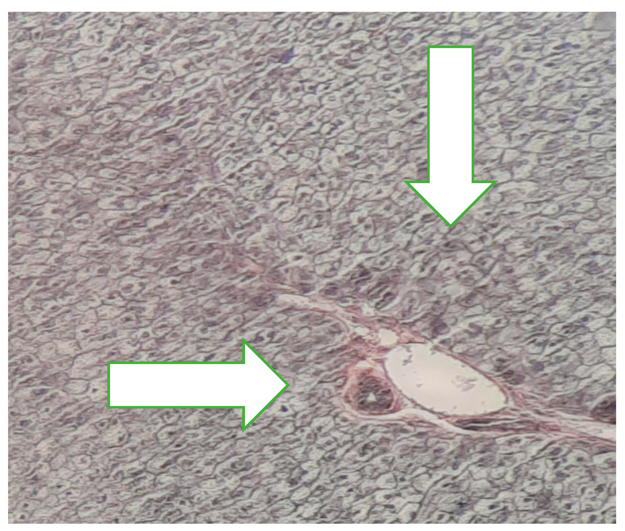
Figure 2.
Normal liver x 40 Goldner Szekely stain.

Figure 3.
Cirrhosis liver x10 H&E stain.
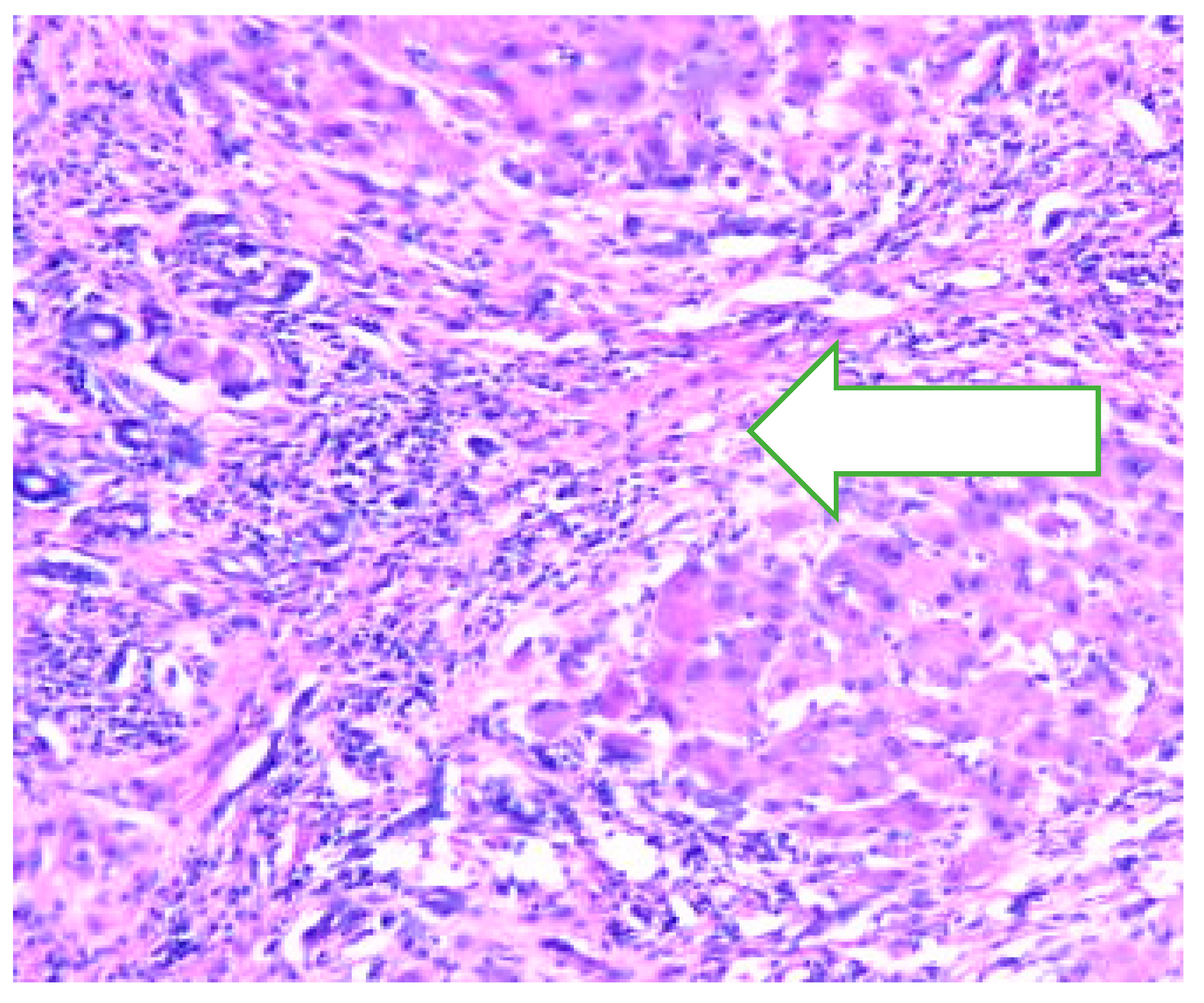
Figure 4.
Cirrhosis liver x10 H&E stain.
Disclaimer/Publisher’s Note: The statements, opinions and data contained in all publications are solely those of the individual author(s) and contributor(s) and not of MDPI and/or the editor(s). MDPI and/or the editor(s) disclaim responsibility for any injury to people or property resulting from any ideas, methods, instructions or products referred to in the content. |
© 2025 by the authors. Licensee MDPI, Basel, Switzerland. This article is an open access article distributed under the terms and conditions of the Creative Commons Attribution (CC BY) license (http://creativecommons.org/licenses/by/4.0/).
Copyright: This open access article is published under a Creative Commons CC BY 4.0 license, which permit the free download, distribution, and reuse, provided that the author and preprint are cited in any reuse.


