
Submitted:
09 October 2023
Posted:
16 October 2023
You are already at the latest version
Abstract
Introduction Despite efforts to use scaffolds to treat meniscus tears, minimal progress has been made in facilitating meniscus regeneration and return of function. Our research objective was to develop a meniscus repair and regeneration implant by applying a resorbable scaffold in combination with cells and growth factors. We report here the results of using platelet-rich plasma (PRP) as a source of growth factors to induce fibrochondrogenic differentiation of human adipose-derived mesenchymal stem cells (hADSC) in a three-dimensional (3D) Type I collagen-based scaffold in vitro. Methods Scaffold Preparation. Type I collagen scaffolds were prepared following a protocol previously published. Two different densities of scaffolds, high density (HD) and low density (LD), were produced for in vitro study. hADSC and PRP Preparation. hADSCs were cultured to the fifth passage to reach the desired number for experimentation. PRP was collected from human blood and activated. Cell Culture Procedure. Effects of PRP on hADSC proliferation and differentiation into fibrochondrogenic cells were examined in four scaffold groups: LD, HD, LD+PRP and HD+PRP. hADSCs were seeded onto scaffolds (n=5) at a concentration of 2 × 10⁶ cells/scaffold. 1% of PRP was added to the experimental media. Cellular proliferation was assessed at 1, 7, 14, and 21 days. Differentiation was measured using qRT-PCR on Days 14 and 21. qRT-PCR analysis of gene expression was completed with primers for COLLAGEN 1 and AGGRECAN. Data Analysis. ANOVAs were conducted (two-tailed tests) at the .05 significance level. Results Cellular proliferation of hADSCs seeded on each scaffold increased over time. Similar trend was observed for cells seeded on HD scaffolds with and without PRP. hADSC showed significant increase in cellular proliferation on the LD scaffolds at Days 1 and 7. At Day 21, PRP treatment and LD scaffold had a synergistic positive effect on Type I collagen gene expression. PRP did not elevate type I collagen gene in the HD group, the HD scaffold alone had the same level of type I collagen gene expression as LD+PRP. Aggrecan expression was elevated in the presence of PRP in both the HD and LD scaffold groups, indicating enhanced fibrochondrogenic differentiation of hADSCs. Effective cell infiltration was observed across both HD and LD scaffolds with and without PRP treatment. HD scaffolds displayed larger cell clusters and more extensive cell migration over time compared to LD scaffolds. However, LD scaffolds resulted a more uniform cellular distribution than HD scaffolds. Conclusion Our study demonstrates that PRP can play an important role in directing hADSCs towards fibrochondrogenic differentiation in Type I collagen-based scaffolds in vitro. Additionally, our study shows that collagen scaffold density can influence the spatial distribution and cellular behavior of infiltrated cells.
Keywords:
meniscectomy
; PRP
; meniscus repair
; meniscus implants
; tissue engineering
; 3D collagen scaffolds
; stem cells
Introduction
The menisci of the knee are two (medial and lateral meniscus) crescent shaped pads of fibrocartilaginous tissue attached inferiorly to the fossae between the femoral condyles and the tibial plateaus. The cellular components of the meniscus are termed fibrochondrocytes, which are more fibroblast-like at the outer vascular region, and more rounded, like the chondrocytes found in articular cartilage morphology in the inner and middle regions [1]. These cells interact with an extracellular matrix (ECM) component that is composed of collagen, glycosaminoglycans (GAGs), and water. In contrast to articular cartilage, where collagen II is most abundant, collagen I is the predominant collagen type in the meniscus [2]. During weight-bearing movements, the menisci of the knee articulate with the femoral condyle to reduce and disperse forces across the joint surfaces. They also produce lubricating factors to minimize friction during movement and provide nutrients to the articular cartilage of the knee joint. Hence, each meniscus is critical to the joint’s structural integrity, function, and homeostasis. Damage to the meniscus leads to aberrant load distribution within the joint and pathological “wear and tear” changes to the articulating surfaces of the joint, which predispose patients to accelerated articular cartilage degeneration (i.e., accelerated osteoarthritis). Such alterations increase joint instability and progressively lead to a plethora of orthopedic sequelae associated with compensatory gait changes and altered movement patterns [3]. There are two broad categories of meniscus tears: acute and chronic, with the former being associated with a traumatic event and the latter associated with gradual wear and tear on the fibrocartilage structure.
Meniscus tears are among the most common orthopedic injuries with an incidence of roughly one million per year in the US alone [4]. Despite the recent advancements in the field of orthopedic medicine, which allow for longer health and activity spans, the prevalence of meniscus tears, either acute traumatic tears that occur more typically in younger patients or chronic overuse tears occurring in older patients, is increasing [3]. Hence, there is a need for effective, non-invasive means for treating meniscus injuries.
Healing of a meniscus tear depends on the amount of blood supply in the area. Blood is supplied to the meniscal tissue from the periphery to the center. Hence, the outer “red zone,” which receives adequate blood supply is most likely to be healed by repair and regeneration without the need for surgical intervention. The middle “red-white zone” has moderate capacity to heal, and finally the avascular “white zone” at the center of the meniscus, has the least capacity to heal properly [5].
Management of meniscus tears differs depending on a variety of factors ranging from the type of tear, the age and activity level of the patient, and their symptoms. In any event however, the surgeon’s priority is to try to preserve the meniscal tissue as much as possible. Acute symptomatic tears are typically managed conservatively with rest, ice, elevation, and oral non-steroidal anti-inflammatory medications. Recent research has shown that supervised physical therapy focused on strengthening the muscle groups around the knee (particularly the quadriceps muscles) results in the same clinical outcomes as the most common operation for meniscus tears, an arthroscopic partial meniscectomy (APM) [6,7,8]. Therefore, surgical management of meniscal tears is mostly indicated for patients who don’t respond to conservative treatments, have diminished quality of life, or have concurrent anterior cruciate ligament injuries. However, long term clinical studies showed that APM still pose greater risk of OA development and rapid chondrolysis with loss of joint space [9].
Intra-articular injections of hyaluronic acid (HA) and or corticosteroids are also commonly used in conjunction with physical therapy to accelerate the healing of damaged meniscus tissue or provide symptomatic relief [3]. Recently, cell-based tissue engineering and orthobiologics, which encompass all biological therapies used in orthopedic medicine, including scaffolds, cell-seeded or growth factor seeded scaffolds, and platelet-rich-plasma (PRP) have shown promise as augmentative treatments for promoting tissue regeneration and meniscus repair. Injections of mesenchymal stem cells (MSCs are typically derived from adipose tissue, bone marrow, synovial tissue, or embryonic tissue) have shown the most promise of the purely cell based injectable orthobiologics for the purpose of regenerating native meniscus tissue [10].
PRP is an injectable orthobiologic, consisting of concentrated platelets and various growth factors, such as fibroblast growth factor 2 (FGF2), platelet derived growth factor (PDGF) and transforming growth factor beta (TGFb), which has the potential to enhance the regeneration of native meniscus tissue [10]. There is a lack of large-scale clinical evidence demonstrating its efficacy in accelerating healing when administered in conjunction with physical therapy, surgery, or both. However, a recent meta-analysis study found that PRP leads to a significant reduction in meniscectomy failure rates and improved overall functional outcomes in patients who received PRP with surgery compared to their non PRP treated counterparts [6].
Scaffold-based orthobiologic treatments (e.g., Collagen Meniscus Implants, CMI or Actifit) for meniscus repair after partial meniscectomy have been shown to restore the meniscal tissue, and clinically reestablish knee function, reduce pain, and prevent or delay the development of OA. CMI was developed in the 1990s. It is the only FDA-approved meniscus implant which has more than 10 years of long-term clinical follow-up [11,12,13,14,15]. However, studies show some patients experienced smaller regenerated meniscus size and incomplete mechanical function with the CMI implants [16]. For total meniscus replacements, meniscal allograft transplantation (MAT) has also demonstrated clinically significant alleviation of pain in young patients 10 years post-surgery [17,18]. However, MAT involves risks of disease transmission from a donor and issues with long-term outcomes regarding allograft deterioration and articular damage [19]. There is currently no treatment modality that ideally restores the biomechanical function of the meniscus. Nevertheless, scaffolds offer structural support, mechanical stability, and a suitable attachment site for seeded cells. This environment facilitates stem cell differentiation leading to the deposition of an extracellular matrix that closely resembles the native meniscus ECM, including proper collagen and glycosaminoglycan organization [20,21]. Efforts to improve the biomimetic and mechanical performance of biodegradable scaffolds for meniscus tear treatment must favor the right microenvironment to allow for meniscal tissue regeneration and repair.
Hence, in the current study, we aimed to develop a more durable bioresorbable meniscus implant with a tissue engineering approach to enhance healing from meniscus injury. This involved a resorbable scaffold made of Type I collagen (the most abundant collagen in meniscus ECM), seeded with hADSCs. The main objective of the study was to investigate the in-vitro cellular growth characteristics and infiltration of hADSCs as well as their fibrochondrogenic differentiation potential with and without PRP stimulation on two different densities of the novel collagen Type I scaffold.
We report here the preliminary results of using PRP as a source of growth factors to induce differentiation of hADSCs on these 3D Type I collagen-based scaffolds in vitro for the potential to promote meniscus tissue repair.
Methods
Scaffold Preparation
Type I collagen from bovine deep flexor tendon was prepared in-house, using the purifying procedure developed by Li and Stone [22,23]. Type I collagen-based meniscus scaffolds were prepared as described previously [23]: 7g of purified Type I collagen fibers were swollen in 1L 0.07M lactic acid, pH 2.5 overnight at 4℃, followed by homogenization to produce a homogeneous dispersion of Type I collagen fibrils. Two different densities of scaffolds were prepared by introducing different amounts of collagen dispersion (120g for the high-density scaffolds and 80g for the low-density scaffolds). Prior to engineering the collagen meniscus scaffolds, the pH of both dispersions was adjusted to 4.8 (the isoelectric point of the collagen) to reconstitute the collagen fibers. The reconstituted fibers were then de-gassed and wound on a mandrel to create fiber orientation in the circumferential direction. Afterwards, the wound collagen scaffolds were molded, dehydrated, lyophilized, crosslinked, rinsed, re-lyophilized, packaged and ETO sterilized. Finally, both high density (HD) and low density (LD) meniscus scaffolds were stored on the shelf for further in vitro studies.
Density Measurement
The meniscus implant was cut in half along the circumferential direction to separate the inner rim and outer rim. The density of the meniscus implant was calculated by using the implant’s dry weight divided by the implant’s total volume.
hADSC and PRP Preparation
hADSCs were obtained from ATCC and cultured in Mesenchymal Stem Cell Basal medium supplemented with Mesenchymal Stem Cell Growth Kit for Adipose and Umbilical-derived (ATCC, Manassas, Virginia) and 1% antibiotic-antimycotic (ThermoFisher Scientific, Waltham, Massachusetts). Cells were plated in T150 cell culture flasks and incubated at 37°C, 21% O2, and 5% CO2 and passaged until the fifth passage for experimentation. PRP was collected from human blood using RegenKit-BCT and prepared according to the manufacturer’s protocol (RegenLab, Brooklyn, New York). Briefly, 10 mL of blood was collected and spun for 5 minutes at 1500g. Platelet-Poor-Plasma was carefully drawn from the tube into an empty 15 mL conical tube until 1 mL remained. This was gently mixed up and down with the platelets sedimented on the surface of the gel separator contained in the tube to obtain Platelet-Rich-Plasma.
Cell Culture Procedure
The effect of PRP on hADSC proliferation and differentiation into fibrochondrogenic cells was investigated. A total of four groups of scaffolds were studied, LD alone, HD alone, LD+PRP and HD+PRP. hADSCs were seeded onto scaffolds (n=5) at a concentration of 2 × 10⁶ cells/scaffold. Cells in (2D) monolayer culture were used as positive control to normalize all measurements during experimentation. 1% of PRP was added to the experimental media. The media was changed every other day. Quantitative measurement of cell proliferation was performed using the alamarBlue assay according to the manufacturer’s protocol (Thermo Fisher Scientific, Waltham, Massachusetts). Since resazurin, the colorimetric indicator of cellular metabolism in AlamarBlue, is a non-toxic reagent, repeated measurements were taken over time on the same sample for the following timepoints: Day 1, 7, 14, 21. After 4 hours of incubation with 10% (v/v) AlamarBlue added in culture media, absorbances were measured at 570 and 600 nm as reference wavelengths per the manufacturer’s protocol with a standard spectrophotometer to determine the amount of AlamarBlue reduced for each sample. Absorbance values were corrected using absorbance of control wells with no cells, with media only, and media with AlamarBlue. Metabolic activity normalized to control was indicated as a percentage of AlamarBlue reduction using the following equations provided by the manufacturer’s protocol.
Alw: absorbance at lower wavelength minus the media blank
Ahw: absorbance at higher wavelength minus the media blank
Ro: Correction factor
On Days 14 and 21, samples were additionally harvested for gene expression studies to assess cellular differentiation. Messenger RNA was collected from cells after lysis using the RNeasy Plus Mini kit (QIAGEN, Venlo, Netherlands). The reverse transcription of mRNA to cDNA was done using the iScript™ cDNA Synthesis Kit and a T100 Thermal Cycler (Bio-Rad Laboratories, Inc., Hercules, California). The relative gene expression levels of the two ECM proteins (Aggrecan and collagen Type I) most predominantly associated with meniscus and cartilage matrix synthesis were measured by qPCR with QuantStudio™ 3 Real-time PCR System (Applied Biosystems, Waltham, Massachusetts) using PowerUp™ SYBR™ Green Master Mix (ThermoFisher Scientific, Waltham, Massachusetts) following standard protocol. The relative fold change of each target primer was calculated using the 2-∆∆CT method and normalized to the housekeeping gene, Glyceraldehyde 3-phosphate dehydrogenase GAPDH. Target sequences for each primer are shown in Table 1.
Histological study
Scaffolds were fixed in 10% formalin (ThermoFisher Scientific, Waltham, Massachusetts) at Days 1, 7, 14, 21 for histology to evaluate cell infiltration and distribution within the scaffolds over time. All fixed samples were subsequently dehydrated before paraffin embedding. After paraffin embedding, sections were cut for each sample at 7 μm thick. Four sections from each group at each time point were stained with hematoxylin and eosin. Images of each section were taken for qualitative evaluation of cell migration within the scaffold overtime.
Statistical Analysis
An analysis of variance (ANOVA) was conducted with a two-factor repeated measures ANOVA where time was one factor and PRP treatment was another factor to test for the effect of PRP treatment of hADSCs on each scaffold over time. Similar analysis was conducted to test the effect of the LD and HD scaffold density in the absence or presence of PRP over time. Sidak’s multiple comparison tests were performed to test for differences between groups. Statistical significance was set at the 0.05 alpha level for all tests. Statistical analysis was performed using GraphPad Prism 8.4.3 (GraphPad Software Inc., San Diego, California).
Results
Density measurement
Table 2 shows the overall average density of HD scaffolds (25% increase) was significantly higher (p=0.0036) than LD scaffolds. When comparing LD and HD scaffolds’ partial density at the inner rim and outer rim, HD scaffolds revealed a significant difference at the inner rim with a 35% increase (p=0.001) and at the outer rim with a 23% increase (p=0.039).
In-vitro cellular proliferation of hADSCs on 3D LD and HD scaffolds
hADSCs seeded on each scaffold showed increases in cellular proliferation with and without PRP treatment. A similar trend in cellular proliferation was observed for hADSCs seeded on HD scaffolds with and without PRP stimulation. Proliferation was highest in both groups at Day 14 (61% ±4 for HD with PRP and 63% ±10 for HD without PRP) with statistically significant increases (p=0.0134, 0.0005, respectively) relative to Day 1 (29%±17, 30%±7 for HD with and without PRP respectively) (Figure 1A). Comparative analysis of the cellular proliferation of hADSCs seeded on LD and HD scaffolds revealed a significant increase for LD group on Day 1 (p=0.0074) and Day 7 (p<0.0001) (Figure 1B). When examining the trend in cellular proliferation of hADSCs seeded on LD scaffolds over time, a transient increase was observed for LD group without PRP stimulation. This increase was determined to be statistically significant at Day 7 (88%±11). This contrasts with a gradual increase over time seen in the LD group stimulated with PRP. Initial cellular proliferation of hADSCs seeded on LD scaffolds without PRP on Day 1 (49%±10) and Day 7 (88%±11) was observed to be higher when compared to LD samples with PRP stimulation (34%±10 and 38%±4, p=0.0003, <0.0001 respectively) (Figure 1C). hADSCs in the LD scaffold group exhibited the highest cellular proliferation on Day 21 with PRP treatment (65%±10) (p<0.0001) (Figure 1D). There was no significant difference in cellular proliferation between LD and HD scaffolds in the presence of PRP.
Fibrochondrogenic potential of hADSCs cultured on 3D scaffolds for meniscus tissue repair
To examine the fibrochondrogenic differentiation potential of hADSCs cultured on LD and HD scaffolds for meniscal tissue repair, relative gene expression changes for aggrecan (ACAN) and collagen I (COL I) were quantified for Day 14 and Day 21. No difference was found in ACAN gene expression level between all groups on Day 14 (Figure 2). However, hADSCs with PRP treatment seeded on both scaffolds demonstrated higher gene expression for ACAN compared to untreated groups at Day 21 (Figure 2A and 2C). This increase was found to be more statistically significant for HD scaffolds (p<0.0001) than LD scaffolds (p=0.0055). When analyzing the effect of PRP treatment over time within groups, the increase of ACAN expression in groups treated with PRP was statistically more significant for HD than LD scaffolds (p=0.0006, 0.0256, respectively) (Figure 2A and 2C). No significant comparative difference was found in ACAN gene expression between hADSCs seeded on LD and HD scaffolds with PRP (Figure 2D). Quantification of gene expression level for COL I for HD groups revealed an increase without PRP stimulation on Days 14 and 21 (Figure 3A). However, this increase was not found to be statistically significant. The opposite trend was observed in the LD groups treated and untreated with PRP. A significant increase in COL I gene expression was found in hADSCs seeded on LD scaffolds with PRP stimulation at Day 21 (p=0.0030) (Figure 3C). Comparison of COL I gene expression in hADSCs seeded on LD and HD scaffolds showed higher levels of expression in HD groups at Day 14 and 21 (Figure 3B). However, the increase was only found to be statistically significant at Day 21 (p=0.0349). The effect of PRP treatment on COL I expression over time was not found to be significant for all groups. In addition, there was no significant difference found between HD and LD scaffolds with the presence of PRP (Figure 3D).
In-vitro cell infiltration of hADSCs seeded on 3D LD and HD collagen scaffolds
To visualize the cellular infiltration and morphology of the hADSCs on LD and HD scaffolds, micrographs of samples were taken after hematoxylin and eosin staining. From Day 1 to Day 21, a progressive cellular infiltration was observed for all groups with and without PRP treatment. hADSCs seeded on HD scaffolds without PRP treatment showed a very thick and dense layer of cells on the surface after 24 hours of cell seeding; in contrast to LD scaffolds with and without PRP treatment, which demonstrated numerous cell clusters migrating through the pores of the scaffold (Figure 4). hADSCs seeded on HD scaffolds with PRP treatment showed a similar trend. Cell penetration became uniform at Day 7 across all groups. Cells on LD scaffolds without PRP treatment showed an increased migratory potential from Day 7 through Day 14 in contrast to those on HD scaffolds with and without PRP treatment. However, on Day 21, HD groups with and without PRP treatment exhibited the greatest cellular infiltration and largest cell aggregation when compared to LD scaffolds with and without PRP treatment.
Discussion
In this in vitro proof of concept study, Type I collagen-based scaffolds with high (HD) and low (LD) densities were developed to evaluate their effect on cellular behavior for meniscus tissue regeneration. After initial seeding, we observed that LD and HD scaffolds allowed for cellular infiltration and aggregation within the scaffold. LD scaffolds resulted in a uniform cell distribution and infiltration over time. HD scaffolds allowed for deeper cellular migration but an uneven spatial distribution of cells. This distinctive pattern between LD and HD scaffolds persisted throughout the study. Micrographs of cell-seeded scaffolds at Day 1 and Day 21 particularly highlighted this difference. Micrographs of both scaffolds on Day 1 revealed the pore size and pore interconnectivity were distinguishable. LD scaffolds had smaller pore sizes and higher interconnectivity. In contrast, HD scaffolds appeared to have larger pore size and limited pore interconnectivity. Therefore, it is possible that the difference in the degree of cell density and migration observed between LD and HD scaffolds resulted from the intrinsic variations in these parameters between the two scaffolds. This finding suggests that cell infiltration and cellular spatial distribution can be controlled by the internal pore architecture of the scaffold. This is consistent with previous findings [24,25,26]. Scaffold composition and pore size are determining factors in regulating the biological activities and adaptation of infiltrated cells [27,28]. In the present study, cellular activity of hADSCs was observed to be significantly higher in LD scaffolds than in HD scaffolds on Days 1 and 7. These findings corroborate previous knowledge that there may be an optimal pore size that is suitable for cell growth and penetration [29,30]. In fact, Han et al. have suggested that optimal pore size may be cell-type dependent [31].
When assessing the fibrochondrogenic potential of hADSCs in both scaffold groups, our results indicated that, in the absence of PRP, the HD scaffolds were more effective in driving fibrochondrogenic differentiation than the LD scaffolds. However, in the presence of PRP, that trend was different. LD scaffolds displayed similar fibrochondrogenic potential with HD scaffolds. PRP upregulated ACAN gene expression regardless of scaffold density. However, scaffold density in combination with PRP modulated the fibrochondrogenic potential of hADSCs especially as we saw upregulation in COL I gene expression only in LD groups. Cell differentiation is a precise dialogue of complex and dynamic spatial-temporal intracellular regulatory networks that is driven by cell-to-cell communication via autocrine and paracrine signaling. The topology of cell-to-cell communication relies on spatial-temporal regulations of ligand-receptor interactions [32]. At the cell-surface level, the interaction of ligand-receptors is dependent on ligand concentrations, spatial organization of receptors and gradient sensing sensitivity of cell-surface receptors [33,34]. Each of these elements can be modulated by the 3D microenvironment of the cell. Therefore, it is possible that the cellular milieu imparted by each Scaffold in combination with PRP influenced the biochemical and biophysical elements governing cell-to-cell communication and the downstream signaling response that drives hADSCs toward fibrochondrogenic differentiation. It should be noted that scaffold porosity can regulate the diffusion of oxygen, nutrients, and other soluble factors in a 3D microenvironment. It was already noted that cellular distribution within the LD scaffolds was distinguishable from HD scaffolds due to scaffold porosity. Therefore, we suspect that the synergistic effect of porosity and PRP may explain the observed changes in the fibrochondrogenic potential of hADSCs on LD scaffolds. The precise mechanism of interaction of these two factors remains to be fully elucidated. Ultimately, these findings reveal a regenerative potential of PRP in the 3D microenvironment of hADSCs that is dependent on the scaffold porosity and density. Stessuk et al. have demonstrated the potential influence of PRP in enhancing migration of ADSCs in-vitro. In our study, PRP did not significantly increase cellular migration within the scaffold [35]. The culture system used in our study may have contributed to the discrepancy between our findings as they seeded hADSCs on a cell culture plate as opposed to a collagen scaffold. Overall, the present study demonstrates proof-of-concept evidence of these cell-seeded and adjuvant treated tissue engineered scaffold as a template for directing cellular differentiation of hADSCs towards a fibrochondrogenic lineage in vitro.
Other researchers have pointed to the importance of scaffold density in promoting cellular infiltration, attachment and then driving the desired phenotype of endogenous or seeded cells for meniscus regeneration [36,37]. In their in vitro study, Ruprecht et al. investigated the migration potential of endogenous meniscus cells or exogenous human bone marrow derived MSCs on a meniscus derived matrix scaffold [38]. They demonstrated that scaffolds seeded with native meniscus cells led to accelerated cellular infiltration and tissue growth and promoted meniscus healing. They also found that highly concentrated scaffolds promoted greater cellular infiltration. In alignment with our study, high-density scaffolds provided more cellular migration compared to low-density over time. Additionally, in our study, variation in cell distribution between low-density and high-density scaffolds was evident through histology.
Other studies have also investigated the ability of Type I collagen scaffolds and other biodegradable scaffolds in combination with PRP to enhance meniscus tissue regeneration and have found that these scaffolds enhance healing in animal models compared to untreated controls [39,40,41,42,43,44,45,46,47,48]. However, the current work is the first study to our knowledge that combines a focus on comparing between Type I collagen scaffold densities and comparing between untreated and PRP treated scaffolds. In fact, this is the first in vitro study to investigate the ability of PRP to stimulate fibrochondrogenic differentiation of hASDCs seeded on Type I collagen scaffold.
Future research
Our findings showed that these scaffolds are conducive to cellular attachment, proliferation, migration, and differentiation. Cell differentiation was assessed using gene expression changes in a static culture. It is well known that the chondrogenic and osteogenic potential of MSCs is affected by mechanical stimuli [49]. The application of compression combined with fluid flow may create a microenvironment more physiologically relevant to the meniscus. Thus, the next near-term goal is to confirm gene expression changes in a dynamic in-vitro culture prior to a preclinical study. The 1-step tissue engineering approach (incorporation of the cells and PRP into the implant at the point of surgery) will be tested to see whether the regeneration of meniscus tissue is accelerated in a large animal model. Ultimately, the clinical application of this scaffold coupled with the regenerative potential of PRP, and the 1-step stem cell seeding is to provide a custom-designed implant to a meniscal defect that is suitable for meniscal tissue repair and regeneration after partial or subtotal meniscectomy.
Limitations of the study
The greatest challenge to translating in vitro, preclinical research in meniscus repair such as this study, is the inherent complexity that comes with the many permutations of scaffold composition and density, cell-scaffold pairing, and biologic adjuvant. The effects of each of these elements are poorly understood let alone in combination with each other. For example: (1) The sulfated glycosaminoglycan (GAG) plays a critical role in the native meniscus as well as in driving the chondrogenic differentiation of MSCs. However, the sulfated GAG was not included in the current scaffold design. (2) The static seeding method used in this study can introduce local variations in cell density resulting in non-uniformity in cell distribution. This could limit cell differentiation. Other methods such as needle injection, vacuum seeding, or rotational seeding could be better suited for enhancing uniformity and reproducibility. (3) A low dose of PRP was used in our study. We have not explored if higher doses induce a higher regenerative potential in hADSCs seeded on these scaffolds. Further research is needed to compare these different elements to elucidate which combinations hold the most promise.
Conclusion
Our findings add to the growing body of preclinical evidence suggesting that stem cell-seeded Type I collagen scaffolds, in combination with PRP has the ability to drive the fibrochondrogenic differentiation of hADSCs in vitro, and potentially enhance the regeneration of damaged meniscal tissue.
Acknowledgements
The work was supported by the Shu-Tung and Alice Li Foundation.
References
- Tan GK, Cooper-White JJ. Interactions of meniscal cells with extracellular matrix molecules: towards the generation of tissue engineered menisci. Cell Adh Migr. 2011 May-Jun;5(3):220-6. [CrossRef]
- Eyre DR, Wu JJ. Collagen of fibrocartilage: a distinctive molecular phenotype in bovine meniscus. FEBS Lett. 1983 Jul 25;158(2):265-70. [CrossRef]
- Luvsannyam E, Jain MS, Leitao AR, Maikawa N, Leitao AE. Meniscus Tear: Pathology, Incidence, and Management. Cureus. 2022;14(5): e25121. [CrossRef]
- UCSF Health. Overview Orthopedics: Meniscus Tear. https://www.ucsfhealth.org/conditions/meniscus-tear#:~:text=Meniscus%20tears%20are%20among%20the,shaped%20structures%20made%20of%20cartilage; 2023 Accessed 5 May 2023. 5 May.
- Jacob G, Shimomura K, Krych AJ, Nakamura N. The Meniscus Tear: A Review of Stem Cell Therapies. Cells. 2019;9(1):92. [CrossRef]
- Li, Z, Weng, X. Platelet-rich plasma use in meniscus repair treatment: a systematic review and meta-analysis of clinical studies. J Orthop Surg Res. 2022;17(01). [CrossRef]
- Freymann U, Degrassi L, Krüger J, Metzlaff S, Endres M, Petersen W. Effect of serum and platelet-rich plasma on human early or advanced degenerative meniscus cells. Connect Tissue Res. 2017;58:6,509-519. [CrossRef]
- Özyalvaç O, Tuzuner T, Gurpinar T, Obut A, Acar B, Emre Y. Radiological and functional outcomes of ultrasound-guided PRP injections in intrasubstance meniscal degenerations. J of Orthop Surg. 2019;27:2. [CrossRef]
- Sihvonen R, Paavola M, Malmivaara A, Itälä A, Joukainen A, Kalske J, Nurmi H, Kumm J, Sillanpää N, Kiekara T, Turkiewicz A, Toivonen P, Englund M, Taimela S, Järvinen TLN; FIDELITY (Finnish Degenerative Meniscus Lesion Study) Investigators. Arthroscopic partial meniscectomy for a degenerative meniscus tear: a 5 year follow-up of the placebo-surgery controlled FIDELITY (Finnish Degenerative Meniscus Lesion Study) trial. Br J Sports Med. 2020;54(22):1332-1339.
- Alessio-Mazzola M, Felli L, Trentini R. et al. Efficacy of Autologous Platelet-Rich Plasma Injections for Grade 3 Symptomatic Degenerative Meniscal Lesions: A 1-Year Follow-up Prospective Study. Sports Health. 2021;14:2. [CrossRef]
- Freyman U, Metzlaff S, Kruger J-P. et al. Effect of Human Serum and 2 Different Types of Platelet Concentrates on Human Meniscus Cell Migration, Proliferation, and Matrix Formation. Arthroscopy. 2016;32:6,1106-1116. [CrossRef]
- Rodkey, W.G., et al., Comparison of the collagen meniscus implant with partial meniscectomy: a prospective randomized trial. J Bone Joint Surg Am. 2008. 90(7): p. 1413-1426.
- Zaffagnini, S., et al., Two-year clinical results of lateral collagen meniscus implant: a multicenter study. Arthroscopy: The Journal of Arthroscopic & Related Surgery, 2015. 31(7): p. 1269-1278.
- Li, S.-T., et al., Type I collagen-based template for meniscus regeneration. Tissue engineering and biodegradable equivalents. Scientific and clinical applications, 2002: p. 237-266.
- Stone, K.R., et al., Regeneration of meniscal cartilage with use of a collagen scaffold. Analysis of preliminary data. JBJS, 1997. 79(12): p. 1770.
- Monllau, J.C., et al., Outcome after partial medial meniscus substitution with the collagen meniscal implant at a minimum of 10 years’ follow-up. Arthroscopy: The Journal of Arthroscopic & Related Surgery, 2011. 27(7): p. 933-943.
- Verdonk PC, Demurie A, Almqvist KF, Veys EM, Verbruggen G, Verdonk R. Transplantation of viable meniscal allograft. Survivorship analysis and clinical outcome of one hundred cases. J Bone Joint Surg Am. 2005. 87(4):715-24.
- Verdonk PC, Verstraete KL, Almqvist KF, De Cuyper K, Veys EM, Verbruggen G, Verdonk R. Meniscal allograft transplantation: long-term clinical results with radiological and magnetic resonance imaging correlations. Knee Surg Sports Traumatol Arthrosc. 2006 Aug;14(8):694-706.
- Lee SR, Kim JG, Nam SW. The tips and pitfalls of meniscus allograft transplantation. Knee Surg Relat Res. 2012 Sep;24(3):137-45.
- Pereira, H, et al. Tissue engineering and regenerative medicine strategies in meniscus lesions. Arthroscopy: The journal of arthroscopic & related surgery, 27.12 (2011): 1706-1719. [CrossRef]
- Muran A, Schaffler B, Wong. et al. Effect of increasing hyaluronic acid content in collagen scaffolds on the maintenance of chondrogenic phenotype in chondrocytes and mesenchymal stem cells. JCJP. 2023:100099. [CrossRef]
- Li, ST and Stone KR, Meniscal Augmentation Device (2000), U.S. Patent No. 6,042,610.
- Li ST. Biopolymer-based meniscus implant. 17/625,825. 2022. U.S. Patent Application.
- Harley BA, Kim HD, Zaman MH, Yannas IV, Lauffenburger DA, Gibson LJ. Microarchitecture of three-dimensional scaffolds influences cell migration behavior via junction interactions. Biophys J. 2008; 95(8):4013-24.
- Murphy CM, O’Brien FJ. Understanding the effect of mean pore size on cell activity in collagen-glycosaminoglycan scaffolds. Cell Adh Migr. 2010;4(3):377-81.
- Somo SI, Akar B, Bayrak ES, Larson JC, Appel AA, Mehdizadeh H, Cinar A, Brey EM. Pore Interconnectivity Influences Growth Factor-Mediated Vascularization in Sphere-Templated Hydrogels. Tissue Eng Part C Methods. 2015;21(8):773-85.
- Yang YL, Motte S, Kaufman LJ. Pore size variable type I collagen gels and their interaction with glioma cells. Biomaterials. 2010;31(21):5678-88.
- Buenzli PR, Lanaro M, Wong CS, McLaughlin MP, Allenby MC, Woodruff MA, Simpson MJ. Cell proliferation and migration explain pore bridging dynamics in 3D printed scaffolds of different pore size. Acta Biomater. 2020;114:285-295.
- O’Brien FJ. Biomaterials & scaffolds for tissue engineering. Mater Today. 2011;14 (3):88-95.
- Bružauskaitė I, Bironaitė D, Bagdonas E, Bernotienė E. Scaffolds and cells for tissue regeneration: different scaffold pore sizes-different cell effects. Cytotechnology. 2016 May;68(3):355-69.
- Han Y, Lian M, Wu Q, Qiao Z, Sun B, Dai K. Effect of Pore Size on Cell Behavior Using Melt Electrowritten Scaffolds. Front Bioeng Biotechnol. 2021;9:629270.
- Armingol E, Officer A, Harismendy O, Lewis NE. Deciphering cell-cell interactions and communication from gene expression. Nat Rev Genet. 2021;22(2):71-88.
- Goldstein B, Dembo M. Approximating the effects of diffusion on reversible reactions at the cell surface: ligand-receptor kinetics. Biophys J. 1995;68(4):1222-30.
- Endres RG, Wingreen NS. Accuracy of direct gradient sensing by single cells. Proc Natl Acad Sci USA. 2008;105(41):15749-54.
- Stessuk T, Puzzi MB, Chaim EA, Alves PC, de Paula EV, Forte A, Izumizawa JM, Oliveira CC, Frei F, Ribeiro-Paes JT. Platelet-rich plasma (PRP) and adipose-derived mesenchymal stem cells: stimulatory effects on proliferation and migration of fibroblasts and keratinocytes in vitro. Arch Dermatol Res. 2016;308(7):511-20.
- Akhmanova M, Osidak E, Domogatsky S, Rodin S, Domogatskaya A. Physical, spatial, and molecular aspects of extracellular matrix of in vivo niches and artificial scaffolds relevant to stem cells research. Stem Cells Int. 2015 Aug 16;2015.
- Matsiko A, Levingstone T, O’Brien F, Gleeson J. Addition of hyaluronic acid improves cellular infiltration and promotes early-stage chondrogenesis in a collagen-based scaffold for cartilage tissue engineering. J Mech Behav Biomed Mater. 2012;11:41-52. [CrossRef]
- Ruprecht JC, Waanders T, Rowland, C. et al. Meniscus-Derived Matrix Scaffolds Promote the Integrative Repair of Meniscal Defects. Sci Rep. 2019;9, 8719.
- Oda S, Otsuki S, Kurokawa Y. et al. A new method for meniscus repair using type I collagen scaffold and infrapatellar fat pad. J. Biomater. Appl. 2015;29(10). [CrossRef]
- Li ST, Yuen D, Li PC, Rodkey WG, Stone KR. Collagen as a biomaterial: an application in knee meniscal fibrocartilage regeneration. MRS Online Proceedings Library (OPL). 1993;331:25.
- Warth, R.J. and W.G. Rodkey, Resorbable collagen scaffolds for the treatment of meniscus defects: a systematic review. Arthroscopy: The Journal of Arthroscopic & Related Surgery 2015. 31(5): p. 927-941.
- Korpershoek J, de Windt T, Hagmeijer M, Vonk L, Saris D. Cell-based meniscus repair and regeneration: at the brink of clinical translation? A systematic review of preclinical studies. Orthop J Sports Med. 2017 5(2). [CrossRef]
- Hutchinson I, Rodeo S, Perrone G, Murray M. Can Platelet-Rich Plasma Enhance Anterior Cruciate Ligament and Meniscal Repair. J Knee Surg 2015; 28(01): 019-028. [CrossRef]
- Kwak H, Nam J, Lee J, Kim H, Yoo, J. Meniscal repair in vivo using human chondrocyte-seeded PLGA mesh scaffold pretreated with platelet-rich plasma. J Tissue Eng Regen Med. 2014;(2):471-480. [CrossRef]
- Ishida K, Kuroda R, Miwa, M. et al. The regenerative effects of platelet-rich plasma on meniscal cells in vitro and its in vivo application with biodegradable gelatin hydrogel. Tissue Eng. 2007;13(5):1103-1112. [CrossRef]
- Blough C, Bobba C, DiBartola A. et al. Biologic augmentation during meniscal repair. J. Knee Surg. 2021;36(05):498-506. [CrossRef]
- Migliorini F, Cuozzo F, Cipollaro L, Oliva F, Hildebrand F, Maffulli N. Platelet-rich plasma (PRP) augmentation does not result in more favourable outcomes in arthroscopic meniscal repair: a meta-analysis. JOTR 2022;23(1). [CrossRef]
- Belk JW, Kraeutler MJ, Thon SG, Littlefield CP, Smith JH, McCarty EC. Augmentation of meniscal repair with platelet-rich plasma: A systematic review of comparative studies. Orthop J Sports Med. 2020 Jun 17;8(6):2325967120926145. [CrossRef]
- Kelly, J, Jacobs R. The role of mechanical signals in regulating chondrogenesis and osteogenesis of mesenchymal stem cells. Birth Defects Res C Embryo Today 2010 Mar;90(1):75-85. [CrossRef]
Figure 1.
Cellular proliferation (n=5): Quantification of cellular activity using absorbance values detected after Alamar Blue Reduction at the different time points (Days 1, 7, 14, 21). Note: All values are represented as means and standard deviations. Statistical significance: *,#p ≤ 0.05, **,##p ≤ 0.01, ***,###p ≤ 0.001. ****,####p ≤ 0.0001. *Significant difference between groups; # Significant difference across time.
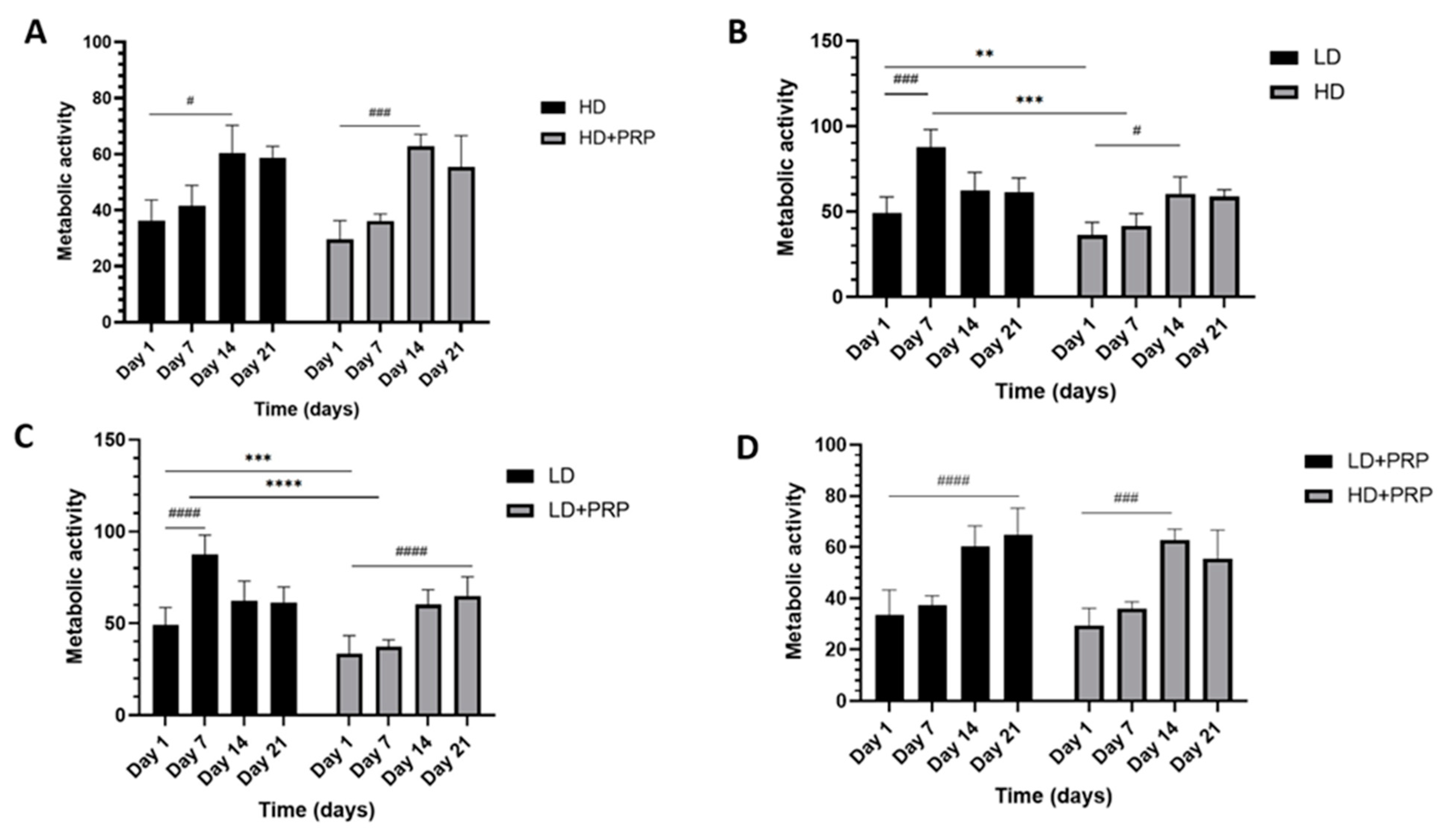
Figure 2.
Gene expression analysis (n=5) Relative fold change in aggrecan (ACAN) gene expression, a chondrogenic ECM marker found in meniscal tissue and articular cartilage.
Figure 2.
Gene expression analysis (n=5) Relative fold change in aggrecan (ACAN) gene expression, a chondrogenic ECM marker found in meniscal tissue and articular cartilage.
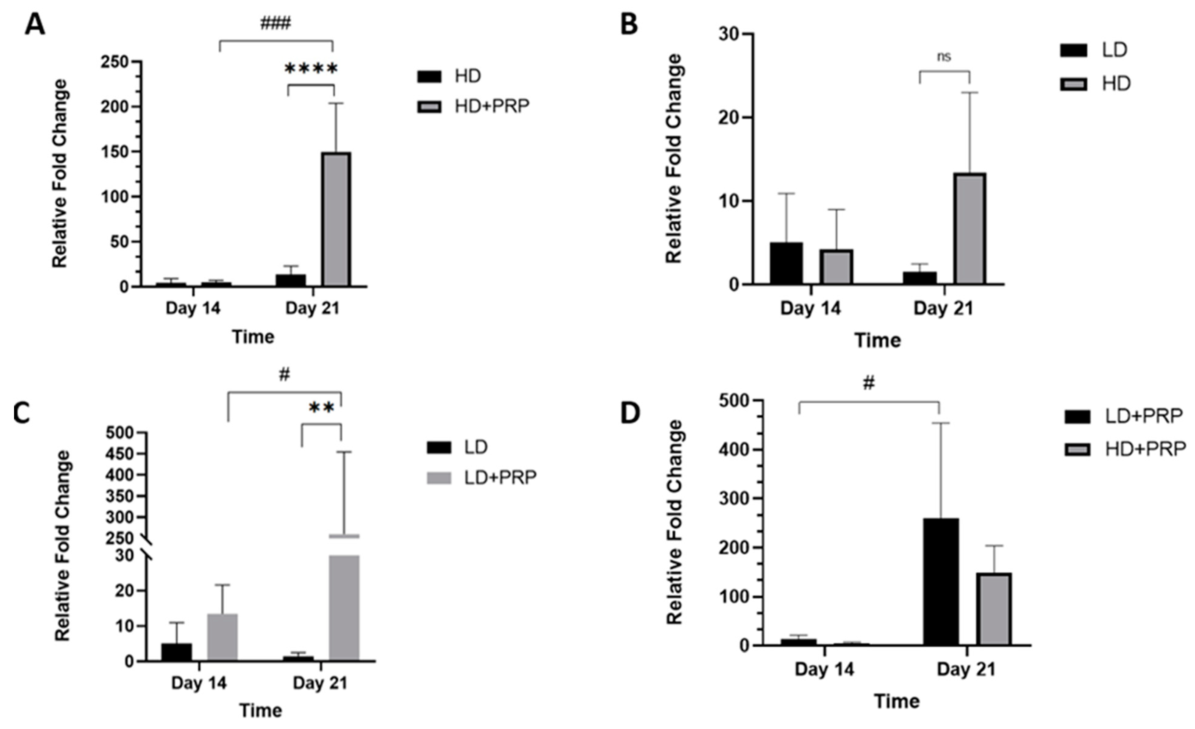
Figure 3.
Gene expression analysis (n=5) Relative fold change in type I Collagen (COL I) gene expression, the most abundant collagen in the meniscus.
Figure 3.
Gene expression analysis (n=5) Relative fold change in type I Collagen (COL I) gene expression, the most abundant collagen in the meniscus.
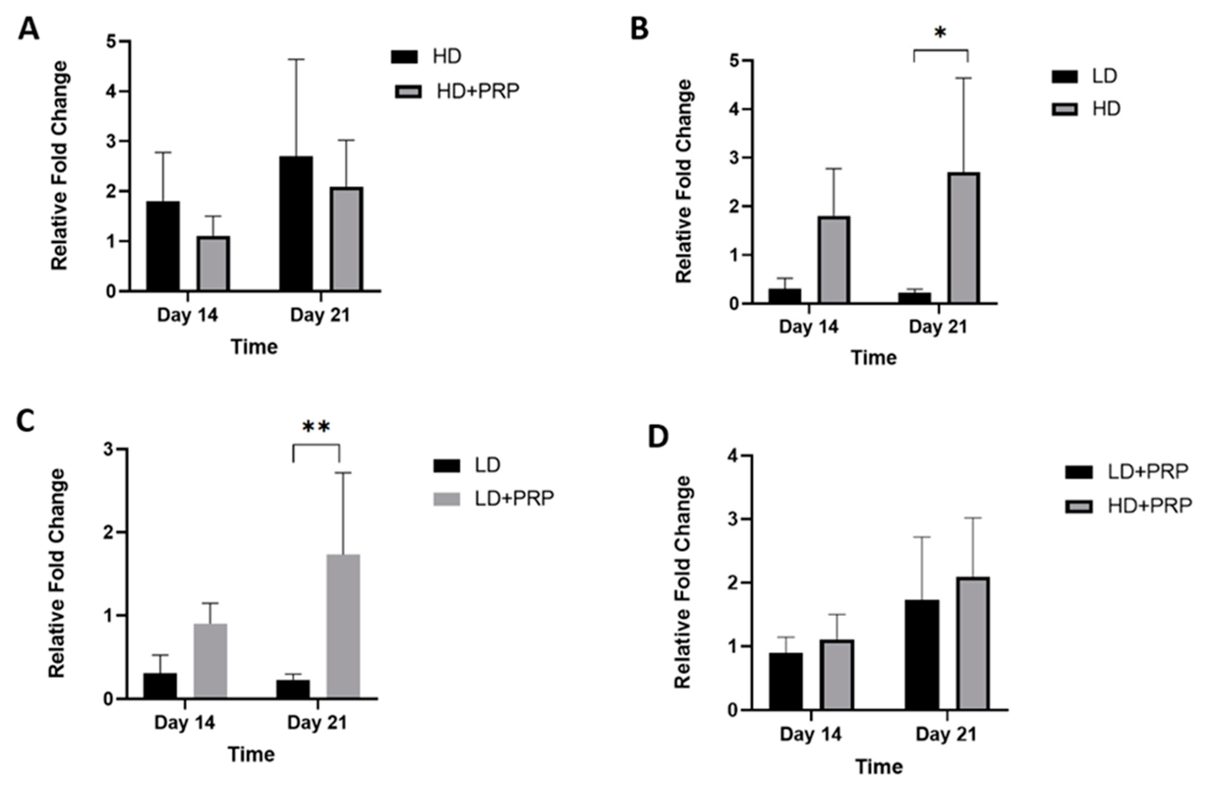
Figure 4.
Micrographs of H&E stained samples of biodegradable collagen type I scaffolds low (LD) and high (HD) densities seeded with hADSCs at Days 1, 7,14, 21 in an in-vitro culture system with and without PRP stimulation. Dark purple (hematoxylin) stains represent cell nuclear components, and pink (eosin) stains display ECM features including collagen scaffold. Scale bar: 100 µm.
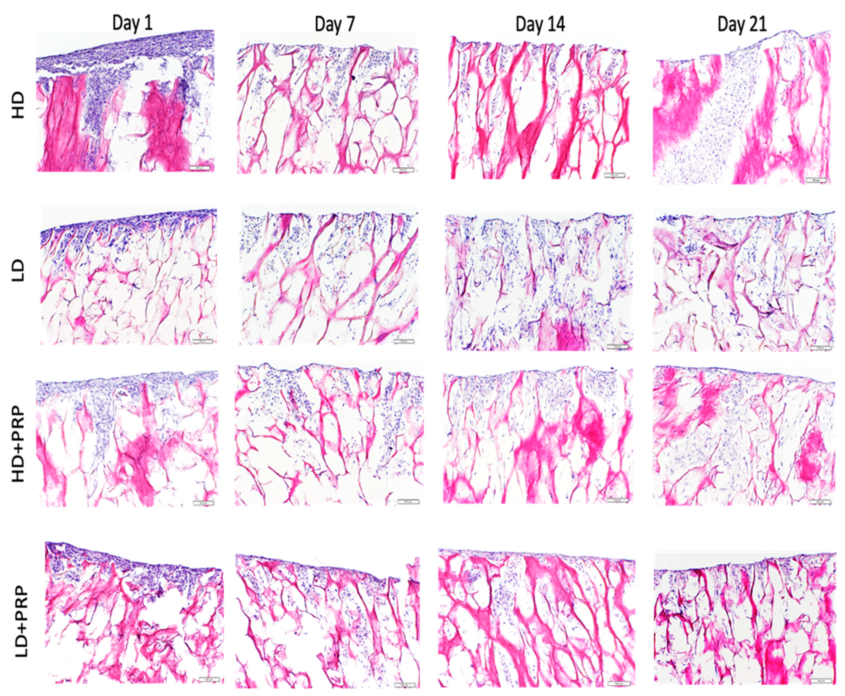

Table 1.
List of the primers and their sequences.
| Primers | Sequences |
|---|---|
| TYPE I COLLAGEN | 5′-ATG CCA TCA AAG TCT TCT GCA A-3′ Forward$$$5′-CTT-GGG-GTT-CTT-GCT-GAT-GTA-C-3′ Reverse |
| AGGRECAN | 5′-CTA-CAT-TGG-TGG-AAG-TGG-TCA-C3′ Forward$$$5′-CCA CTA-GCT-CTC-CCA-CTA-ATG-T-3′ Reverse |
| GAPDH | 5′-ACC-CAG-AAG-ACT-GTG-GAT-GG-3′ Forward$$$5′-GAG-GCA-GGG-ATG-ATG-TTC-TG-3′ Reverse |
Table 2.
Summary of density.
| Low Density (LD) | High Density (HD) | |||
|---|---|---|---|---|
| Overall Density (g/cm3) | 0.15±0.01 | 0.19±0.01 | ||
| Partial Density (g/cm3) | Inner rim | Outer rim | Inner rim | Outer rim |
| 0.19±0.01 | 0.13±0.01 | 0.26±0.01 | 0.16±0.01 | |
Disclaimer/Publisher’s Note: The statements, opinions and data contained in all publications are solely those of the individual author(s) and contributor(s) and not of MDPI and/or the editor(s). MDPI and/or the editor(s) disclaim responsibility for any injury to people or property resulting from any ideas, methods, instructions or products referred to in the content. |
© 2023 by the authors. Licensee MDPI, Basel, Switzerland. This article is an open access article distributed under the terms and conditions of the Creative Commons Attribution (CC BY) license (http://creativecommons.org/licenses/by/4.0/).
Copyright: This open access article is published under a Creative Commons CC BY 4.0 license, which permit the free download, distribution, and reuse, provided that the author and preprint are cited in any reuse.


