Submitted:
06 October 2023
Posted:
09 October 2023
You are already at the latest version
Abstract
Purpose The objective of this study was to eliminate any ambiguity by examining the correlation between gut microbiota and both AMD and glaucoma.
Methods Mendelian randomization studies were conducted utilizing the data sourced from the GWAS database for the gut microbiome, AMD, and glaucoma. SNP estimates were summarized through five MR methods. We utilized Cochran's Q statistic to evaluate the heterogeneity of the instrumental variables. Additionally, we employed a "leave-one-out" approach to verify the stability of our findings.
Results IVW suggests that Eubacterium (oxidoreducens group) and Parabacteroides had a protective effect on AMD. Both weighted median and IVW suggests that Lachnospiraceae (NK4A136 group) and Ruminococcaceae (UCG009) had a protective effect on AMD. However, both weighted median and IVW suggests that Dorea had a risk effect on AMD. Similarly, The IVW of Eubacterium (ventriosum group) showed a risk effect on AMD. The weighted median of Eubacterium (nodatum group), Lachnospiraceae (NC2004 group), and Roseburia had a risk effect on glaucoma. IVW suggested that Ruminococcaceae (UCG004) had a risk effect on glaucoma. Reverse MR analysis found a causal link between Eubacterium (nodatum group) and glaucoma. No causal relationships were found between AMD or glaucoma and the other mentioned bacterial groups. No significant heterogeneity or evidence of horizontal pleiotropy was detected.
Conclusions This study found that certain gut bacteria had protective effects on AMD, while others may be risk factors for AMD or glaucoma. Besides, reverse MR found that glaucoma led to increased abundance of certain gut bacteria. Further trials are needed to clarify the specific mechanisms involved.
Keywords:
Age-related macular degeneration
; Glaucoma
; Gut microbiota
; Mendelian randomization
; Gut-retina axis
1. Introduction
Age-related macular degeneration (AMD) and glaucoma are prevalent eye diseases that can lead to blindness on a global scale. AMD is a progressive damage to the structure and function of the macula, leading to central vision loss and is one of the main causes of blindness among the elderly. The pathogenesis of AMD is complex and influenced by genetic polymorphism, immune, metabolic, light damage, nutrition, and other factors. During the progression of AMD, microglia and macrophages migrate to the subretinal and choroidal regions, causing local imbalance in the immune microenvironment of the retinal pigment epithelial cell layer, but the specific mechanism is still unclear [1,2]. Glaucoma is a progressive neurodegenerative disorder that is characterized by the gradual deterioration of retinal ganglion cells and their axons [3]. The pathogenesis of glaucoma is still being delineated. Nowadays, there are no satisfactory strategies for the prevention and treatment of AMD and glaucoma.
The gut microbiota refers to the diverse microbial community that resides in the human intestine, and it plays a crucial role in host metabolism, immune defense, and immune tolerance. In fact, Vujkovic-Cvijin et al. [4] introduced the concept of the gut-eye axis, highlighting the role of the gut microbiota in the development of various eye diseases, including dry eye syndrome, glaucoma, AMD, uveitis, and diabetic retinopathy (DR) [5]. Zinkernagel et al. [6] conducted a study in which they sequenced the gut microbiota of both patients with wet AMD and a control group. The results showed significant differences in the abundance of specific gut microbiota between the two groups. Another clinical study on AMD analyzed the Kyoto Encyclopedia of Genes and Genomes (KEGG) database and found 72 metabolic pathways with notable variations in gut microbiota between individuals with AMD and those in the control group, revealing the pathogenesis of gut microbiota involvement in AMD [7]. Gong et al. [8] sequenced the bacterial genomes of fecal samples from patients with primary open-angle glaucoma (POAG) using 16S rRNA V4 gene sequencing and found that the abundance of Prevotella and Escherichia coli was notably higher in POAG patients than in healthy individuals. Furthermore, the development of angle-closure glaucoma is also influenced by the gut microbiota. Gong et al. [9] compared the distribution of gut microbiota between POAG and primary angle-closure glaucoma patients and found differences between the two groups. But most previous studies have struggled to confirm exposure duration and outcomes. Moreover, the correlation between gut microbiota and AMD or glaucoma may be affected by various confounding factors, including environmental factors, age, lifestyle, and dietary habits. Therefore, the causal relationship between gut microbiota and both AMD and glaucoma is limited by these confounding factors.
Mendelian randomization (MR) is an innovative method for investigating the potential causal links between gut microbiota and the development of AMD or glaucoma. MR employs genetic variation as exposure instrumental variables (IVs) to evaluate the causal relationship between exposure and the outcome of the disease [10]. As the transmission of genotypes from parents to offspring is random, the relationship between genetic variation and outcome is not influenced by common confounding factors, and the causal pathway is plausible [11]. MR has been extensively used to determine the causal relationships between gut microbiota and ocular diseases [12-14]. In this study, the MR analysis was conducted using genome-wide association study (GWAS) data to estimate the causal relationships between gut microbiota and both AMD and glaucoma.
2. Materials and Methods
2.1. Data Sources
2.1.1. Gut Microbiota
Genetic variations for gut microbiota are available from the MRC Integrative Epidemiology Unit (IEU) Open GWAS database (http://gwas.mrcieu.ac.uk). The research involved 18,340 participants from 24 cohorts, primarily of European descent (n=13,266). The study employed direct taxonomic binning to screen and categorize microbiota composition based on variable regions V4, V3-V4, and V1-V2 of the 16S rRNA gene. The researchers performed microbiota quantitative trait loci (mbQTL) mapping analysis in order to detect genetic variations in the host and determine their location on genetic loci linked to the abundance levels of bacterial taxa in the gut microbiota. The study identified a total of 131 genera with an average abundance greater than 1% at the lowest taxonomic level, including 12 unknown genera [15]. Therefore, the analysis included 119 genus-level taxonomic units.
2.1.2. AMD
Genetic variations of AMD are also available from the IEU Open GWAS database. A total of 105,248 individuals from 11 different data sources, such as the International AMD Genomics Consortium (IAMDGC) and UK Biobank (UKBB), were involved in the research. The participants were European descent and consisted of 14,034 cases and 91,214 controls. The study utilized both GWAS and a candidate approach based on 14 early AMD variants to identify early AMD loci [16]. This study merged significant genome-wide mutations (P<5×10⁻⁸) into independent loci. The genes that overlapped with the specified loci were utilized for further biological investigation. In addition, this study also used GCTA for approximate conditional analysis based on meta-analysis to find independent secondary signals in new AMD loci.
2.1.3. Glaucoma
Genetic variations of glaucoma are also obtained from the IEU Open GWAS database. The study performed GWAS on glaucoma and its key endophenotypes (including vertical cup-disc ratio and intraocular pressure) and included 351,696 individuals, these participants were European descent, including 133,492 cases and 90,939 controls [17]. This study first conducted a GWAS analysis on glaucoma and its main phenotypes, including vertical cup-disc ratio (VCDR) and intraocular pressure. The data was combined through multiple trait analysis of GWAS (MTAG) to identify new loci. The reliability of the new loci was validated by two independent POAG cohorts. In addition, this study created a polygenic risk score (PRS) based on MTAG summary data and further confirmed its clinical significance in early and late glaucoma cohorts.
2.1.4. IVs
IVs were selected based on the following selection criteria: (1) For the forward MR analysis, potential IVs for each genus were identified as single nucleotide polymorphisms (SNPs) within the locus range, with a significant threshold of P < 5.0×10⁻⁶. For the reversed MR analysis, SNPs associated with each genus were selected as potential IVs at a significant threshold (P < 5.0×10⁻⁸) within the locus range. (2) In order to determine the linkage disequilibrium (LD) between SNPs, the reference panel used was the European sample data from the 1000 Genomes project. SNPs with an R2<0.001 and a cluster window size of 10000 kb were retained, with only the SNP having the smallest P value. (3) Remove palindromic SNPs with intermediate allele frequencies.
2.2. Statistical Analysis
This study used five methods, including MR Egger, weighted median, inverse variance weighted (IVW), simple mode, and weighted mode to test the causal relationship between gut microbiota and AMD and glaucoma. The MR-Egger method is one of the commonly used randomization patterns in Mendelian randomization, which evaluates the impact of a factor on a disease based on a linear regression model. Egger regression is used to estimate bias and correct the results, resulting in more accurate causal estimates [18]. The Weighted median method is mainly used to handle biased samples, effectively reducing sample bias and improving the reliability and accuracy of randomized experiments [19]. The IVW approach employs meta-analysis to merge the Wald estimates of each SNP, resulting in a comprehensive estimation of the impact of gut microbiota on AMD and glaucoma. Its advantage is that it can simultaneously consider the effects of multiple genotypes on the study factor, thereby improving the accuracy of causal inference. The IVW result will be unbiased if there is no horizontal pleiotropy [20]. The simple mode and weighted mode are both commonly used randomization patterns that eliminate interference factors in experimental results by implementing random grouping [21]. Cochran's IVW Q statistic was applied in this study to quantify the heterogeneity of IVs. The MR-PRESSO analysis is designed to identify and mitigate the effects of horizontal pleiotropy by eliminating notable outliers. Furthermore, we conducted the "leave-one-out" analysis to detect potential heterogeneous SNPs by sequentially excluding each instrumental SNP. In order to evaluate the potential causal relationship between gut microbiota and both AMD and glaucoma, a reverse MR analysis of the two eye diseases with gut microbiota was performed. The method is consistent with the forward MR, and the threshold for significant gene locus selection is P < 5 ×10⁻⁸. R version 4.2.2 was utilized for all statistical analyses. The TwosampleMR [22] and MR-PRESSO [23] were employed for conducting the MR analyses. The flowchart is presented in Figure 1.
3. Results
119 bacterial genera were analyzed using 774 SNPs as instrumental variables (IVs) based on the selection criteria. Tables S1 and S2 provides detail information of the selected IVs. As shown in Table 1, , , Figure 2 and Figure 3, six bacterial genera, specifically, Dorea, Eubacterium (oxidoreducens group), Eubacterium (ventriosum group), Lachnospiraceae (NK4A136 group), Parabacteroides and Ruminococcaceae (UCG009), were found to be linked to AMD at least one MR method. Besides, Eubacterium (nodatum group), Lachnospiraceae (NC2004 group), Roseburia and Ruminococcaceae (UCG004), were found to be linked to glaucoma at least one MR method. IVW estimated indicates that Eubacterium (oxidoreducens group) (OR = 0.84, 95% CI, 0.70–1.00, P = 0.049) and Parabacteroides (OR = 0.70, 95% CI, 0.51–0.96, P = 0.025) had a protective effect on AMD. Both weighted median and IVW estimation suggests that Lachnospiraceae (NK4A136 group) (weighted median OR = 0.81, 95% CI, 0.66–0.99, P = 0.041; IVW OR = 0.84, 95% CI, 0.71–0.98, P = 0.031) and Ruminococcaceae (UCG009) (weighted median OR = 0.76, 95% CI, 0.62–0.94, P = 0.011; IVW OR = 0.83, 95% CI, 0.70–0.99, P = 0.036) had a protective effect on AMD. However, both weighted median and IVW estimation suggests that Dorea had a risk effect on AMD (weighted median OR = 1.50, 95% CI, 1.08–2.08, P = 0.02; IVW OR = 1.46, 95% CI, 1.15–1.85, P = 0.002). Similarly, The IVW estimation of Eubacterium (ventriosum group) (OR = 1.23, 95% CI, 1.01–1.50, P = 0.038) showed a risk effect on AMD. The weighted median estimation of Eubacterium (nodatum group) (OR = 1.16, 95% CI, 1.01–1.35, P = 0.041), Lachnospiraceae (NC2004 group) (OR = 1.24, 95% CI, 1.03–1.51, P = 0.026), and Roseburia (OR = 1.28, 95% CI, 1.03–1.59, P = 0.028) had a risk effect on glaucoma. IVW estimation suggested that Ruminococcaceae (UCG004) had a risk effect on glaucoma (OR = 1.21, 95% CI, 1.02–1.43, P = 0.029).
According to Table S5, Cochran's IVW Q test did not reveal any significant heterogeneity. Additionally, according to Table S6, the results of the MR-Egger regression intercept analysis showed no obvious directional horizontal pleiotropy. MR-PRESSO method did not detect any noteworthy exceptional values. Thus, there was not enough evidence to support horizontal pleiotropy in the relationship between these gut microbiota compositions and the two diseases. Leave-one-out method was conducted to confirm the impact of each SNP on the overall causal estimate. After removing each SNP, MR analysis was systematically performed again on the remaining SNPs (Figure 4 and Figure 5). The findings remained consistent, highlighting a significant causal connection among the computed results of all SNPs.
Reverse MR analysis was conducted on the gut microbiota compositions that were identified to have a causal link with AMD and glaucoma in the forward MR analysis. The results indicated a correlation between Eubacterium (nodatum group) and glaucoma. As shown in Table 4, the weighted median estimate suggested that glaucoma had a risk effect on Eubacterium (nodatum group) (OR = 1.15, 95% CI, 1.04–1.28, P = 0.006). Nevertheless, no significant causal association was found between the two eye diseases and other gut microbiota mentioned above (Table 3 and Table 4). The Cochran’s IVW Q test results indicated no significant heterogeneity of these IVs. Furthermore, the results of the MR-Egger regression intercept analysis revealed no significant directional horizontal pleiotropy (Tables S7 and S8).
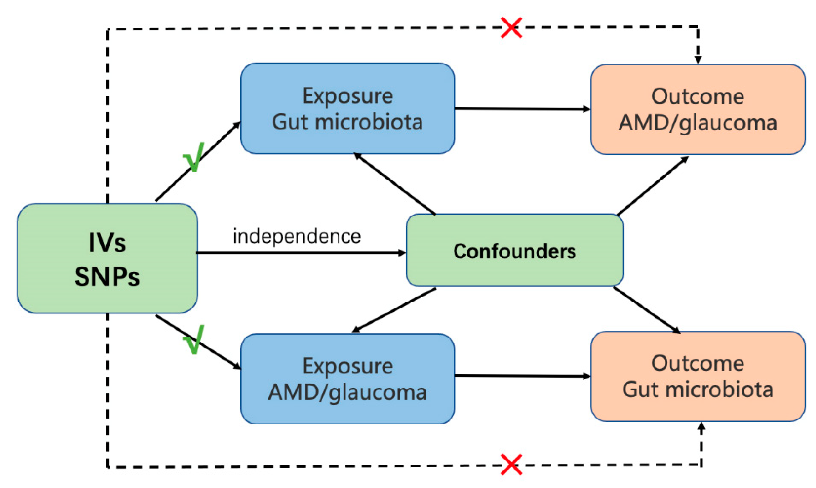
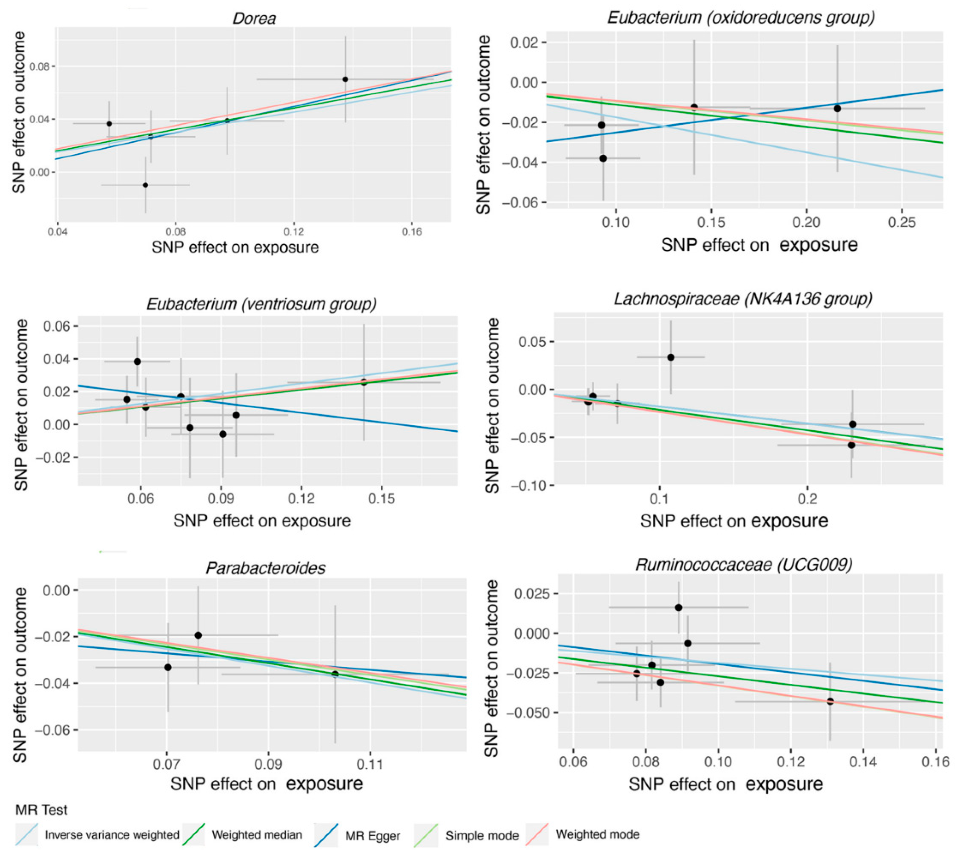
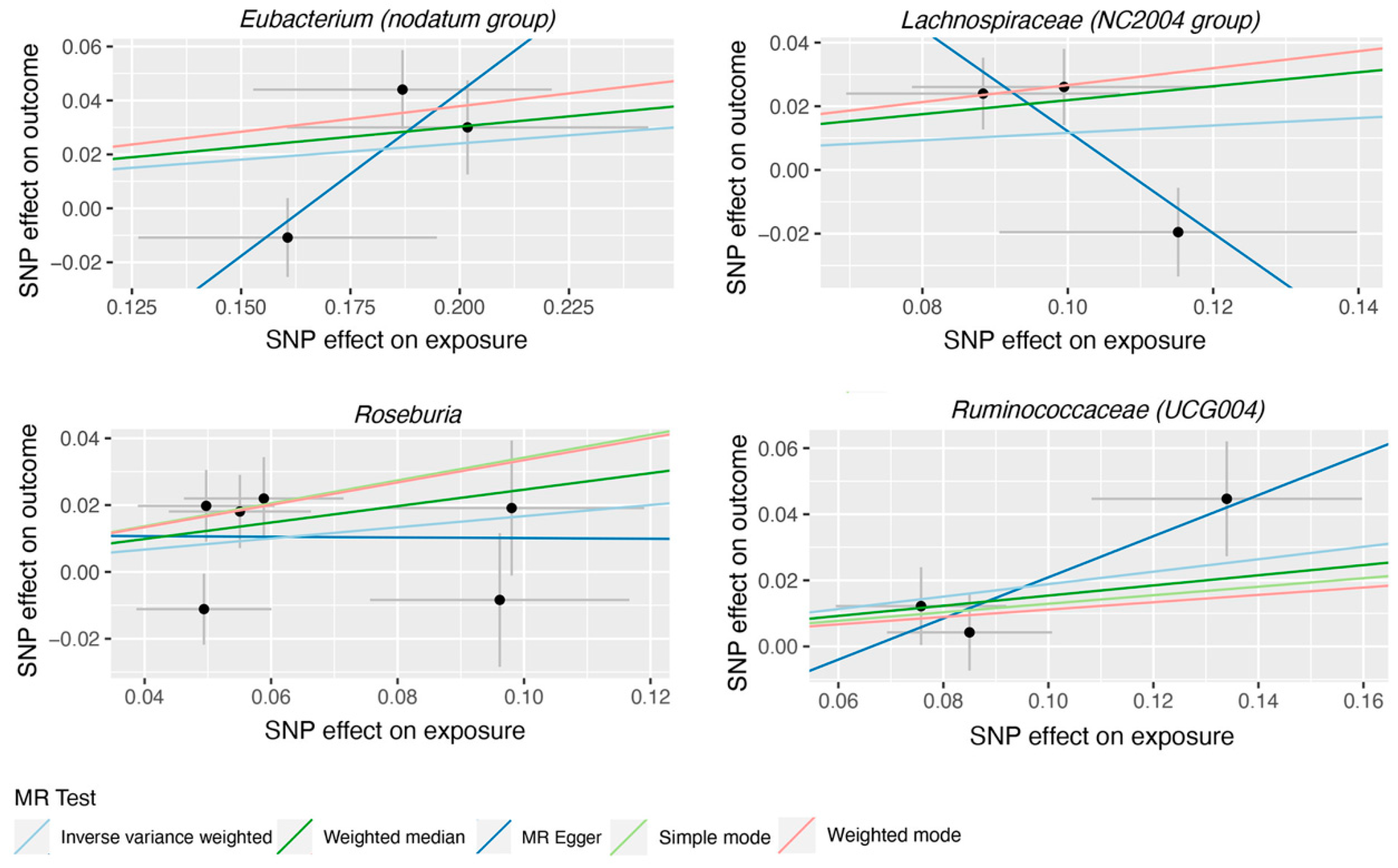
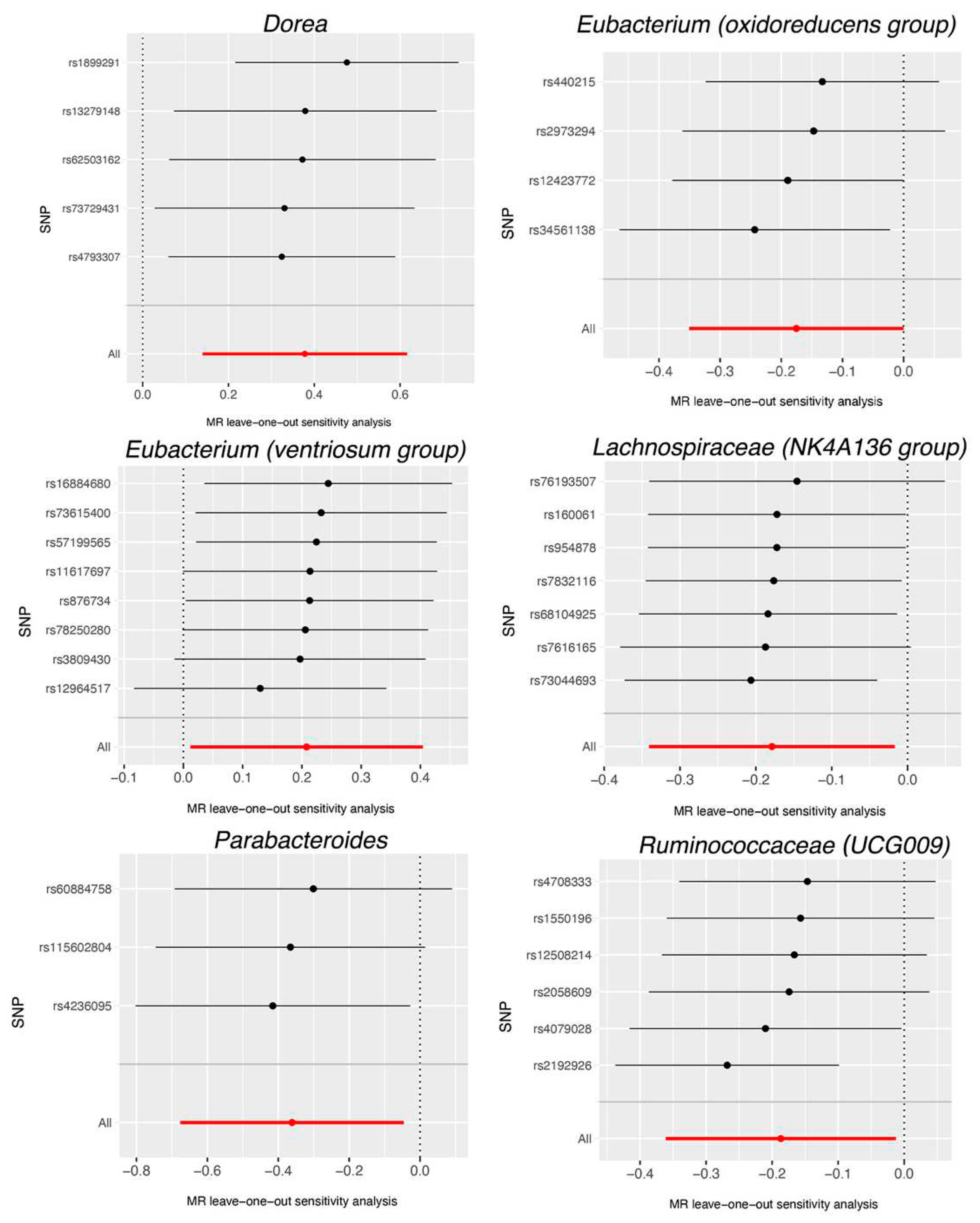
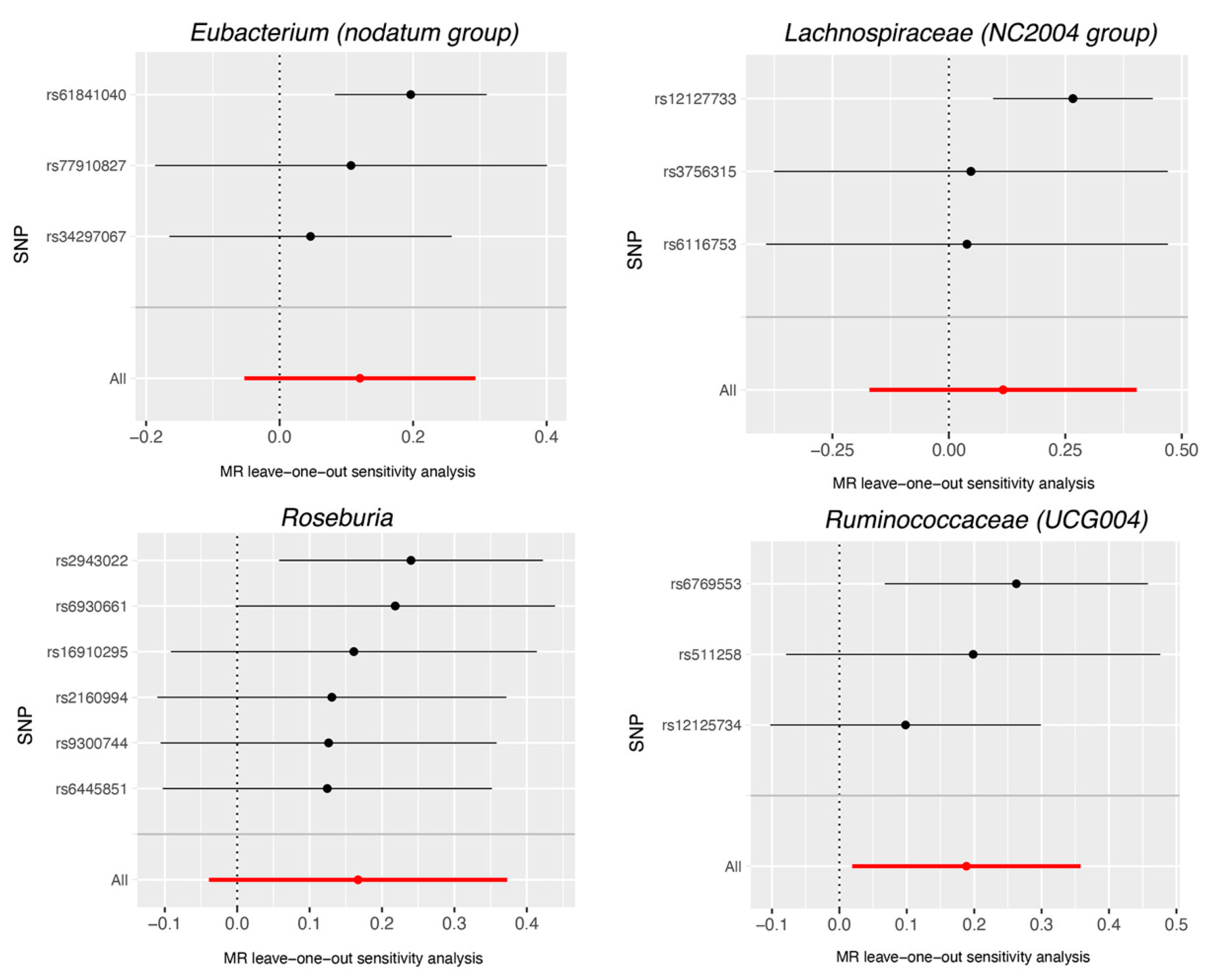

Table 1.
MR estimate for the association between gut microbiota and AMD.
| Bacterial taxa (Exposures) | Methods | SNPs | OR | 95%CI | P |
|---|---|---|---|---|---|
| Dorea | MR Egger | 5 | 1.64 | 0.64-4.22 | 0.382 |
| Weighted median | 5 | 1.50 | 1.08-2.08 | 0.016 | |
| IVW | 5 | 1.46 | 1.15-1.85 | 0.002 | |
| Simple mode | 5 | 1.55 | 1.01-2.39 | 0.116 | |
| Weighted mode | 5 | 1.55 | 1.04-2.33 | 0.099 | |
| Eubacterium (oxidoreducens group) | MR Egger | 4 | 1.13 | 0.67-1.90 | 0.687 |
| Weighted median | 4 | 0.89 | 0.72-1.11 | 0.318 | |
| IVW | 4 | 0.84 | 0.70-1.00 | 0.049 | |
| Simple mode | 4 | 0.91 | 0.68-1.22 | 0.572 | |
| Weighted mode | 4 | 0.91 | 0.70-1.18 | 0.547 | |
| Eubacterium (ventriosum group) | MR Egger | 8 | 0.82 | 0.41-1.66 | 0.602 |
| Weighted median | 8 | 1.19 | 0.92-1.54 | 0.175 | |
| IVW | 8 | 1.23 | 1.01-1.50 | 0.038 | |
| Simple mode | 8 | 1.20 | 0.82-1.76 | 0.380 | |
| Weighted mode | 8 | 1.20 | 0.84-1.73 | 0.355 | |
| Lachnospiraceae (NK4A136 group) | MR Egger | 7 | 0.84 | 0.63-1.11 | 0.277 |
| Weighted median | 7 | 0.81 | 0.66-0.99 | 0.041 | |
| IVW | 7 | 0.84 | 0.71-0.98 | 0.031 | |
| Simple mode | 7 | 0.79 | 0.61-1.04 | 0.143 | |
| Weighted mode | 7 | 0.79 | 0.63-1.00 | 0.093 | |
| Parabacteroides | MR Egger | 3 | 0.84 | 0.10-6.75 | 0.896 |
| Weighted median | 3 | 0.71 | 0.48-1.04 | 0.080 | |
| IVW | 3 | 0.70 | 0.51-0.96 | 0.025 | |
| Simple mode | 3 | 0.72 | 0.45-1.13 | 0.290 | |
| Weighted mode | 3 | 0.72 | 0.47-1.11 | 0.280 | |
| Ruminococcaceae (UCG009) | MR Egger | 6 | 0.77 | 0.21-2.80 | 0.709 |
| Weighted median | 6 | 0.76 | 0.62-0.94 | 0.011 | |
| IVW | 6 | 0.83 | 0.70-0.99 | 0.036 | |
| Simple mode | 6 | 0.72 | 0.53-0.98 | 0.093 | |
| Weighted mode | 6 | 0.72 | 0.52-0.99 | 0.101 |
MR, Mendelian randomization; AMD, age-related macular degeneration; SNP, single nucleotide polymorphism; OR, odds ratio; CI, confidence interval; IVM, inverse variance weighted.
Table 2.
MR estimate for the association between gut microbiota and glaucoma.
| Bacterial taxa (Exposures) | Methods | SNPs | OR | 95%CI | P |
|---|---|---|---|---|---|
| Eubacterium (nodatum group) | MR Egger | 3 | 3.37 | 0.70-16.2 | 0.371 |
| Weighted median | 3 | 1.16 | 1.01-1.35 | 0.041 | |
| IVW | 3 | 1.13 | 0.95-1.34 | 0.173 | |
| Simple mode | 3 | 1.21 | 1.01-1.45 | 0.176 | |
| Weighted mode | 3 | 1.21 | 1.01-1.45 | 0.179 | |
| Lachnospiraceae (NC2004 group) | MR Egger | 3 | 0.20 | 0.03-1.19 | 0.328 |
| Weighted median | 3 | 1.24 | 1.03-1.51 | 0.026 | |
| IVW | 3 | 1.12 | 0.84-1.50 | 0.427 | |
| Simple mode | 3 | 1.31 | 1.01-1.68 | 0.175 | |
| Weighted mode | 3 | 1.31 | 1.02-1.68 | 0.172 | |
| Roseburia | MR Egger | 6 | 0.99 | 0.41-2.41 | 0.984 |
| Weighted median | 6 | 1.28 | 1.03-1.59 | 0.028 | |
| IVW | 6 | 1.18 | 0.96-1.45 | 0.112 | |
| Simple mode | 6 | 1.41 | 0.98-2.03 | 0.124 | |
| Weighted mode | 6 | 1.40 | 0.95-2.05 | 0.146 | |
| Ruminococcaceae (UCG004) | MR Egger | 3 | 1.86 | 0.93-3.72 | 0.328 |
| Weighted median | 3 | 1.17 | 0.94-1.45 | 0.161 | |
| IVW | 3 | 1.21 | 1.02-1.43 | 0.029 | |
| Simple mode | 3 | 1.14 | 0.88-1.47 | 0.482 |
MR, Mendelian randomization; SNP, single nucleotide polymorphism; OR, odds ratio; CI, confidence interval; IVM, inverse variance weighted.
Table 3.
MR estimate for the association between AMD and the above gut microbiota.
| Bacterial taxa (Exposures) | Methods | SNPs | OR | 95%CI | P |
|---|---|---|---|---|---|
| Dorea | MR Egger | 8 | 0.96 | 0.89-1.03 | 0.274 |
| Weighted median | 8 | 0.96 | 0.92-1.01 | 0.152 | |
| IVW | 8 | 0.96 | 0.92-1.01 | 0.098 | |
| Simple mode | 8 | 0.96 | 0.88-1.04 | 0.319 | |
| Weighted mode | 8 | 0.96 | 0.92-1.01 | 0.191 | |
| Eubacterium (oxidoreducens group) | MR Egger | 8 | 1.10 | 0.96-1.26 | 0.209 |
| Weighted median | 8 | 1.02 | 0.93-1.11 | 0.713 | |
| IVW | 8 | 1.02 | 0.95-1.10 | 0.579 | |
| Simple mode | 8 | 1.05 | 0.91-1.23 | 0.518 | |
| Weighted mode | 8 | 1.03 | 0.95-1.13 | 0.471 | |
| Eubacterium (ventriosum group) | MR Egger | 8 | 1.03 | 0.95-1.12 | 0.467 |
| Weighted median | 8 | 1.03 | 0.97-1.09 | 0.330 | |
| IVW | 8 | 1.03 | 0.99-1.08 | 0.163 | |
| Simple mode | 8 | 0.99 | 0.90-1.08 | 0.807 | |
| Weighted mode | 8 | 1.02 | 0.97-1.08 | 0.488 | |
| Lachnospiraceae (NK4A136 group) | MR Egger | 8 | 0.93 | 0.87-1.01 | 0.124 |
| Weighted median | 8 | 0.96 | 0.91-1.01 | 0.114 | |
| IVW | 8 | 0.96 | 0.92-1.01 | 0.086 | |
| Simple mode | 8 | 1.00 | 0.92-1.09 | 0.970 | |
| Weighted mode | 8 | 0.95 | 0.91-1.00 | 0.103 | |
| Parabacteroides | MR Egger | 8 | 0.94 | 0.85-1.04 | 0.302 |
| Weighted median | 8 | 0.98 | 0.94-1.03 | 0.483 | |
| IVW | 8 | 1.00 | 0.94-1.06 | 0.990 | |
| Simple mode | 8 | 0.96 | 0.88-1.04 | 0.307 | |
| Weighted mode | 8 | 0.98 | 0.93-1.03 | 0.419 | |
| Ruminococcaceae (UCG009) | MR Egger | 8 | 1.00 | 0.97-1.16 | 0.964 |
| Weighted median | 8 | 1.03 | 0.95-1.12 | 0.486 | |
| IVW | 8 | 1.04 | 0.96-1.12 | 0.308 | |
| Simple mode | 8 | 0.95 | 0.83-1.08 | 0.430 |
MR, Mendelian randomization; AMD, age-related macular degeneration; SNP, single nucleotide polymorphism; OR, odds ratio; CI, confidence interval; IVM, inverse variance weighted.
Table 4.
MR estimate for the association between glaucoma and the above gut microbiota.
| Bacterial taxa (Exposures) | Methods | SNPs | OR | 95%CI | P |
|---|---|---|---|---|---|
| Eubacterium (nodatum group) | MR Egger | 81 | 1.06 | 0.87-1.28 | 0.578 |
| Weighted median | 81 | 1.15 | 1.04-1.28 | 0.005 | |
| IVW | 81 | 1.07 | 1.00-1.14 | 0.052 | |
| Simple mode | 81 | 1.27 | 0.97-1.67 | 0.086 | |
| Weighted mode | 81 | 1.24 | 0.99-1.55 | 0.071 | |
| Lachnospiraceae (NC2004group) | MR Egger | 75 | 0.89 | 0.77-1.03 | 0.113 |
| Weighted median | 75 | 0.95 | 0.88-1.03 | 0.217 | |
| IVW | 75 | 1.01 | 0.95-1.06 | 0.845 | |
| Simple mode | 75 | 0.93 | 0.78-1.11 | 0.433 | |
| Weighted mode | 75 | 0.92 | 0.81-1.06 | 0.263 | |
| Roseburia | MR Egger | 84 | 0.96 | 0.89-1.04 | 0.309 |
| Weighted median | 84 | 1.00 | 0.96-1.05 | 0.906 | |
| IVW | 84 | 1.00 | 0.97-1.03 | 0.996 | |
| Simple mode | 84 | 0.95 | 0.87-1.03 | 0.236 | |
| Weighted mode | 84 | 0.96 | 0.90-1.03 | 0.293 | |
| Ruminococcaceae (UCG004) | MR Egger | 83 | 0.99 | 0.89-1.11 | 0.906 |
| Weighted median | 83 | 1.05 | 0.99-1.12 | 0.113 | |
| IVW | 83 | 1.00 | 0.96-1.04 | 0.886 | |
| Simple mode | 83 | 1.08 | 0.93-1.25 | 0.322 |
MR, Mendelian randomization; SNP, single nucleotide polymorphism; OR, odds ratio; CI, confidence interval; IVM, inverse variance weighted.
Figure 1.
Overview of MR analyses process and major assumptions.
Figure 2.
Scatter plots for the casual association between gut microbiota and AMD.
Figure 3.
Scatter plots for the casual association between gut microbiota and glaucoma.
Figure 4.
Leave-one-out plots for the causal association between gut microbiota and AMD.
Figure 5.
Leave-one-out plots for the causal association between gut microbiota and glaucoma.
4. Discussion
In the present study, we utilized summary statistics data sourced from the IEU Open GWAS Project on gut microbiome, AMD, and glaucoma to conduct a MR analysis to find the causal relationship between gut microbiome and AMD/glaucoma. It was found some gut microbiotas (including Eubacterium (oxidoreducens group), Parabacteroides, Lachnospiraceae (NK4A136 group), and Ruminococcaceae (UCG009)) that had a significant protective effect on AMD, while Dorea and Eubacterium (ventriosum group) had a risk effect on AMD. Gut microbiotas (including Eubacterium (nodatum group), Lachnospiraceae (NC2004 group), Roseburia, and Ruminococcaceae (UCG004)) were all found to have a risk effect on glaucoma. Previous observational studies have found a link between the gut microbiome and the development of AMD and glaucoma [7,24,25].
Dorea is the main gas-producing bacterium in the gut. Previous metabolomics analysis has shown that Dorea is involved in the formation of the gut barrier, affects innate immunity, participates in the regulation of malignant tumor cell cycle and host adaptive immunity [26]. In eye disease research, Dorea is associated with the onset of fungal keratitis (FK), and its abundance is significantly higher in FK samples when compared to the control group [27]. However, in individuals with Sjögren's syndrome (SS) or dry eye syndrome (DES) as compared to healthy individuals, the abundance of the Dorea genus is significantly reduced [28]. Eubacterium is one of the core genera of human gut microbiota, playing a crucial role in nutrient metabolism and maintaining gut balance. Eubacterium can produce short-chain fatty acids (SCFAs), especially butyrate, which can provide nutrients and energy to the intestinal epithelium. Previous studies have found causal relationships between Eubacterium and diabetic retinopathy (DR) and optic neuritis (ON) [12,14]. In addition, there have been reports of interactions between Eubacterium and AMD, but its correlation with glaucoma has not been reported.5 Lachnospiraceae and Ruminococcaceae are both core genera of human gut microbiota and may be potential beneficial bacteria involved in carbohydrate metabolism, producing butyrate as the main source of energy for the host. The increase in their abundance is related to aging [29]. In eye diseases, Lachnospiraceae and Ruminococcaceae are related to bacterial keratitis and uveitis [30,31] and Lachnospiraceae is also related to fungal keratitis and mucous membrane pemphigoid [27,32]. In addition, studies have reported interactions between Lachnospiraceae, Ruminococcaceae, and AMD, but their causal relationship with AMD is still unknown [5]. Besides, their interaction with glaucoma has not been reported yet. Parabacteroides has been less studied in eye diseases, and the relative abundance of Parabacteroides in the feces of SS subjects was markedly less compared to the control group [33]. In the study of demyelinating optic neuritis (DON), the abundance of Parabacteroides was lower in DON than in the normal control group [34]. Zysset-Burri et al. [6] compared 57 patients with neovascular AMD to 58 healthy controls in a study investigating the correlation between gut microbiota and AMD. The results showed that Oscillibacter and Bacteroides were significantly higher in non-AMD patients. However, the current research did not find a causal relationship between Oscillibacter or Bacteroides and AMD, possibly due to different types of AMD. Zinkernagel et al. [35] sequenced the gut microbiome of AMD patients and healthy controls and found that Oscillibacter, Anaerotruncus, Eubacterium ventriosum, and Ruminococcus torques were relatively more abundant in AMD patients, whereas Bacteroides eggerthii was enriched in the control group. The present study also showed that Eubacterium ventriosum had a risk effect on AMD. Another study showed that the abundance of Oscillospira, Blautia, and Dorea was decreased in AMD patients [36]. This study also found that Dorea had a risk effect on AMD. It was reported that the abundance of Bacteroides and Prevotella is associated with POAG. Gong et al. [8] used 16S rRNA sequencing to detect the fecal microbiota of 30 POAG patients and 30 healthy individuals, and found that Escherichia coli, g unidentified Enterobacteriaceae, and Prevotellaceae were the most increased in POAG, while Bacteroides plebeius and Megamonas were notably reduced. Another study demonstrated a noteworthy reduction in the distribution of Blautia and Fusicatenibacter in fecal samples of patients with primary angle-closure glaucoma (PACG) [9]. However, these bacterial genera did not show a significant causal relationship with glaucoma in this study, possibly due to differences in the ethnicity of the study population and disease classification. The above studies mostly focused on Chinese people, while the present study mainly included Europeans.
There are several advantages to this study. By MR analyzing, the causal relationship between gut microbiota and both AMD and glaucoma can be determined, which eliminates confounding factors and reverses causal inference. We obtained genetic variation data of gut microbiota from the largest GWAS meta-analysis to ensure the reliability of the MR analysis instrument. To detect and eliminate horizontal pleiotropy, we utilized MR-PRESSO and MR-Egger regression interval trial tests. Additionally, we used a MR design and non-overlapping exposure and outcome summary-level data to avoid bias [37]. Nonetheless, this study also has some limitations. Since summary statistics data were used in the analysis instead of raw data, subgroup analysis, such as distinguishing early AMD from late AMD, and different types of glaucoma cannot be performed. As the exposure dataset only provides genus-level classification, we are unable to investigate the potential causal link between gut microbiota and AMD/glaucoma at the species level. In order to perform sensitivity analysis and horizontal pleiotropy testing, additional genetic variations must be incorporated as IVs. The sample size for gut microbiota is limited, and the reverse MR analysis results may be weakly influenced by instrument bias and cannot completely exclude reverse causality. Although most of the participants in the GWAS gut microbiota data meta-analysis were of European descent, population stratification may still pose a potential confounding factor, rendering the study results not entirely generalizable to non-European populations. To enhance the generalizability of deeper explorations investigating the causal relationships between gut microbiota and AMD/glaucoma, it is recommended to conduct studies in diverse European and non-European populations.
5. Conclusions
In conclusion, the present study identified causal relationships between gut microbiota including Dorea, Eubacterium (oxidoreducens group), Eubacterium (ventriosum group), Lachnospiraceae (NK4A136 group), Parabacteroides, and Ruminococcaceae (UCG009) with AMD, and Eubacterium (nodatum group), Lachnospiraceae (NC2004 group), Roseburia, and Ruminococcaceae (UCG004) with glaucoma. The protective or risk mechanisms of these gut microbiota on AMD and glaucoma require further RCT studies. In addition, reverse MR found a causal relationship between glaucoma and Eubacterium (nodatum group), and no causal relationships were found between AMD or glaucoma and the other mentioned bacterial groups. However, it cannot be ruled out that AMD or glaucoma may affect the gut microbiota, and further research is needed to confirm these conclusions.
Supplementary Materials
The following supporting information can be downloaded at the website of this paper posted on Preprints.org, Table S1: SNPs information of bacteria with AMD; Table S2: SNPs information of bacteria with glaucoma; Table S3: MR estimates of bacteria on AMD; Table S4: MR estimates of bacteria on glaucoma. Table S5: Heterogeneity tests of bacteria on AMD and glaucoma; Table S6: Horizontal pleiotropy tests of bacteria on AMD and glaucoma; Table S7: Heterogeneity and horizontal pleiotropy tests of AMD on bacteria. Table S8: Heterogeneity and horizontal pleiotropy tests of glaucoma on bacteria.
Author Contributions
Conceptualization C.L. and P.R.L.; methodology, C.L.; software, C.L.; validation, C.L. and P.R.L.; resources, C.L.; data curation, X.X.; writing—original draft preparation, C.L. and P.R.L.; writing—review and editing, C.L. and P.R.L.; visualization, C.L.; supervision, P.R.L.; funding acquisition, P.R.L.. All authors have read and agreed to the published version of the manuscript.
Funding
This research was funded by This study was supported by Jiangsu Provincial Medical Innovation Team (grant No. CXTDA2017039) and the National Natural Science Foundation in China (grant No. 81671641, 82271113).
Institutional Review Board Statement
This article does not contain any studies involving human participants performed by any of the authors.
Informed Consent Statement
Informed consent was obtained from all subjects involved in the study.Written informed consent has been obtained from the patients to publish this paper.
Data Availability Statement
Data are available on reasonable request.
Conflicts of Interest
The authors declare no conflict of interest.
References
- Ambati, J.; Atkinson, J.P.; Gelfand, B.D. Immunology of age-related macular degeneration. Nat Rev Immunol 2013, 13, 438–451. [Google Scholar] [CrossRef] [PubMed]
- Mitchell, P.; Liew, G.; Gopinath, B.; et al. Age-related macular degeneration. Lancet 2018, 392, 1147–1159. [Google Scholar] [CrossRef] [PubMed]
- Tham, Y.C.; Li, X.; Wong, T.Y.; et al. Global prevalence of glaucoma and projections of glaucoma burden through 2040: a systematic review and meta-analysis. Ophthalmology 2014, 121, 2081–2090. [Google Scholar] [CrossRef] [PubMed]
- Vujkovic-Cvijin, I.; Sklar, J.; Jiang, L.; et al. Host variables confound gut microbiota studies of human disease. Nature 2020, 587, 448–454. [Google Scholar] [CrossRef] [PubMed]
- Rowan, S.; Taylor, A. The Role of Microbiota in Retinal Disease. Adv Exp Med Biol 2018, 1074, 429–435. [Google Scholar] [CrossRef]
- Zysset-Burri, D.C.; Keller, I.; Berger, L.E.; et al. Associations of the intestinal microbiome with the complement system in neovascular age-related macular degeneration. NPJ Genom Med 2020, 5, 34. [Google Scholar] [CrossRef] [PubMed]
- Lima-Fontes, M.; Meira, L.; Barata, P.; et al. Gut microbiota and age-related macular degeneration: A growing partnership. Surv Ophthalmol 2022, 67, 883–891. [Google Scholar] [CrossRef] [PubMed]
- Gong, H.; Zhang, S.; Li, Q.; et al. Gut microbiota compositional profile and serum metabolic phenotype in patients with primary open-angle glaucoma. Exp Eye Res 2020, 191, 107921. [Google Scholar] [CrossRef]
- Gong, H.; Zeng, R.; Li, Q.; et al. The profile of gut microbiota and central carbon-related metabolites in primary angle-closure glaucoma patients. Int Ophthalmol 2022, 42, 1927–1938. [Google Scholar] [CrossRef]
- Greenland, S. An introduction to instrumental variables for epidemiologists. Int J Epidemiol 2000, 29, 722–729. [Google Scholar] [CrossRef]
- Burgess, S.; Thompson, S.G. Mendelian randomization: methods for causal inference using genetic variants: CRC Press.2021.
- Liu, K.; Zou, J.; Fan, H.; et al. Causal effects of gut microbiota on diabetic retinopathy: A Mendelian randomization study. Front Immunol 2022, 13, 930318. [Google Scholar] [CrossRef] [PubMed]
- Nusinovici, S.; Li, H.; Thakur, S.; et al. High-Density Lipoprotein 3 Cholesterol and Primary Open-Angle Glaucoma: Metabolomics and Mendelian Randomization Analyses. Ophthalmology 2022, 129, 285–294. [Google Scholar] [CrossRef] [PubMed]
- Liu, K.; Wu, P.; Zou, J.; et al. Mendelian randomization analysis reveals causal relationships between gut microbiome and optic neuritis. Hum Genet 2022, 28. [Google Scholar] [CrossRef] [PubMed]
- Kurilshikov, A.; Medina-Gomez, C.; Bacigalupe, R.; et al. Large-scale association analyses identify host factors influencing human gut microbiome composition. Nat Genet 2021, 53, 156–165. [Google Scholar] [CrossRef] [PubMed]
- Winkler, T.W.; Grassmann, F.; Brandl, C.; et al. Genome-wide association meta-analysis for early age-related macular degeneration highlights novel loci and insights for advanced disease. BMC Med Genomics 2020, 13, 120. [Google Scholar] [CrossRef] [PubMed]
- Craig, J.E.; Han, X.; Qassim, A.; et al. Multitrait analysis of glaucoma identifies new risk loci and enables polygenic prediction of disease susceptibility and progression. Nat Genet 2020, 52, 160–166. [Google Scholar] [CrossRef]
- Bowden, J.; Davey Smith, G.; Burgess, S. Mendelian randomization with invalid instruments: effect estimation and bias detection through Egger regression. Int J Epidemio. 2015, 44, 512–525. [Google Scholar] [CrossRef]
- Pocock, S.J.; Simon, R. Sequential treatment assignment with balancing for prognostic factors in the controlled clinical trial. Biometrics 1975, 31, 103–115. [Google Scholar] [CrossRef]
- Burgess, S.; Dudbridge, F.; Thompson, S.G. Combining information on multiple instrumental variables in Mendelian randomization: comparison of allele score and summarized data methods. Stat Med 2016, 35, 1880–1906. [Google Scholar] [CrossRef]
- Scott, N.W.; McPherson, G.C.; Ramsay, C.R.; et al. The method of minimization for allocation to clinical trials. a review. Control Clin Trials 2002, 23, 662–674. [Google Scholar] [CrossRef]
- Hemani, G.; Tilling, K.; Davey Smith, G. Orienting the causal relationship between imprecisely measured traits using GWAS summary data. PLoS Genet 2017, 13, e1007081. [Google Scholar] [CrossRef]
- Verbanck, M.; Chen, C.Y.; Neale, B.; et al. Detection of widespread horizontal pleiotropy in causal relationships inferred from Mendelian randomization between complex traits and diseases. Nat Genet 2018, 50, 693–698. [Google Scholar] [CrossRef] [PubMed]
- Rinninella, E.; Mele, M.C.; Merendino, N.; et al. The Role of Diet, Micronutrients and the Gut Microbiota in Age-Related Macular Degeneration: New Perspectives from the Gut⁻Retina Axis. Nutrients 2018, 10, 1677. [Google Scholar] [CrossRef] [PubMed]
- Zhang, Y.; Zhou, X.; Lu, Y. Gut microbiota and derived metabolomic profiling in glaucoma with progressive neurodegeneration. Front Cell Infect Microbiol 2022, 12, 968992. [Google Scholar] [CrossRef]
- Chaput, N.; Lepage, P.; Coutzac, C.; et al. Baseline gut microbiota predicts clinical response and colitis in metastatic melanoma patients treated with ipilimumab. Ann Oncol 2017, 28, 1368–1379. [Google Scholar] [CrossRef] [PubMed]
- Kalyana Chakravarthy, S.; Jayasudha, R.; et al. Alterations in the gut bacterial microbiome in fungal Keratitis patients. PLoS One 2018, 13, e0199640. [Google Scholar] [CrossRef]
- Moon, J.; Choi, S.H.; Yoon, C.H.; et al. Gut dysbiosis is prevailing in Sjögren's syndrome and is related to dry eye severity. PLoS One 2020, 15, e0229029. [Google Scholar] [CrossRef]
- Wang, J.; Qie, J.; Zhu, D.; et al. The landscape in the gut microbiome of long-lived families reveals new insights on longevity and aging - relevant neural and immune function. Gut Microbes 2022, 14, 2107288. [Google Scholar] [CrossRef]
- Jayasudha, R.; Chakravarthy, S.K.; Prashanthi, G.S.; et al. Alterations in gut bacterial and fungal microbiomes are associated with bacterial Keratitis, an inflammatory disease of the human eye. J Biosci 2018, 43, 835–856. [Google Scholar] [CrossRef]
- Kalyana Chakravarthy, S.; Jayasudha, R.; Sai Prashanthi, G.; et al. Dysbiosis in the Gut Bacterial Microbiome of Patients with Uveitis, an Inflammatory Disease of the Eye. Indian J Microbiol 2018, 58, 457–469. [Google Scholar] [CrossRef]
- Low, L.; Suleiman, K.; Shamdas, M.; et al. Gut Dysbiosis in Ocular Mucous Membrane Pemphigoid. Front Cell Infect Microbiol 2022, 12, 780354. [Google Scholar] [CrossRef] [PubMed]
- de Paiva, C.S.; Jones, D.B.; Stern, M.E.; et al. Altered Mucosal Microbiome Diversity and Disease Severity in Sjögren Syndrome. Sci Rep 2016, 6, 23561. [Google Scholar] [CrossRef] [PubMed]
- Liu, Y.; Fan, H.; Shao, Y.; et al. Gut microbiota dysbiosis associated with different types of demyelinating optic neuritis in patients. Mult Scler Relat Disord 2023, 72, 104619. [Google Scholar] [CrossRef] [PubMed]
- Zinkernagel, M.S.; Zysset-Burri, D.C.; Keller, I.; et al. Association of the Intestinal Microbiome with the Development of Neovascular Age-Related Macular Degeneration. Sci Rep 2017, 7, 40826. [Google Scholar] [CrossRef]
- Lin, P. Importance of the intestinal microbiota in ocular inflammatory diseases: A review. Clin Exp Ophthalmol 2019, 47, 418–422. [Google Scholar] [CrossRef]
- Burgess, S.; Davies, N.M.; Thompson, S.G. Bias due to participant overlap in two-sample Mendelian randomization. Genet Epidemiol 2016, 40, 597–608. [Google Scholar] [CrossRef]
Disclaimer/Publisher’s Note: The statements, opinions and data contained in all publications are solely those of the individual author(s) and contributor(s) and not of MDPI and/or the editor(s). MDPI and/or the editor(s) disclaim responsibility for any injury to people or property resulting from any ideas, methods, instructions or products referred to in the content. |
© 2023 by the authors. Licensee MDPI, Basel, Switzerland. This article is an open access article distributed under the terms and conditions of the Creative Commons Attribution (CC BY) license (http://creativecommons.org/licenses/by/4.0/).
Copyright: This open access article is published under a Creative Commons CC BY 4.0 license, which permit the free download, distribution, and reuse, provided that the author and preprint are cited in any reuse.


