Submitted:
05 October 2023
Posted:
06 October 2023
You are already at the latest version
Abstract
Criminal investigations aimed to track the route walked by Missing Persons and Fugitives (MPFs) usually involve Intelligence analysts, military planners, experts in mobile forensics, traditional investigative methods, and sniffer dog handlers. Notwithstanding, when MPFs are devoid of any technological device and move in uninhabited rural areas devoid of tele cameras and densely covered by vegetation, tracking backwards the route walked by MPFs may be a much more arduous task. In such complex cases, a very efficient approach may consist in comparing the geological traces found on the MPFs with soils and plants exposed in the event scenes. In particular, the search for peculiar or rare particles and aggregates may strengthen the weight of the geological evidence comparisons. A match of mineralogical, textural, and botanical data may demonstrate the provenance of the traces from the soil of a specific site, linking in this way the MPFs to the scene of events. Based on the above, the present paper reports geological and botanical determinations accomplished for a “mediatic” casework. Results allowed to ascertain a general high degree of compatibility among traces collected on the MPFs and on the soil from the scene of events. The most significant positive matches, based on the finding of a ten of peculiar and rare particles and assemblages, allowed reconstructing a route about 1.1 km long, as the crow flies, on the event site. Notwithstanding this procedure was extremely time consuming and available only in a backwards reconstruction linked to the MPFs’ findings, it was of uttermost importance in strengthen the inferences proposed, where other methods could not provide any information.
Keywords:
Geoforensics (Forensic Geology)
; Sedimentology
; Provenance
; Comparative analyses
; Optical Microscopy
; SEM-EDS
; Image Analysis
; Missing Persons and Fugitives.
1. Introduction
Since the 2001 September 11 attacks and the Global War on Terror, the organizational structures and procedures devoted to the search for persons of national and international interest assumed an important role in the global counterterrorism strategies [1,2].
In particular, criminal investigations, aimed to track the route walked by Missing Persons and Fugitives (MPFs) in the urban and rural territory, usually involve Intelligence analysts (IMINT - Imagery Intelligence, OSINT – Open Source Intelligence, etc.), military planners, experts in mobile forensics sniffer, dog handlers, and traditional investigative methods [1,3]. Notwithstanding, to track backwards the route traversed by the MPFs may be a very arduous task, when the MPFs do not bring with them mobiles, GPS or any other technological devices and they move in uninhabited rural areas of the countryside devoid of security or private tele cameras and covered by dense vegetation. In such peculiar circumstances, as demonstrated since the XVIII century [4,5,6] the mapping of the paths walked by the actors of crimes can be realized thanks to comparative analyses based on geological and soil evidence collected on the crime actors and the scenes of the events, respectively. In Germany, the study of the stains of stratified soil on the footwears and trousers of a homicide suspect allowed the expert Georg Popp to link him to a sequence of different sites of the crime scenes and suspect’s home [4]. Nowadays, once the MPFs are found, dead or alive, a very efficient backwards criminalistic approach may consist in comparing both the geological traces and micro traces found on the MPFs’ belongings (unknown samples, i.e. of unknown provenance) with soils/sediments and plants exposed in the sites of finding, last sighting, alibi, and investigative interest (known samples, i.e. of known provenance). A match of the comparative analysis data may demonstrate the provenance of the unknown traces from a site showing analogous microenvironmental characteristics [7].
With this in mind, the present research reports the scientific method and main results of geological comparative analyses, requested by the judicial authority for solving a “mediatic” forensic casework occurred in Italy in the countryside a few years ago. Geological data allowed tracking the route walked by the two MPFs revealing very useful information for the consequent criminological and criminalistic implications.
Notwithstanding the criminal proceeding was archived, the Author preferred to deal with the data and inferences described in the following, presenting MPFs/victims, samples, and sites in anonymously way.
2. Geoforensics
Forensic Sciences, based on a holistic approach, use different multidisciplinary and transdisciplinary disciplines, such as criminology and criminalistics (forensic medicine, forensic pathology, dentistry, toxicology, serology, anthropology, archaeology, entomology, physics, biology, chemistry, computer science, geology, and botany) [8,9].
Geoforensics (or Forensic Geology) is a discipline 150 years old, probably originated also before during the Roman empire period. In the historical criminological/criminalistic scenario, a few examples of worldwide mediatic serious crimes solved by forensic eologists may be related to cases of kidnapping and homicide of victims, as in the case of the honorable Aldo Moro (1978, Italy) [10] or the agent of the DEA (Drug Enforcement Administration) Enrique Camarena Salazar (1985, Mexico) [4], or cases of searches for MPFs, such as the terrorist Osama bin Laden (2001, Afghanistan) [11].
Forensic Geology applies principles, methods, and techniques of the earth sciences for solving criminal cases [12,13,14,15,16,17,18,19,20,21,22,23,24,25,26,27,28,29]. These latter mostly concern crimes against human being (homicides, kidnappings, sexual violence, robbery) and animals, property (burglary, damaging), environment (environmental disaster or pollution) [30,31,32,33,34,35,36,37], and counterterrorism [11]. In particular, in serious crimes occurred outdoor in the countryside, such as homicides or kidnappings, it is high the probability that useful info-investigative diversified data may be obtained especially in ascertainments aimed to define:
- Their pre-mortem presence on the scene.
- Their walking route on the site.
- The possible transfer of the victim’s corpse in secondary crimes scenes.
- The modality of victim’s death.
The stratigraphical approach may be of paramount importance when micro-stratigraphy sequences on belongings of the suspects are recognized and sub-sampled. In such circumstances, the stratigraphy principle of superposition will assist in the reconstruction of the timing of the events (Somma 2023a).
Main forensic analyses and activities devoted to criminal cases may involve:
- Comparative analyses and provenance studies, based on mineralogical, petrographic, sedimentological, palaeontological, and geochemical investigations [38,39,40,41,42,43,44,45,46,47,48,49,50,51,52,53,54]. Forensic comparisons between two or multiple samples of geological and soil traces and micro traces are aimed to ascertain whether they originated from different sources [51]. When specimens result indistinguishable, the possibility that a single source is the provenance area of the samples cannot be excluded [51]. Such investigations may allow to link the actors of a crime (suspect and victim of a homicide) to the crime scene or the scene of events.
-
Mineralogical, geochemical, and palaeontological analyses.
-
Remote sensing, geological, geochemical activities, together with geophysical shallow prospections and applied geology investigations.These investigations [60,61,62,63,64,65,66,67,68,69,70,71,72,73], also based on comparative analyses, may allow to characterize the environmental matrices (soil and water) and the related underground in cases of environmental crimes. In particular, the remote sensing surveys for localizing MPFs, dens of terrorists, or in general illicit activities, are carried out on photos, ortho imaging, videos, and photograms, in the visible, ultraviolet, infrared, elaborated in GIS systems [61,62,64,65,66,67,68,69,70]. Such investigations may also be applied to depict and search for shallow clandestine gravesites and concealments [74,75,76,77,78,79,80,81,82,83,84,85,86,87,88,89,90,91,92].
Main comparative analytical methods are used for characterizing composition, textures, and structures of the inorganic, organic, and anthropogenic components of the geological and soil evidence (Table 1).
The main parameters and characteristics investigated in the inorganic and anthropogenic fractions may be synthesized as follow (Table 1):
- Colour.
- Particle size.
- Structure and texture.
- Fossil content.
- Mineralogy.
- Chemical and chemical-physical composition.
The organic component, vegetal and animal, may be very abundant and important in the sample and once identified must be separated and submitted to the specialistic analyses of forensic botanists and entomologists, respectively. In particular, the finding of plant remains such as seeds, thorns, leaves, and pollens or their associations demonstrated to be very useful for linking evidence to a specific environment of provenance and for dating the period of transfer the trace [10,93,94,95,96,97,98,99]. New advances on forensic biology on the vegetal DNA identification may demonstrate the transfer of a specific plant DNA to an actor of crime. Such multi-disciplinary scientific approach on inorganic and organic evidence based on standardized protocols [100] and procedures reported in the international scientific literature may provide very useful data especially in crime scenes occurred in the countryside.
On the base of the above, it is evident that the ascertainment of the compatibility or similarity among geological and soil traces and micro traces of unknown provenance and soils and sediments of known provenance may assume a fundamental role in the geoforensics investigations. Notwithstanding, a simple compatibility among forensic specimens may not be decisive if not supported by strong geological, biological, physical, and chemical evidence. As a matter of facts, in contrast with individual characteristics (DNA, fingerprints), the geological and soil evidence being provided of class characteristics [9] needs to be carefully investigated for obtaining assessable data. The main task for achieving a high probatory value of the evidence presented to the court should be aimed to gain the highest number of geological characteristics and peculiar particles [4] to compare.
If the wide range of compared characteristics will provide evidence that there is a match or not among compared forensic specimens, it is possible to link or exclude with a high level of probability, the actors of a crime with or from the criminal act ascertaining that the compared specimens possess features analogous to those of a specific microenvironment from which these may derive [19,23].
2. Criminal Casework
A few years ago, an Italian locality of the countryside was the sad scenario of a criminal case of presumed kidnapping, concluded after sometimes with the finding of the human remains of two subjects. One day, the two left their home, and after a car accident, they quickly abandoned their car and belongings, departing the site. They disappeared into thin air, and nobody saw them in the surrounding areas. A few days after their disappearance, the two MPFs were found lifeless in two separate sites not too far from the site of the car accident (Figure 1). Victim 1 was found about 1.1 km away, as the crow flies, from the place of the last sighting (Figure 1), under an infrastructure, a few meters away. One shoe was found on the ground near the corpse, the other one in suspension attached to a shrub. Victim 2 remains were approximately at 0.5 km from the site of the car accident, halfway between it and the place where victim 1 was found (Figure 1). The body was found skeletonized and bones were found dispersed in an area (wide about 800 m2) of the dense Mediterranean maquis.
The shoes of victim 2 were found at a few tens of meters of distance from the remains, at higher altitude.
The judicial authority disposed an impressive investigation, covering a wide spectrum of traditional and scientific investigations in the field of inter- and trans-disciplines, usually used for characterizing at 360° serious crimes happened outdoor in the countryside, such as legal medicine, forensic pathology, odontology, entomology, toxicology, veterinary, psychiatry, engineering, computer science, physics, geology, and botany.
In particular, considering that the two MPFs had not with them any mobile, GPS, or electronical devise, the judicial authority gave to the Author the task to ascertain the active pre-mortem presence of the two MPFs in the event site and reconstruct the route walked by them, if possible.
2. Materials and Methods
Notwithstanding geological and botanical traces are mostly provided of class characteristics, a sort of “fingerprint” of the specimens [101] may be identified by means of a careful examination of both peculiar and rare grains [4], minerals, composition, textural features and peculiar assemblages of minerals and vegetal remains studied in both the unknown and known specimens to be compared. A clever expert should be able to carry out comparative analyses based on a wide range of parameters and characteristics.
With this in mind, the comparative analyses were performed tracing the “fingerprints” of both geological trace evidence (related to the two MPFs and their belongings) and soils (related to the event scene and localities of investigative interest).
Geological trace evidence (unknown samples) was sampled searching for traces under magnifying glass (10x, 20x, 30x) on the two cadavers during the autopsy, and successively on their belongings and skeletal remains by using a stereomicroscope in the laboratory of forensic geology. Special attention was devoted to the footwears, being these latter in strictly contact with the topsoil during the movement of the MPFs. Such contact, as stated by the Locard exchange principle, may allow an easy transfer of inorganic and organic (plant and small animal remains) particles from the topsoil to the footwears, linking in such a way the subject to a specific site [102,103].
The MPF 1’s shoes resulted to be very dirty and scratched (Figure 2A,B), very rich in vegetal material (mostly seeds and thorns inside the shoes and fixed on the soles and laces, Figure 2C) and with inorganic traces (Figure 2C).
The MPF 2’s shoes were intact, pair up, and with inorganic and vegetal traces (mostly thorns fixed on the soles).
Multiple soil evidence (known specimens) was collected on the sites of the human remains’ findings (known samples) and the localities trampled by the victims with the same footwears, in the days immediately before the disappearance event, for exclusion purposes. For the same aim, footwears of the two MPFs were also sized for characterizing the usual type of traces present.
A few hundreds of samples were collected overall.
Multiple geological trace and soil evidence was firstly analysed under stereomicroscope [4], as it was. Specimens were treated, sieved, and separated. Soil samples were sieved mechanically or analysed by laser diffraction technique. Trace samples were separated on the base of the diameter by means of image analyses. Different sub-samples on the base of the grain size were obtained.
Optical analyses in stereomicroscopy and SEM-EDS were aimed at investigating the following main parameters and characteristics: color, coating, shape, habitus, luster, particle size, texture, fossil content, mineralogy. Stubs of the samples with selected particles were prepared for the SEM-EDS forensic characterization, in order to investigate both morphological features at higher magnification and composition. Smear slides of the specimens were observed under petrographic microscope for an expeditious characterization of mineralogy and fossil content. The different classes of parameters and characteristics were quantitatively characterized counting each particle of specimen (at least 100 particles for sample) for the most widespread grain sizes. Particle morphometry was determined by means of image analysis software. The Riley Sphericity (√) (Di: the diameter of the largest inscribed circle, Dc i: the diameter of the smallest circumscribing circle) was applied for the two-dimensional sphericity measurement. The roundness was determined by means of comparative charts.
The protocols used for geological analyses were conformed with how willing in Bourguignon et al. (2019) [100] and the international literature.
Botanical evidence was collected on the victims, their belongings, or separated from the geological trace evidence. Vegetal elements were sampled on the plants growing on the sites of the events and other sites of investigative interest or separated from the soil evidence. Morphological determinations on plants (algae included) were accomplished under microscope by experts in systematic botany.
Particle size analyses and separations were performed by using:
- Mechanical siever (Retsch AS 200 control model) with sieves (2000 µ, 1000 µ, 500 µ, 250 µ, 125 µ, 63 µ).
- Laser diffraction granulometer (Malvern Instruments Mastersizer 2000) equipped with workstation (Malvern Instruments) (Figure 3A).
- Motorized stereomicroscope equipped with Zeiss digital camera and workstation (image analysis software for morphometric investigations, Zeiss AXIOVISION) (Figure 3B).
Analyses were performed on particles contained in Petri capsules, glass slides, smear slides, stubs with carbon adhesive, by using the instruments provided of the following characteristics:
- Stereomicroscope (Leica MZ 12, magnifications from 8X to 100X).
- Motorized stereomicroscope with reflected and transmitted polarized light (Zeiss Stereo Discovery.V20; magnification from 3.8X to 530X with optical zoom) equipped with Zeiss digital camera and workstation.
- Motorized petrographic optical microscope with reflected and transmitted polarized light (Zeiss Imager.M2m model, magnifications from 25X to 500X) equipped with Zeiss tele camera and workstation (Figure 3B).
- Optical microscope for biological use (Leitz Laborlux 12, magnifications from 40X to 1000X equipped with 12 MP digital camera (Apple Inc.).
- SEM (FEI QUANTA FEG 450 model, operating in low vacuum, chamber pressure of 50 Pa at 20.00 kV equipped with an energy dispersive X-ray analyser (SEM-EDS) and workstation (AMETEK) (Figure 3C).
3. Results on the Comparative Analyses
3.1. Geological Evidence
Samples resulted to be made of sandy to silty hyaline siliciclastic grains mainly composed of mono-mineral grains/clasts of quartz with different intensity yellow to orange Fe-coatings, minor opaque yellow ocher lithoclasts of quartzarenites, and microfossils (benthic foraminifera).
Mineralogical, petrographic, and sedimentological examinations on the geological trace and soil evidence revealed that these were mainly composed of an analogous mineral assemblage. Notwithstanding the mineralogical homogeneity, seven main classes of grain typologies were identified on the base of different mineralogical and textural characteristics and parameters (luster, coating, color, shape, habitus, roundness, and sphericity) examined under stereobinocular reflected and transmitted light microscope, scanning electron microscope with an energy dispersive (SEM-EDS) X-ray analyser for X-ray microanalyses.
Figure 3.
Instrumentations. A) Laser diffraction granulometer (Malvern Instruments Mastersizer 2000). B). Workstation - Motorized stereomicroscope with reflected and transmitted polarized light (Zeiss Stereo Discovery.V20, on the left), motorized petrographic optical microscope with reflected and transmitted polarized light (Zeiss Imager.M2m mode, on the right), and image analysis software (Zeiss AXIOVISION, in the center) - Forensic Geology laboratory, MIFT Department, University of Messina. C) SEM (FEI QUANTA FEG 450 model) equipped with EDS system and workstation (AMETEK) - Laboratory of microscopic analyses, Engineering Department, University of Messina. Source: Author.
The identified grain characteristics and parameters were described for each class. About one thousand of medium-fine to very fine sandy grains were analysed in the unknown samples as well as in the known samples, in the complex (Table 2, Figure 4).
Grains appeared mostly rounded with minor percentage of sub-angular clasts whereas the Riley sphericity resulted to be equal to 0.8 both in both the unknown and known samples.
Additional comparisons, accomplished in order to exclude possible previous traces in the victims’ questioned samples related to their staying with the same footwears in the sites visited in the days immediately before the tragic event, allowed to ascertain the absence of particles composed of mono- and polymineral assemblages of metamorphic rocks typical of the soils collected in these other locations of investigative interest.
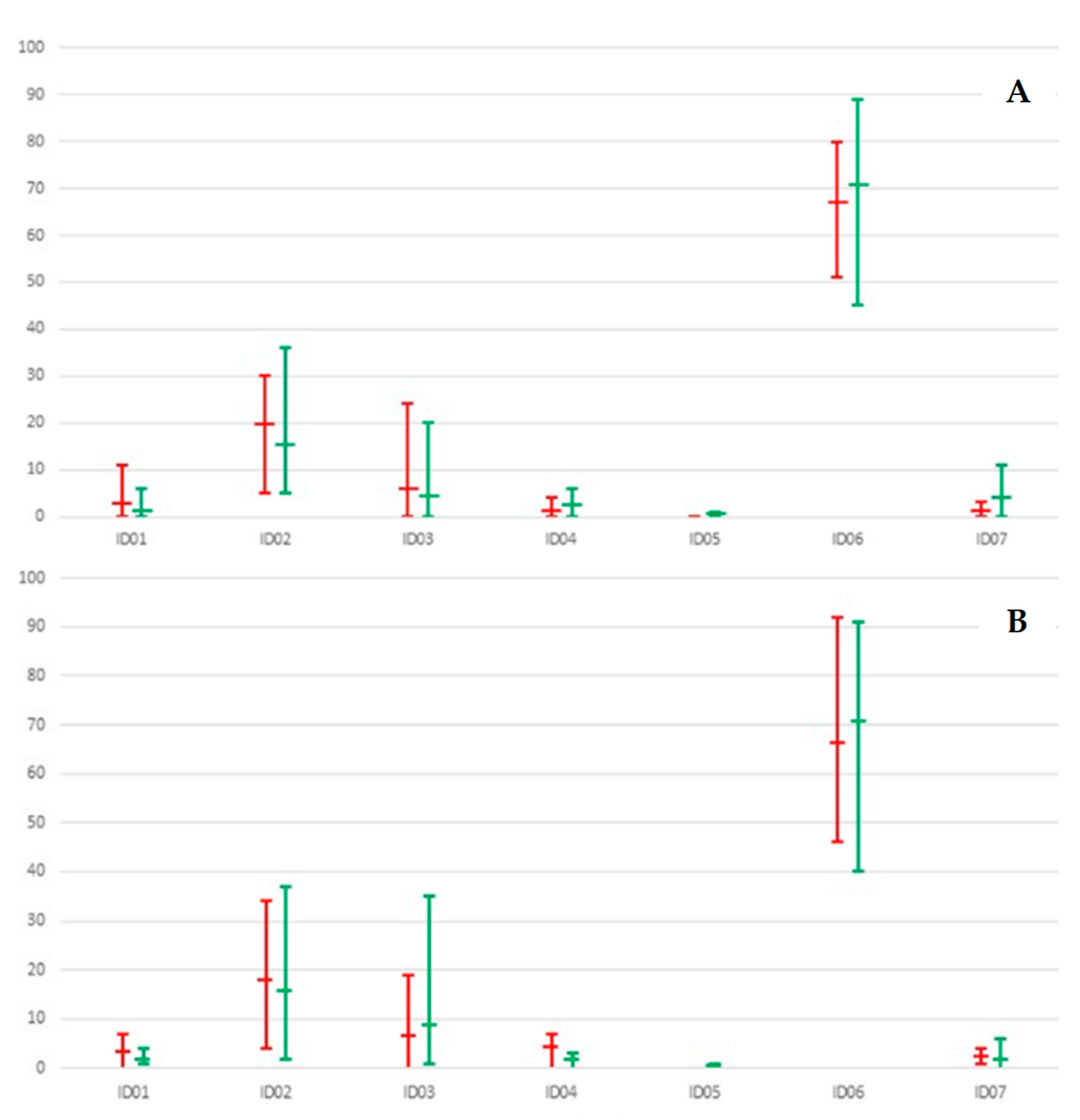
Two (250-125 µ, 125-63 µ) and three (250-125 µ, 125-63 µ, <63 µ) grain sizes were identified as the most representative ones of both the unknown and known samples. The distribution of the quantitative data related to the seven class characteristics of the unknown and known samples, reported for both the 250-125 µ and the 125-63 µ grain sizes, showed an optimal overlaps and trends (Figure 5). In particular, three main classes were prevalent in both unknown and known samples. These were in order of abundance (Table 2, Figure 5):
- i
- ID06 - Rounded and spherical hyaline clasts with yellow/orange coating with a percentage of sub-angular and lamellar grains.
- ii
- ID02 - Rounded hyaline grains with evidence of the original crystalline habitus with yellow/orange coating.
- iii
- ID03 - Rounded and spherical hyaline grains without coating.
Figure 4.
A-D) Microphotographs taken under stereobinocular microscope with reflected light of sandy grains of quarts. A) Unknown trace evidence observed with dark background. B) Known evidence observed with dark background. C) ID1-4 ID6 class grains of quarts observed with mm-sized background. D) SEM micrograph of unknown sample with ID02 class grain. E) SEM-EDS spectrum of quarts grain with Fe coating (spot 2 in Figure 4D). Source: Author.
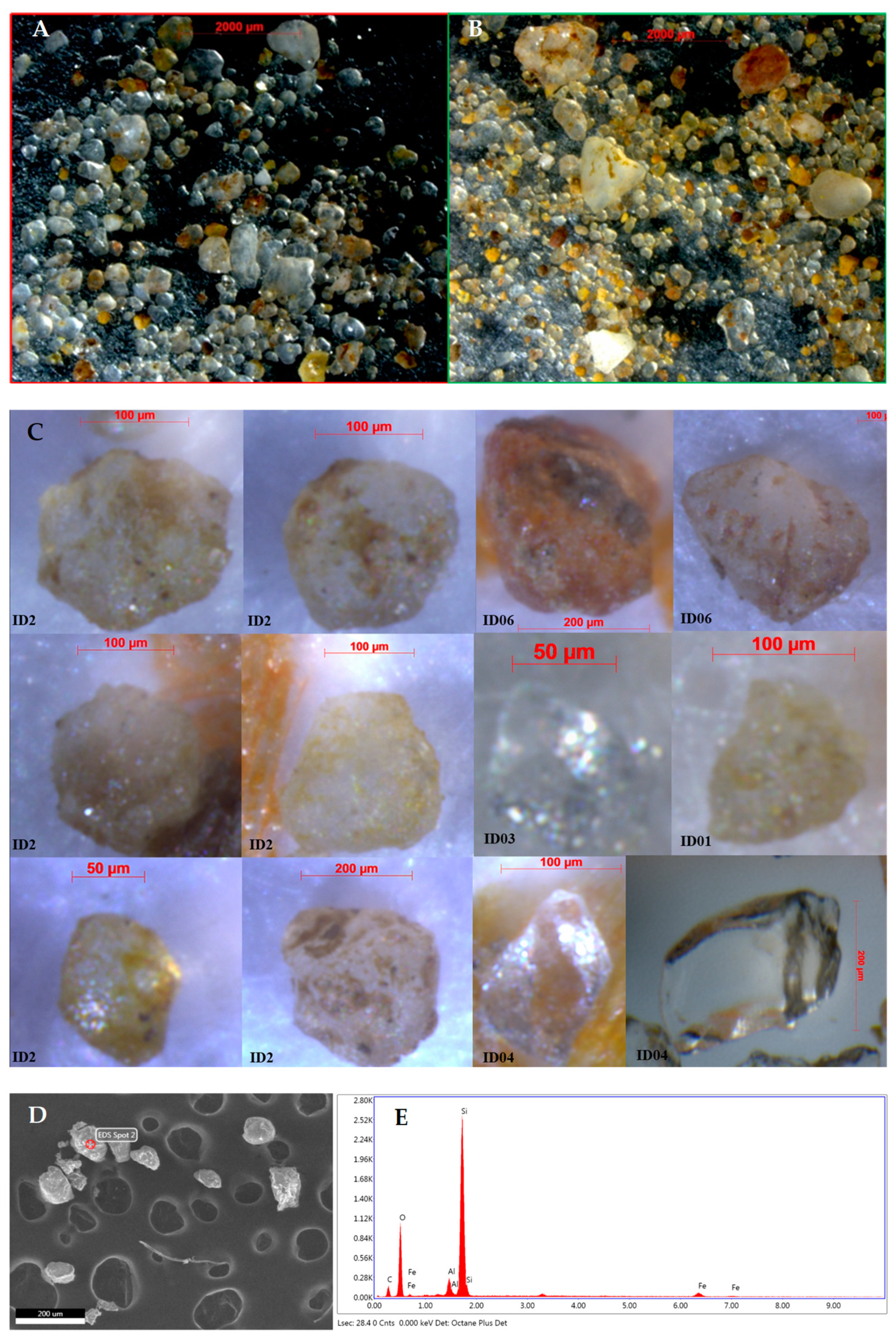
Figure 5.
Comparisons of the percentages of the 7 class characteristics recognized in hundreds of unknown (red line) and known (green line) samples (from the event sites), reported for the 250-125 µm grain size (A) and the 125-63 µm grain size (B). Minimum, medium, and maximum values (horizontal traits) are also shown. Source: Author.
3.2. Botanical Evidence
Forensic botany was applied for the present casework in order to corroborate the forensic geology investigations. The amount of the vegetal material was prevailing on the inorganic grains. The vegetal fraction separated from the geological traces related to the unknown samples from the MPFs and their belongings and observed under stereomicroscopy, microscopy, and SEM-EDS system, resulted composed of plant fragments or entire elements of branches, twigs, leaves, thorns, capsules, fruits, seeds, pollen, herbaceous fragments, wood, vegetable debris, decomposing organic material (humus), and algae (including diatoms).
The main 12 species of remnants of traces of the identified terrestrial plants were [98]:
- Erica arborea (leaves, capsules, seeds).
- Quercus suber (leaves, flowers, seeds).
- Olea europaea (leaves, seeds).
- Cistus monspeliensis (leaves, seeds, capsules, etc.).
- Pistacia lentiscus (leaves, seeds).
- Myrtus communis (leaves, seeds).
- Cytisus infestus (branches, legume, thorns).
- Smilax aspera (leaves, thorns).
- Rosa sempervirens (thorns).
- Rubus ulmifolius (thorns).
- Rosacea Amygdaloidea (thorns).
- Cynara cardunculus (thorns).
Peculiar particles were considered the thorns and seeds being the most abundant vegetal component sampled on bodies, clothing, and footwears of both MPFs.
Over 522 seeds of Erica arborea and 81 thorns ascribable to Rosa sempervirens, Rubus ulmifolius, Rosacea Amygdaloidea, Cynara cardunculus, Cytisus infestus, and Smilax aspera were found in the shoes of victim 1, mostly inside the shoes and planted on the soles, respectively. Another peculiar material was represented by an aggregate made up of remnants of freshwater algae found in the sole of one victim 1’s footwear [98].
Only 16 thorns ascribable to Rosa sempervirens or Rubus ulmifolius, Cynara cardunculus, Cytisus infestus, and Smilax aspera were found planted on the soles of the shoes of victim 2. No traces of algae were found except a unique diatom [97,98].
The same 12 species of terrestrial plants above reported were also found in the vegetation present in the sites of the events and in the vegetal component of the soils collected from this area.
On the base of the above, four main macro-areas with different botanical characteristics were distinguished during remote sensing and field work. The identified macro-areas were [98]:
- Shrub formation with Mediterranean maquis (prevailing macro area).
- Area inside the highway perimeter.
- Meadow area with anthropic pressure from pasture with some puddles of freshwater with algae.
- Tree formation dominated by Sughera (Quercus suber L.) with a circumscribed zone showing abundant concentration of Erica arborea shrubs wood and soils rich in fresh to decomposed seeds of Erica arborea (about 6000 seeds of Erica arborea were examined and counted in the soils).
3.3. Tracing the Route Walked by the Two MPFs
During investigations, great attention was also devoted to the specific search for rare or peculiar particles or assemblages of inorganic and vegetal origin. This examen allowed to recognize in the unknown samples:
- Rare particles of calcite and dolomite.
- Peculiar compositions of clay minerals rich in calcium phosphate.
- Peculiar assemblages of different classes of mineral grains.
- Vegetal remains of mm-sized thorns of Cynara cardunculus and Rosa sempervirens, seeds of Erica arborea, and assemblages of algae.
The same rare and peculiar materials were also recognized in some specific sites of the scene of events, as specified in the following. Geobotanic data on the unknown samples resulted to be typical of microenvironments very similar to those identified in the scene of events. These results were of paramount importance in allowing investigators and judicial authority to link the MPFs to specific sites of the scene of events and in tracing the route of both the MPFs, obtained from 9 specific microenvironments recognized in the scene of events. Figure 6 and Table 3 illustrate the sequence and distribution of the match points and match linear belts (M), from which the path was delineated by means interpolation of matching sites and 4 exit/entry points (E1-4), recognized in the event scene, starting from the site of the car accident site (CA). The localizations of the two different finding sites of the human remains (F1 and F2) were also reported.
3.3.1. Car Accident (Table 3, Figure 6)
The car accident occurred among two vehicles inside a tunnel of the highway. After the crash, the two subjects abandoned their vehicle and went out of the tunnel, reaching a lateral gate of the highway.
3.3.2. Match Point 1 (M1) (Table 3, Figure 6)
Inorganic micro traces (calcite) from MPF 1’s ring.
These micro traces were comparable with the calcareous composition of the top of a ~ 1 m high perimeter wall delimiting the lateral gate of the highway (Figure 7). The transfer from the wall to the ring could realize when the MPF 1, in order to reach the raised ground on the back of the highway, passed on the wall touching it with the hands to lift him-herself from the roadway in order to continue to escape from the car accident site.
3.3.3. Exit (E1) (Table 3, Figure 6)
Two MPFs passed through a rudimental wood gate delimiting the area surrounding the highway tunnels.
3.3.4. Match Linear Belt 2 (M2) (Table 3, Figure 6)
Organic traces (thorns of Cynara cardunculus) from MPF 1’s sock and MPF 2’s shoes.
This evidence was comparable with some thorny plants growing on an uncultivated field in the meadow area with anthropic pressure from pasture (Figure 8). The transfer from the plants to the shoes could realize when the subject walked on these thorny plants distributed in the linear belt reported in Figure 6.
3.3.5. Match Point 3 (M3) (Table 3, Figure 6)
Organic traces (algae) from MPF 1’s shoes.
These were comparable with algae present on freshwaters of a muddy puddle (Figure 9). The transfer from the puddle to the shoes’ soles could realize when the subject walked on this specific puddle.
3.3.6. Match Point 4 (M4) (Table 3, Figure 6)
Inorganic micro traces of P-rich clays from MPFs’ shoes.
These were comparable with a wet soil present on the above reported muddy puddle (Figure 10). The transfer from the P-rich clayish wet soil to the victim could realize when the MPF 1 and 2’s shoes walked on the wet area.
3.3.7. Match Point 5 (M5) (Table 3, Figure 6)
Inorganic traces from MPF 2’s shoes.
This particle assemblage was comparable with the sandy and silty soil present in a specific site of an uncultivated field in the meadow area with anthropic pressure from pasture (Figure 11). The transfer of the soil to the shoes could realize when the victim walked on this site.
3.3.8. Exit Point (E2) (Table 3, Figure 6)
The two MPFs passed through a rudimental wood gate delimiting an uncultivated field in the meadow area with anthropic pressure from pasture.
3.3.9. Match Point 6 (M6) (Table 3, Figure 6)
Inorganic traces (dolomite) from MPF 2’s shoes.
These were comparable with clasts of pinkish dolostones artificially reported on a dirt road present on an uncultivated field, near a wood rudimentary gate (Figure 12). The transfer to the shoes could realize when the victim walked on this road.
3.3.10. Entry Point (E3) (Table 3, Figure 6)
The two MPFs passed through a passage in the barbed wire delimiting the Sughera wood with Erica arborea plants, entering in this cover and isolated area.
3.3.11. Match Linear Belt 7 (M7) (Table 3, Figure 6)
Organic traces (Erica arborea seeds) from MPF 1’s shoes (Figure 13) and socks.
The freshness state of the Erica arborea seeds in the MPF 1’s shoes (Figure 13A) were comparable with seeds present in the soil present in the wood with Sughera (Quercus suber L.) associated with Erica arborea shrubs above cited (Figure 13). The transfer to the internal parts of shoes and socks could realize when the victim walked on this area, along the identified linear belt, infilling the shoe internal parts with Erica arborea seed-rich soil and seeds fallen by the shrubs due to the impact of the body with Erica arborea shrubs.
3.3.12. Match Point 8 (M8) (Table 3, Figure 6)
Inorganic traces from MPF 1’s shoes.
This particle assemblage was comparable with the sandy and silty soil present in the area of the wood with Sughera (Quercus suber L.) and Erica arborea shrubs (Figure 14). The transfer of the soil to the shoes could realize when the MPF1 passed on this area.
3.3.13. Finding Site (F2) (Table 3, Figure 6)
The skeletonized human remains of victim 2 were found distributed in an articulated area of the dense Mediterranean maquis.
3.3.14. Exit (E4) (Table 3, Figure 6)
The MPF 1 passed through a passage in the barbed wire delimiting the Sughera wood with Erica arborea plants, abandoning this area.
3.3.15. Match Point 9 (M9) (Table 3, Figure 6)
Organic traces (Rosa sempervirens thorns) from MPF 1’s shoes, socks, and clothing.
3.3.16. Finding Site (F1) (Table 3, Figure 6)
The manner of death of the MPF 1 for precipitation was ascertained by the medico legal investigations. The prone position of the MPF 1’s body, its distance from the infrastructure, as well as its relationships with the plants present on the ground univocally supported this reconstruction. In particular, the injuries on the posterior and lateral sides of the limbs could be dependent on a dynamic impact due to the body’s precipitation on the plants growing on the ground.
4. Discussion and Conclusions
The wide spectrum of mineralogical, morphological, morphoscopic, textural, and botanical determinations accomplished for the present criminal case work allowed to ascertain a general high degree of compatibility among samples collected on the MPFs and on the scene of events, and the exclusion of possible previous traces related to their staying with the same footwears in other sites in the days immediately before the tragic event.
These comparative analyses provided fundamental info-investigative data of paramount importance for establishing the active pre-mortem presence of the MPFs on the scene, dealing with a criminal case where other methods were not very useful and inapplicable. Nobody saw the MPFs walking in the countryside, MPFs had not any mobiles, GPS, or technological devices, no tele cameras were in the scene.
The most significant positive matches, based on the finding of peculiar and rare particles and assemblages, allowed reconstructing a very detailed walking carried out by the two MPFs on the event site. As a matter of facts, the finding of a ten of different points and linear belts of positive match, from 1 (near the car accident site) to 9 (site of the finding of victim 1), allowed to reconstruct and tracking the route walked by the two MPFs in the hours immediately preceding their death (Table 3, Figure 6).
These inferences were very useful for the judicial system. A few criminological and criminalistic inferences based on the investigations, carried out in laboratory and on field, allowed to hypothesize that:
- Both MPFs actively interacted with the natural and anthropogenic microenvironments recognized in the scene of events (Figure 6).
- Both MPFs actively walked in the scene of events along a specific route stretched in areas with different degrees of vegetation density and difficulty to pass through (Figure 6).
- MPF 2 walked for a shorter path than that of MGF 1; this path was reconstructed up to the dirty path with detritus of dolostones (M6); it may be hypothesized that MPF 2 was carried out in the MPF 1’s arms in the Sughera woody area with Erica arborea, from the surrounding of M6 site up to M8 site, i.e. in an area very next to the site of finding of victim 2 (F2 in Figure 6).
- MPF 1 walked for a longer path than that of MPF 2 up to the site of finding of the body (F1 in Figure 6).
- MPF 1 actively climbed on the infrastructure (M9 in Figure 6). This action supported evaluations made by the coroner on injuries due to precipitation from a high infrastructure.
In conclusion, the peculiar and rare grains, minerals, composition, textures together the peculiar and rare assemblages analysed in the unknown and known specimens from victims and event scene permitted to identify a “fingerprint” of most of the examined specimens. Most of these fingerprints were obtained by means of image analysis and the manual counting of grain characteristics. This procedure, also if it was extremely time consuming and relevant only in cases after the MPF’s finding, it was of uttermost importance in strengthen the reconstructions here proposed. In such way, investigators revealed to be able to provide very strong geological and botanical evidence for supporting criminal investigations.
The here presented method for tracking the route walked by MPFs could fall back in the strategies to contrast the Al-Qaeda terrorism. When a terrorist is captured in the countryside, it may be useful or necessary to track backwards the route walked by him/her to search for a terrorist den or a site of investigative interest (burial of firearms, explosives, etc.).
Notwithstanding these encouraging and promising results and the great potentiality of this method, in Italy Forensic Geology still represents a minor discipline of the Forensic Sciences [104]. In the Italian police criminal laboratories, the analysis of geological and botanical traces actually rarely occurs, differently from the realities of other countries (Federal Bureau of Investigation -FBI- Laboratory of Trace Evidence, Quantico, USA [51,105]; Institute of Criminalistics -ICP- Prague, Czech Republic [106,107]; Australia [50]).
On the basis of the above, for the next generations of forensic experts in geology and botany, it should be desirable that the academics and researchers:
- Promote initiatives for introducing the master’s degree in geology in the police applications for assuming Geologists/Forensic Examiners for the police forensic bureau (Carabinieri, Police).
- Develop forensic protocols envisaging all possible technical procedures / operations to accurately apply on the crime or event scene to preserve evidence made of inorganic and organic materials and strengthen greater interaction and collaboration between forensic geologists, botanists, and experts in legal medicine directly on the crime scene, to arrange all the activities aimed at preserving these traces as far as possible, even during necropsy operations.
Funding
This research received no external funding.
Acknowledgments
The Author is deeply grateful to the systematic botanists Prof. PhD Marina Morabito, Dr. PhD Fabio Mondello, and Dr PhD Angelo Troia for their expertise applied for the determination of plant remains. The Author thanks Prof. PhD Elpida Piperopoulos and Dr PhD Giuseppe Sabatino for their assistance during the SEM-EDS analyses and the reviewers and editors for strongly improving the quality of the present paper.
Conflicts of Interest
The author declares no conflict of interest.
References
- Nilson, M. T.; Marks, S.; Meer, T. Manhunting: a methodology for finding persons of national interest. 2005. PhD Thesis. Monterey, California. Naval Postgraduate School.
- Somma, R. Unraveling crimes with geosciences. AAPP Atti Accad. Peloritana dei Pericolanti Cl. Sci. Fis. Mat. Nat. 2023, 101, 1–23. [Google Scholar] [CrossRef]
- Richards, Julian. Intelligence and counterterrorism. New York: Routledge, 2018.
- Murray, R. C. Evidence from the earth: forensic geology and criminal investigation; Mountain Press Publishing Company, Missoula, Montana, 2004; pp. 226.
- Murray, R. C. Forensic geology: yesterday, today and tomorrow. Geol. Soc. Spec. Publ. 2004, 232, 7–9. [Google Scholar] [CrossRef]
- Murray, R.C.; Tedrow, J.C. Forensic geology: Earth sciences and criminal investigation. Rutgers University Press Address, Piscataway, United States, 1975; pp. 232.
- Pirrie, D.; Dawson, L.; Graham, G. Predictive geolocation: forensic soil analysis for provenance determination. Episodes 2017, 40, 141–147. [Google Scholar] [CrossRef]
- Intini, A.; Picozzi, M. Scienze forensi. Teoria e prassi dell’investigazione scientifica. UTET Giuridica Publisher, Milano, Italy, EAN: 9788859803973, ISBN: 8859803977, 2009; pp. 544.
- Saferstein, R. Criminalistics: An Introduction to Forensic Science. Pearcon, 2017. ISBN-13: 978-0134477596, ISBN-10: 0134477596.
- Lombardi, G. The contribution of forensic geology and other trace evidence analysis to the investigation of the killing of Italian Prime Minister Aldo Moro. J. Forensic Sci. 1999, 44, 634–642. [Google Scholar] [CrossRef] [PubMed]
- Shroder, J.J. Remote Sensing and GIS as Counterterrorism Tools for Homeland Security: The case of Afghanistan. In: Sui, D.Z. (eds) Geospatial Technologies and Homeland Security. The GeoJournal Library, vol 94. Springer, Dordrecht, 2008. [CrossRef]
- Tindall, C. G. Forensic Geology. Soil Sci. 1994, 157, 128. [Google Scholar] [CrossRef]
- Pye, K.; Croft, D. J. (eds.) Forensic geoscience: Principles, techniques and applications; Geol. Soc. Spec. Publ., England, 2004; pp. 232.
- Pye, K. Forensic geology. In Encyclopedia of Geology. Selley, R.C., Cocks, L.R.M., Plimer, I.R., Eds; Elsevier Ltd., The Boulevard, Lanford Lane, Kidlington, Oxford, UK Amsterdam, Netherlands, 2005; Volume 2, pp. 261-273.
- Ruffell, A.; McKinley, J. Forensic Geology & Geoscience. Earth-Sci. Rev. 2005, 69, 235–247. [Google Scholar] [CrossRef]
- Morgan, R.M.; Wiltshire, P.E.J.; Parker, A.; Bull, P. The role of forensic geoscience in wildlife crime detection. Forensic Sci. Int. 2006, 162, 152–162. [Google Scholar] [CrossRef] [PubMed]
- Morgan, R. M.; Bull, P. A. Forensic geoscience and crime detection. Minerva Med. 2007, 127, 73–89. [Google Scholar]
- Pye, K. Geological and Soil Evidence, 1st ed.; CRC Press, Boca Raton, Oxford, 2007; pp. 356. [CrossRef]
- Ruffell, A.; McKinley, J. Geoforensics; John Wiley & Sons Ltd, The Atrium, Southern Gate, Chichester, West Sussex, England, 2008; pp. 332.
- Fitzpatrick, R.W.; Raven, M.D.; Forrester, S.T. A systematic approach to soil forensics: criminal case studies involving transference from crime scene to forensic evidence. In Criminal and environmental soil forensics; Springer Science & Business Media B.V., Netherlands, 2009; pp. 105-127.
- Pirrie, D. Forensic geology in serious crime investigation. Geol. Today 2009, 25, 188–192. [Google Scholar] [CrossRef]
- Ruffell, A. Forensic pedology, forensic geology, forensic geoscience, geoforensics and soil forensics. Forensic Sci. Int. 2010, 202, 9–12. [Google Scholar] [CrossRef]
- Di Maggio, R. M.; Barone, P. M.; Pettinelli, E.; Mattei, E.; Lauro, S. E.; Banchelli, A. Geologia Forense. Geoscienze e indagini giudiziarie, 1st ed.; Dario Flaccovio Editore: Palermo, Italia, 2013; p. 319. [Google Scholar]
- Sangwan, P.; Nain, T.; Singal, K.; Hooda, N.; Sharma, N. Soil as a tool of revelation in forensic science: a review. Analytical Methods 2020, 12, 5150–5159. [Google Scholar] [CrossRef]
- Donnelly, L. J.; Pirrie, D.; Harrison, M.; Ruffell, A.; Dawson, L. A. (eds.) A guide to forensic geology, 1st ed.; Geol. Soc. London, England, 2021; pp. 217.
- Fitzpatrick, R. W.; Donnelly, L. J. An introduction to forensic soil science and forensic geology: a synthesis. Geol. Soc. Spec. Publ. 2021, 492, 1–32. [Google Scholar] [CrossRef]
- Somma, R.; Maniscalco, R. Forensic geology applied to criminal investigation: a case report. AAPP Atti Accad. Peloritana dei Pericolanti Cl. Sci. Fis. Mat. Nat. 2023, 101, A9–1. [Google Scholar] [CrossRef]
- Somma, R.; Trombino, L. Introducing “Advances and applications in Geoforensics: Unraveling crimes with Geology. AAPP Atti Accad. Peloritana dei Pericolanti Cl. Sci. Fis. Mat. Nat. 2023, 101, E1–1. [Google Scholar] [CrossRef]
- Spoto, S. E. , Barone, S., and Somma, R. An introduction to forensic geosciences. AAPP Atti Accad. Peloritana dei Pericolanti Cl. Sci. Fis. Mat. Nat. 2023, 101, A1–1. [Google Scholar] [CrossRef]
- Oivanki, S. M. Forensic geology: geologic investigation as a tool for enforcement of environmental regulations. Mississippi Geology 1996, 17, 45–63. [Google Scholar]
- Ritz, K.; Dawson, L.A.; Miller, D. Criminal and Environmental Soil Forensics. Springer 2009, pp. 519. [CrossRef]
- Ruffell, A.; Dawson, L. Forensic geology in environmental crime: Illegal waste movement & burial in Northern Ireland. Environ. Forensics 2009, 10, 208–213. [Google Scholar] [CrossRef]
- Ruffell, A.; Kulessa, B. Application of geophysical techniques in identifying illegally buried toxic waste. Environ. Forensics 2009, 10, 196–207. [Google Scholar] [CrossRef]
- Pirrie, D.; Ruffell, A.; Dawson, L. A. Environmental and criminal geoforensics: an introduction. Geol. Soc. Spec. Publ. 2013, 384, 1–7. [Google Scholar] [CrossRef]
- Ruffell, A. (2013) Soil and drift geology in forensic investigations. In Pirrie, D.; Ruffell, A.; Dawson, L. A. (Eds.) Environmental and criminal geoforensics: an introduction. Geol. Soc. Spec. Publ. 2013, 384, 163–172. [Google Scholar] [CrossRef]
- Ruffell, A.; Pringle, J. K.; Graham, C.; Langton, M.; Jones, G. M. Geophysical assessment of illegally buried toxic waste for a legal enquiry: A case study in Northern Ireland (UK). Environ. Forensics 2018, 19, 239–252. [Google Scholar] [CrossRef]
- Ruffell, A.; Barry, L. The desktop study an essential element of geoforensic search: homicide and environmental cases (west Belfast, Northern Ireland, UK). Geol. Soc. Spec. Publ. 2021, 492, 39–53. [Google Scholar] [CrossRef]
- Locard, R. The analysis of dust traces. Revue Internationale de Criminalistique 1929, I, 4–5. [Google Scholar]
- Graves, W. J. A Mineralogical Soil Classification Technique for the Forensic Scientist. J. Forensic Sci. 1979, 24, 323–338. [Google Scholar] [CrossRef]
- Palenik, S. Microscopic trace evidence – the overlooked clue. Part III Max Frei – Sherlock Holmes with microscope. Microscope 1982, 30, 93–100. [Google Scholar]
- Sugita, R.; Marumo, Y. Validity of color examination for forensic soil identification. Forensic Sci. Int. 1996, 83, 201–210. [Google Scholar] [CrossRef]
- Pirrie, D.; Butcher, A.R.; Power, M.R.; Gottlieb, P.; Miller, G.L. Rapid quantitative mineral and phase analysis using automated scanning electron microscopy (QemSCAN); potential applications in forensic geoscience. In Forensic geoscience: Principles, techniques and applications; Geol. Soc. Spec. Publ. Pye, K.; Croft, D. J. (eds.), London, England, 2004; 123–136.
- Ruffell, A.; Wiltshire, P. Conjunctive use of quantitative and qualitative X-ray diffraction analysis of soils and rocks for forensic analysis”. Forensic Sci. Int. 2004, 145, 13–23. [Google Scholar] [CrossRef] [PubMed]
- Bull, P.A.; Morgan, R.M.; Dunkerley, S. SEM-EDS analysis and discrimination of forensic soil by Cengiz et al., A comment. Forensic Sci. Int. 2005, 155, 222–224. [Google Scholar] [CrossRef]
- Morgan, R.M.; Bull, P.A. Data interpretation in forensic sediment and soil geochemistry. Environ. Forensics 2006, 7, 325–334. [Google Scholar] [CrossRef]
- McKinley, J.; Ruffell, A. Contemporaneous spatial sampling at scenes of crime: advantages and disadvantages. Forensic Sci. Int. 2007, 172, 196–202. [Google Scholar] [CrossRef]
- Morgan, R. M.; Bull, P. A. The philosophy, nature and practice of forensic sediment analysis. Prog. Phys. Geogr. 2007, 31, 43–58. [Google Scholar] [CrossRef]
- Fitzpatrick, R.W. Nature, Distribution, and Origin of Soil Materials in the Forensic Comparison of Soils. In: Soil analysis in forensic taphonomy: chemical and biological effects of buried human remains. Ed. by M. Tibbett and D. Carter. CRC Press 2008. [CrossRef]
- Ruffell, A.; Sandiford, A. Maximising trace soil evidence: An improved recovery method developed during investigation of a $26 million bank robbery. Forensic Sci. Int. 2011, 209, e1–e7. [Google Scholar] [CrossRef]
- Fitzpatrick, R. W.; Raven, M. D. How Pedology and Mineralogy Helped Solve a Double Murder Case: Using Forensics to Inspire Future Generations of Soil Scientists. Soil Horizons 2012, 53, 14. [Google Scholar] [CrossRef]
- Webb, J. B.; Bottrell, M.; Stern, L. A.; Saginor, I. Geology of the FBI lab and the challenge to the admissibility of forensic geology in US court. Episodes 2017, 40, 118–119. [Google Scholar] [CrossRef]
- Fitzpatrick, R. W. Soil: Forensic Analysis. In: Wiley Encyclopedia of Forensic Science. Fitzpatrick, R. W.; Donnelly, L. J. An introduction to forensic soil science and forensic geology: A synthesis. Geol. Soc. Spec. Publ. 2021, 492, 1–32. [Google Scholar] [CrossRef]
- Somma, R. A multidisciplinary approach based on the cooperation of forensic geologists, botanists, and engineers: Computed Axial Tomography applied to a case work. AAPP Atti Accad. Peloritana dei Pericolanti Cl. Sci. Fis. Mat. Nat. 2023, 101, 1–10. [Google Scholar] [CrossRef]
- Somma, R.; Spoto, S. E.; Raffaele, M.; Salmeri, F. Measuring color techniques for forensic comparative analyses of geological evidence. AAPP Atti Accad. Peloritana dei Pericolanti Cl. Sci. Fis. Mat. Nat. 2023, 101, 1–16. [Google Scholar] [CrossRef]
- Ruffell, A.; Majury, N.; Brooks, W. E. Geological fakes and frauds. Earth-Sci. Rev. 2012, 111, 224–231. [Google Scholar] [CrossRef]
- Barume, B.; Naeher, U.; Ruppen, D.; Schütte, P. Conflict minerals (3TG): Mining production, applications and recycling. Current Opinion in Green and Sustainable Chemistry 2016, 1, 8–12. [Google Scholar] [CrossRef]
- Ruffell, A.; Schneck, B. International case studies in forensic geology: fakes and frauds, homicides and environmental crime. Episodes Int. J. Geosci. 2017, 40, 172–175. [Google Scholar] [CrossRef]
- Marra., A.C.; Di Silvestro, G.; Somma, R. Palaeontology applied to criminal investigation. AAPP Atti Accad. Peloritana dei Pericolanti Cl. Sci. Fis. Mat. Nat. 2023, 101, 1–16. [Google Scholar] [CrossRef]
- Spoto, S. E. Illicit trafficking of diamonds: new frontiers. AAPP Atti Accad. Peloritana dei Pericolanti Cl. Sci. Fis. Mat. Nat. 2023, 101, 1–9. [Google Scholar] [CrossRef]
- Davenport, G.C. Remote sensing applications in forensic investigations. Hist. Archaeol. 2001, 35, 87–100. [Google Scholar] [CrossRef]
- Manhein, M.H.; Listi, G.A.; Leitner, M. The application of geographic information systems and spatial analysis to assess dumped and subsequently scattered human remains. J. Forensic Sci. 2006, 51, 469–74. [Google Scholar] [CrossRef] [PubMed]
- Herrmann, N.P.; Devlin, J.B. Assessment of commingled human remains using a GIS-based approach. In Recovery, analysis, and identification of commingled human remains, Adams, B.J., Byrd, J.E. eds.; Humana Press Publisher, Totowa, New Jersey, 2008, 257–270.
- Donnelly, L.; Harrison, M. Geomorphological and geoforensic interpretation of maps, aerial imagery, conditions of diggability and the colour-coded RAG prioritization system in searches for criminal burials. Geol. Soc. Spec. Publ. 2013, 384, 173–194. [Google Scholar] [CrossRef]
- Elmes, G.A.; Roedl, G.; Conley, J. (eds.). Forensic GIS: the role of geospatial technologies for investigating crime and providing evidence. Springer Press Publisher, Dordrecht, Netherlands, 2014, pp. 320.
- Ruffell, A.; McAllister, S. A RAG system for the management forensic and archaeological searches of burial grounds. Int. J. Archaeol. 2015, 3, 1–8. [Google Scholar] [CrossRef]
- Bunch, A.W.; Kim, M.; Brunelli, R. Under our nose: the use of GIS technology and case notes to focus search efforts. J. Forensic Sci. 2017, 62, 92–98. [Google Scholar] [CrossRef] [PubMed]
- Somma, R.; Cascio, M.; Silvestro, M.; Torre, E. A GIS-based quantitative approach for the search of clandestine graves, Italy. J. Forensic Sci. 2018, 63, 882–898. [Google Scholar] [CrossRef] [PubMed]
- Somma, R.; Costa, N. Unraveling Crimes with Geology: As Geological and Geographical Evidence Related to Clandestine Graves May Assist the Judicial System. Geosciences 2022, 12, 339. [Google Scholar] [CrossRef]
- Somma, R. The space and time dimensions in the criminal behaviour of lust murderers. AAPP Atti Accad. Peloritana dei Pericolanti Cl. Sci. Fis. Mat. Nat. 2023, 101, 1–36. [Google Scholar] [CrossRef]
- Somma, R.; Costa, N. GIS-based RAG-coded search priority scenarios for predictive maps to prevent future serial serious crimes: the case study of the Florence Monster. AAPP Atti Accad. Peloritana dei Pericolanti Cl. Sci. Fis. Mat. Nat. 2023, 101, 1–17. [Google Scholar] [CrossRef]
- Wolff, M.; Asche, H. Towards geovisual analysis of crime scenes – a 3D crime mapping approach. In Advances in GIScience, Sester, M., Bernard, L., Paelke, V. eds.; Germany: Springer-Verlag Publisher, Berlin/Heidelberg, Germany, 2009; pp. 429–448. [Google Scholar] [CrossRef]
- Baldino, G.; Ventura Spagnolo, E.; Fodale, V.; Pennisi, C.; Mondello, C.; Altadonna, A.; Raffaele, M.; Salmeri, F.; Somma, R.; Asmundo, A.; Sapienza, D. The application of 3D virtual models in the judicial inspection of indoor and outdoor crime scenes. AAPP Atti Accad. Peloritana dei Pericolanti Cl. Sci. Fis. Mat. Nat. 2023, 101, 1–21. [Google Scholar] [CrossRef]
- Somma, R.; Altadonna, A.; Cucinotta, F.; Raffaele, M.; Salmeri, F.; Baldino, G.; Ventura Spagnolo, E.; Sapienza, D. The technologies of Laser Scanning and Structured Blue Light Scanning applied to criminal investigation: case studies. AAPP Atti Accad. Peloritana dei Pericolanti Cl. Sci. Fis. Mat. Nat. 2023, 101, 1–18. [Google Scholar] [CrossRef]
- France, D.L.; Griffin, T.J.; Swanburg, J.G.; Lindemann, J.W.; Davenport, G.C.; Trammell, V.; et al. Necrosearch revisited: further multidisciplinary approaches to the detection of clandestine graves. In Forensic taphonomy: the postmortem fate of human remains, Haglund, W.D., Sorg, M.H., Eds.; CRC Press Publisher, New York, USA, 1997; pp. 497–509. [CrossRef]
- Ruffell, A.; Wilson, J. Shallow ground investigation using radiometrics and spectral gamma-ray data. Archaeol. Prospect. 1998, 5, 203–215. [Google Scholar] [CrossRef]
- Ruffell, A. Remote detection and identification of organic remains. Archaeol. Prospect. 2002, 9, 115–122. [Google Scholar] [CrossRef]
- Ruffell, A. Burial location using cheap and reliable quantitative probe measurements. Diversity in forensic anthropology. Spec. Publ. Forensic Sci. Int. 2004, 151, 207–211. [Google Scholar] [CrossRef]
- Ruffell, A. Searching for the I.R.A. Disappeared: ground-penetrating radar investigation of a churchyard burial site, Northern Ireland. J. Forensic Sci. 2005, 50, 414–424. [Google Scholar] [CrossRef]
- Salsarola, D.; Cattaneo, C. Archeologia forense. In Scienze Forensi – Teoria e prassi dell’investigazione scientifica; Intini, A., Picozzi, M. Eds.; UTET Giuridica Publisher, Milano, Italy, 2009, pp. 207–226.
- Pringle, J.K.; Jervis, J.R. Electrical resistivity survey to search for a recent clandestine burial of a homicide victim, UK. Forensic Sci. Int. 2010, 202, 1–7. [Google Scholar] [CrossRef]
- Harrison, M. Grave concerns, locating and unearthing human bodies. Aust. J. Forensic Sci. 2011, 43, 324–325. [Google Scholar] [CrossRef]
- Larson, D.O.; Vass, A.A.; Wise, M. Advanced scientific methods and procedures in the forensic investigation of clandestine graves. J. Contemp. Crim. Justice 2011, 27, 149–182. [Google Scholar] [CrossRef]
- Pringle, J.K.; Ruffell, A.; Jervis, J.R.; Donnelly, L.; McKinley, J.; Hansen, J.; et al. The use of geoscience methods for terrestrial forensic searches. Earth-Sci. Rev. 2012, 114, 108–123. [Google Scholar] [CrossRef]
- Sagripanti, G. L.; Villalba, D.; Aguilera, D.; Giaccardi, A. Advances of forensic geology in Argentina: search with non-invasive methods for victims of enforced disappearance. Boletin de Geol. 2017, 39, 55–69. [Google Scholar] [CrossRef]
- López Batista, M.; Rodríguez López, S.; Fieguth Batista, A. The Use of GIS in Forensic Archaeology to Search Clandestine Graves in Uruguay. Sci. Technol. Archaeol. Res. 2018, 2, 61–74. [Google Scholar] [CrossRef]
- Kamaluddin, M.R.; Mahat, N.A.; Mat Saat, G.A.; Othman, A.; Anthony, I.L.; Kumar, S.; Wahab, S.; Meyappan, S.; Rathakrishnan, B.; Ibrahim, F. The Psychology of Murder Concealment Acts. Int. J. Environ. Res. Public Health 2021, 18, 3113. [Google Scholar] [CrossRef] [PubMed]
- Rocke, B.; Ruffell, A.; Donnelly, L. Drone aerial imagery for the simulation of a neonate burial based on the geoforensic search strategy (GSS). J. Forensic Sci. 2021, 66, 1506–1519. [Google Scholar] [CrossRef] [PubMed]
- Rocke, B.; Ruffell, A. Detection of Single Burials Using Multispectral Drone Data: Three Case Studies. J. Forensic Sci. 2022, 2, 72–87. [Google Scholar] [CrossRef]
- Byrd, H. J.; Sutton, L. The Use of Forensic Entomology within Clandestine Gravesite Investigations. AAPP Atti Accad. Peloritana dei Pericolanti Cl. Sci. Fis. Mat. Nat. 2023, 101, 1–13. [Google Scholar] [CrossRef]
- Somma, R.; Sutton, L.; Byrd, J. H. Forensic geology applied to the search for homicide graves. AAPP Atti Accad. Peloritana dei Pericolanti Cl. Sci. Fis. Mat. Nat. 2023, 101, 1–20. [Google Scholar] [CrossRef]
- Tagliabue, G.; Masseroli, A.; Ern, S. I. E.; Comolli, R.; Tambone, F.; Cattaneo, C.; Trombino, L. The Fate of Phosphorus in Experimental Burials: Chemical and Ultramicroscopic Characterization and Environmental Control of Its Persistency. Geosciences 2023, 13, 24. [Google Scholar] [CrossRef]
- Tagliabue, G.; Masseroli, A.; Mattia, M.; Sala, C.; Belgiovine, E.; Capuzzo, D.; Galimberti, P.; Slavazzi, F.; Cattaneo, C.; Trombino, L. Thanatogenic Anthrosols: a geoforensic approach to the exploration of the Sepolcreto of the Ca’ Granda (Milan). AAPP Atti Accad. Peloritana dei Pericolanti Cl. Sci. Fis. Mat. Nat. 2023, 101, 1–21. [Google Scholar] [CrossRef]
- Brown, A. G. The use of forensic botany and geology in war crimes investigations in NE Bosnia. Forensic Sci. Int. 2006, 163, 204–210. [Google Scholar] [CrossRef]
- Jones, G. D.; Bryant, V. M. A comparison of pollen counts: Light versus scanning electron microscopy. Grana 2007, 46, 20–33. [Google Scholar] [CrossRef]
- Caccianiga, M.; Bottacin, S.; Cattaneo, C. Vegetation Dynamics as a Tool for Detecting Clandestine Graves. J. Forensic Sci. 2012, 57, 983–988. [Google Scholar] [CrossRef]
- Scott, K. R.; Morgan, R. M.; Jones, V. J.; Cameron, N. G. The transferability of diatoms to clothing and the methods appropriate for their collection and analysis in forensic geoscience. Forensic Sci. Int. 2014, 241, 127–137. [Google Scholar] [CrossRef]
- Morabito, M.; Somma, R. The crucial role of Forensic Botany in the solution of judicial cases. AAPP Atti Accad. Peloritana dei Pericolanti Cl. Sci. Fis. Mat. Nat. 2023, 101, 1–16. [Google Scholar] [CrossRef]
- Morabito, M.; Mondello, F.; Somma, R. Macrobotanic data implementing Forensic Geology investigations. AAPP Atti Accad. Peloritana dei Pericolanti Cl. Sci. Fis. Mat. Nat. 2023, 101, 1–15. [Google Scholar] [CrossRef]
- Somma, R.; Cascio, M.; Cucinotta, F.; Mondello, F.; Morabito, M. Recent advances in forensic geology and botany for the reconstruction of event dynamics in outdoor crime scenes: a case study”. AAPP Atti Accad. Peloritana dei Pericolanti Cl. Sci. Fis. Mat. Nat. 2023, 101, 1–21. [Google Scholar] [CrossRef]
- Bourguignon, L.; Hellmann, A.; Marlof, A.; Fernadez Rodriquez, A.; Repele, M.; Utehaag, S.; Dawson, L. Best practice manual for the forensic comparison of soil traces. Best Practise Manual, ENFI-BPM-APS-02, Version 1, December 2019.
- Bull, P.A.; Morgan, R.M. Sediment fingerprints: A forensic technique using quartz sand grains. Sci. Justice 2006, 46, 64–68. [Google Scholar] [CrossRef] [PubMed]
- Bull, P.A.; Morgan, R.M.; Wilson, H.E.; Dunkerely, S. Multi-technique comparison of source and primary transfer soil samples: an experimental investigation by Croft, D. J. and Pye, K. A comment. Sci. Justice 2004, 44, 173–176. [Google Scholar]
- Werner, D.; Burnier, C.; Yu, Y.; Marolf, A. R.; Wang, Y.; Massonnet, G. Identification of some factors influencing soil transfer on shoes. Sci. Justice 2019, 59, 643–653. [Google Scholar] [CrossRef] [PubMed]
- Galloway, A. Foreword. AAPP Atti Accad. Peloritana dei Pericolanti Cl. Sci. Fis. Mat. Nat. 2023, 101, E1. [Google Scholar] [CrossRef]
- FBI laboratory division. Available online: https://www.fbi.gov/investigate/how-we-investigate/laboratory-division (accessed on 3 October 2023).
- Police Czech Republic. Available online: https://www.policie.cz/default.aspx (accessed on 3 October 2023).
- Antušková, V.; Šefců, R.; Šulcová, P.; Dohnalová, Ž.; Luxová, J.; Bajeux Kmoníčková, M.; Turková, I.; Kotrlý, M. Spectroscopic characterisation of Naples yellow variations in paintings from the turn of the 20th century. J. Raman Spectrosc. 2023, 54, 171. [Google Scholar] [CrossRef]
- Pringle, J. K. Forensic geology: getting geological principles and practices into the classroom. Sch. Sci. Rev. 2007, 89, 1–4. [Google Scholar]
- Williams, T. J. Sherlock Holmes to CSI: Microscopy in the Forensic Geology Classroom. Microsc. Microanal. 2008, 14, 862–863. [Google Scholar] [CrossRef]
- Spoto, S. E.; Boncaldo, A.; Capodivento, A.; Di Agosto, M.; Maccarone, D.; Scibilia, D. Aviation and Volcanic Ash Hazards: A Flipped Classroom Approach To Study Complex Systems. AAPP Atti Accad. Peloritana dei Pericolanti Cl. Sci. Fis. Mat. Nat. 2021, 99, 1–10. [Google Scholar] [CrossRef]
- Spoto, S. E.; Somma, R.; Crea, F. Using a forensic-based learning approach to teach geochemistry. AAPP Atti Accad. Peloritana dei Pericolanti Cl. Sci. Fis. Mat. Nat. 2021, 99, 1–9. [Google Scholar] [CrossRef]
- Somma, R. Advances in Flipped Classrooms for Teaching and Learning Forensic Geology. Educ. Sci. 2022, 12, 403. [Google Scholar] [CrossRef]
- Somma, R.; Baldino, G.; Altadonna, A.; Asmundo, A.; Fodale, V.; Gualniera, P.; Mondello, C.; Pennisi, C.; Raffaele, M.; Salmeri, F.; Ventura Spagnolo, E.; Sapienza, D. Education and training activities in forensic and biomedical sciences: The Laser scanner technology. AAPP Atti Accad. Peloritana dei Pericolanti Cl. Sci. Fis. Mat. Nat. 2023, 101, 1–18. [Google Scholar] [CrossRef]
Figure 1.
3D model of the slope, elaborated in ArcGis, showing the localization of the car accident site (green sphere), the victim 2’s finding site (red sphere), and the victim 1’s finding site (whitish sphere). Legend: Ranges of inclinations of the slope. Source: Author.
Figure 1.
3D model of the slope, elaborated in ArcGis, showing the localization of the car accident site (green sphere), the victim 2’s finding site (red sphere), and the victim 1’s finding site (whitish sphere). Legend: Ranges of inclinations of the slope. Source: Author.
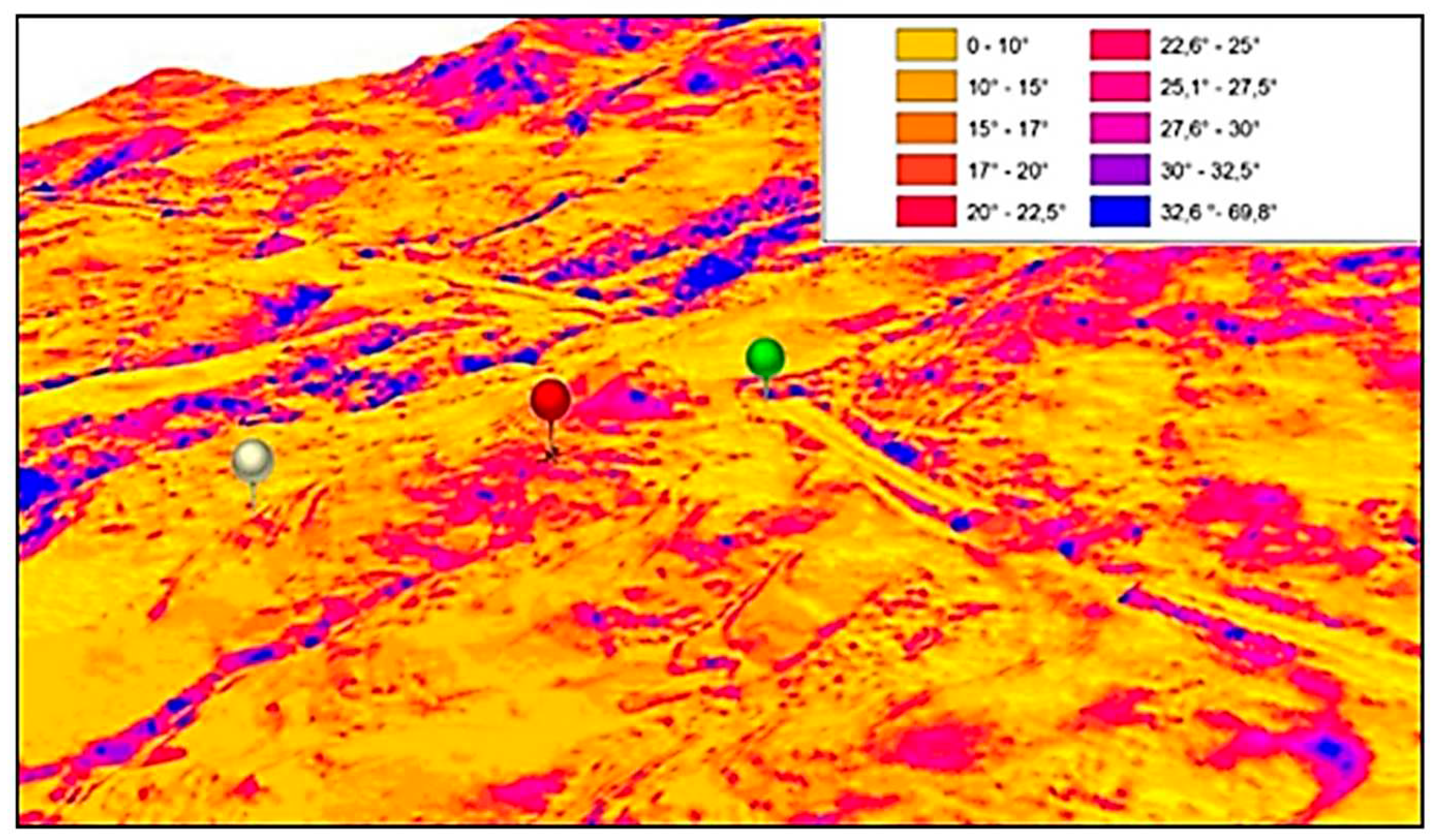
Figure 2.
Microphotographs taken under stereobinocular microscope and reflected light. A-B) Deep scratches of the leather of the MPF 1’s shoes upper part, presumably due to the impact and abrasion of thorny plants during the walking of the MPF 1. C) Most common appearance of questioned trace showing a very abundant vegetal component (Erica arborea leaves, capsules, and seeds, branches, thorns, and humus collected in the internal part of the shoes, see later), transferred to MPF1’s shoes inner part during the walking in a woody area. D) Two thorns of Cytisus infestus fixed in the sole of the MPF 2’s shoes, transferred during the walking of the MPF 2’s shoes during the walking in the scene of events. Plants determined by Fabio Mondello. Source: Author.
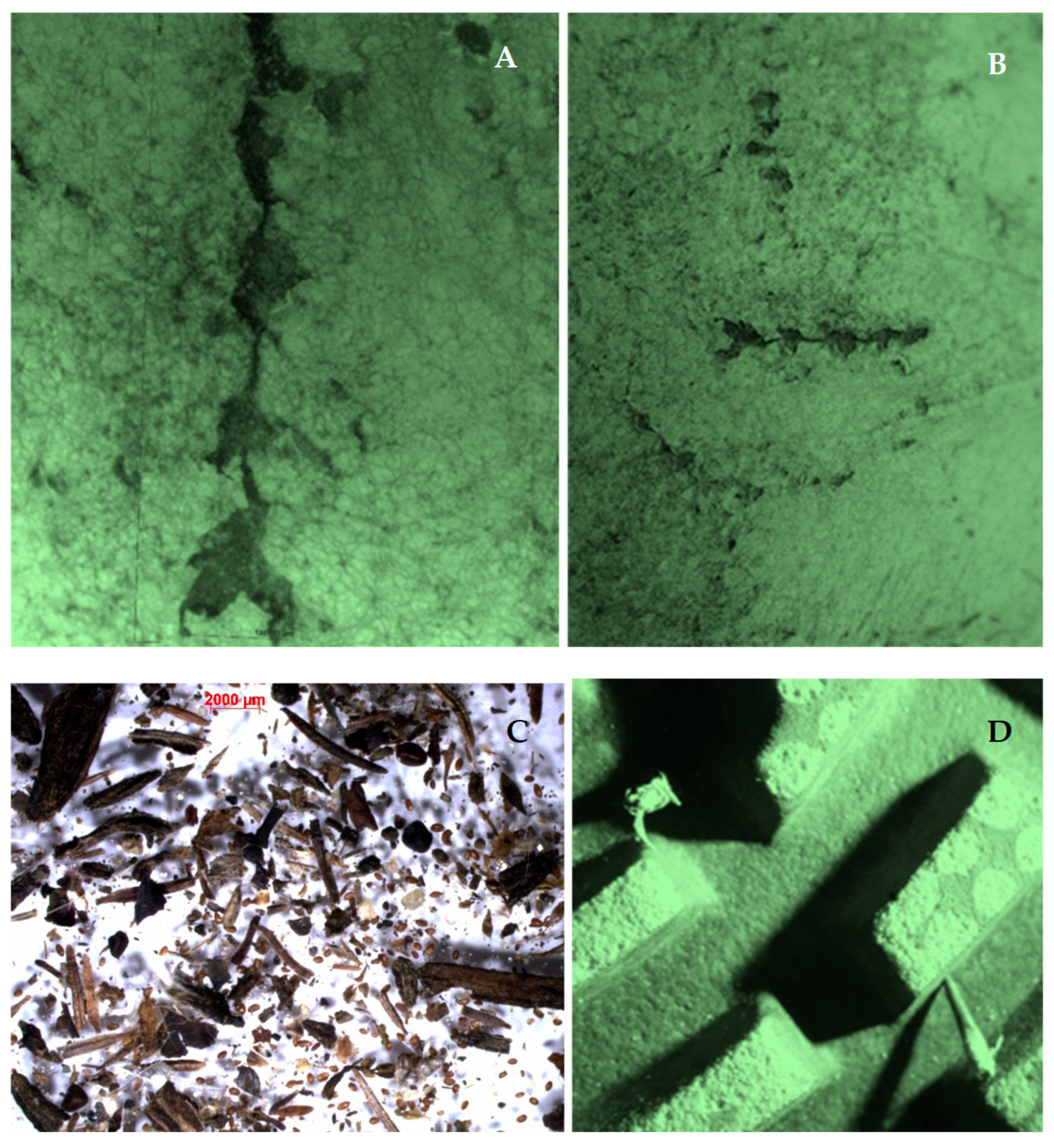
Figure 6.
Route walked by the two MPFs in the scene of events. The route was reconstructed interpolating the starting point of the car accident (CA) with the 9 match points and linear belts (M), and the Exit/Entry sites (E). Legend: 1) CA – Car Accident; 2) M1 – Match point (MPF 1); 3) M6 – Match point (MPF 2); 4) M4 – Match point (MPFs 1-2); 5) - Match linear belt; 6) F2 - Finding site of victim 2; 7) F1 - Finding site of victim 1; 8) E1 – Exit/Entry (rudimental wood gates and holes in barber wire); 9) Physical barrier due to climbing thorny plants; 10) Sughera wood with Erica arborea plants. 11) Gate; 12) Limit of property; 13) Dirty roads. Source: Author.
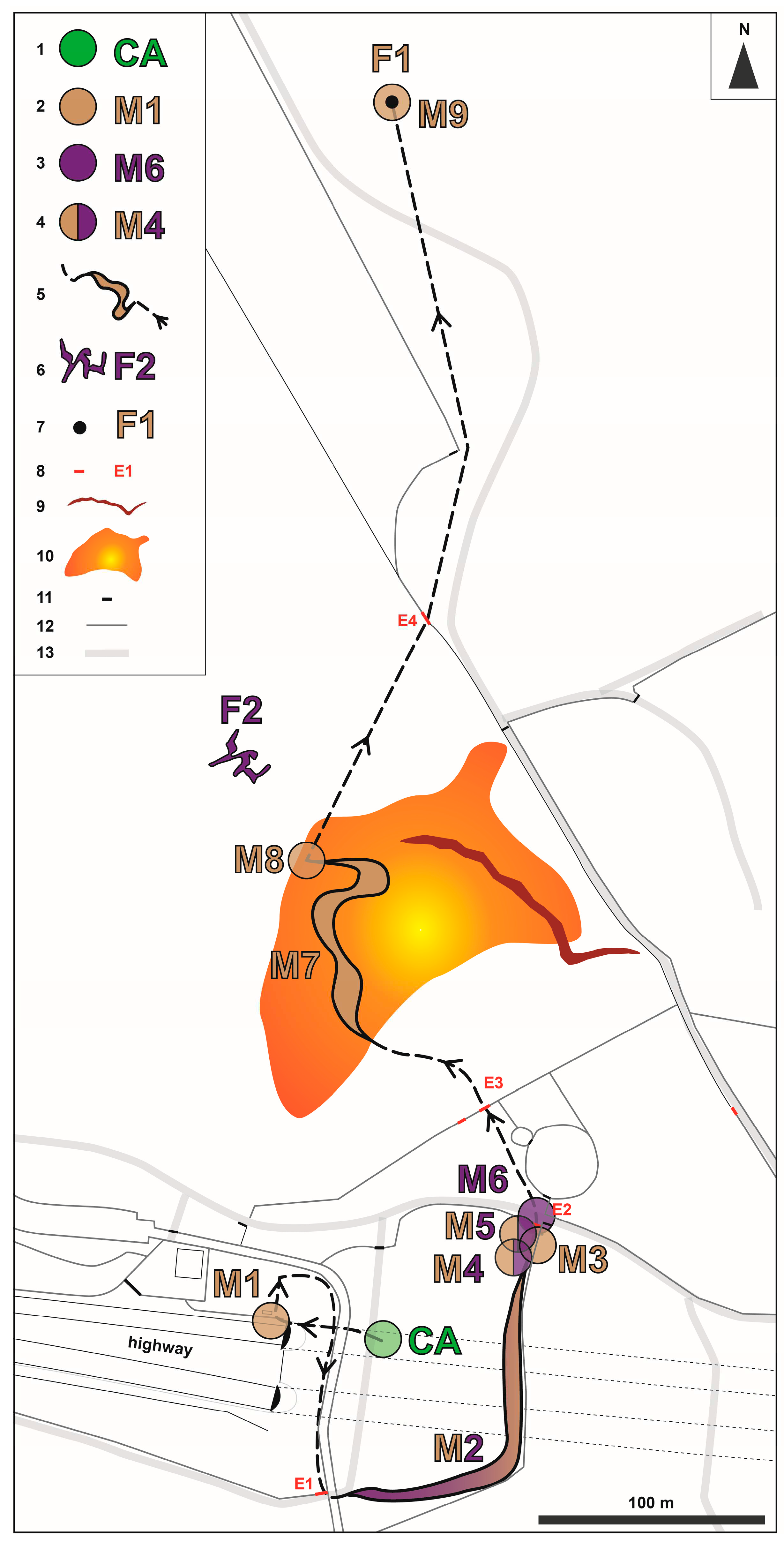
Figure 7.
A) SEM micrograph of calcite traces in a groove on the gold ring of MPF 1. B - SEM micrograph of calcium carbonate fragments scraped from the upper part of the wall. C) X-ray microanalysis (SEM-EDS) compatible with calcium carbonate from the traces (spot 1 in Figure 7A). D) X-ray microanalysis (SEM-EDS) compatible with calcium carbonate from the fragments (spot 12 in Figure 7B) (M1 in Figure 6). Source: Author.
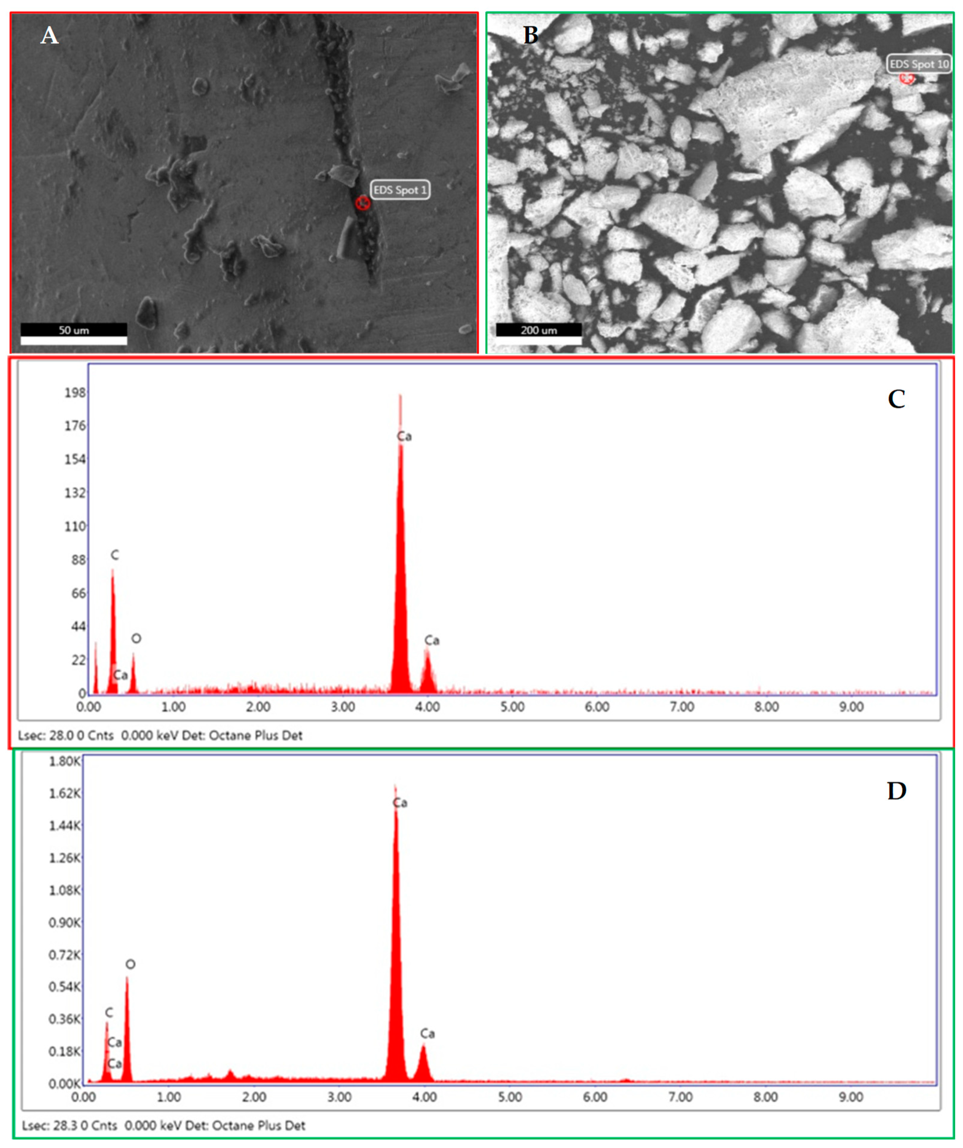
Figure 8.
Microphotographs taken under stereobinocular microscope and reflected light of Cynara cardunculus. A) Thorn from the sock of MPF1. B) Thorn extracted from the sole of the shoe of MPF 2. The hole in the sole is observable near the pointed termination. C) Thorns collected from a plant in the event site. D) Thorns and leaves collected from a plant in the event site (M2 in Figure 6). Plants determined by Fabio Mondello. Source: Author.
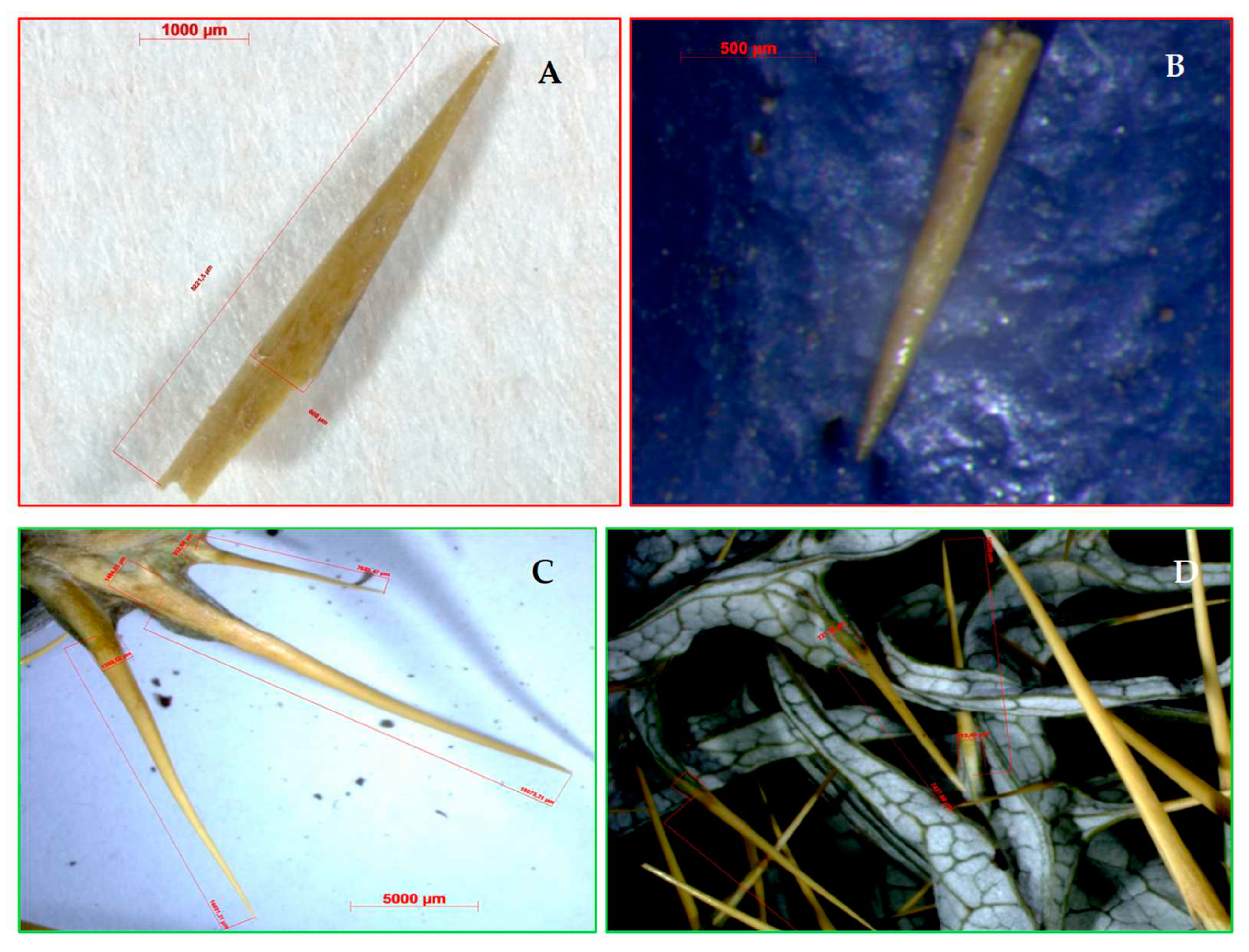
Figure 9.
A) Algae and soil aggregate imbedded in the space among adjacent circular cleats on the sole of the shoes observed under stereobinocular microscope with reflected light. B) Chlorellales or Chlamydomonadales (Chlorophyta) from the aggregate of Figure 9A observed under microscope. C) Puddle with freshwaters in the scene of events. D) Chlorellales or Chlamydomonadales (Chlorophyta) from the puddle with freshwaters of Figure 9C (M3 in Figure 6). Scale bar:100 µm. Algae determined by Marina Morabito. Source: Author.
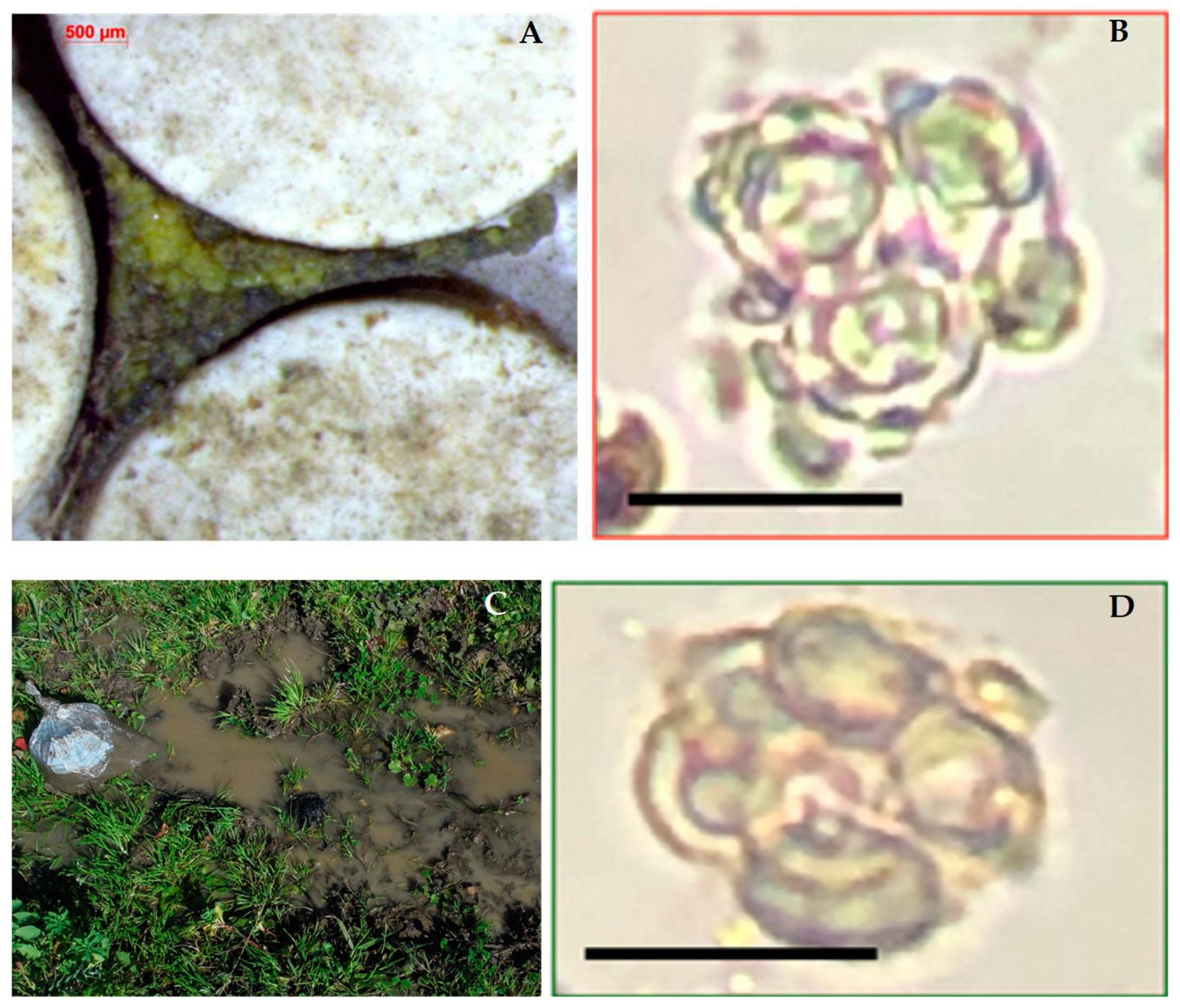
Figure 10.
A) SEM micrograph image of P-rich clayey minerals from the MPF 1’s shoes (unknown sample. B) SEM micrograph image of P-rich clayey soil from a puddle in the event scene. C-D) X-ray microanalyses (SEM-EDS) related to P-rich clayey minerals (spot 3 in Figure 10A) (C) and soil (Figure 10B) (D) (M4 - spot 1 in Figure 6). Source: Author.
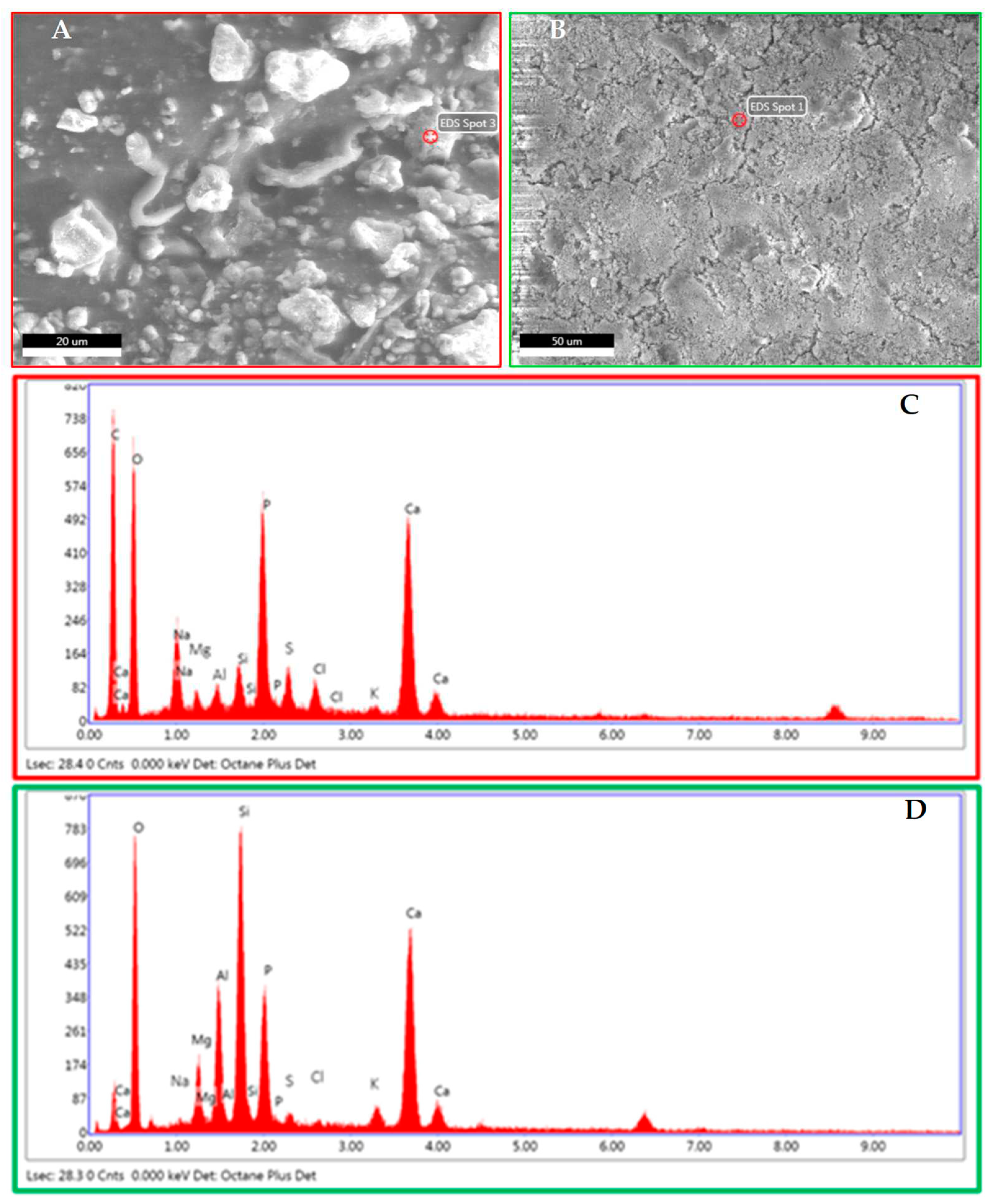
Figure 11.
A) Sandy grains from the MPF 2’s shoes (unknown sample) under stereomicroscope. B) Sandy grains from a soil sample (unknown sample) from the event scene under stereomicroscope. C-D) Comparative analyses (sieve size is expressed in µm). Histograms of the 7 grain types related to the percentages of the sandy grains from the MPF 2’s shoes (in Figure 11A) (C) and the soil (Figure 11B) (D) (M5 in Figure 6). Source: Author.
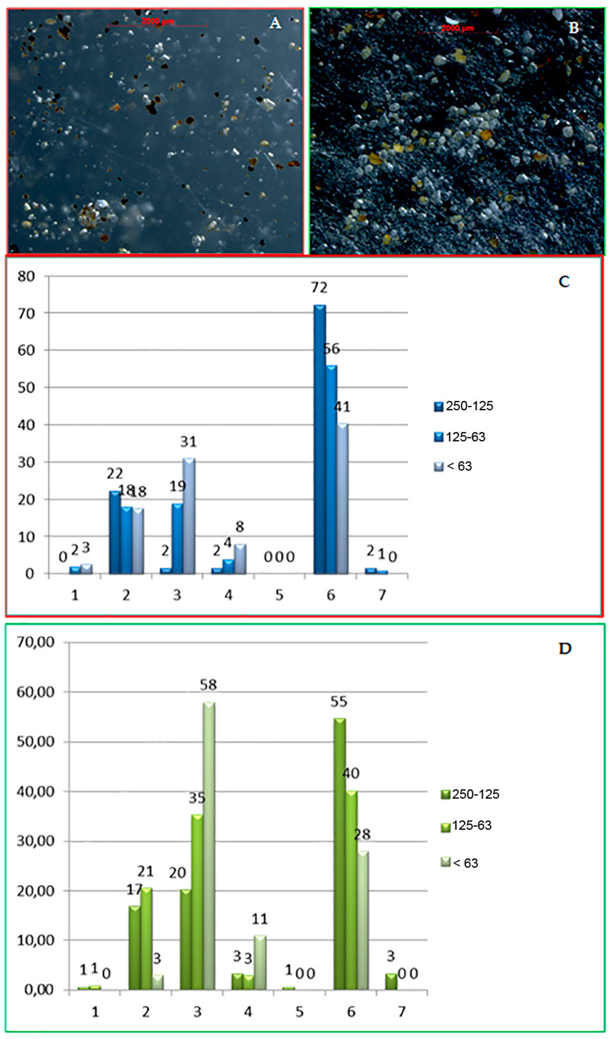
Figure 12.
A) SEM micrograph image of dolomitic clast from the MPF 2’s shoes (unknown sample. B) Microphotograph under stereomicroscope of clast of pinkish dolostone (known sample) from a dirty road in the event scene. C-D) X-ray microanalyses (SEM-EDS) of dolomite minerals from the MPF 2’s shoes (C) and dolostone from the event scene (D) (M6 in Figure 6). Source: Author.
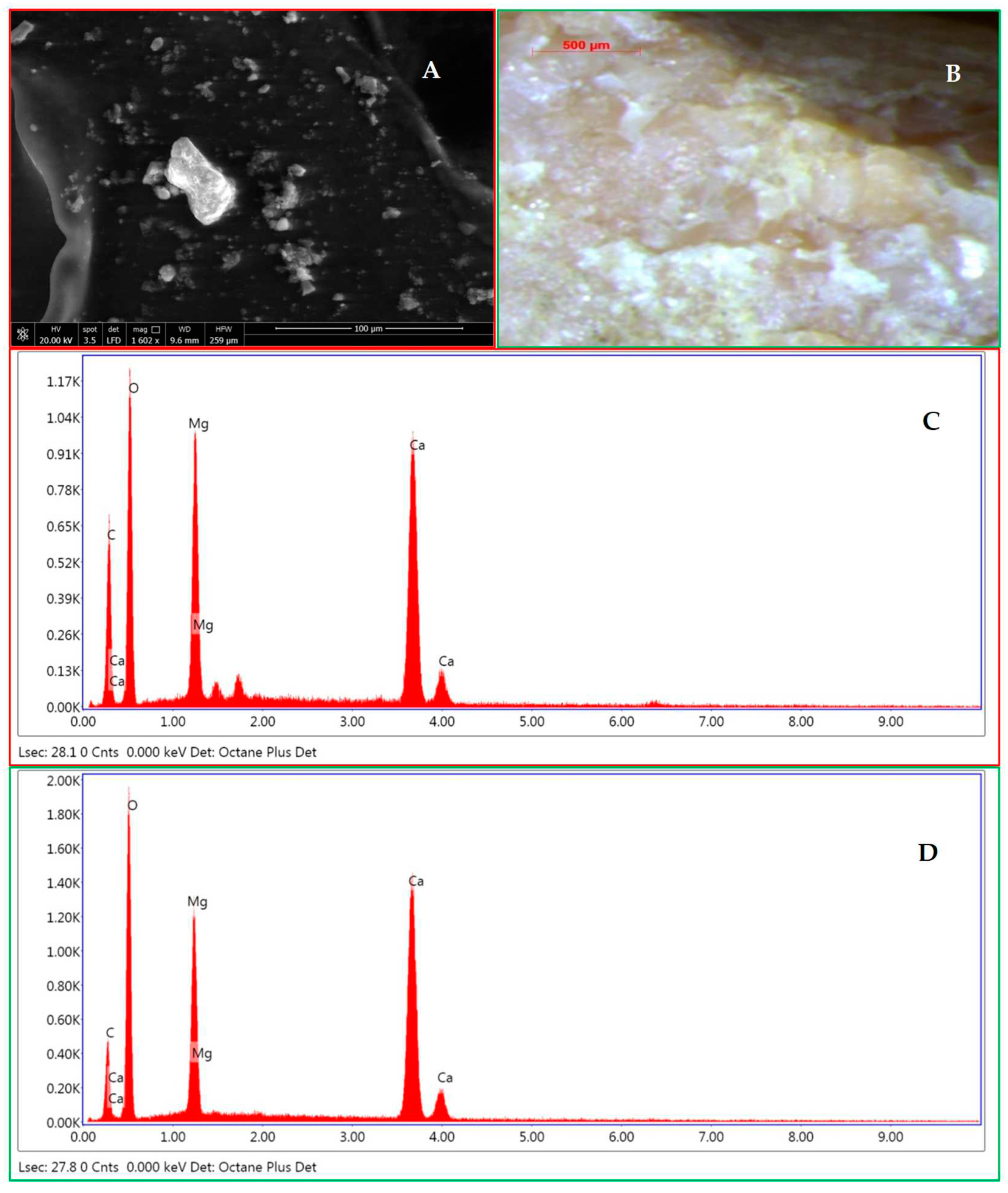
Figure 13.
A) Dirty internal side of the upper part of the MPF 1’s shoes showing geological evidence with a very abundant vegetal component made up of Erica arborea seeds (unknown sample), transferred to the shoes during the walking of MPF 1 in the Sughera woody area with Erica arborea. B) Erica arborea seeds and humus from the victim 1’s shoes (unknown sample), under stereomicroscope. C) Erica arborea seeds separated from a soil sample (known sample) from the Sughera woody area with Erica arborea, under stereomicroscope. D) SEM micrograph of an Erica arborea seed (Figure 13C). E) Seed of Erica arborea, under stereomicroscope (Figure 13D). (M7 in Figure 6). Erica arborea determined by Fabio Mondello and Angelo Troia. Source: Author.
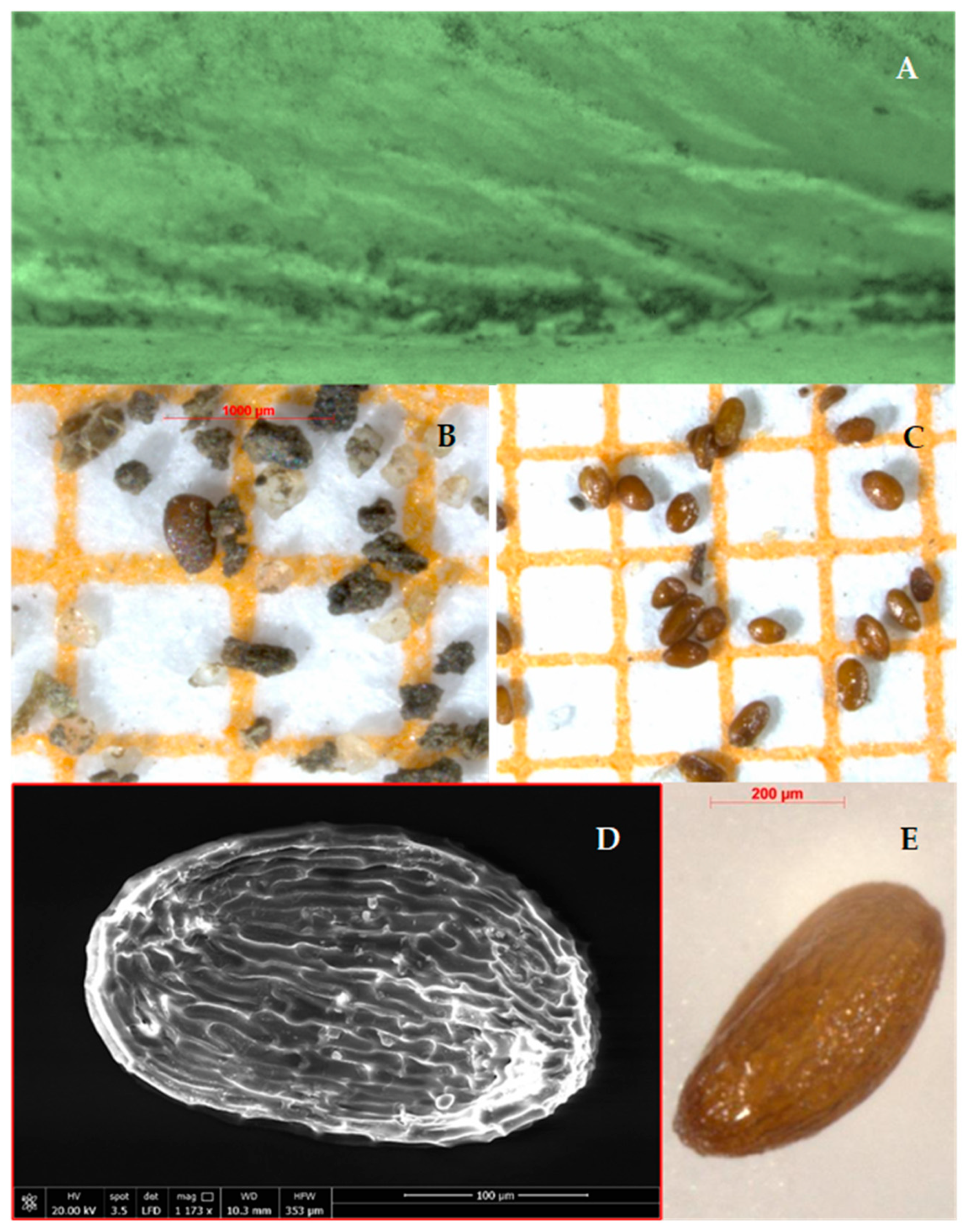
Figure 14.
A) Sandy grains from the MPF 1’s shoes (unknown sample) under stereomicroscope. B) Sandy grains from a soil sample (unknown sample) from the event scene under stereomicroscope. C-D) Comparative analyses (sieve size is expressed in µm). Histograms of the 7 grain types related to the percentages of the sandy grains from the MPF 1’s shoes (in Figure 14A) (C) and the soil (Figure 14B) (D) (M8 in Figure 6). Source: Author.
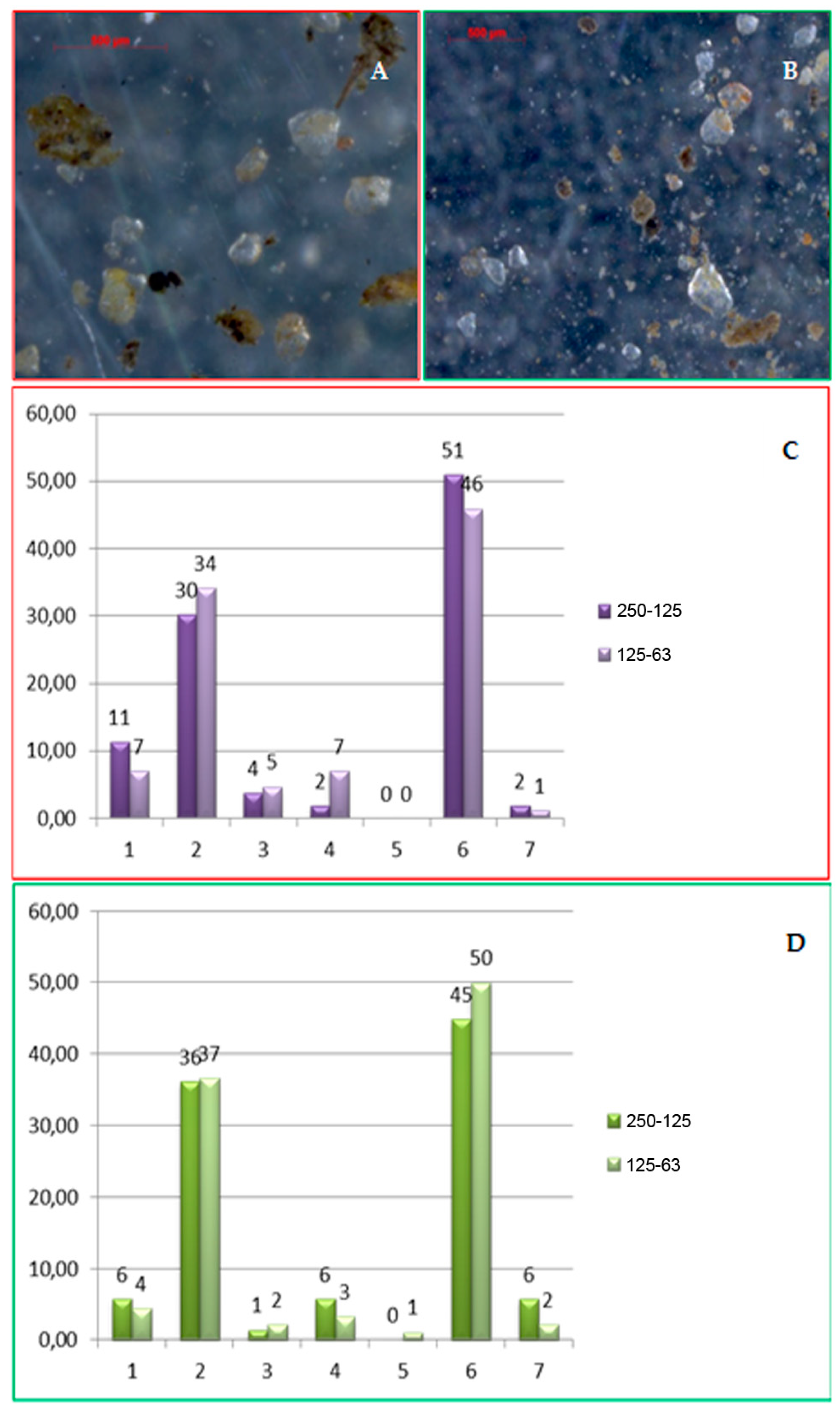
Figure 15.
A) Thorn of Rosa sempervirens L. catch in the lace of the MPF 1’s shoes (in the bottom left). B) Thorn of Rosa sempervirens L. (Figure 15A), under stereomicroscope. C) Twig with a thorn of Rosa sempervirens L. collected from the climbing plant grown up on the infrastructure present in the scene of events (M9 in Figure 6). Plants determined by Fabio Mondello. Source: Author.
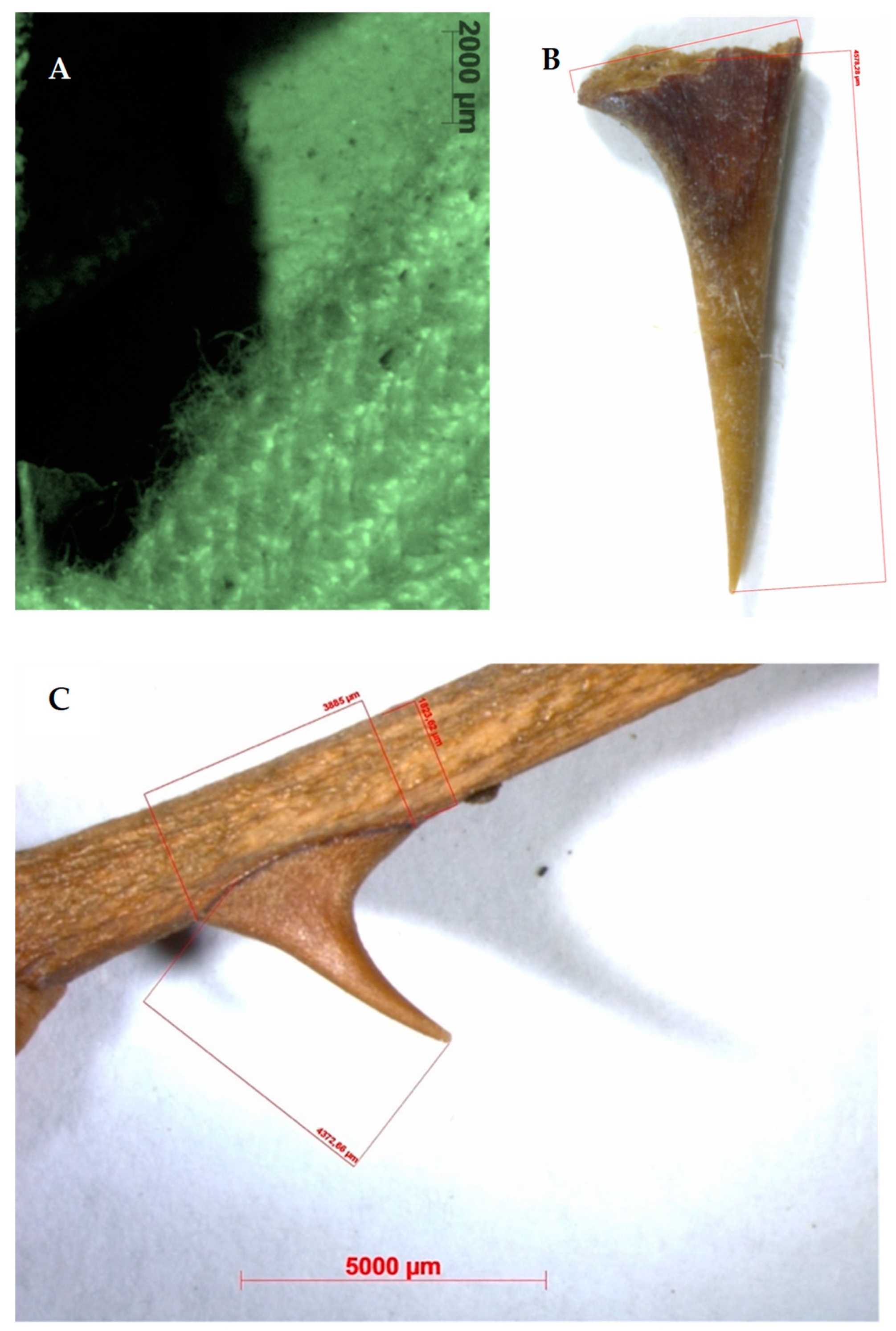

Table 1.
Main methods and techniques usually applied for comparative analyses of forensic geological evidence in laboratory. * Instruments also portable in the field.
Table 1.
Main methods and techniques usually applied for comparative analyses of forensic geological evidence in laboratory. * Instruments also portable in the field.
| Matrices | Methods and Techniques | Analysed Characteristics |
|---|---|---|
| Geological samples | Spectrophotometers / Munsell charts / Computational Methods | Colour |
| Geological samples | Mechanical siever / Laser Diffraction Particle Size Analyzer / Coulter counter / Particle Size Analyzer through automated microscopy and image analysis for measuring particle size and particle shape | Texture (grain size, morphology) |
| Geological samples | Optical Microscopy (OM) by stereo microscope*, in transmitted and reflected light, with tele camera and workstation for image analyses | Texture / Structure |
| Geological samples | Optical Microscopy (OM) by polarizing mi-croscope, in transmitted and reflected light, with tele camera and workstation for image analyses | Mineral composition / Texture / Structure |
| Geological samples | Powder X-Ray Diffractometry (PXRD) | Mineral composition |
| Geological samples | Scanning Electron Microscopy with Energy Dispersion System (SEM-EDS) / Quantitative Evaluation of Minerals by Scanning Electron Microscopy (QUEMSCAN) | Composition / Texture / Structure |
| Geological samples | Scanning Electron Microscopy (SEM) | Composition / Morphology |
| Geological samples (Fossils) | X-Ray Fluorescence (XRF)* | Elemental qualitative determination |
| Geological samples | µ-RAMAN spectroscopy* / FTIR spectroscopy | Molecular qualitative determination |
| Geological samples | Inductively Coupled Plasma-Mass Spectrometry (ICP-MS) / Inductively Coupled Plasma – Optical Emission Spectroscopy (ICP-OES) / Instrumental Neutron Activation Analysis (INAA) | Elemental quantitative determination |
Table 2.
Characterization of the different types of grains identified in the forensic unknown and known samples of the case work.
Table 2.
Characterization of the different types of grains identified in the forensic unknown and known samples of the case work.
| Class ID | Description |
|---|---|
| ID01 | Hyaline grains predominantly rounded and triangular in shape with yellow/orange coating, which gives the grains a hyaline appearance with more or less marked yellow/orange “spots” up to straw yellow / orange / reddish. |
| ID02 | Rounded hyaline grains with evidence of the original crystalline habitus with yellow/orange coating, which gives the grains a hyaline appearance with more or less marked yellow/orange “spots” up to straw yellow/orange/reddish with minor percentage of sub-angular clasts. |
| ID03 | Predominantly rounded and spherical hyaline grains without coating. |
| ID04 | Hyaline grains with rounded tabular crystalline habitus and without coating. |
| ID05 | Rare hyaline grains of smoky gray color and without coating. |
| ID06 | Rounded and spherical hyaline clasts with yellow/orange coating, which gives the clasts a hyaline appearance with more or less marked yellow/orange “spots” up to straw yellow / orange / reddish with a smaller percentage of sub-angular and lamellar grains. |
| ID07 | Opaque grains mainly yellow ocher and fossil forms (mainly benthic foraminifera) with a smaller percentage of opaque brown or light grains. |
Table 3.
Sites selected for linking MPFs to the event sites and reconstructing the route walked by them.
Table 3.
Sites selected for linking MPFs to the event sites and reconstructing the route walked by them.
| MPFs/Victims | Acronyms | Typology of Sites for Linking MPFs to Event Site |
|---|---|---|
| MPF 1 – MPF 2 | CA | Car accident site (Route start point) |
| MPF 1 | M1 | Match point |
| MPF 1 – MPF 2 | M2 | Match linear belt |
| MPF 1 – MPF 2 | E1 | Exit (rudimental wood gate) |
| MPF 1 | M3 | Match point |
| MPF 1 – MPF 2 | M4 | Match point |
| MPF 1 – MPF 2 | M5 | Match point |
| MPF 1 – MPF 2 | E2 | Exit (rudimental wood gate) |
| MPF 2 | M6 | Match point |
| MPF 1 – MPF 2 | E3 | Entry (hole in the barber wire perimeter) |
| MPF 1 | M7 | Match linear belt |
| MPF 1 | M8 | Match point |
| Victim 2 | F2 | Finding site of skeletonized human remains |
| MPF 1 | E4 | Exit (hole in the barber wire perimeter) |
| MPF 1 | M9 | Match point |
| Victim 1 | F1 | Finding site of human remains (Route end point) |
Disclaimer/Publisher’s Note: The statements, opinions and data contained in all publications are solely those of the individual author(s) and contributor(s) and not of MDPI and/or the editor(s). MDPI and/or the editor(s) disclaim responsibility for any injury to people or property resulting from any ideas, methods, instructions or products referred to in the content. |
© 2023 by the authors. Licensee MDPI, Basel, Switzerland. This article is an open access article distributed under the terms and conditions of the Creative Commons Attribution (CC BY) license (http://creativecommons.org/licenses/by/4.0/).
Copyright: This open access article is published under a Creative Commons CC BY 4.0 license, which permit the free download, distribution, and reuse, provided that the author and preprint are cited in any reuse.


