Submitted:
02 October 2023
Posted:
03 October 2023
You are already at the latest version
Abstract
Pathogenic microorganisms can cause a range of infectious diseases and are often treated with antimicrobial agents. The rise of antimicrobial resistance caused by inappropriate use of these agents in various settings has become a global health threat. There is, therefore, a need to find new and effective agents to combat infections caused by resistant pathogens. Nanotechnology has seen significant growth and development in recent years and has a wide range of applications in various fields, including health, agriculture, and industry. This novel area offers the potential for the synthesis of nanoparticles (NPs) with antimicrobial activity, such as iron oxide nanoparticles (IONPs). The use of IONPs is a promising way to overcome antimicrobial resistance or pathogenicity because of their ability to interact with several biological molecules and to inhibit microbial growth. This review focuses on the most common green synthesis methods for producing IONPs using bacteria, fungi, plants, and organic waste, as well as the most common methods for the characterization of these IONPs. Finally, it summarizes their application as promising antibacterial, antifungal, antiparasitic, and antiviral agents.
Keywords:
antimicrobial resistance
; green synthesis
; IONPs
; antibacterial activity
; antifungal activity
; antiparasitic
; antiviral activity
1. Introduction
Microbial agents such as fungi, bacteria, parasites, and viruses cause various infectious diseases and can be treated with antimicrobial agents. The main concern in treating infectious diseases is that many pathogenic microorganisms have developed resistance against antimicrobial agents, for example, antibiotics [1]. The development of antibiotic resistance is the result of the inappropriate use of antibiotics in community, clinical, and agricultural settings. Consequently, multidrug-resistant bacteria have become a global health threat [2]. Therefore, there is an urgent need to find new effective agents to overcome drug resistance. In this regard, nanotechnology reveals new opportunities for the biosynthesis of nanoparticles with antibacterial properties to solve these challenges [3]. Nanoparticles are materials that show uniqueness in size (generally 1 to 100 nm), structure, physicochemical, electric, magnetic, thermal, mechanical, catalytic, optical scattering properties, and shape [4]. Green synthesis creates metallic nanoparticles through a bottom-up approach that includes a chemical reduction in which a natural product extract replaces a chemical-reducing agent [2]. Iron oxide nanoparticles (IONPs) are defined as “Nanozymes” due to their inherent enzyme-like activities and catalytic properties that are comparable to those of different oxidases such as peroxidases, catalase, sulfite oxidase, and superoxide dismutase [5]. Another feature of these particles is their potential to bind different biological molecules like peptides, enzymes, nucleic acids, lipids, fatty acids, and various metabolites. These nanoparticles have applications in optics, mechanics, biotechnology, engineering, remediation, microbiology, environmental medicine, and electronics, among others [6,7]. This review focuses on green methods for synthesizing iron oxide nanoparticles using bacteria, fungi, yeasts, plant extract, and organic waste. In addition, IONP's prospective antimicrobial applications are discussed in detail. This review will provide compelling information for researchers in the field of nanobiotechnology.
2. Green synthesis of iron oxide nanoparticles
2.1. Synthesis of iron oxide nanoparticles using microorganisms
The synthesis of iron oxide nanoparticles (IONPs) using microorganisms has several advantages over other methods due to the relative abundance of microorganisms, the generation of less toxic by-products for biomedical purposes, and lower cost [8]. Several microorganisms have the ability to accumulate and detoxify metals, and this property can be exploited to convert iron ions from metal salts into nanoparticles [9] using a variety of enzymes, cofactors, proteins, and secondary metabolites of the microorganism [9,10]. These molecules can act not only as reducing agents but also play an important role as stabilizers and protectors of nanoparticles [9]. Microbial proteins containing functional groups, such as hydroxyl, thiol, carboxylic acid, and amine, have been demonstrated to play a role in the stabilization of metal nanoparticles. These functional groups serve as binding sites for metal ions, which are subsequently reduced to form nanoparticles within the cell wall or the periplasmic space [11].
The synthesis of nanoparticles using microorganisms such as bacteria, fungi, yeasts, and algae can be carried out by intracellular or extracellular mechanisms [10]. The extracellular mechanism consists of the reduction of metal ions, such as iron, by proteins, enzymes, or components of the cell wall of microorganisms [12]. These components work as reducing agents and stabilizers and prevent the agglomeration of iron oxide nanoparticles [11]. For this type of synthesis, the microorganisms are grown under optimal conditions, the supernatant is collected by centrifugation and mixed with an aqueous solution of the metal salt, and the presence of nanoparticles is evidenced by the change in color of the medium [9]. Metal nanoparticles can also be formed when metal ions are trapped on the cell surface by electrostatic interaction and are reduced by enzyme action [11].
Intracellular mechanisms involve enzymatic reduction of metal oxide ions through electrostatic binding of metal ions to -COOH groups of the microbial cell wall, metal ions diffuse into the cell and interact with enzymes to form metal oxide nanoparticles [13,14]. For this type of synthesis, the microorganisms are grown under optimal conditions, then the biomass is washed with sterile water and incubated with a solution of metal salts until a color change indicates the nanoparticle synthesis [9] (Figure 1).
The reduction of metal ions is under the control of several factors, such as functional groups in the cell wall, type of strain, pH, temperature, culture medium, and metal salt concentration. These factors directly or indirectly affect the size, shape, composition, and performance of nanoparticles [15].
2.2. Synthesis of iron oxide nanoparticles using bacteria
The synthesis of iron oxide nanoparticles generally begins with the selection and isolation of bacteria, this includes culturing the strain on specific media such as BHI agar or nutrient agar, followed by incubation under controlled conditions at 35–37 °C for 24–48 h. Additionally, conditions such as carbon dioxide atmosphere may be required. The obtained individual colonies are transferred to the nutrient medium, such as Luria Bertani or nutrient broth, followed by incubation and centrifugation [16]. The supernatant (extracellular method) or the cell pellet (intracellular method) should be taken depending on the synthesis mechanism. The supernatant or sediment suspension is combined with the precursor salts, such as FeCl3.6H2O or Fe2O3, in the desired volume and concentration. The reaction conditions are optimized by adjusting the pH to the preferred range (5–9), and the mixture is then incubated at different temperatures depending on the strain of bacteria used. After incubation, the color of the medium changes, for example, from dark red to dark brown, indicating the production of IONP’s nanoparticles [17]. When using the intracellular approach, an additional step is required to remove the nanoparticles from inside the cell; for this, a heat shock must be performed by resuspending the cells in buffer, centrifuging, and then using liquid nitrogen followed by a steam bath at 37 °C. The supernatant is centrifuged and collected as a cytoplasmic extract [9,18]. Finally, the synthesized IONPs are dried and characterized. The synthesis of IONPs via different bacterial species is summarized in Table 1.
2.3. Synthesis of iron oxide nanoparticles using fungi
The production of iron oxide nanoparticles via fungal synthesis is considered an attractive option due to its simplicity of scaling, the low cost of the materials necessary for growth, the capacity to produce high amounts of biomass even at an industrial scale, and the low toxicity of the residue [41,42]. In addition, fungal species have higher metal tolerance and bioaccumulation properties, facilitating the synthesis of metal nanoparticles [43]. Fungi are known to secrete large amounts of proteins, enzymes (i.e reductase enzymes [44]), and metabolites that can convert metal salts into various metal nanoparticles [13,45]. Researchers discovered that chitin, a key component of the fungal wall system, is involved in the formation of heavy metal complexes, which are linked to the synthesis of nanoparticles [46]. However, the mechanisms behind metal nanoparticle synthesis using fungi still need to be fully understood.
To optimize the synthesis, some factors must be considered, including the microorganism strain, growth parameters, sample preparation technique, metallic precursor, metallic salt concentration, pH, and reaction temperature [47]. Metal nanoparticles may be synthesized using fungi in a variety of ways, including employing fungal biomass, cell-free filtrate (CFF) from fungal culture, or fungal cell filtrate (FCF) [10]. The use of mycelium is one of the simplest approaches; this process involves culturing the selected fungus strain in an appropriate medium and then separating the mycelium by centrifugation. The mycelium is combined with a metal precursor salt solution and incubated for 4–5 days at 28 °C. The solution is then filtered to produce a colloidal suspension containing the synthesized nanoparticles [22,48]. Nanoparticles can also be synthesized using the cell-free filtrate, which is obtained by growing fungi in a suitable medium and separating the mycelium by filtration. After washing and resuspending the fungal biomass in distilled water, it is incubated on a rotary shaker for 1–5 days, and the filtrate is collected by separating the mycelium with filter paper. Finally, the cell-free filtrate is combined with a solution of iron precursor salts such as ferric chloride and incubated for a few hours on the rotary shaker to achieve IONPs synthesis [45]. For the IONPs synthesis method based on fungal cell filtrate (FFC), the fungal strain is cultivated in the appropriate medium, after which the culture medium is filtered, and the filtrate obtained is centrifuged and filtered again to obtain the final FFC. Finally, a FeCl3 solution is stirred at room temperature for 5 min. An immediate visual change in color reveals IONPs synthesis [49].
On the other hand, relatively little is known about IONPs biosynthesis from yeast. This type of fungus has a widely varied metabolic and proteomic profile, which means it can produce a wide variety of compounds that have the potential to act as reducing agents [50]. However, more research is required to evaluate its potential and biosynthetic mechanism. Recent reports on the synthesis of IONPs by yeast and fungi are shown in Table 2.
2.4. Synthesis of iron oxide nanoparticles using algae
Algae (microalgae and macroalgae) are commonly used to produce different metallic nanoparticles, and this has become an environmentally friendly option because it is a clean, non-toxic, and sustainable process with the potential to produce a wide variety of sizes, shapes, compositions, and physical and chemical properties [56]. Glycoproteins and different functional groups, such as carbonyl, hydroxyl, and carboxyl, as well as proteins, enzymes, alkaloids, carotenoids, vitamins, terpenes, and cell wall polysaccharides [57,58], are just a few of the biomolecules found in algae. Furthermore, algae can also accumulate heavy metals and produce secondary metabolites with anti-biological fouling effects [56,59]. These characteristics, together with the components they produce, are critical in the reduction, protection, manufacture, and stabilization of [10] nanoparticles [60]. For example, an aqueous extract of Sargassum muticum was used to synthesize cubic iron oxide nanoparticles from FeCl3 at a temperature of 25 °C, and it was determined that the polysaccharides in the algae extract were the main factor in the reduction of FeCl3 to Fe3O4 nanoparticles with an average size of 18 ± 4 nm [58].
To synthesize nanoparticles from algae, the samples are washed with distilled water and then dried for 72 h under light [58]. The dried samples are then powdered, freeze-dried, weighed, and boiled by mixing the powder with the necessary volume of distilled water. To produce the extract, the boiled extract is subjected to a filtration step. The synthesis of the IONPs begins by combining the extract with a known amount of iron salts, such as FeCl3 [58,61]. The color change in the media indicates that nanoparticle nucleation has occurred [56,62]. This process is facilitated by the biomolecules found in algae extract, which facilitate the aggregation and self-assembly of nucleonic particles. During this process, the most energetically favorable and stable particle shapes are generated, with cubes, hexagons, pentagons, bars, spheres, triangles, and wires being the most prevalent [63]. Table 3 summarizes information on nanoparticles obtained from various algae extracts.
2.5. Synthesis of iron oxide nanoparticles using plants
Because microorganism-based synthesis requires long incubation periods, extensive research has been conducted on plant-based synthesis of iron oxide nanoparticles since plants, on the other hand, reduce metal salts in a shorter time [9]. Plant extracts used for the biosynthesis of nanoparticles can be obtained from the leaves, flowers, bark, roots, fruits, and stems [10]. Bioactive compounds of these extracts, including mainly tannins, phenols, alkaloids, saponins, organic acids, flavonoids, and vitamins, work as reducing agents for metal salts and stabilizers of nanoparticles [10,71].
Depending on the iron precursor used to form the Fe0 metal, electron-rich biomolecules containing hydroxyl groups (-OH) have the ability to effectively reduce iron ions from the divalent or trivalent oxidation state (Fe2+ or Fe3+) [72,73]. The characteristics of the synthesized nanoparticles are mostly determined by discrete parameters such as plant extract type, volume ratio of the extract, metal salt solutions, and reaction variables such as pH, temperature, and incubation time [72,74].
To obtain the plant extract, the first step is to collect the desired part of the plant of interest, which is then washed with distilled water to remove any contaminants, dried at room temperature, and then ground into a fine powder to provide a good surface for extraction [73]. The active compounds of this powder are extracted by mixing it with the appropriate solvent, either hot or cold; the most frequently employed solvents are water, methanol, ethanol, or aqueous dilutions of these [75,76]. Finally, it is filtered, and the clear solution is mixed with the iron precursor salts (iron chloride, iron nitrate, or iron sulfate) to produce IONPs. In this case, the chemical components in the extract also stabilize the nanoparticles, and the change in color of the solution, which depends on the kind of salt used, intensifies as the nanoparticles are synthesized [7,73,77]. As shown in Table 4, various parts of plants have been studied for the environmentally safe synthesis of iron oxide nanoparticles.
2.6. Green synthesis of iron oxide nanoparticles using from biological waste products
Agro-wastes are abundantly produced during the processing of agricultural products and are frequently discarded into the environment, causing pollution [114]. Agro-waste has been found to be rich in biomolecules such as flavonoids, phenolics, and proteins that can serve as bioreductant agents in the green synthesis of diverse metallic nanoparticles [115]. A new effort to expand the use of agro-wastes as sources of biomolecules in green nanotechnology have received considerable attention, and various agro-wastes have been documented for their relevance in nanobiotechnology [115]. As a result, a range of metallic nanoparticles have been successfully synthesized from agro-industrial wastes, and extracts from these residues have been employed as reducing and stabilizing agents in the majority of cases [116]. The nanoparticles produced in this way have a wide range of activities, including antimicrobial, antioxidant, anticorrosive, and cytotoxic against cancer cells, among others [116,117,118,119]. In general, extracts containing active biomolecules that promote the formation of nanoparticles can be prepared using a simple hot water extraction process of dried and ground agro-waste materials [115]. Information on the nanoparticles obtained from biological waste is highlighted in Table 5.
3. Characterization methods
The green synthesis methods of metallic nanoparticles are continually being improved and optimized to achieve comparable results (size and morphology) to physical and chemical methods. Usually, a change in the color of the reaction mixture indicates the formation of nanoparticles; however, this does not guarantee their formation, so additional processes must be used to identify the particles formed. Continuous UV spectra of the reaction mixture are one of the most frequently applied methods to confirm the formation of nanoparticles [92,132,133] Currently, a variety of characterization methods are useful for evaluating the properties and uses of synthesized nanoparticles, as well as optimizing synthesis procedures [134]. However, no single instrument can characterize all material properties. As a result, numerous complementary and confirmatory techniques for nanoparticle characterization should be applied (Table 6).
The most common nanoparticle parameters studied include size, shape, charge, and surface topology [134]. However, characterizing nanoparticles remains difficult due to the sample preparation procedure, and the characterization technique utilized is directly dependent on the properties required [135].
One of the most important parameters to evaluate is particle size and size distribution, which correlates to the particle's external dimensions. Because this property can determine other properties and the behavior of the final product [136,137], it is important for biological and biomedical applications. This property frequently differs depending on the method employed to evaluate it, which can be attributable to a variety of factors. For example, different methods evaluate different aspects of nanoparticle dimensions, or the method can induce a change in the effective particle size [138]. Among the most common methods for determining the size of iron oxide nanoparticles are: Transmission Electron Microscopy (TEM) [119,139,140,141], Scanning Electron Microscopes (SEM) [71,119,142,143], X-ray Diffraction (XRD) [142,144,145,146], Dynamic Light Scattering (DLS) [147,148,149], High-resolution Transmission Electron Microscopy (HRTEM) [150,151,152], Scanning Transmission Electron Microscopy (STEM) [92].
The physical appearance of a particle (morphology), which comprises shape and surface topography, is important to the behavior of iron oxide nanoparticles. Morphology affects different properties like dispersion, functionality, and toxicity. This property, like the size, is evaluated through direct viewing of the particles, so commonly, electron microscopy techniques are utilized. Among the various forms of iron oxide nanoparticles that have been reported are: round (SEM) [153], spherical (SEM) [142,143], (STEM) [92], (TEM) [146], spheroidal (HRTEM) [152], cubic (SEM) [143], rectangular and triangular (HRTEM) [151], rhombohedral [119], and needle-like (FESEM and HRTEM) [154].
Another important property to determine is the chemical composition of iron oxide nanoparticles. It can be characterized both in terms of surface and bulk chemistry and can be measured at the ensemble or single nanoparticle level. It is mostly determined using spectroscopic techniques, which may be paired with microscopy to attain single-particle level resolution, with the most often used methods being XRD [140,152,155,156], EDS [92,155,157] and EDX [144,158,159,160]. Because the nanoparticle's external layer interacts with the surrounding environment, determining its composition (surface chemistry) is also important and can be obtained using different techniques like Auger electron spectroscopy [161,162], X-ray photoelectron spectroscopy [163,164,165], or inductively coupled plasma mass spectrometry [166].
Along with the chemical composition, another property of importance in the study of IONPs is the crystalline structure of the nanoparticles. In general, XRD is used to determine the three-dimensional arrangement of the atoms in nanoparticles as well as the different phases. However, it has been found that nanoparticles smaller than 5 nm have an impact on the XRD-observed patterns [167]. SAED [168], which is being used increasingly commonly, is a method for a more accurate representation of the crystalline structure of iron oxide nanoparticles [146,149,155].
The charge of nanoparticles in suspension is also an important property to consider. The surface charge is the charge formed on the surface of the nanoparticle as a result of proton adsorption or desorption at hydroxylated sites, and due to the difficulties of directly measuring this charge, the particle's zeta potential is frequently determined. The zeta potential is the difference in potential between the stationary layer of the dispersion medium attached to the dispersed particle and the mobile dispersion medium [168] and can be measured by titration [169] or electrophoresis [92,170,171]. It is important because it is used as an indicator of colloidal stability, which is a critical parameter for many emulsions or dispersions; additionally, it has been used to investigate the interaction of iron oxide nanoparticles with various biological systems, including human cells [172], bacteria [173], and proteins [174].
Nanoparticles' magnetic behavior differs from that of bulk materials, as do other properties. Furthermore, the synthesis method, size, and shape of IONPs all affect the magnetic properties of the nanoparticles. When the particle size is between nano and micro, it exhibits superparamagnetism, resulting in low remanent magnetization and a coercive field. As a result, the nanoparticles can interact with an external magnetic field, but in the absence of this field, the particles exhibit no magnetism. Because of this property, superparamagnetic iron oxide nanoparticles (SPIONs) have a high potential for use in biomedical applications, including controlled drug delivery, chemotherapy, drug formulations, hyperthermia treatment, and radioimmunotherapy, among others [136]. Magnetometry techniques such as VSM (vibrating sample magnetometer) [80,148,158] and SQUID (superconducting quantum interference device) magnetometry [175] are used to determine the net magnetization of nanoparticles.
3.1. UV–VIS spectroscopy
UV–vis spectroscopy, which is based on the Beer-Lambert law, is a common method for investigating the optical and structural properties of nanomaterials, such as shape, size, agglomeration state, concentration, refractive index, and colloidal stability [176,177,178,179,180]. This technique, as previously stated, can be used to confirm the formation of IONPs. Due to their surface plasmon resonance (the resonance of the metal's conduction electrons), metal nanoparticles with a diameter of less than 100 nm efficiently scatter optical light, however, as the particle size increases, the absorption spectra can change, shifting toward longer wavelengths. The nanoparticle's composition, environment, form, and size affect the plasmon resonance's bandwidth, magnitude, and wavelength peak [181]. Greenly synthesized iron oxide nanoparticles typically have absorption spectra with peaks in the 290–370 nm region and an estimated energy band gap from 2.9 to 4.22 eV [82,182,183,184].
3.2. TEM and HRTEM
Transmission electron microscopy (TEM) is a characterization technique used to study the dynamics of physical and chemical processes of the nanostructure at an atomic resolution [185]. An electron beam interacts with the sample, and the transmitted electrons are used to form the image by magnifying and focusing them using an objective lens. The interaction of the electron beam with the sample in the TEM image results in diffraction processes as opposed to absorption processes, which occur in the light microscopic image. Because the sample will be exposed to a high vacuum during the analysis, it must be prepared to endure both the electron beam and the high vacuum chamber [186]. The difference in electron densities of the structures, the density and thickness of the sample, the objective aperture´s diameter, and the energy of the electrons all influence TEM images [187]. Additionally, the contrast image can be generated using either unscattered electrons (light field image) or deflected electrons (dark field image). This technique is widely used to characterize the morphology of nanostructures, such as particle size [188,189], agglomeration state [107], electronic state, and chemical composition [190]. Furthermore, depending on the sample, the electron energies for the beam are generally 100 keV for conventional images and 1 MeV for high-resolution imaging [186]. High-resolution transmission electron microscopy (HRTEM) is a TEM imaging mode that provides atomic-level imaging of a nanoparticle's crystalline structure. With this level of detail, it is feasible to image crystal structures, crystal defects, and individual atoms [181].
3.3. SEM
Scanning Electron Microscopy (SEM) is a non-destructive analytical method used for surface and microstructural analysis that also uses an electron beam, typically ranging from 1 to 30 keV, that scans the surface of the sample. [186,191]. This technique is also useful for determining the purity, homogeneity, and degree of dispersion [192]. When the electron beam strikes a nanoparticle, it emits X-rays as well as three types of electrons: backscattered (or primary) electrons, secondary electrons, and Auger electrons. SEM makes use of both primary and secondary electrons, with the latter being used to generate high-resolution images that reveal features as small as 1-5 nm [192]. An advantage of SEM characterization is that it also helps to study the surface characteristics of the sample of IONPs, given the three-dimensional appearance of the surface structure [100].
Nanoparticles do not require pretreatment since metals conduct electricity when struck by an electron beam, whereas non-metallic materials do. To do this, they are often coated with gold to create a thin coating of conductive material. In standard SEM, however, water molecules evaporate in a vacuum, producing an impediment for the electron beams and obscuring image clarity; hence water removal is important.
3.4. EDX
Energy-dispersive X-ray spectroscopy (EDX) is a technique for determining the purity and elemental composition of synthesized nanoparticles [193]. As previously stated, when an electron beam hits a nanoparticle, X-rays are released along with the three types of electrons, and the energy of the generated X-rays depends on the material under investigation, hence EDX is a technique used in combination with SEM. An image of each element in the sample may be obtained by moving the electron beam over the material, giving an overall mapping of the sample by examining near and at the surface elements and estimating its proportion at different points [181].
3.5. XRD
X-ray diffraction (XRD) is a non-destructive technique used to analyze the crystallographic structure of a material. It can also be used to determine element proportions, the position of atoms in the unit cell, unit cell characteristics, and the number of atoms in the unit cell [194]. The interaction of the X-ray beam with the atomic planes results in partial beam transmission, with the remainder absorbed, refracted, scattered, and diffracted by the sample [181]. High-resolution powder X-ray diffraction (HRXRD) can be used to obtain finer information about the structure of nanoparticles, such as ion spacing and bond length of related substances in single crystal cells, and it is one of the most widely used techniques for determining crystalline perfection and defect studies in single crystals [195], as well as detailed studies of structure and phase transformation in materials [196].
The lattice and structural characteristics may be determined by measuring the diffraction angle obtained when an X-ray beam is incident on the nanoparticles. For example, the crystal size may be determined by applying the original Scherrer equation or a modified version (1).
L= k/βcosθ or lnβ=ln k/L+ln 1/cosθ
Where L = average crystallite size, k = constant related to shape (0.94), λ = wavelength of X-ray, β = peak width of the diffraction peak profile at half maximum height, and θ = Bragg’s angle [197].
3.6. DLS
Dynamic light scattering (DLS), also known as photon correlation spectroscopy or quasi-elastic light scattering, is a frequently employed technique for determining the size distribution profile of NPs in colloidal solutions in the nano- and submicrometer ranges [134]. When a monochromatic laser beam strikes a suspension of tiny particles, the light scatters in all directions, depending on the size and shape of the nanoparticles [198]. Given that the nanoparticles in suspension are in continuous motion due to Brownian motion, the scattering intensity changes over time and offers information on particle size, allowing the hydrodynamic diameter of NPs in solution to be determined. The hydrodynamic diameter is the diameter of the NPs and solvent molecules that diffuse at the same rate as the colloid [199,200]. DLS can also measure the stability of nanoparticles over time, which is useful for understanding how they could behave in different environments and applications [201].
4. Antimicrobial properties of green-mediated synthesis iron oxide nanoparticles
4.1. Antibacterial activity.
Considering the emergence of antibiotic-resistant strains as the leading public health threat worldwide, the search for novel molecules with antibacterial potential is of particular interest. Free iron ions and various metal and metal oxide NPs like silver, ZnO, Al2O3, and TiO2 have been screened as bactericidal or antibacterial agents [202]. However, the main concern when using nanoparticles with antibacterial properties is their high toxicity, which has been reported for ZnO [63, 64]; thus, a nanoparticle that is biologically active against bacteria and biocompatible is needed [61,62]. In this context, IONPs have recently emerged as an alternative due to their bactericide properties and biocompatibility in vivo and in vitro. Despite the potential of nanoparticles as antimicrobial agents, some risks have been reported concerning human health and the environment. In contrast to this, IONPs have been shown to be safe and do not cause toxicity in mammalian cell cultures [203,204], a highly desired characteristic to use in biomedical, clinical, and pharmaceutical applications. The most relevant IONPs with biological activity are Fe2O3, magnetite Fe3O4, and limonite Fe2O3*H2O [205].
4.1.1. Mechanisms of Antibacterial IONPs Activity.
The primary mechanism by which metal oxide nanoparticles exhibit bactericidal activity is through the regeneration of reactive oxygen species (ROS) [206,207]. ROS species, such a as superoxide radicals (O2-), hydroxyl radicals (OH-), hydrogen peroxide (H
2
O
2
), and singlet oxygen (
1
O
2
) are involved in this process. It has been proposed that the combined interaction between IONPs and ROS species might be responsible for the bactericidal activity [208]. Furthermore, the interaction of Fe
2+
ions and ROS species leads to the formation of free hydroxyl radicals and hydrogen peroxide, ultimately resulting in bacterial death. Various mechanisms of bactericidal action, which have recently been reported, are described below [209].
4.1.2. Membrane disruption
As mentioned, iron oxide nanoparticles can interact with bacterial cell membranes via electrostatic interactions, producing harmful oxidative stress in the bacterium by generating free radicals [210]. Additionally, intracellular oxygen in aerobic bacteria oxidizes zero-valent iron, causing irreversible oxidative damage to the cell. Moreover, because of their small size, IONPs can enter the cell and cause physical damage to the cell wall, limiting cellular growth, replication, and even communication [206].
In E. coli, electron microscope analysis revealed that Fe
2
O
3
nanoparticles bind to the cell wall and even penetrate into the cytoplasm, where they accumulate, causing vacuole formation and cell wall disruption [211,212]. Fe
3
O
4
nanoparticles bind to the formate hydrogenlyase complex (FHL) found in Gram-negative bacteria cell walls, generating a gradient in the inner membrane that has a strong antimicrobial effect on these bacteria [213]. Another mechanism that causes membrane disruption is disturbing the activity of F0/F1-ATPase, which reduces the membrane redox potential and, as a result, the H
+
rate [213]. The size and shape of IONPs have been suggested as key factors in elucidating their bactericidal activity [214].
4.1.3. Protein and DNA damage
ROS damage DNA molecules through their genotoxic action [215]. A decrease in the activity of antioxidant system enzymes (SOD, catalase, and glutathione reductase) might cause an increase in ROS concentration [67]. Metal ions are able to bind the mercapto (-SH), amino (–NH), and carboxyl (–COOH) groups of proteins, including enzymes, causing inactivation or partial inhibition [214]. IONPs can reduce the expression of antibiotic-resistance genes (ARGs) in antibiotic-resistant bacteria found in operating rooms [205]. The capacity of nanoparticles with small sizes to inhibit DNA replication by inactivating topoisomerase has also been reported [216]. ROS interacts with thiol groups in membrane proteins, generating oxidative stress, which denatures the protein, resulting in membrane impermeability and, finally, microbial death [22].
4.1.4. Antibiofilm activities
The physicochemical properties of nanoparticles, such as surface charge and hydrophobicity, are important when evaluating their antibiofilm activity because they influence their capacity to adsorb and penetrate biofilms [217,218]. Positively charged and neutral Fe
3
O
4
nanoparticles have been shown to reduce biofilm formation in Streptococcus mutans, the primary cause of caries, as compared to negatively charged IONPs [219]. It has also been observed that IONPs decreases alginate production, which is a key component of the exopolysaccharide of P. aeruginosa biofilm. This reduction is dose-dependent at concentrations ranging from 0.8 to 64 μg/mL [220]. Superparamagnetic iron oxide nanoparticles (SPIONs) cause cell death and biofilm destruction due to vibration damage, local hyperthermia, and ROS generation [221]. These factors lead to the dissociation of bacteria from a biofilm, damage to the bacterial cell wall, membrane rupture, the fusion of different cells with each other, and death [222].
A list of relevant bacteria in the medical field that are susceptible to IONPs is presented in Table 7.
4.2. Antifungal activity
Iron oxide nanoparticles are a promising and safe antifungal agent with many applications, including medicinal, agricultural, and environmental applications [235]. Due to the benefits of sustainability, environmental considerations, and less toxic waste production, green synthesis of nanoparticles used for biomedical applications is gaining popularity over chemically synthesized agents. Research on the antifungal activity of IONPs derived from green synthesis is still limited, although results against a wide range of pathogenic fungi are promising [236] (see also Table 8).
For instance, IONPs (mixed Fe2O3 and Fe3O4) synthesized using tannic acid in a green method showed antimycotic activity against Trichothecium roseum, Cladosporium herbarum, Penicillium chrysogenum, Alternaria alternata, and Aspergillus niger [236]. The antifungal activity was measured by inhibiting spore germination and determining the zone of inhibition (agar well diffusion assay) of fungal pathogens using a concentration of 0.5 mg/ml in both assays, with an inhibition range of 87.74% – 70.67% and an inhibition zone ranging from 28.67 mm – 18.00 mm, respectively (Table 8). The minimum inhibitory concentration (MIC) varied between 0.063 mg/mL and 0.016 mg/mL for the most sensitive pathogens, Penicillium chrysogenum, and Aspergillus niger, and was comparable with the MIC value shown by the conventional fungicide hexaconazole, which was used as a standard control [236].
Mixed α-Fe2O3 and γ-Fe2O3 IONPs synthesized from Fe(NO3)3·9H2O, using Platanus orientalis leaf extract in an aqueous phase green synthesis approach also showed promising antifungal activity against Aspergillus niger and even stronger activity against Mucor piriformis, with inhibition zones in the agar well diffusion assay of 16 mm and 26 mm, respectively, using 75 μL of 0.10 mg/mL NPs. These inhibition zones were also equivalent to hexaconazole (Table 8) [105]. The authors attributed the high activity against Mucor piriformi to the fungus's stronger surface interaction with the IONPs.
IONPs generated from FeCl3.6H2O with green and black tea leaf extracts were tested against Aspergillus flavus and Aspergillus parasiticus strains and compared with the antifungal activity of copper and silver nanoparticles. Furthermore, their effects on aflatoxin production and adsorption were also evaluated. While silver nanoparticles had greater antimicrobial activity, IONPs displayed the highest adsorption capabilities of aflatoxin B1, giving them an advantage in applications such as food safety. A concentration of 100 µg/mL IONPs reduced the aflatoxin production by 39.2 – 43.5% in Aspergillus flavus and 47.1 – 51.6% in Aspergillus parasiticus, suggesting significant inhibition of the fungal pathogens (Table 8) [237].
Several other studies have been performed using a variety of biological source materials and green synthesis approaches for IONPs and tested against various fungal strains, which are summarized in Table 8. Briefly, Laurus nobilis L. extracts for IONPs generation results in antifungal activity against Aspergillus flavus and Penicillium spinulosum [3]; leaf extract of Euphorbia helioscopia aids IONPs generation with inhibitory activity against Cladosporium herbarum [238]; and Euphorbia hirta leaf extract can be used to generate IONPs with antifungal activity against Aspergillus fumigatus, Aspergillus niger and Arthogrophis cuboida (Table 8) [94].
It is worth noting that nanoparticles generally allow for surface modification to either change the physicochemical properties or even for targeted drug or bio-cargo delivery via coating/adsorption/binding of therapeutic and bioactive molecules. One such example is chitosan-coated IONPs with antimycotic activity against Fusarium solani, Aspergillus niger, and Candida albicans [239]. While these IONPs were not synthesized in a green approach, the same method used for their modification should be applicable to IONPs from green synthesis. Labeling IONPs from green synthesis with approved fungicidal drugs should also be viable, allowing for more targeted delivery with dual antifungal effects. However, studies using this strategy are currently insufficient, and the safety of such modified IONPs will need to be carefully investigated [240].
The mechanism of action of IONPs for their antifungal activity has not been investigated in detail; however, it is expected to involve the same mechanisms that are responsible for their antibacterial activity. This includes the effects on cell membrane permeability and the generation of radical oxygen species (ROS) via Fenton reactions or photocatalytic reactions with dissolved oxygen, which cause damage to the cell's DNA, cell wall components, and proteins, resulting in cell growth inhibition and cell death. The nanoparticles' high surface-to-volume ratio allows them to strongly adhere to the cell surface of fungi, while their small size allows them to penetrate and damage the cell wall, directing their effects toward the microbial cells [105]. To fully understand the mechanism of action for the antimycotic activity of IONPs derived from green synthesis, more research will be required, which will also help us better understand the different activity profiles of varying IONPs against different fungal strains.
A summary of the described studies is provided in Table 8 and reveals that the antimicrobial activity of IONPs generated by green synthesis can vary depending on the biological source material and synthesis approach used.
4.3. Antiparasitic activity
IONPs derived either from green or chemical synthesis have been used as antiplasmodial and antileishmanial agents; however, most studies have used chemically synthesized NPs [241,242], and only few reports exist of the use of green synthesized IONPs (Table 9). Furthermore, the IONPs have been used in combination with known antiparasitic drugs (i.e., artesunate, albendazole) to enhance their inhibitory effect [242,243,244]. Nevertheless, to the best of our knowledge, none of the studies have used green synthesized IONPs. A key feature is that IONPs have not shown cytotoxic effects in vitro, making them attractive for pharmaceutical and medical applications [14].
When the antileishmanial activity (IC50) of chemically synthesized IONPs was compared to that of biologically synthesized IONPs from Trigonella foenum-graecum plant extract, no greater difference against both promastigotes and amastigotes of Leishmania tropica was observed, despite the fact that the IC50s of biologically synthesized IONPs were slightly lower. However, an enhanced inhibitory effect was observed under LED exposure [245]. High levels of reactive oxygen species were found to be responsible for parasite DNA damage and membrane integrity loss. Notably, these green synthesized IONPs proved to be biocompatible with human erythrocytes. Overall, the results showed that biologically produced IONPs had promising antiprotozoal agent capabilities [245]. In another study, IONPs synthesized from FeCl3.6H2O and Nephrolepis exaltata aqueous extract showed favorable parasite inhibitory activity against Plasmodium at 25 μg/mL (62 % inhibition) when compared to chloroquine control samples (70 % inhibition) at the same concentration [14]; however, the mechanism of action was not described.
In general, these IONPs show encouraging antiparasitic properties. Nevertheless, most studies (although limited) are based on non-green synthesized IONPs. As a result, further research should be done using IONPs obtained through green synthesis to better understand their potential to inhibit parasite growth.
4.4. Antiviral activity
Nanotechnology and its applications have shown great potential in the development of novel diagnostic and therapeutic approaches against viral infections [247]. In particular, literature has reported exciting discoveries regarding the antiviral activity of metal complexes and metal-based nanomaterials [248]. Most of these studies involve both the use of the nanoparticles alone or the coating of nanoparticle surfaces with known antiviral compounds [249,250]. The main antiviral mechanisms observed in metal and metal oxide nanoparticles are: inhibition of viral attachment and uncoating by viral-attachment-protein hijacking, disruption of glycoprotein disulfide bonds, and viral structure denaturation via oxidation (reactive oxygen species or ROS) [4]. However, the study of iron oxide nanoparticles (IONPs) has received little attention as potential antivirals. Chemically synthesized IONPs had antiviral activity against the H1N1 Influenza A virus [251,252]. The use of approximately 4.25 pg of IONPs reduced viral titer by 8-fold while maintaining 50% of cell viability [252]. However, the human and environmental effects of the inherent toxicity associated with laboratory chemical synthesis and its byproducts are a serious issue [253]. A recent study described a moderate inhibition of polio virus-1 and polio virus-2 after treatment of infected L20B (mice cell line transfected with poliovirus receptor) and RD (human rhabdomyosarcoma) cells with IONPs synthesized from the aqueous fruit extracts of Hyphaene thebaica (Table 9) [246]. Overall, the antiviral potential of IONPs is vast and still remains unexplored, which poses a fundamental question to be fulfilled by future studies regarding green-synthesized nanoparticles.
5. Conclusions and future prospects
The present review provides a thorough examination of Iron Oxide Nanoparticles (IONPs), specifically focusing on their synthesis through environmentally sustainable methods, their notable antimicrobial capabilities, and the significance of the most prominent characterization methods. This combination of factors sheds light on both the potential benefits and challenges associated with the utilization of IONPs as antimicrobial agents.
The green synthesis of IONPs, frequently involving the utilization of plant extracts, microorganisms, or biocompatible materials, aligns cohesively with the contemporary emphasis on sustainability and eco-friendly practices within the realm of nanotechnology. Such an environmentally conscious approach not only minimizes the ecological footprint but also engenders the development of nanomaterials that are safer and more biocompatible.
The demonstrated antimicrobial efficacy of IONPs, spanning a broad spectrum of microorganisms encompassing bacteria, fungi, parasites and viruses, represents a promising avenue for addressing the escalating global challenge of antimicrobial resistance. The observed synergistic effects when IONPs are combined with traditional antibiotics present a potential solution for countering resistant strains.
Nevertheless, it is imperative to acknowledge and confront the constraints and apprehensions surrounding the use of IONPs. Cytotoxicity remains a substantial challenge, necessitating meticulous optimization of dosage, surface functionalization, and comprehensive assessments of biocompatibility. Furthermore, the long-term consequences of IONPs exposure and the prospect of their accumulation in biological systems demand further rigorous investigation.
Looking forward, the prospects of IONPs as antimicrobial agents appear promising. Advancements in surface modification techniques, innovative strategies for targeted delivery, and rigorous regulatory approval procedures are poised to unlock their full potential. Additionally, mindful consideration of their potential environmental implications and the implementation of responsible disposal methodologies are integral components of future research and applications.
In essence, the combination of green synthesis methodologies, potent antimicrobial attributes, and rigorous characterization of IONPs has unveiled a promising trajectory for combating microbial infections. While challenges persist, the convergence of research endeavors, accompanied by an unwavering commitment to safety and sustainability, offers a glimpse into a future where IONPs stand poised to revolutionize the landscape of antimicrobial therapeutics, thereby mitigating the escalating global threat of antimicrobial resistance.
Author Contributions
Conceptualization J.Z.-M.; writing—original draft preparation J.Z.-M., J.G., J.H.-M., C.B.-O., S.E.C.-P., A.M., A.M.-R., L.P.-G, writing—review and editing J.Z.-M., J.G., J.H.-M., C.B.-O., S.E.C.-P., A.M., A.M.-R., L.P.-G; supervision J.Z.-M.; project administration J.Z.-M, L.P.-G. All authors have read and agreed to the published version of the manuscript.
Funding
This research received no external funding.
Institutional Review Board Statement
Not applicable.
Informed Consent Statement
Not applicable.
Data Availability Statement
All tables were created by the authors. All sources of information were adequately referenced. There was no need to obtain copyright permission.
Acknowledgments
All the figures in the article were created with Biorender.com
Conflicts of Interest
The authors declare no conflict of interest
References
- Aisida, S.O.; Madubuonu, N.; Alnasir, M.H.; Ahmad, I.; Botha, S.; Maaza, M.; Ezema, F.I. Biogenic synthesis of iron oxide nanorods using Moringa oleifera leaf extract for antibacterial applications. Appl. Nanosci. 2020, 10, 305–315. [Google Scholar] [CrossRef]
- Hussain, I.; Singh, N.B.; Singh, A.; Singh, H.; Singh, S.C. Green synthesis of nanoparticles and its potential application. Biotechnol. Lett. 2016, 38, 545–560. [Google Scholar] [CrossRef] [PubMed]
- Jamzad, M.; Kamari Bidkorpeh, M. Green synthesis of iron oxide nanoparticles by the aqueous extract of Laurus nobilis L. leaves and evaluation of the antimicrobial activity. J. Nanostruct. Chem. 2020, 10, 193–201. [Google Scholar] [CrossRef]
- Alavi, M.; Kamarasu, P.; McClements, D.J.; Moore, M.D. Metal and metal oxide-based antiviral nanoparticles: Properties, mechanisms of action, and applications. Adv. Colloid Interface Sci. 2022, 306, 102726. [Google Scholar] [CrossRef]
- Gao, L.; Fan, K.; Yan, X. Iron oxide nanozyme: A multifunctional enzyme mimetic for biomedical applications. Theranostics 2017, 7, 3207–3227. [Google Scholar] [CrossRef]
- Bhuiyan, M.S.H.; Miah, M.Y.; Paul, S.C.; Aka, T.D.; Saha, O.; Rahaman, M.M.; Sharif, M.J.I.; Habiba, O.; Ashaduzzaman, M. Green synthesis of iron oxide nanoparticle using Carica papaya leaf extract: application for photocatalytic degradation of remazol yellow RR dye and antibacterial activity. Heliyon 2020, 6, e04603. [Google Scholar] [CrossRef] [PubMed]
- Siddiqi, K.S.; Ur Rahman, A.; Tajuddin; Husen, A. Biogenic fabrication of iron/iron oxide nanoparticles and their application. Nanoscale Res. Lett. 2016, 11, 498. [Google Scholar] [CrossRef]
- Ali, A.; Zafar, H.; Zia, M.; Ul Haq, I.; Phull, A.R.; Ali, J.S.; Hussain, A. Synthesis, characterization, applications, and challenges of iron oxide nanoparticles. Nanotechnol. Sci. Appl. 2016, 9, 49–67. [Google Scholar] [CrossRef] [PubMed]
- Singh, P.; Kim, Y.-J.; Zhang, D.; Yang, D.-C. Biological Synthesis of Nanoparticles from Plants and Microorganisms. Trends Biotechnol. 2016, 34, 588–599. [Google Scholar] [CrossRef]
- Priya; Naveen; Kaur, K. ; Sidhu, A.K. Green Synthesis: An Eco-friendly Route for the Synthesis of Iron Oxide Nanoparticles. Front. Nanotechnol. 2021, 3. [Google Scholar] [CrossRef]
- Lahiri, D.; Nag, M.; Sheikh, H.I.; Sarkar, T.; Edinur, H.A.; Pati, S.; Ray, R.R. Microbiologically-Synthesized Nanoparticles and Their Role in Silencing the Biofilm Signaling Cascade. Front. Microbiol. 2021, 12, 636588. [Google Scholar] [CrossRef] [PubMed]
- Fatemi, M.; Mollania, N.; Momeni-Moghaddam, M.; Sadeghifar, F. Extracellular biosynthesis of magnetic iron oxide nanoparticles by Bacillus cereus strain HMH1: Characterization and in vitro cytotoxicity analysis on MCF-7 and 3T3 cell lines. J. Biotechnol. 2018, 270, 1–11. [Google Scholar] [CrossRef] [PubMed]
- Nadeem, M.; Khan, R.; Shah, N.; Bangash, I.R.; Abbasi, B.H.; Hano, C.; Liu, C.; Ullah, S.; Hashmi, S.S.; Nadhman, A.; Celli, J. A review of microbial mediated iron nanoparticles (ionps) and its biomedical applications. Nanomaterials (Basel) 2021, 12. [Google Scholar] [CrossRef]
- Nadeem, F.; Fozia, F.; Aslam, M.; Ahmad, I.; Ahmad, S.; Ullah, R.; Almutairi, M.H.; Aleya, L.; Abdel-Daim, M.M. Characterization, Antiplasmodial and Cytotoxic Activities of Green Synthesized Iron Oxide Nanoparticles Using Nephrolepis exaltata Aqueous Extract. Molecules 2022, 27. [Google Scholar] [CrossRef] [PubMed]
- Iravani, S.; Varma, R.S. Greener synthesis of lignin nanoparticles and their applications. Green Chem. 2020, 22, 612–636. [Google Scholar] [CrossRef]
- Fani, M.; Ghandehari, F.; Rezaee, M. Biosynthesis of Iron Oxide Nanoparticles by Cytoplasmic Extract of Bacteria Lactobacillus Fermentum. Journal of Medicinal and Chemical Sciences 2018. [Google Scholar]
- Sundaram, P.A.; Augustine, R.; Kannan, M. Extracellular biosynthesis of iron oxide nanoparticles by Bacillus subtilis strains isolated from rhizosphere soil. Biotechnol. Bioprocess Eng. 2012, 17, 835–840. [Google Scholar] [CrossRef]
- Torabian, P.; Ghandehari, F.; Fatemi, M. Biosynthesis of iron oxide nanoparticles by cytoplasmic extracts of bacteria lactobacillus casei. Asian Journal of Green Chemistry 2018. [Google Scholar]
- Dlamini, N.G.; Basson, A.K.; Pullabhotla, R.V. Wastewater Treatment by a Polymeric Bioflocculant and Iron Nanoparticles Synthesized from a Bioflocculant. Polymers (Basel) 2020, 12. [Google Scholar] [CrossRef]
- Bharde, A.A.; Parikh, R.Y.; Baidakova, M.; Jouen, S.; Hannoyer, B.; Enoki, T.; Prasad, B.L.V.; Shouche, Y.S.; Ogale, S.; Sastry, M. Bacteria-mediated precursor-dependent biosynthesis of superparamagnetic iron oxide and iron sulfide nanoparticles. Langmuir 2008, 24, 5787–5794. [Google Scholar] [CrossRef]
- Bharde, A.; Wani, A.; Shouche, Y.; Joy, P.A.; Prasad, B.L.V.; Sastry, M. Bacterial aerobic synthesis of nanocrystalline magnetite. J. Am. Chem. Soc. 2005, 127, 9326–9327. [Google Scholar] [CrossRef]
- Kaul, R.K.; Kumar, P.; Burman, U.; Joshi, P.; Agrawal, A.; Raliya, R.; Tarafdar, J.C. Magnesium and iron nanoparticles production using microorganisms and various salts. Mater Sci-Pol 2012, 30, 254–258. [Google Scholar] [CrossRef]
- Daneshvar, M.; Hosseini, M.R. From the iron boring scraps to superparamagnetic nanoparticles through an aerobic biological route. J. Hazard. Mater. 2018, 357, 393–400. [Google Scholar] [CrossRef]
- Das, K.R.; Kerkar, S. Biosynthesis of iron nanoparticles by sulphate reducing bacteria and its application in remediating chromium from water. Int. J. Pharma Bio Sci. 2017, 8. [Google Scholar] [CrossRef]
- Crespo, K.A.; Baronetti, J.L.; Quinteros, M.A.; Páez, P.L.; Paraje, M.G. Intra- and Extracellular Biosynthesis and Characterization of Iron Nanoparticles from Prokaryotic Microorganisms with Anticoagulant Activity. Pharm. Res. 2017, 34, 591–598. [Google Scholar] [CrossRef] [PubMed]
- Byrne, J.M.; Telling, N.D.; Coker, V.S.; Pattrick, R.A.D.; van der Laan, G.; Arenholz, E.; Tuna, F.; Lloyd, J.R. Control of nanoparticle size, reactivity and magnetic properties during the bioproduction of magnetite by Geobacter sulfurreducens. Nanotechnology 2011, 22, 455709. [Google Scholar] [CrossRef]
- Arias, S.L.; Shetty, A.R.; Senpan, A.; Echeverry-Rendón, M.; Reece, L.M.; Allain, J.P. Fabrication of a Functionalized Magnetic Bacterial Nanocellulose with Iron Oxide Nanoparticles. J. Vis. Exp. 2016. [Google Scholar] [CrossRef] [PubMed]
- Raĭkher, Yu.L.; Stepanov, V.I.; Stolyar, S.V.; Ladygina, V.P.; Balaev, D.A.; Ishchenko, L.A.; Balasoiu, M. Magnetic properties of biomineral particles produced by bacteria Klebsiella oxytoca. Phys. Solid State 2010, 52, 298–305. [Google Scholar] [CrossRef]
- Hashimoto, H.; Yokoyama, S.; Asaoka, H.; Kusano, Y.; Ikeda, Y.; Seno, M.; Takada, J.; Fujii, T.; Nakanishi, M.; Murakami, R. Characteristics of hollow microtubes consisting of amorphous iron oxide nanoparticles produced by iron oxidizing bacteria, Leptothrix ochracea. J. Magn. Magn. Mater. 2007, 310, 2405–2407. [Google Scholar] [CrossRef]
- Obayemi, J.D.; Dozie-Nwachukwu, S.; Danyuo, Y.; Odusanya, O.S.; Anuku, N.; Malatesta, K.; Soboyejo, W.O. Biosynthesis and the conjugation of magnetite nanoparticles with luteinizing hormone releasing hormone (LHRH). Mater. Sci. Eng. C Mater. Biol. Appl. 2015, 46, 482–496. [Google Scholar] [CrossRef] [PubMed]
- Rosenfeldt, S.; Mickoleit, F.; Jörke, C.; Clement, J.H.; Markert, S.; Jérôme, V.; Schwarzinger, S.; Freitag, R.; Schüler, D.; Uebe, R.; Schenk, A.S. Towards standardized purification of bacterial magnetic nanoparticles for future in vivo applications. Acta Biomater. 2021, 120, 293–303. [Google Scholar] [CrossRef] [PubMed]
- Mukherjee, P. Stenotrophomonas and microbacterium: mediated biogenesis of copper, silver and iron nanoparticles—proteomic insights and antibacterial properties versus biofilm formation. J. Clust. Sci. 2017, 28, 331–358. [Google Scholar] [CrossRef]
- Li, J.; Menguy, N.; Gatel, C.; Boureau, V.; Snoeck, E.; Patriarche, G.; Leroy, E.; Pan, Y. Crystal growth of bullet-shaped magnetite in magnetotactic bacteria of the Nitrospirae phylum. J. R. Soc. Interface 2015, 12. [Google Scholar] [CrossRef] [PubMed]
- Desai, M.P.; Pawar, K.D. Immobilization of cellulase on iron tolerant Pseudomonas stutzeri biosynthesized photocatalytically active magnetic nanoparticles for increased thermal stability. Mater. Sci. Eng. C Mater. Biol. Appl. 2020, 106, 110169. [Google Scholar] [CrossRef] [PubMed]
- de França Bettencourt, G.M.; Degenhardt, J.; Zevallos Torres, L.A.; de Andrade Tanobe, V.O.; Soccol, C.R. Green biosynthesis of single and bimetallic nanoparticles of iron and manganese using bacterial auxin complex to act as plant bio-fertilizer. Biocatal. Agric. Biotechnol. 2020, 30, 101822. [Google Scholar] [CrossRef]
- Zaki, S.A.; Eltarahony, M.M.; Abd-El-Haleem, D.A. Disinfection of water and wastewater by biosynthesized magnetite and zerovalent iron nanoparticles via NAP-NAR enzymes of Proteus mirabilis 10B. Environ. Sci. Pollut. Res. Int. 2019, 26, 23661–23678. [Google Scholar] [CrossRef]
- Bose, S.; Hochella, M.F.; Gorby, Y.A.; Kennedy, D.W.; McCready, D.E.; Madden, A.S.; Lower, B.H. Bioreduction of hematite nanoparticles by the dissimilatory iron reducing bacterium Shewanella oneidensis MR-1. Geochim. Cosmochim. Acta 2009, 73, 962–976. [Google Scholar] [CrossRef]
- Haikarainen, T.; Paturi, P.; Lindén, J.; Haataja, S.; Meyer-Klaucke, W.; Finne, J.; Papageorgiou, A.C. Magnetic properties and structural characterization of iron oxide nanoparticles formed by Streptococcus suis Dpr and four mutants. J. Biol. Inorg. Chem. 2011, 16, 799–807. [Google Scholar] [CrossRef]
- Moon, J.-W.; Rawn, C.J.; Rondinone, A.J.; Love, L.J.; Roh, Y.; Everett, S.M.; Lauf, R.J.; Phelps, T.J. Large-scale production of magnetic nanoparticles using bacterial fermentation. J. Ind. Microbiol. Biotechnol. 2010, 37, 1023–1031. [Google Scholar] [CrossRef]
- Elcey; Kuruvilla, A.; Thomas, D. Synthesis of magnetite nanoparticles from optimized iron reducing bacteria isolated from iron ore mining sites. undefined, 2014.
- Chatterjee, S.; Mahanty, S.; Das, P.; Chaudhuri, P.; Das, S. Biofabrication of iron oxide nanoparticles using manglicolous fungus Aspergillus niger BSC-1 and removal of Cr(VI) from aqueous solution. Chemical Engineering Journal 2020, 385, 123790. [Google Scholar] [CrossRef]
- Bharde, A.; Rautaray, D.; Bansal, V.; Ahmad, A.; Sarkar, I.; Yusuf, S.M.; Sanyal, M.; Sastry, M. Extracellular biosynthesis of magnetite using fungi. Small 2006, 2, 135–141. [Google Scholar] [CrossRef] [PubMed]
- Agarwal, H.; Venkat Kumar, S.; Rajeshkumar, S. A review on green synthesis of zinc oxide nanoparticles – An eco-friendly approach. Resource-Efficient Technologies 2017, 3, 406–413. [Google Scholar] [CrossRef]
- Alghuthaymi, M.A.; Almoammar, H.; Rai, M.; Said-Galiev, E.; Abd-Elsalam, K.A. Myconanoparticles: synthesis and their role in phytopathogens management. Biotechnol. Biotechnol. Equip. 2015, 29, 221–236. [Google Scholar] [CrossRef] [PubMed]
- Zakariya, N.A.; Majeed, S.; Jusof, W.H.W. Investigation of antioxidant and antibacterial activity of iron oxide nanoparticles (IONPS) synthesized from the aqueous extract of Penicillium spp. Sensors International 2022, 3, 100164. [Google Scholar] [CrossRef]
- Wang, L.; Liu, X.; Lee, D.-J.; Tay, J.-H.; Zhang, Y.; Wan, C.-L.; Chen, X.-F. Recent advances on biosorption by aerobic granular sludge. J. Hazard. Mater. 2018, 357, 253–270. [Google Scholar] [CrossRef]
- Silva, L.P.; Reis, I.G.; Bonatto, C.C. Green synthesis of metal nanoparticles by plants: current trends and challenges. In Green processes for nanotechnology; Basiuk, V. A., Basiuk, E. V., Eds.; Springer International Publishing: Cham, 2015; pp. 259–275; ISBN 978-3-319-15460-2. [Google Scholar]
- Bhargava, A.; Jain, N.; Barathi L., M. ; Akhtar, Mohd.S.; Yun, Y.-S., Panwar, J. Synthesis, characterization and mechanistic insights of mycogenic iron oxide nanoparticles. In Nanotechnology for sustainable development, Diallo, M. S., Fromer, N. A., Jhon, M. S., Eds.; Springer International Publishing: Cham, 2014; pp. 337–348; ISBN 978-3-319-05040-9. [Google Scholar]
- Mahanty, S.; Bakshi, M.; Ghosh, S.; Chatterjee, S.; Bhattacharyya, S.; Das, P.; Das, S.; Chaudhuri, P. Green Synthesis of Iron Oxide Nanoparticles Mediated by Filamentous Fungi Isolated from Sundarban Mangrove Ecosystem, India. Bionanoscience 2019, 9, 637–651. [Google Scholar] [CrossRef]
- Baccile, N.; Noiville, R.; Stievano, L.; Van Bogaert, I. Sophorolipids-functionalized iron oxide nanoparticles. Phys. Chem. Chem. Phys. 2013, 15, 1606–1620. [Google Scholar] [CrossRef] [PubMed]
- Sarkar, J.; Mollick, M.M.R.; Chattopadhyay, D.; Acharya, K. An eco-friendly route of γ-Fe2O3 nanoparticles formation and investigation of the mechanical properties of the HPMC-γ-Fe2O3 nanocomposites. Bioprocess Biosyst. Eng. 2017, 40, 351–359. [Google Scholar] [CrossRef]
- Abdeen, M.; Sabry, S.; Ghozlan, H.; El-Gendy, A.A.; Carpenter, E.E. Microbial-Physical Synthesis of Fe and Fe3 O4 Magnetic Nanoparticles UsingAspergillus niger YESM1 and Supercritical Condition of Ethanol. J. Nanomater. 2016, 2016, 1–7. [Google Scholar] [CrossRef]
- Mishra, A.; Ahmad, R.; Sardar, M. Biosynthesized iron oxide nanoparticles mimicking peroxidase activity: application for biocatalysis and biosensing. J Nanoengng Nanomfg 2015, 5, 37–42. [Google Scholar] [CrossRef]
- Vainshtein, M.; Belova, N.; Kulakovskaya, T.; Suzina, N.; Sorokin, V. Synthesis of magneto-sensitive iron-containing nanoparticles by yeasts. J. Ind. Microbiol. Biotechnol. 2014, 41, 657–663. [Google Scholar] [CrossRef] [PubMed]
- Li, Q.; Liu, D.; Wang, T.; Chen, C.; Gadd, G.M. Iron coral: Novel fungal biomineralization of nanoscale zerovalent iron composites for treatment of chlorinated pollutants. Chemical Engineering Journal 2020, 402, 126263. [Google Scholar] [CrossRef]
- Fawcett, D.; Verduin, J.J.; Shah, M.; Sharma, S.B.; Poinern, G.E.J. A Review of Current Research into the Biogenic Synthesis of Metal and Metal Oxide Nanoparticles via Marine Algae and Seagrasses. Journal of Nanoscience 2017, 2017, 1–15. [Google Scholar] [CrossRef]
- de Morais, M.G.; Vaz, B. da S.; de Morais, E.G.; Costa, J.A.V. Biologically active metabolites synthesized by microalgae. Biomed Res. Int. 2015, 2015, 835761. [Google Scholar] [CrossRef]
- Mahdavi, M.; Namvar, F.; Ahmad, M.B.; Mohamad, R. Green biosynthesis and characterization of magnetic iron oxide (Fe₃O₄) nanoparticles using seaweed (Sargassum muticum) aqueous extract. Molecules 2013, 18, 5954–5964. [Google Scholar] [CrossRef]
- Sathishkumar, R.S.; Sundaramanickam, A.; Srinath, R.; Ramesh, T.; Saranya, K.; Meena, M.; Surya, P. Green synthesis of silver nanoparticles by bloom forming marine microalgae Trichodesmium erythraeum and its applications in antioxidant, drug-resistant bacteria, and cytotoxicity activity. Journal of Saudi Chemical Society 2019, 23, 1180–1191. [Google Scholar] [CrossRef]
- Subramaniyam, V.; Subashchandrabose, S.R.; Ganeshkumar, V.; Thavamani, P.; Chen, Z.; Naidu, R.; Megharaj, M. Cultivation of Chlorella on brewery wastewater and nano-particle biosynthesis by its biomass. Bioresour. Technol. 2016, 211, 698–703. [Google Scholar] [CrossRef]
- Salem, D.M.S.A.; Ismail, M.M.; Aly-Eldeen, M.A. Biogenic synthesis and antimicrobial potency of iron oxide (Fe3O4) nanoparticles using algae harvested from the Mediterranean Sea, Egypt. The Egyptian Journal of Aquatic Research 2019, 45, 197–204. [Google Scholar] [CrossRef]
- Sandhya, J.; Kalaiselvam, S. Biogenic synthesis of magnetic iron oxide nanoparticles using inedible borassus flabellifer seed coat: characterization, antimicrobial, antioxidant activity and in vitro cytotoxicity analysis. Mater. Res. Express 2020, 7, 015045. [Google Scholar] [CrossRef]
- Sicard, C.; Brayner, R.; Margueritat, J.; Hémadi, M.; Couté, A.; Yéprémian, C.; Djediat, C.; Aubard, J.; Fiévet, F.; Livage, J.; Coradin, T. Nano-gold biosynthesis by silica-encapsulated micro-algae: a “living” bio-hybrid material. J. Mater. Chem. 2010, 20, 9342. [Google Scholar] [CrossRef]
- Siji, S.; Njana, J.; AmritaP, J.; Vishnudasan, D. Biogenic Synthesis of Iron oxide Nanoparticles from Marine Algae. 2018.
- Win, T.T.; Khan, S.; Bo, B.; Zada, S.; Fu, P. Green synthesis and characterization of Fe3O4 nanoparticles using Chlorella-K01 extract for potential enhancement of plant growth stimulating and antifungal activity. Sci. Rep. 2021, 11, 21996. [Google Scholar] [CrossRef]
- El-Sheekh, M.M.; El-Kassas, H.Y.; Shams El-Din, N.G.; Eissa, D.I.; El-Sherbiny, B.A. Green synthesis, characterization applications of iron oxide nanoparticles for antialgal and wastewater bioremediation using three brown algae. Int. J. Phytoremediation 2021, 23, 1538–1552. [Google Scholar] [CrossRef] [PubMed]
- Yew, Y.P.; Shameli, K.; Miyake, M.; Kuwano, N.; Bt Ahmad Khairudin, N.B.; Bt Mohamad, S.E.; Lee, K.X. Green Synthesis of Magnetite (Fe3O4) Nanoparticles Using Seaweed (Kappaphycus alvarezii) Extract. Nanoscale Res. Lett. 2016, 11, 276. [Google Scholar] [CrossRef] [PubMed]
- Arularasu, M.V.; Devakumar, J.; Rajendran, T.V. An innovative approach for green synthesis of iron oxide nanoparticles: Characterization and its photocatalytic activity. Polyhedron 2018, 156, 279–290. [Google Scholar] [CrossRef]
- El-Kassas, H.Y.; Aly-Eldeen, M.A.; Gharib, S.M. Green synthesis of iron oxide (Fe3O4) nanoparticles using two selected brown seaweeds: Characterization and application for lead bioremediation. Acta Oceanol. Sin. 2016, 35, 89–98. [Google Scholar] [CrossRef]
- Shalaby, S.M.; Madkour, F.F.; El-Kassas, H.Y.; Mohamed, A.A.; Elgarahy, A.M. Green synthesis of recyclable iron oxide nanoparticles using Spirulina platensis microalgae for adsorptive removal of cationic and anionic dyes. Environ. Sci. Pollut. Res. Int. 2021, 28, 65549–65572. [Google Scholar] [CrossRef]
- Bibi, I.; Nazar, N.; Ata, S.; Sultan, M.; Ali, A.; Abbas, A.; Jilani, K.; Kamal, S.; Sarim, F.M.; Khan, M.I.; Jalal, F.; Iqbal, M. Green synthesis of iron oxide nanoparticles using pomegranate seeds extract and photocatalytic activity evaluation for the degradation of textile dye. Journal of Materials Research and Technology 2019, 8, 6115–6124. [Google Scholar] [CrossRef]
- Saif, S.; Tahir, A.; Chen, Y. Green synthesis of iron nanoparticles and their environmental applications and implications. Nanomaterials (Basel) 2016, 6. [Google Scholar] [CrossRef]
- Bolade, O.P.; Williams, A.B.; Benson, N.U. Green synthesis of iron-based nanomaterials for environmental remediation: A review. Environmental Nanotechnology, Monitoring & Management 2020, 13, 100279. [Google Scholar] [CrossRef]
- Makarov, V.V.; Makarova, S.S.; Love, A.J.; Sinitsyna, O.V.; Dudnik, A.O.; Yaminsky, I.V.; Taliansky, M.E.; Kalinina, N.O. Biosynthesis of stable iron oxide nanoparticles in aqueous extracts of Hordeum vulgare and Rumex acetosa plants. Langmuir 2014, 30, 5982–5988. [Google Scholar] [CrossRef] [PubMed]
- Karunakaran, S.; Ramanujam, S.; Gurunathan, B. Green synthesised iron and iron-based nanoparticle in environmental and biomedical application: - a review. IET Nanobiotechnol. 2018, 12, 1003–1008. [Google Scholar] [CrossRef] [PubMed]
- Singh, K.K.; Senapati, K.K.; Sarma, K.C. Synthesis of superparamagnetic Fe 3 O 4 nanoparticles coated with green tea polyphenols and their use for removal of dye pollutant from aqueous solution. Journal of Environmental Chemical Engineering 2017, 5, 2214–2221. [Google Scholar] [CrossRef]
- Prasad, A.S. Iron oxide nanoparticles synthesized by controlled bio-precipitation using leaf extract of Garlic Vine (Mansoa alliacea). Mater. Sci. Semicond. Process. 2016, 53, 79–83. [Google Scholar] [CrossRef]
- Herrera-Becerra, R.; Zorrilla, C.; Ascencio, J.A. Production of iron oxide nanoparticles by a biosynthesis method: an environmentally friendly route. J. Phys. Chem. C 2007, 111, 16147–16153. [Google Scholar] [CrossRef]
- Anwer, Z.; Jamali, A.R.; Khan, W.; Bhatti, J.; Akhter, F.; Batool, M. Green synthesis of active Fe2O3 nanoparticles using Aloe barbadensis and Camellia sinensis for efficient degradation of malachite green and Congo red dye. Biomass Conv. Bioref. 2022. [Google Scholar] [CrossRef]
- Narayanan, K.B.; Han, S.S. One-Pot Green Synthesis of Hematite (α-Fe2O3) Nanoparticles by Ultrasonic Irradiation and Their In Vitro Cytotoxicity on Human Keratinocytes CRL-2310. J. Clust. Sci. 2016, 27, 1763–1775. [Google Scholar] [CrossRef]
- Moacă, E.-A.; Watz, C.G.; Flondor Ionescu, D.; Păcurariu, C.; Tudoran, L.B.; Ianoș, R.; Socoliuc, V.; Drăghici, G.-A.; Iftode, A.; Liga, S.; Dragoș, D.; Dehelean, C.A. Biosynthesis of Iron Oxide Nanoparticles: Physico-Chemical Characterization and Their In Vitro Cytotoxicity on Healthy and Tumorigenic Cell Lines. Nanomaterials (Basel) 2022, 12. [Google Scholar] [CrossRef]
- Karpagavinayagam, P.; Vedhi, C. Green synthesis of iron oxide nanoparticles using Avicennia marina flower extract. Vacuum 2019, 160, 286–292. [Google Scholar] [CrossRef]
- Zambri, N.D.S.; Taib, N.I.; Abdul Latif, F.; Mohamed, Z. Utilization of neem leaf extract on biosynthesis of iron oxide nanoparticles. Molecules 2019, 24. [Google Scholar] [CrossRef]
- Sirdeshpande, K.D.; Sridhar, A.; Cholkar, K.M.; Selvaraj, R. Structural characterization of mesoporous magnetite nanoparticles synthesized using the leaf extract of Calliandra haematocephala and their photocatalytic degradation of malachite green dye. Appl. Nanosci. 2018, 1–9. [Google Scholar] [CrossRef]
- Singh, K.; Chopra, D.S.; Singh, D.; Singh, N. Optimization and ecofriendly synthesis of iron oxide nanoparticles as potential antioxidant. Arabian Journal of Chemistry 2020, 13, 9034–9046. [Google Scholar] [CrossRef]
- Thakur, S.; Karak, N. One-step approach to prepare magnetic iron oxide/reduced graphene oxide nanohybrid for efficient organic and inorganic pollutants removal. Mater. Chem. Phys 2014, 144, 425–432. [Google Scholar] [CrossRef]
- Groiss, S.; Selvaraj, R.; Varadavenkatesan, T.; Vinayagam, R. Structural characterization, antibacterial and catalytic effect of iron oxide nanoparticles synthesised using the leaf extract of Cynometra ramiflora. J. Mol. Struct 2017, 1128, 572–578. [Google Scholar] [CrossRef]
- Ruíz-Baltazar, Á. de J.; Reyes-López, S.Y.; Mondragón-Sánchez, M. de L.; Robles-Cortés, A.I.; Pérez, R. Eco-friendly synthesis of Fe3O4 nanoparticles: Evaluation of their catalytic activity in methylene blue degradation by kinetic adsorption models. Results in Physics 2019, 12, 989–995. [Google Scholar] [CrossRef]
- Beheshtkhoo, N.; Kouhbanani, M.A.J.; Savardashtaki, A.; Amani, A.M.; Taghizadeh, S. Green synthesis of iron oxide nanoparticles by aqueous leaf extract of Daphne mezereum as a novel dye removing material. Appl. Phys. A 2018, 124, 363. [Google Scholar] [CrossRef]
- Wei, Y.; Fang, Z.; Zheng, L.; Tsang, E.P. Biosynthesized iron nanoparticles in aqueous extracts of Eichhornia crassipes and its mechanism in the hexavalent chromium removal. Appl. Surf. Sci. 2017, 399, 322–329. [Google Scholar] [CrossRef]
- Balamurugan, M.; Saravanan, S.; Soga, T. Synthesis of iron oxide nanoparticles by using eucalyptus globulus plant extract. e-J. Surf. Sci. Nanotech. 2014, 12, 363–367. [Google Scholar] [CrossRef]
- Andrade-Zavaleta, K.; Chacon-Laiza, Y.; Asmat-Campos, D.; Raquel-Checca, N. Green Synthesis of Superparamagnetic Iron Oxide Nanoparticles with Eucalyptus globulus Extract and Their Application in the Removal of Heavy Metals from Agricultural Soil. Molecules 2022, 27. [Google Scholar] [CrossRef]
- Atarod, M.; Nasrollahzadeh, M.; Sajadi, S.M. Green synthesis of a Cu/reduced graphene oxide/Fe3 O4 nanocomposite using Euphorbia wallichii leaf extract and its application as a recyclable and heterogeneous catalyst for the reduction of 4-nitrophenol and rhodamine B. RSC Adv. 2015, 5, 91532–91543. [Google Scholar] [CrossRef]
- Ahmad, W.; Kumar Jaiswal, K.; Amjad, M. Euphorbia herita leaf extract as a reducing agent in a facile green synthesis of iron oxide nanoparticles and antimicrobial activity evaluation. Inorganic and Nano-Metal Chemistry, 2020, 1–8. [CrossRef]
- Karade, V.C.; Parit, S.B.; Dawkar, V.V.; Devan, R.S.; Choudhary, R.J.; Kedge, V.V.; Pawar, N.V.; Kim, J.H.; Chougale, A.D. A green approach for the synthesis of α-Fe2O3 nanoparticles from Gardenia resinifera plant and it’s In vitro hyperthermia application. Heliyon 2019, 5, e02044. [Google Scholar] [CrossRef]
- Narayanan, S.; Sathy, B.N.; Mony, U.; Koyakutty, M.; Nair, S.V.; Menon, D. Biocompatible magnetite/gold nanohybrid contrast agents via green chemistry for MRI and CT bioimaging. ACS Appl. Mater. Interfaces 2012, 4, 251–260. [Google Scholar] [CrossRef] [PubMed]
- Plachtová, P.; Medříková, Z.; Zbořil, R.; Tuček, J.; Varma, R.S.; Maršálek, B. Iron and iron oxide nanoparticles synthesized using green tea extract: improved ecotoxicological profile and ability to degrade malachite green. ACS Sustain. Chem. Eng. 2018, 6, 8679–8687. [Google Scholar] [CrossRef]
- Razack, S.A.; Suresh, A.; Sriram, S.; Ramakrishnan, G.; Sadanandham, S.; Veerasamy, M.; Nagalamadaka, R.B.; Sahadevan, R. Green synthesis of iron oxide nanoparticles using Hibiscus rosa-sinensis for fortifying wheat biscuits. SN Appl. Sci. 2020, 2, 898. [Google Scholar] [CrossRef]
- Khalil, A.T.; Ovais, M.; Ullah, I.; Ali, M.; Shinwari, Z.K.; Maaza, M. Biosynthesis of iron oxide (Fe2 O3 ) nanoparticles via aqueous extracts ofSageretia thea (Osbeck.) and their pharmacognostic properties. Green Chemistry Letters and Reviews 2017, 10, 186–201. [Google Scholar] [CrossRef]
- Kanagasubbulakshmi, S.; Kadirvelu, K. Green synthesis of Iron oxide nanoparticles using Lagenaria siceraria and evaluation of its Antimicrobial activity. Def. Life. Sc. Jl. 2017, 2, 422. [Google Scholar] [CrossRef]
- Nithya, K.; Sathish, A.; Senthil Kumar, P.; Ramachandran, T. Fast kinetics and high adsorption capacity of green extract capped superparamagnetic iron oxide nanoparticles for the adsorption of Ni(II) ions. Journal of Industrial and Engineering Chemistry 2018, 59, 230–241. [Google Scholar] [CrossRef]
- Chauhan, S.; Upadhyay, L.S.B. Biosynthesis of iron oxide nanoparticles using plant derivatives of Lawsonia inermis (Henna) and its surface modification for biomedical application. Nanotechnol. Environ. Eng. 2019, 4, 8. [Google Scholar] [CrossRef]
- Pattanayak, M.; Nayak, P.L. Ecofriendly green synthesis of iron nanoparticles from various plants and spices extract. International Journal of Plant, Animal and Environmental Sciences, 2013.
- Lohrasbi, S.; Kouhbanani, M.A.J.; Beheshtkhoo, N.; Ghasemi, Y.; Amani, A.M.; Taghizadeh, S. Green Synthesis of Iron Nanoparticles Using Plantago major Leaf Extract and Their Application as a Catalyst for the Decolorization of Azo Dye. Bionanoscience 2019, 9, 1–6. [Google Scholar] [CrossRef]
- Devi, H.S.; Boda, M.A.; Shah, M.A.; Parveen, S.; Wani, A.H. Green synthesis of iron oxide nanoparticles using Platanus orientalis leaf extract for antifungal activity. Green Processing and Synthesis 2019, 8, 38–45. [Google Scholar] [CrossRef]
- Madubuonu, N.; Aisida, S.O.; Ahmad, I.; Botha, S.; Zhao, T.; Maaza, M.; Ezema, F.I. Bio-inspired iron oxide nanoparticles using Psidium guajava aqueous extract for antibacterial activity. Appl. Phys. A 2020, 126, 72. [Google Scholar] [CrossRef]
- Vasantharaj, S.; Sathiyavimal, S.; Senthilkumar, P.; LewisOscar, F.; Pugazhendhi, A. Biosynthesis of iron oxide nanoparticles using leaf extract of Ruellia tuberosa: Antimicrobial properties and their applications in photocatalytic degradation. J Photochem Photobiol B, Biol 2019, 192, 74–82. [Google Scholar] [CrossRef] [PubMed]
- Rajendran, S.P.; Sengodan, K. Synthesis and Characterization of Zinc Oxide and Iron Oxide Nanoparticles Using Sesbania grandiflora Leaf Extract as Reducing Agent. Journal of Nanoscience 2017, 2017, 1–7. [Google Scholar] [CrossRef]
- Cai, Y.; Shen, Y.; Xie, A.; Li, S.; Wang, X. Green synthesis of soya bean sprouts-mediated superparamagnetic Fe3O4 nanoparticles. J. Magn. Magn. Mater. 2010, 322, 2938–2943. [Google Scholar] [CrossRef]
- Ahmad, W.; Khan, A.U.; Shams, S.; Qin, L.; Yuan, Q.; Ahmad, A.; Wei, Y.; Khan, Z.U.H.; Ullah, S.; Rahman, A.U. Eco-benign approach to synthesize spherical iron oxide nanoparticles: A new insight in photocatalytic and biomedical applications. J Photochem Photobiol B, Biol 2020, 205, 111821. [Google Scholar] [CrossRef]
- Akhter, S.M.H.; Mohammad, F.; Ahmad, S. Terminalia belerica Mediated Green Synthesis of Nanoparticles of Copper, Iron and Zinc Metal Oxides as the Alternate Antibacterial Agents Against some Common Pathogens. Bionanoscience 2019, 9, 1–8. [Google Scholar] [CrossRef]
- EBSCOhost | 88941904 | Biogenic synthesis of fe3o4 nanoparticles using tridax procumbens leaf extract and its antibacterial activity on pseudomonas aeruginosa. Available online: https://web.p.ebscohost.com/abstract?direct=true&profile=ehost&scope=site&authtype=crawler&jrnl=18423582&asa=Y&AN=88941904&h=NhJp6M8b1WHqokbPkvy5F0nS964DyVei%2fYOlyPXMzZ%2blxWEL%2bup4DlP5C6UyJ0YZzT9E2pRtdXzXFwEIzdyKMQ%3d%3d&crl=c&resultNs=AdminWebAuth&resultLocal=ErrCrlNotAuth&crlhashurl=login.aspx%3fdirect%3dtrue%26profile%3dehost%26scope%3dsite%26authtype%3dcrawler%26jrnl%3d18423582%26asa%3dY%26AN%3d88941904 (accessed on 31 December 2022).
- Manquián-Cerda, K.; Cruces, E.; Angélica Rubio, M.; Reyes, C.; Arancibia-Miranda, N. Preparation of nanoscale iron (oxide, oxyhydroxides and zero-valent) particles derived from blueberries: Reactivity, characterization and removal mechanism of arsenate. Ecotoxicol. Environ. Saf. 2017, 145, 69–77. [Google Scholar] [CrossRef] [PubMed]
- Lateef, A.; Oloke, J.K.; Gueguim Kana, E.B.; Oyeniyi, S.O.; Onifade, O.R.; Oyeleye, A.O.; Oladosu, O.C.; Oyelami, A.O. Improving the quality of agro-wastes by solid-state fermentation: enhanced antioxidant activities and nutritional qualities. World J. Microbiol. Biotechnol. 2008, 24, 2369–2374. [Google Scholar] [CrossRef]
- Adelere, I.A.; Lateef, A. A novel approach to the green synthesis of metallic nanoparticles: the use of agro-wastes, enzymes, and pigments. Nanotechnol. Rev. 2016, 5. [Google Scholar] [CrossRef]
- García, D.G.; Garzón-Romero, C.; Salazar, M.A.; Lagos, K.J.; Campaña, K.O.; Debut, A.; Vizuete, K.; Rivera, M.R.; Niebieskikwiat, D.; Benitez, M.J.; Romero, M.P. Bioinspired Synthesis of Magnetic Nanoparticles Based on Iron Oxides Using Orange Waste and Their Application as Photo-Activated Antibacterial Agents. Int. J. Mol. Sci. 2023, 24. [Google Scholar] [CrossRef]
- Akpomie, K.G.; Ghosh, S.; Gryzenhout, M.; Conradie, J. Ananas comosus peel–mediated green synthesized magnetite nanoparticles and their antifungal activity against four filamentous fungal strains. Biomass Conv. Bioref. 2021. [Google Scholar] [CrossRef]
- Periakaruppan, R.; Chen, X.; Thangaraj, K.; Jeyaraj, A.; Nguyen, H.H.; Yu, Y.; Hu, S.; Lu, L.; Li, X. Utilization of tea resources with the production of superparamagnetic biogenic iron oxide nanoparticles and an assessment of their antioxidant activities. J. Clean. Prod. 2021, 278, 123962. [Google Scholar] [CrossRef]
- Elizondo-Villarreal, N.; Verástegui-Domínguez, L.; Rodríguez-Batista, R.; Gándara-Martínez, E.; Alcorta-García, A.; Martínez-Delgado, D.; Rodríguez-Castellanos, E.A.; Vázquez-Rodríguez, F.; Gómez-Rodríguez, C. Green Synthesis of Magnetic Nanoparticles of Iron Oxide Using Aqueous Extracts of Lemon Peel Waste and Its Application in Anti-Corrosive Coatings. Materials (Basel) 2022, 15. [Google Scholar] [CrossRef] [PubMed]
- Khan, M.Y.; Mangrich, A.S.; Schultz, J.; Grasel, F.S.; Mattoso, N.; Mosca, D.H. Green chemistry preparation of superparamagnetic nanoparticles containing Fe3O4 cores in biochar. J. Anal. Appl. Pyrolysis 2015, 116, 42–48. [Google Scholar] [CrossRef]
- Sebastian, A.; Nangia, A.; Prasad, M.N.V. A green synthetic route to phenolics fabricated magnetite nanoparticles from coconut husk extract: Implications to treat metal contaminated water and heavy metal stress in Oryza sativa L. J. Clean. Prod. 2018, 174, 355–366. [Google Scholar] [CrossRef]
- Krishna Murthy, T.P.; Gowrishankar, B.S.; Krishna, R.H.; Chandraprabha, M.N.; Mathew, B.B. Magnetic modification of coffee husk hydrochar for adsorptive removal of methylene blue: Isotherms, kinetics and thermodynamic studies. Environmental Chemistry and Ecotoxicology 2020, 2, 205–212. [Google Scholar] [CrossRef]
- Bishnoi, S.; Kumar, A.; Selvaraj, R. Facile synthesis of magnetic iron oxide nanoparticles using inedible Cynometra ramiflora fruit extract waste and their photocatalytic degradation of methylene blue dye. Mater. Res. Bull. 2018, 97, 121–127. [Google Scholar] [CrossRef]
- Izadiyan, Z.; Shameli, K.; Miyake, M.; Hara, H.; Mohamad, S.E.B.; Kalantari, K.; Taib, S.H.M.; Rasouli, E. Cytotoxicity assay of plant-mediated synthesized iron oxide nanoparticles using Juglans regia green husk extract. Arabian Journal of Chemistry 2018. [Google Scholar] [CrossRef]
- Bano, S.; Nazir, S.; Nazir, A.; Munir, S.; Mahmood, T.; Afzal, M.; Ansari, F.L.; Mazhar, K. Microwave-assisted green synthesis of superparamagnetic nanoparticles using fruit peel extracts: surface engineering, T 2 relaxometry, and photodynamic treatment potential. Int. J. Nanomedicine 2016, 11, 3833–3848. [Google Scholar] [CrossRef]
- Venkateswarlu, S.; Rao, Y.S.; Balaji, T.; Prathima, B.; Jyothi, N.V.V. Biogenic synthesis of Fe3O4 magnetic nanoparticles using plantain peel extract. Mater. Lett. 2013, 100, 241–244. [Google Scholar] [CrossRef]
- Yusefi, M.; Shameli, K.; Ali, R.R.; Pang, S.-W.; Teow, S.-Y. Evaluating Anticancer Activity of Plant-Mediated Synthesized Iron Oxide Nanoparticles Using Punica Granatum Fruit Peel Extract. J. Mol. Struct 2020, 1204, 127539. [Google Scholar] [CrossRef]
- Yuvakkumar, R.; Hong, S.I. Green synthesis of spinel magnetite iron oxide nanoparticles. AMR 2014, 1051, 39–42. [Google Scholar] [CrossRef]
- Areeshi, M.Y. Rice straw mediated green synthesis and characterization of iron oxide nanoparticles and its application to improve thermal stability of endoglucanase enzyme. Int. J. Food Microbiol. 2022, 374, 109722. [Google Scholar] [CrossRef] [PubMed]
- Ehrampoush, M.H.; Miria, M.; Salmani, M.H.; Mahvi, A.H. Cadmium removal from aqueous solution by green synthesis iron oxide nanoparticles with tangerine peel extract. J. Environ. Health Sci. Eng. 2015, 13, 84. [Google Scholar] [CrossRef] [PubMed]
- Prasad, Ch.; Gangadhara, S.; Venkateswarlu, P. Bio-inspired green synthesis of Fe3O4 magnetic nanoparticles using watermelon rinds and their catalytic activity. Appl. Nanosci. 2016, 6, 797–802. [Google Scholar] [CrossRef]
- Buarki, F.; AbuHassan, H.; Al Hannan, F.; Henari, F.Z. Green Synthesis of Iron Oxide Nanoparticles Using Hibiscus rosa sinensis Flowers and Their Antibacterial Activity. J. Nanotechnol. 2022, 2022, 1–6. [Google Scholar] [CrossRef]
- Demirezen, D.A.; Yılmaz, Ş.; Yılmaz, D.D.; Yıldız, Y.Ş. Green Synthesis of Iron Oxide Nanoparticles Using Ceratonia Siliqua L. Aqueous Extract: Optimization, Characterization, Stabilization and Evaluation of Its Antibacterial Activity Against Gram-Positive and Gram-Negative Bacteria. Res. Sq. 2022. [CrossRef]
- Mourdikoudis, S.; Pallares, R.M.; Thanh, N.T.K. Characterization techniques for nanoparticles: comparison and complementarity upon studying nanoparticle properties. Nanoscale 2018, 10, 12871–12934. [Google Scholar] [CrossRef]
- Kumar, A.; Dixit, C.K. Methods for characterization of nanoparticles. In Advances in nanomedicine for the delivery of therapeutic nucleic acids; Elsevier, 2017; pp. 43–58. ISBN 9780081005576.
- Mohammed, L.; Gomaa, H.G.; Ragab, D.; Zhu, J. Magnetic nanoparticles for environmental and biomedical applications: A review. Particuology 2017, 30, 1–14. [Google Scholar] [CrossRef]
- Ali, A.; Shah, T.; Ullah, R.; Zhou, P.; Guo, M.; Ovais, M.; Tan, Z.; Rui, Y. Review on recent progress in magnetic nanoparticles: synthesis, characterization, and diverse applications. Front. Chem. 2021, 9, 629054. [Google Scholar] [CrossRef]
- Latham, A.H.; Wilson, M.J.; Schiffer, P.; Williams, M.E. TEM-induced structural evolution in amorphous Fe oxide nanoparticles. J. Am. Chem. Soc. 2006, 128, 12632–12633. [Google Scholar] [CrossRef]
- Sharma, D.; Ledwani, L.; Mehrotra, T.; Kumar, N.; Pervaiz, N.; Kumar, R. Biosynthesis of hematite nanoparticles using Rheum emodi and their antimicrobial and anticancerous effects in vitro. J Photochem Photobiol B, Biol 2020, 206, 111841. [Google Scholar] [CrossRef]
- Ashraf, I.; Singh, N.B.; Agarwal, A. Green synthesis of iron oxide nanoparticles using Amla seed for methylene blue dye removal from water. Materials Today: Proceedings 2023, 72, 311–316. [Google Scholar] [CrossRef]
- Calderón Bedoya, P.A.; Botta, P.M.; Bercoff, P.G.; Fanovich, M.A. Influence of the milling materials on the mechanochemical synthesis of magnetic iron oxide nanoparticles. J Alloys Compd 2023, 939, 168720. [Google Scholar] [CrossRef]
- Gindaba, G.T.; Demsash, H.D.; Jayakumar, M. Green synthesis, characterization, and application of metal oxide nanoparticles for mercury removal from aqueous solution. Environ. Monit. Assess. 2022, 195, 9. [Google Scholar] [CrossRef]
- Sasani, M.; Fataei, E.; Safari, R.; Nasehi, F.; Mosayyebi, M. Antimicrobial Potentials of Iron Oxide and Silver Nanoparticles Green-Synthesized in Fusarium solani. Journal of Chemical Health Risks 2023. [Google Scholar]
- Saleem, S.; Khan, M.S. Phyto-interactive impact of green synthesized iron oxide nanoparticles and Rhizobium pusense on morpho-physiological and yield components of greengram. Plant Physiol. Biochem. 2023, 194, 146–160. [Google Scholar] [CrossRef] [PubMed]
- Bansal, K.; Singh, J.; Dhaliwal, A.S. Green synthesis and characterization of superparamagnetic nanocomposite based on reduced graphene oxide/Fe3 O4 prepared using leaf extract of Azadirachta indica. Inorganic and Nano-Metal Chemistry 2023, 1–9. [Google Scholar] [CrossRef]
- Nizam, A.; Warrier, V.G.; Devasia, J.; Ganganagappa, N. Magnetic iron oxide nanoparticles immobilized on microporous molecular sieves as efficient porous catalyst for photodegradation, transesterification and esterification reactions. J. Porous Mater. 2022, 29, 119–129. [Google Scholar] [CrossRef]
- Afrouz, M.; Ahmadi-Nouraldinvand, F.; Elias, S.G.; Alebrahim, M.T.; Tseng, T.M.; Zahedian, H. Green synthesis of spermine coated iron nanoparticles and its effect on biochemical properties of Rosmarinus officinalis. Sci. Rep. 2023, 13, 775. [Google Scholar] [CrossRef]
- Green synthesis and characterization of iron oxide nanoparticles using Coriandrum sativum L. leaf extract. IJBB 2022. [Google Scholar] [CrossRef]
- Rizvi, M.; Bhatia, T.; Gupta, R. Green & sustainable synthetic route of obtaining iron oxide nanoparticles using Hylocereus undantus (pitaya or dragon fruit). Materials Today: Proceedings 2022, 50, 1100–1106. [Google Scholar] [CrossRef]
- Khan, Z.U.H.; Latif, S.; Abdulaziz, F.; Shah, N.S.; Imran, M.; Muhammad, N.; Iqbal, J.; Shahid, M.; Salam, M.A.; Khasim, S.; Khan, H.U. Photocatalytic response in water pollutants with addition of biomedical and anti-leishmanial study of iron oxide nanoparticles. J Photochem Photobiol B, Biol 2022, 234, 112544. [Google Scholar] [CrossRef] [PubMed]
- K, V.; G, P.; S, M.; G, R.; S, S. Echinochloa frumentacea grains extract mediated synthesis and characterization of iron oxide nanoparticles: A greener nano drug for potential biomedical applications. J. Drug Deliv. Sci. Technol. 2022, 76, 103799. [Google Scholar] [CrossRef]
- Daniel, A.I.; Umar, M.B.; Tijani, O.J.; Muhammad, R. Antidiabetic potentials of green-synthesized alpha iron oxide nanoparticles using stem extract of Securidaca longipedunculata. Int. Nano Lett. 2022, 12, 281–293. [Google Scholar] [CrossRef]
- Javad, S.; Maqsood, S.; Shah, A.A.; Singh, A.; Noor Shah, A.; Nawaz, M.; Bashir, M.A.; M. El Nashar, E.; A. Alghamdi, M.; F.El-kott, A.; Mosa, W.F.A. Iron nanoparticles mitigates cadmium toxicity in Triticum aestivum; Modulation of antioxidative defense system and physiochemical characteristics. Journal of King Saud University - Science 2023, 35, 102498. [Google Scholar] [CrossRef]
- Prabhu, P.; Rao, M.; Murugesan, G.; Narasimhan, M.K.; Varadavenkatesan, T.; Vinayagam, R.; Lan Chi, N.T.; Pugazhendhi, A.; Selvaraj, R. Synthesis, characterization and anticancer activity of the green-synthesized hematite nanoparticles. Environ. Res. 2022, 214, 113864. [Google Scholar] [CrossRef]
- Green synthesis of iron oxide nanoparticles by using ficus carica leaf extract and its antioxidant activity. Biointerface Res. Appl. Chem. 2021, 12, 2108–2116. [CrossRef]
- (2) (PDF) Green Synthesis-Mediated Iron Oxide Nanoparticles using. Available online: https://www.researchgate.net/publication/371416474_Green_Synthesis-Mediated_Iron_Oxide_Nanoparticles_using (accessed on 19 September 2023).
- Hazel, D.; Gobi, N. One-Pot Facile Green Synthesis of Iron Oxide Nanoparticles Using Aqueous Stem Extract of Amaranthus Campestris and Comparison of its Characteristics with Chemically Synthesized Iron Oxide Nanoparticles. Res. Sq. 2022. [Google Scholar] [CrossRef]
- Kiwumulo, H.F.; Muwonge, H.; Ibingira, C.; Lubwama, M.; Kirabira, J.B.; Ssekitoleko, R.T. Green synthesis and characterization of iron-oxide nanoparticles using Moringa oleifera: a potential protocol for use in low and middle income countries. BMC Res. Notes 2022, 15, 149. [Google Scholar] [CrossRef]
- Ananthi, S.; Kavitha, M.; Kumar, E.R.; Balamurugan, A.; Al-Douri, Y.; Alzahrani, H.K.; Keshk, A.A.; Habeebullah, T.M.; Abdel-Hafez, S.H.; El-Metwaly, N.M. Natural tannic acid (green tea) mediated synthesis of ethanol sensor based Fe3O4 nanoparticles: Investigation of structural, morphological, optical properties and colloidal stability for gas sensor application. Sensors and Actuators B: Chemical 2022, 352, 131071. [Google Scholar] [CrossRef]
- Saad AlGarni, T.; Ali, M.H.H.; Al-Mohaimeed, A.M. Green biosynthesis of Fe3O4 nanoparticles using Chlorella vulgaris extract for enhancing degradation of 2,4 dinitrophenol. Journal of King Saud University - Science 2023, 35, 102426. [Google Scholar] [CrossRef]
- Shahwan, T.; Abu Sirriah, S.; Nairat, M.; Boyacı, E.; Eroğlu, A.E.; Scott, T.B.; Hallam, K.R. Green synthesis of iron nanoparticles and their application as a Fenton-like catalyst for the degradation of aqueous cationic and anionic dyes. Chemical Engineering Journal 2011, 172, 258–266. [Google Scholar] [CrossRef]
- Lesiak, B.; Rangam, N.; Jiricek, P.; Gordeev, I.; Tóth, J.; Kövér, L.; Mohai, M.; Borowicz, P. Surface study of fe3o4 nanoparticles functionalized with biocompatible adsorbed molecules. Front. Chem. 2019, 7, 642. [Google Scholar] [CrossRef]
- Adhikari, A.; Chhetri, K.; Acharya, D.; Pant, B.; Adhikari, A. Green Synthesis of Iron Oxide Nanoparticles Using Psidium guajava L. Leaves Extract for Degradation of Organic Dyes and Anti-microbial Applications. Catalysts 2022, 12, 1188. [Google Scholar] [CrossRef]
- Mundekkad, D.; Alex, A.V. Analysis of structural and biomimetic characteristics of the green-synthesized Fe3O4 nanozyme from the fruit peel extract of Punica granatum. Chem. Pap. 2022, 76, 3863–3878. [Google Scholar] [CrossRef]
- Birusanti, A.B.; Espenti, C.S.; Mala, S. Plant-Extract-Assisted Eco-friendly synthesis of Iron oxide nanoparticles using cape gooseberry Extract and their Antibacterial study. Res. Sq. 2022. [Google Scholar] [CrossRef]
- Roychoudhury, P.; Golubeva, A.; Dąbek, P.; Pryshchepa, O.; Sagandykova, G.; Pomastowski, P.; Gloc, M.; Dobrucka, R.; Kurzydłowski, K.; Buszewski, B.; Witkowski, A. Study on Biogenic Spindle-Shaped Iron-Oxide Nanoparticles by Pseudostaurosira trainorii in Field of Laser Desorption/Ionization Applications. Int. J. Mol. Sci. 2022, 23. [Google Scholar] [CrossRef]
- Vorontsov, A.V.; Tsybulya, S.V. Influence of nanoparticles size on XRD patterns for small monodisperse nanoparticles of cu0 and tio2 anatase. Ind. Eng. Chem. Res. 2018, 57, 2526–2536. [Google Scholar] [CrossRef]
- Weirich, T.E.; Zou, X.; Ramlau, R.; Simon, A.; Cascarano, G.L.; Giacovazzo, C.; Hovmöller, S. Structures of nanometre-size crystals determined from selected-area electron diffraction data. Acta Crystallogr, A, Found Crystallogr 2000, 56, 29–35. [Google Scholar] [CrossRef]
- Charron, G.; Hühn, D.; Perrier, A.; Cordier, L.; Pickett, C.J.; Nann, T.; Parak, W.J. On the use of pH titration to quantitatively characterize colloidal nanoparticles. Langmuir 2012, 28, 15141–15149. [Google Scholar] [CrossRef]
- Aksu Demirezen, D.; Yılmaz, Ş.; Demirezen Yılmaz, D.; Yıldız, Y.Ş. Green synthesis of iron oxide nanoparticles using Ceratonia siliqua L. aqueous extract: improvement of colloidal stability by optimizing synthesis parameters, and evaluation of antibacterial activity against Gram-positive and Gram-negative bacteria. IJMR 2022, 113, 849–861. [Google Scholar] [CrossRef]
- Mohamad Ebrahimzadeh Sepasgozar, S.; Mohseni, S.; Feyzizadeh, B.; Morsali, A. Eco-Friendly Synthesis of Magnetic Iron Oxide Nanoparticles Using Achillea Nobilis Extract and Evaluation of Their Antioxidant and Antibacterial properties. Journal of Food Biosciences and Technology 2022. [Google Scholar]
- Zhang, Y.; Yang, M.; Portney, N.G.; Cui, D.; Budak, G.; Ozbay, E.; Ozkan, M.; Ozkan, C.S. Zeta potential: a surface electrical characteristic to probe the interaction of nanoparticles with normal and cancer human breast epithelial cells. Biomed. Microdevices 2008, 10, 321–328. [Google Scholar] [CrossRef] [PubMed]
- Schwegmann, H.; Feitz, A.J.; Frimmel, F.H. Influence of the zeta potential on the sorption and toxicity of iron oxide nanoparticles on S. cerevisiae and E. coli. J. Colloid Interface Sci. 2010, 347, 43–48. [Google Scholar] [CrossRef] [PubMed]
- Sakulkhu, U.; Mahmoudi, M.; Maurizi, L.; Salaklang, J.; Hofmann, H. Protein corona composition of superparamagnetic iron oxide nanoparticles with various physico-chemical properties and coatings. Sci. Rep. 2014, 4, 5020. [Google Scholar] [CrossRef]
- Samrot, A.V.; Senthilkumar, P.; Rashmitha, S.; Veera, P.; Sahithya, C.S. Azadirachta indica influenced biosynthesis of super-paramagnetic iron-oxide nanoparticles and their applications in tannery water treatment and X-ray imaging. J. Nanostruct. Chem. 2018, 8, 343–351. [Google Scholar] [CrossRef]
- Wypij, M.; Czarnecka, J.; Świecimska, M.; Dahm, H.; Rai, M.; Golinska, P. Synthesis, characterization and evaluation of antimicrobial and cytotoxic activities of biogenic silver nanoparticles synthesized from Streptomyces xinghaiensis OF1 strain. World J. Microbiol. Biotechnol. 2018, 34, 23. [Google Scholar] [CrossRef]
- Ghamipoor, S.; Fayyazi, F.; Bahadorikhalili, S. Phytochemical synthesis of silver nanoparticles using anthemis nobilis extract and its antibacterial activity. Zeitschrift für Physikalische Chemie 2020, 234, 531–540. [Google Scholar] [CrossRef]
- Gu, H.; Chen, X.; Chen, F.; Zhou, X.; Parsaee, Z. Ultrasound-assisted biosynthesis of CuO-NPs using brown alga Cystoseira trinodis: Characterization, photocatalytic AOP, DPPH scavenging and antibacterial investigations. Ultrason. Sonochem. 2018, 41, 109–119. [Google Scholar] [CrossRef]
- Wiesner, M.R.; Lowry, G.V.; Casman, E.; Bertsch, P.M.; Matson, C.W.; Di Giulio, R.T.; Liu, J.; Hochella, M.F. Meditations on the ubiquity and mutability of nano-sized materials in the environment. ACS Nano 2011, 5, 8466–8470. [Google Scholar] [CrossRef]
- Matinise, N.; Fuku, X.G.; Kaviyarasu, K.; Mayedwa, N.; Maaza, M. ZnO nanoparticles via Moringa oleifera green synthesis: Physical properties & mechanism of formation. Appl. Surf. Sci. 2017, 406, 339–347. [Google Scholar] [CrossRef]
- Titus, D.; James Jebaseelan Samuel, E.; Roopan, S.M. Nanoparticle characterization techniques. In Green synthesis, characterization and applications of nanoparticles; Elsevier, 2019; pp. 303–319 ISBN 9780081025796.
- Niraimathee, V.A.; Subha, V.; Ravindran, R.S.E.; Renganathan, S. Green synthesis of iron oxide nanoparticles from Mimosa pudica root extract. IJESD 2016, 15, 227. [Google Scholar] [CrossRef]
- Bouafia, A.; Laouini, S.E. Green synthesis of iron oxide nanoparticles by aqueous leaves extract of Mentha Pulegium L.: Effect of ferric chloride concentration on the type of product. Mater. Lett. 2020, 265, 127364. [Google Scholar] [CrossRef]
- Aziz, W.J.; Abid, M.A.; Kadhim, D.A.; Mejbel, M.K. Synthesis of iron oxide (β-fe2 o3 ) nanoparticles from Iraqi grapes extract and its biomedical application. IOP Conf. Ser.: Mater. Sci. Eng. 2020, 881, 012099. [Google Scholar] [CrossRef]
- Zhang, C.; Firestein, K.L.; Fernando, J.F.S.; Siriwardena, D.; von Treifeldt, J.E.; Golberg, D. Recent progress of in situ transmission electron microscopy for energy materials. Adv. Mater. 2020, 32, e1904094. [Google Scholar] [CrossRef] [PubMed]
- Inkson, B.J. Scanning electron microscopy (SEM) and transmission electron microscopy (TEM) for materials characterization. In Materials characterization using nondestructive evaluation (NDE) methods; Elsevier, 2016; pp. 17–43 ISBN 9780081000403.
- Zheng, H.; Meng, Y.S.; Zhu, Y. Frontiers of in situ electron microscopy. MRS Bull. 2015, 40, 12–18. [Google Scholar] [CrossRef]
- Munawer, U.; Raghavendra, V.B.; Ningaraju, S.; Krishna, K.L.; Ghosh, A.R.; Melappa, G.; Pugazhendhi, A. Biofabrication of gold nanoparticles mediated by the endophytic Cladosporium species: Photodegradation, in vitro anticancer activity and in vivo antitumor studies. Int. J. Pharm. 2020, 588, 119729. [Google Scholar] [CrossRef] [PubMed]
- Jamdagni, P.; Khatri, P.; Rana, J.S. Green synthesis of zinc oxide nanoparticles using flower extract of Nyctanthes arbor-tristis and their antifungal activity. Journal of King Saud University - Science 2016. [Google Scholar] [CrossRef]
- Cheng, Y.; Zhang, L.; Zhang, Q.; Li, J.; Tang, Y.; Delmas, C.; Zhu, T.; Winter, M.; Wang, M.-S.; Huang, J. Understanding all solid-state lithium batteries through in situ transmission electron microscopy. Materials Today 2021, 42, 137–161. [Google Scholar] [CrossRef]
- Vijayaraghavan, K.; Ashokkumar, T. Plant-mediated biosynthesis of metallic nanoparticles: A review of literature, factors affecting synthesis, characterization techniques and applications. Journal of Environmental Chemical Engineering 2017, 5, 4866–4883. [Google Scholar] [CrossRef]
- Patravale, V.; Dandekar, P.; Jain, R. Characterization techniques for nanoparticulate carriers. In Nanoparticulate Drug Delivery; Elsevier, 2012; pp. 87–121 ISBN 9781907568985.
- Yadav, V.K.; Fulekar, M.H. Biogenic synthesis of maghemite nanoparticles (γ-Fe2O3) using Tridax leaf extract and its application for removal of fly ash heavy metals (Pb, Cd). Materials Today: Proceedings 2018, 5, 20704–20710. [Google Scholar] [CrossRef]
- Fouladi-Fard, R.; Aali, R.; Mohammadi-Aghdam, S.; Mortazavi-derazkola, S. The surface modification of spherical ZnO with Ag nanoparticles: A novel agent, biogenic synthesis, catalytic and antibacterial activities. Arabian Journal of Chemistry 2022, 15, 103658. [Google Scholar] [CrossRef]
- Senthil Kumar, K.; Moorthy Babu, S.; Bhagavannarayana, G. Study of the influence of dopants on the crystalline perfection of ferroelectric glycine phosphite single crystals using high-resolution X-ray diffraction analysis. J. Appl. Crystallogr. 2011, 44, 313–318. [Google Scholar] [CrossRef]
- Malinov, S.; Sha, W.; Guo, Z.; Tang, C.C.; Long, A.E. Synchrotron X-ray diffraction study of the phase transformations in titanium alloys. Materials Characterization 2002, 48, 279–295. [Google Scholar] [CrossRef]
- Monshi, A.; Foroughi, M.R.; Monshi, M.R. Modified Scherrer Equation to Estimate More Accurately Nano-Crystallite Size Using XRD. WJNSE 2012, 02, 154–160. [Google Scholar] [CrossRef]
- Bhattacharjee, S. DLS and zeta potential - What they are and what they are not? J. Control. Release 2016, 235, 337–351. [Google Scholar] [CrossRef]
- Stetefeld, J.; McKenna, S.A.; Patel, T.R. Dynamic light scattering: a practical guide and applications in biomedical sciences. Biophys. Rev. 2016, 8, 409–427. [Google Scholar] [CrossRef] [PubMed]
- Kaszuba, M.; McKnight, D.; Connah, M.T.; McNeil-Watson, F.K.; Nobbmann, U. Measuring sub nanometre sizes using dynamic light scattering. J. Nanopart. Res. 2008, 10, 823–829. [Google Scholar] [CrossRef]
- Riedl, J.C.; Sarkar, M.; Fiuza, T.; Cousin, F.; Depeyrot, J.; Dubois, E.; Mériguet, G.; Perzynski, R.; Peyre, V. Design of concentrated colloidal dispersions of iron oxide nanoparticles in ionic liquids: Structure and thermal stability from 25 to 200 °C. J. Colloid Interface Sci. 2022, 607, 584–594. [Google Scholar] [CrossRef]
- Slavin, Y.N.; Asnis, J.; Häfeli, U.O.; Bach, H. Metal nanoparticles: understanding the mechanisms behind antibacterial activity. J. Nanobiotechnology 2017, 15, 65. [Google Scholar] [CrossRef]
- Sihem, L.; Hanine, D.; Faiza, B. Antibacterial Activity of α-Fe2O3 and α-Fe2O3@Ag Nanoparticles Prepared by Urtica Leaf Extract. Nanotechnol. Russ. 2020, 15, 198–203. [Google Scholar] [CrossRef]
- Li, D.; Shen, M.; Xia, J.; Shi, X. Recent developments of cancer nanomedicines based on ultrasmall iron oxide nanoparticles and nanoclusters. Nanomedicine (Lond) 2021, 16, 609–612. [Google Scholar] [CrossRef] [PubMed]
- AlMatar, M.; Makky, E.A.; Var, I.; Koksal, F. The Role of Nanoparticles in the Inhibition of Multidrug-Resistant Bacteria and Biofilms. Curr. Drug Deliv. 2018, 15, 470–484. [Google Scholar] [CrossRef] [PubMed]
- Lee, C.; Kim, J.Y.; Lee, W.I.; Nelson, K.L.; Yoon, J.; Sedlak, D.L. Bactericidal effect of zero-valent iron nanoparticles on Escherichia coli. Environ. Sci. Technol. 2008, 42, 4927–4933. [Google Scholar] [CrossRef] [PubMed]
- Aisida, S.O.; Batool, A.; Khan, F.M.; Rahman, L.; Mahmood, A.; Ahmad, I.; Zhao, T.; Maaza, M.; Ezema, F.I. Calcination induced PEG-Ni-ZnO nanorod composite and its biomedical applications. Mater. Chem. Phys 2020, 255, 123603. [Google Scholar] [CrossRef]
- Touati, D. Iron and oxidative stress in bacteria. Arch. Biochem. Biophys. 2000, 373, 1–6. [Google Scholar] [CrossRef]
- Padmavathy, N.; Vijayaraghavan, R. Enhanced bioactivity of ZnO nanoparticles-an antimicrobial study. Sci. Technol. Adv. Mater. 2008, 9, 035004. [Google Scholar] [CrossRef]
- Jayanthi, S.A. The influence of PEG 20,000 concentration on the size control and magnetic properties of functionalized bio-compatible magnetic nanoparticles | Abstract. Der Pharma Chemica 2013. [Google Scholar]
- Armijo, L.M.; Wawrzyniec, S.J.; Kopciuch, M.; Brandt, Y.I.; Rivera, A.C.; Withers, N.J.; Cook, N.C.; Huber, D.L.; Monson, T.C.; Smyth, H.D.C.; Osiński, M. Antibacterial activity of iron oxide, iron nitride, and tobramycin conjugated nanoparticles against Pseudomonas aeruginosa biofilms. J. Nanobiotechnology 2020, 18, 35. [Google Scholar] [CrossRef]
- Li, Y.; Yang, D.; Wang, S.; Li, C.; Xue, B.; Yang, L.; Shen, Z.; Jin, M.; Wang, J.; Qiu, Z. The Detailed Bactericidal Process of Ferric Oxide Nanoparticles on E. coli. Molecules 2018, 23. [Google Scholar] [CrossRef]
- Gabrielyan, L.; Hovhannisyan, A.; Gevorgyan, V.; Ananyan, M.; Trchounian, A. Antibacterial effects of iron oxide (Fe3O4) nanoparticles: distinguishing concentration-dependent effects with different bacterial cells growth and membrane-associated mechanisms. Appl. Microbiol. Biotechnol. 2019, 103, 2773–2782. [Google Scholar] [CrossRef]
- Yu, J.; Zhang, W.; Li, Y.; Wang, G.; Yang, L.; Jin, J.; Chen, Q.; Huang, M. Synthesis, characterization, antimicrobial activity and mechanism of a novel hydroxyapatite whisker/nano zinc oxide biomaterial. Biomed. Mater. 2014, 10, 015001. [Google Scholar] [CrossRef]
- Kohanski, M.A.; DePristo, M.A.; Collins, J.J. Sublethal antibiotic treatment leads to multidrug resistance via radical-induced mutagenesis. Mol. Cell 2010, 37, 311–320. [Google Scholar] [CrossRef] [PubMed]
- Guittat, L.; Alberti, P.; Rosu, F.; Van Miert, S.; Thetiot, E.; Pieters, L.; Gabelica, V.; De Pauw, E.; Ottaviani, A.; Riou, J.-F.; Mergny, J.-L. Interactions of cryptolepine and neocryptolepine with unusual DNA structures. Biochimie 2003, 85, 535–547. [Google Scholar] [CrossRef] [PubMed]
- Morones, J.R.; Elechiguerra, J.L.; Camacho, A.; Holt, K.; Kouri, J.B.; Ramírez, J.T.; Yacaman, M.J. The bactericidal effect of silver nanoparticles. Nanotechnology 2005, 16, 2346–2353. [Google Scholar] [CrossRef] [PubMed]
- Nel, A.E.; Mädler, L.; Velegol, D.; Xia, T.; Hoek, E.M.V.; Somasundaran, P.; Klaessig, F.; Castranova, V.; Thompson, M. Understanding biophysicochemical interactions at the nano-bio interface. Nat. Mater. 2009, 8, 543–557. [Google Scholar] [CrossRef]
- Javanbakht, T.; Laurent, S.; Stanicki, D.; Wilkinson, K.J. Relating the Surface Properties of Superparamagnetic Iron Oxide Nanoparticles (SPIONs) to Their Bactericidal Effect towards a Biofilm of Streptococcus mutans. PLoS ONE 2016, 11, e0154445. [Google Scholar] [CrossRef]
- Al-Shabib, N.A.; Husain, F.M.; Ahmed, F.; Khan, R.A.; Khan, M.S.; Ansari, F.A.; Alam, M.Z.; Ahmed, M.A.; Khan, M.S.; Baig, M.H.; Khan, J.M.; Shahzad, S.A.; Arshad, M.; Alyousef, A.; Ahmad, I. Low Temperature Synthesis of Superparamagnetic Iron Oxide (Fe3O4) Nanoparticles and Their ROS Mediated Inhibition of Biofilm Formed by Food-Associated Bacteria. Front. Microbiol. 2018, 9, 2567. [Google Scholar] [CrossRef]
- Kolen’ko, Y.V.; Bañobre-López, M.; Rodríguez-Abreu, C.; Carbó-Argibay, E.; Sailsman, A.; Piñeiro-Redondo, Y.; Cerqueira, M.F.; Petrovykh, D.Y.; Kovnir, K.; Lebedev, O.I.; Rivas, J. Large-Scale Synthesis of Colloidal Fe3 O4 Nanoparticles Exhibiting High Heating Efficiency in Magnetic Hyperthermia. J. Phys. Chem. C 2014, 118, 8691–8701. [Google Scholar] [CrossRef]
- Li, W.; Wei, W.; Wu, X.; Zhao, Y.; Dai, H. The antibacterial and antibiofilm activities of mesoporous hollow Fe3O4 nanoparticles in an alternating magnetic field. Biomater. Sci. 2020, 8, 4492–4507. [Google Scholar] [CrossRef]
- Madubuonu, N.; Aisida, S.O.; Ali, A.; Ahmad, I.; Zhao, T.-K.; Botha, S.; Maaza, M.; Ezema, F.I. Biosynthesis of iron oxide nanoparticles via a composite of Psidium guavaja-Moringa oleifera and their antibacterial and photocatalytic study. J Photochem Photobiol B, Biol 2019, 199, 111601. [Google Scholar] [CrossRef] [PubMed]
- Patra, J.K.; Ali, M.S.; Oh, I.-G.; Baek, K.-H. Proteasome inhibitory, antioxidant, and synergistic antibacterial and anticandidal activity of green biosynthesized magnetic Fe3O4 nanoparticles using the aqueous extract of corn (Zea mays L.) ear leaves. Artif. Cells Nanomed. Biotechnol. 2017, 45, 349–356. [Google Scholar] [CrossRef] [PubMed]
- Ebrahiminezhad, A.; Zare, M.; Kiyanpour, S.; Berenjian, A.; Niknezhad, S.V.; Ghasemi, Y. Biosynthesis of xanthangum-coated INPs by using Xanthomonas campestris. IET nanobiotechnol. 2018, 12, 254–258. [Google Scholar] [CrossRef]
- Majeed, S.; Abdullah, M.S. bin; Nanda, A.; Ansari, M.T. In vitro study of the antibacterial and anticancer activities of silver nanoparticles synthesized from Penicillium brevicompactum (MTCC-1999). Journal of Taibah University for Science 2016, 10, 614–620. [Google Scholar] [CrossRef]
- Sathishkumar, G.; Logeshwaran, V.; Sarathbabu, S.; Jha, P.K.; Jeyaraj, M.; Rajkuberan, C.; Senthilkumar, N.; Sivaramakrishnan, S. Green synthesis of magnetic Fe3O4 nanoparticles using Couroupita guianensis Aubl. fruit extract for their antibacterial and cytotoxicity activities. Artif. Cells Nanomed. Biotechnol. 2018, 46, 589–598. [Google Scholar] [CrossRef] [PubMed]
- Yoonus, J.; Resmi, R.; Beena, B. Evaluation of antibacterial and anticancer activity of green synthesized iron oxide (α-Fe2O3) nanoparticles. Materials Today: Proceedings 2021, 46, 2969–2974. [Google Scholar] [CrossRef]
- Al-Karagoly, H.; Rhyaf, A.; Naji, H.; Albukhaty, S.; AlMalki, F.A.; Alyamani, A.A.; Albaqami, J.; Aloufi, S. Green synthesis, characterization, cytotoxicity, and antimicrobial activity of iron oxide nanoparticles using Nigella sativa seed extract. Green Processing and Synthesis 2022, 11, 254–265. [Google Scholar] [CrossRef]
- Mirza, A.U.; Kareem, A.; Nami, S.A.A.; Khan, M.S.; Rehman, S.; Bhat, S.A.; Mohammad, A.; Nishat, N. Biogenic synthesis of iron oxide nanoparticles using Agrewia optiva and Prunus persica phyto species: Characterization, antibacterial and antioxidant activity. J Photochem Photobiol B, Biol 2018, 185, 262–274. [Google Scholar] [CrossRef]
- Muhammad, W.; Khan, M.A.; Nazir, M.; Siddiquah, A.; Mushtaq, S.; Hashmi, S.S.; Abbasi, B.H. Papaver somniferum L. mediated novel bioinspired lead oxide (PbO) and iron oxide (Fe2O3) nanoparticles: In-vitro biological applications, biocompatibility and their potential towards HepG2 cell line. Mater. Sci. Eng. C Mater. Biol. Appl. 2019, 103, 109740. [Google Scholar] [CrossRef]
- Qasim, S.; Zafar, A.; Saif, M.S.; Ali, Z.; Nazar, M.; Waqas, M.; Haq, A.U.; Tariq, T.; Hassan, S.G.; Iqbal, F.; Shu, X.-G.; Hasan, M. Green synthesis of iron oxide nanorods using Withania coagulans extract improved photocatalytic degradation and antimicrobial activity. J Photochem Photobiol B, Biol 2020, 204, 111784. [Google Scholar] [CrossRef]
- Ali, K.; Ahmed, B.; Khan, M.S.; Musarrat, J. Differential surface contact killing of pristine and low EPS Pseudomonas aeruginosa with Aloe vera capped hematite (α-Fe2O3) nanoparticles. J Photochem Photobiol B, Biol 2018, 188, 146–158. [Google Scholar] [CrossRef]
- Arasu, M.V.; Arokiyaraj, S.; Viayaraghavan, P.; Kumar, T.S.J.; Duraipandiyan, V.; Al-Dhabi, N.A.; Kaviyarasu, K. One step green synthesis of larvicidal, and azo dye degrading antibacterial nanoparticles by response surface methodology. J Photochem Photobiol B, Biol 2019, 190, 154–162. [Google Scholar] [CrossRef] [PubMed]
- Bachheti, R.K.; Konwarh, R.; Gupta, V.; Husen, A.; Joshi, A. Green synthesis of iron oxide nanoparticles: cutting edge technology and multifaceted applications. In Nanomaterials and plant potential; Husen, A., Iqbal, M., Eds.; Springer International Publishing: Cham, 2019; ISBN 978-3-030-05568-4. [Google Scholar]
- Parveen, S.; Wani, A.H.; Shah, M.A.; Devi, H.S.; Bhat, M.Y.; Koka, J.A. Preparation, characterization and antifungal activity of iron oxide nanoparticles. Microb. Pathog. 2018, 115, 287–292. [Google Scholar] [CrossRef]
- Asghar, M.A.; Zahir, E.; Shahid, S.M.; Khan, M.N.; Asghar, M.A.; Iqbal, J.; Walker, G. Iron, copper and silver nanoparticles: Green synthesis using green and black tea leaves extracts and evaluation of antibacterial, antifungal and aflatoxin B 1 adsorption activity. LWT 2018, 90, 98–107. [Google Scholar] [CrossRef]
- Henam, S.D.; Ahmad, F.; Shah, M.A.; Parveen, S.; Wani, A.H. Microwave synthesis of nanoparticles and their antifungal activities. Spectrochim. Acta A Mol. Biomol. Spectrosc. 2019, 213, 337–341. [Google Scholar] [CrossRef]
- Nehra, P.; Chauhan, R.P.; Garg, N.; Verma, K. Antibacterial and antifungal activity of chitosan coated iron oxide nanoparticles. Br. J. Biomed. Sci. 2018, 75, 13–18. [Google Scholar] [CrossRef] [PubMed]
- Soliman, G.M. Nanoparticles as safe and effective delivery systems of antifungal agents: Achievements and challenges. Int. J. Pharm. 2017, 523, 15–32. [Google Scholar] [CrossRef] [PubMed]
- Dorostkar, R.; Ghalavand, M.; Nazarizadeh, A.; Tat, M.; Hashemzadeh, M.S. Anthelmintic effects of zinc oxide and iron oxide nanoparticles against Toxocara vitulorum. Int. Nano Lett. 2017, 7, 157–164. [Google Scholar] [CrossRef]
- hassan, ahmed; Ramdan, M. ; S.F. A, O.; Elkabawy, L. In vitro anthelmintic effects of iron oxide and zinc oxide nanoparticles against Fasciola Spp. in Dakhla Oasis, Egypt. Benha Veterinary Medical Journal 2021, 41, 144–147. [Google Scholar] [CrossRef]
- Kannan, D.; Yadav, N.; Ahmad, S.; Namdev, P.; Bhattacharjee, S.; Lochab, B.; Singh, S. Pre-clinical study of iron oxide nanoparticles fortified artesunate for efficient targeting of malarial parasite. EBioMedicine 2019, 45, 261–277. [Google Scholar] [CrossRef]
- Abidi, H.; Ghaedi, M.; Rafiei, A.; Jelowdar, A.; Salimi, A.; Asfaram, A.; Ostovan, A. Magnetic solid lipid nanoparticles co-loaded with albendazole as an anti-parasitic drug: Sonochemical preparation, characterization, and in vitro drug release. J. Mol. Liq. 2018, 268, 11–18. [Google Scholar] [CrossRef]
- Ain, Q. ul; Islam, A.; Nadhman, A.; Yasinzai, M. Comparative analysis of chemically and biologically synthesized iron oxide nanoparticles against Leishmania tropica. BioRxiv 2019. [Google Scholar] [CrossRef]
- Mohamed, H.E.A.; Afridi, S.; Khalil, A.T.; Ali, M.; Zohra, T.; Salman, M.; Ikram, A.; Shinwari, Z.K.; Maaza, M. Bio-redox potential of Hyphaene thebaica in bio-fabrication of ultrafine maghemite phase iron oxide nanoparticles (Fe2O3 NPs) for therapeutic applications. Materials Science and Engineering: C 2020, 112, 110890. [Google Scholar] [CrossRef] [PubMed]
- Chakravarty, M.; Vora, A. Nanotechnology-based antiviral therapeutics. Drug Deliv. Transl. Res. 2021, 11, 748–787. [Google Scholar] [CrossRef] [PubMed]
- Abate, C.; Carnamucio, F.; Giuffrè, O.; Foti, C. Metal-Based Compounds in Antiviral Therapy. Biomolecules 2022, 12, 933. [Google Scholar] [CrossRef]
- Galdiero, S.; Falanga, A.; Vitiello, M.; Cantisani, M.; Marra, V.; Galdiero, M. Silver nanoparticles as potential antiviral agents. Molecules 2011, 16, 8894–8918. [Google Scholar] [CrossRef] [PubMed]
- Shahabadi, N.; Khorshidi, A.; Zhaleh, H.; Kashanian, S. Synthesis, characterization, cytotoxicity and DNA binding studies of Fe 3 O 4 @SiO 2 nanoparticles coated by an antiviral drug lamivudine. J. Drug Deliv. Sci. Technol. 2018, 46, 55–65. [Google Scholar] [CrossRef]
- Kumar, R.; Sahoo, G.C.; Chawla-Sarkar, M.; Nayak, M.K.; Trivedi, K.; Rana, S.; Pandey, K.; Das, V.; Topno, R.; Das, P. Antiviral effect of Glycine coated Iron oxide nanoparticles iron against H1N1 influenza A virus. Int. J. Infect. Dis. 2016, 45, 281–282. [Google Scholar] [CrossRef]
- Kumar, R.; Nayak, M.; Sahoo, G.C.; Pandey, K.; Sarkar, M.C.; Ansari, Y.; Das, V.N.R.; Topno, R.K.; Bhawna; Madhukar, M. ; Das, P. Iron oxide nanoparticles based antiviral activity of H1N1 influenza A virus. J. Infect. Chemother. 2019, 25, 325–329. [Google Scholar] [CrossRef]
- Gour, A.; Jain, N.K. Advances in green synthesis of nanoparticles. Artif. Cells Nanomed. Biotechnol. 2019, 47, 844–851. [Google Scholar] [CrossRef]
Figure 1.
Schematic representation of green synthesis of iron oxide nanoparticles.
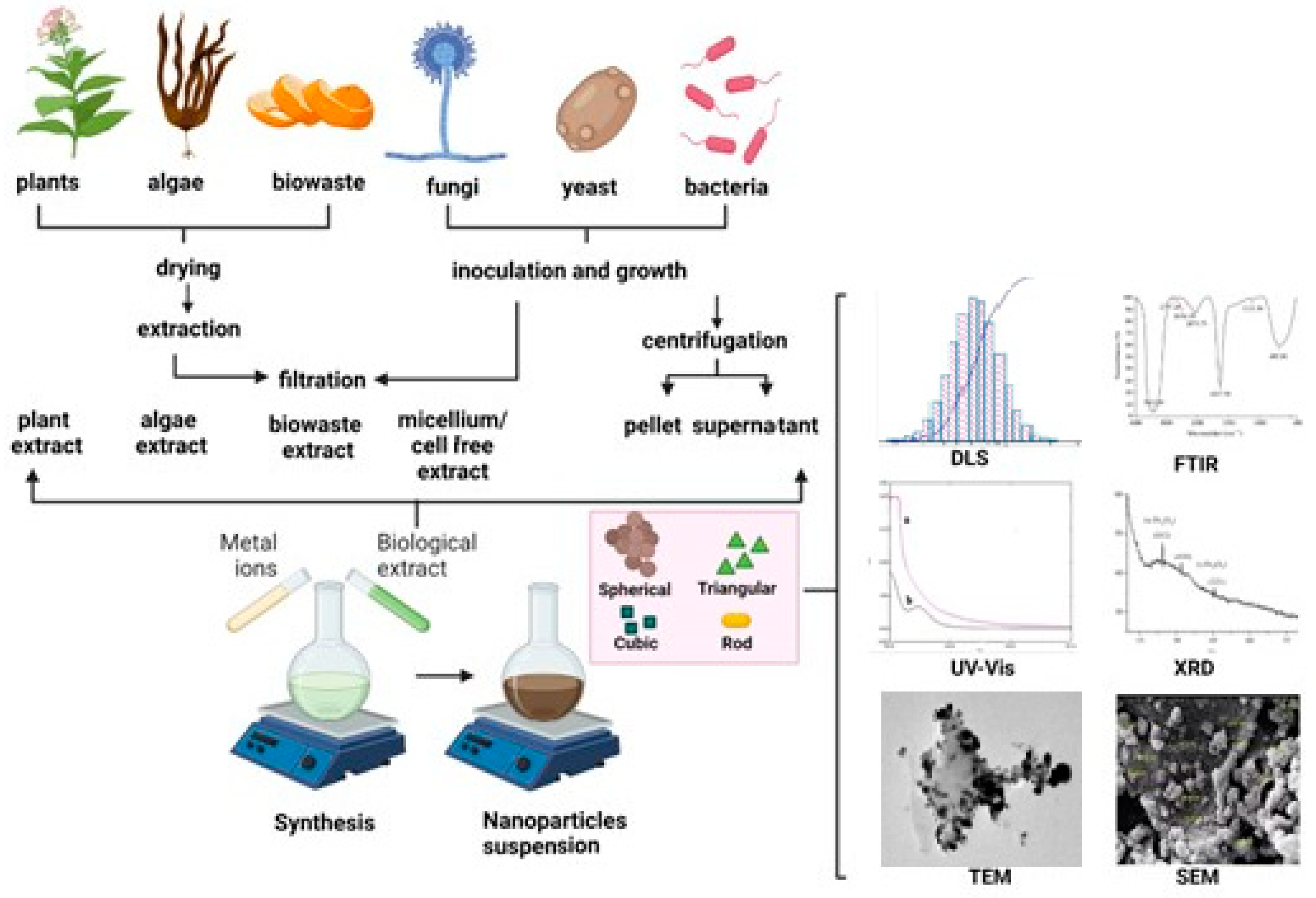

Table 1.
Iron oxide nanoparticles synthesized by bacterial species.
| Bacterial strain | Type of nanoparticle | Mechanism of synthesis | Size of nanoparticle (nm) |
Nanoparticle morphology | Ref. |
|---|---|---|---|---|---|
| Alcaligens faecalis | Fe2O3 | Extracellular | 12.3 | Irregular spherical | [19] |
| Actinobacter spp. | ɣ-Fe2O3 | Extracellular | 10–50 | Crystal | [20] |
| Actinobacter spp. | FeO-NPs | Intracellular | 10–40 | Quasi-spherical | [21] |
| Bacillus cereus | Fe3O4 | Extracellular | 29.3 | Spherical | [12] |
| Bacillus coagulans | FeO-NPs | Extracellular | - | - | [22] |
| Bacillus subtilis | Fe3O4 | Extracellular | 60–80 | Spherical | [17] |
|
Bacillus subtilis Bacillus pasteurii Bacillus licheniformis |
Fe3O4 | Extracellular | 37–97 | Rhombohedral | [23] |
| Desulfotomacculum acetoxidans | Fe3O4 | - | 21 | - | [24] |
| Desulfovibrio, strain LS4 | Fe2O3 | Extracellular | 19 | Round shaped | [24] |
| Escherichia coli | Fe3O4 | Extracellular | 23 ± 1 | Spherical | [25] |
| Geobacter sulfurreducens | Fe3O4 | Extracellular | 10–50 | - | [26] |
| Gluconacetobacter xylinus | Fe3O4 | Intracellular | 50 | - | [27] |
| Klebsiella oxytoca | Ferrihydrite nanoparticles | - | 2–5 | - | [28] |
| Lactobacillus casei | Fe3O4 | Intracellular | 10–15 | Spherical | [18] |
| Lactobacillus fermentum | Fe3O4 | Intracellular | 10–15 | Spherical | [16] |
| Leptothrix ochracea | - | Extracellular | 100 | Hollow tube | [29] |
| Magnetospirillum magneticum | Fe3O4 | Intracellular | 10–60 | Cuboidal, rectangular, and spherical NPs | [30] |
| Magnetospirillum gryphiswaldense | Fe3O4 | Extracellular/ Intracellular | 25–55 | Polydisperse | [31] |
| Microbacterium marinilacus | FeO-NPs | Extracellular | 25–88 | Irregular | [32] |
| Nitrospirae (MYR-1) | Fe2O3 | Intracellular | 40 | Bullet-shaped | [33] |
| Pseudomonas stutzeri | Fe2O3 | - | 10-20 | - | [34] |
| Paenibacillus polymyxa | FeO-NPs | - | 26.65 | Spherical | [35] |
| Proteus mirabilis | Fe3O4 | Intracellular | 1.44–1.92 | Spherical | [36] |
| Pseudomonas aeruginosa | Fe3O4 | Extracellular | 23 | Spherical | [25] |
| Shewanella oneidensis | Fe2O3 | Intracellular | 30–43 | Pseudo hexagonal shape | [37] |
| Streptococcus suis | Fe3O4 | - | - | - | [38] |
| Thermoanaerobacter sp | Fe3O4 | Extracellular | ~13.0 | Spherical | [39] |
| Thiobacillus thioparus | Fe3O4 | Intracellular | - | - | [40] |
Table 2.
Synthesis of iron oxide nanoparticles using fungi and yeast.
| Fungi | Location of synthesis | Type of nanoparticle | Size of nanoparticle (nm) |
Nanoparticle morphology | Ref. |
|---|---|---|---|---|---|
| Alternaria alternate | Cell-free filtrate | γ-Fe2O3 | 75–650 | Cubic | [51] |
| Aspergillus fumigatus | Fungal biomass | Fe2O3 | 42.4 | Irregular spherical | [22] |
| Aspergillus japonicus | Fungal biomass | Fe3O4 | 60–70 | Cubic | [48] |
| Aspergillus niger YESM 1 | Fungal homogenate | Fe3O4 | 18-50 | Spherical | [52] |
| Aspergillus niger BSC-1 | Cell-free filtrate | Fe3O4 | 20–40 | Orthorhombic | [41] |
| Aspergillus wentii | Fungal biomass | Fe2O3 | 46 | - | [22] |
| Baker’syeast | Fungal biomass | Fe2O3 | 2–10 | - | [53] |
| Candida bombicola | Fungal biomass | Fe3O4 | 8.5–4.5 | - | [50] |
| Chaetomium globosum | Fungal biomass | Fe2O3 | 25.3 | Irregular spherical | [22] |
| Cryptococcus humicola | - | Fe3O4 | 8-9 | Spherical | [54] |
| Curvularia lunata | Fungal biomass | Fe2O3 | 20.8 | Irregular spherical | [22] |
| Fusarium incarnatum | Fungal cell filtrate | Fe3O4 | 30.56 ± 8.68 | Spherical | [49] |
| Fusarium oxysporum | Fungal biomass | Fe3O4 | 10-40 | Cube | [42] |
| Neurospora crassa | Fungal biomass | Fe3O4 | 50 | Coralline appearance | [55] |
| Phialemoniopsis ocularis | Fungal cell filtrate | Fe3O4 | 13.13 ± 4.32 | Spherical | [49] |
| Pochonia chlamydosporium | Fungal biomass | Fe2O3 | ~12-50 | Spherical | [22] |
| Thichoderma asperellum | Fungal cell filtrate | Fe3O4 | 25 ± 3.94 | Spherical | [49] |
| Verticillium sp. | Fungal biomass | Fe3O4 | 100–400 | Cubo-octahedrally | [42] |
Table 3.
Synthesis of iron oxide nanoparticles using algae.
| Algae | Location of synthesis | Type of nanoparticle | Size of nanoparticle (nm) |
Nanoparticle morphology | Ref. |
|---|---|---|---|---|---|
| Chaetomorpha antennina | Cell-free extract | Fe3O4 | 9–10 | - | [64] |
| Chlorella K01 | Dried powder | Fe3O4 | 76.5 | Spherical | [65] |
| Colpomenia sinuosa | Dried powder | Fe3O4 | 11.24–33.71 | Spherical | [61] |
| Colpomenia sinuosa | Dried powder | Fe3O4 | 17.78 | Cubic | [66] |
| Kappaphycus alvarezzi | Dried powder | Fe3O4 | 14.7 | Spherical | [67,68] |
| Padina pavonica | Dried powder | Fe3O4 | 31.41 | Spherical | [66] |
| Padina pavonica | Freeze-dried powder | Fe3O4 | 10–19.5 | Spherical | [69] |
| Petalonia fascia | Dried powder | Fe3O4 | 9.42, | Spherical | [66] |
| Pterocladia capillacea | Dried powder | Fe3O4 | 16.85–22.47 | Spherical | [61] |
| Sargassum acinarium | Freeze-dried powder | Fe3O4 | 21.6–27.4 | Spherical | [69] |
| Sargassum muticum | Freeze-dried powder | Fe3O4 | 18 | Cubic | [58] |
|
Spirulina platensis |
Dried powder | Fe3O4 | 10 | - | [70] |
| Turbinaria turbinata | Cell-free extract | Fe3O4 | 8–14 | - | [64] |
Table 4.
Synthesis of iron oxide nanoparticles using plants.
| Plant species | Part of the plant | Type of nanoparticle | Size of nanoparticle (nm) |
Nanoparticle morphology | Ref. |
|---|---|---|---|---|---|
| Medicago sativa | Leaves | Fe2O3/Fe3O4 | 3.1 | Spherical | [78] |
| Aloe barbadensis | Leaves | Fe2O3 | 80–100 | Spherical | [79] |
| Amla | Seeds | Fe2O3 | 2-5 | Spherical | [140] |
| Arisaema amurense | Root | Fe2O3 | 24.55 ± 6.9 | Nearly spherical | [80] |
| Artemisia annua | Leaves | Fe2O3/Fe3O4 | 4.7 ± 0.8 | Spherical | [81] |
| Avecinnia marina | Flowers | FeO-NPs | 10–40 | Non-uniform | [82] |
| Azadirachta indica | Leaves | Fe3O4 | 9-12 | Irregular | [83] |
| Calliandra haematocephala | Leaves | Fe3O4 | 85.4–87.9 | Bead-like spherical | [84] |
| Carica papaya | Leaves | α-Fe2O3 | 2.15 | Spherical to agglomerate | [6] |
| Coriandrum sativum | Leaves | FeO-NPs | 20–90 | Spherical | [85] |
| Colocasia esculenta | Leaves | FeO-NPs | 15 | Spherical | [86] |
| Cynometra ramiflora | Leaves | FeO-NPs | - | Agglomerated | [87] |
| Cynara cardunculus | Leaves | Fe3O4 | 13.5 | Semi-spherical | [88] |
| Daphne mezereum | Leaves | Fe3O4 | 6.5–14.9 | Spherical | [89] |
| Eichhornia crassipes | Leaves | FeO-NPs | >100 | Rod-shaped | [90] |
| Eucalyptus globulus | Leaves | β-Fe2O3 | 100 | Spherical | [91] |
| Eucalyptus globulus | Leaves | Fe3O4 | 2.34 ± 0.53 | Spherical | [92] |
| Euphorbia wallichii | Leaves | FeO-NPs | 10–15 | Spherical | [93] |
| Euphorbia herita | Leaves | Fe3O4 | 25–80 | Spherical | [94] |
| Gardenia resinifera | Leaves | α-Fe2O3 | 3–8 | Spherical | [95] |
| Grapes | Seeds | Fe3O4 | 35 | Spherical | [96] |
| Green tea | Leaves | FeO-NPs | 5.7 ± 4.1 | Spherical | [97] |
| Hibiscus rosasinensis | Flower | FeO-NPs | 65 | Spinel | [98] |
| Sageretia thea | Leaves | Fe2O3 | 29 | Tetragonal crystalline | [99] |
| Lagenaria siceraria | Leaves | Fe3O4 | 30–100 | Cube | [100] |
| Lantana camara | Leaves | FeO-NPs | - | Crystalline nanorods | [101] |
| Laurus nobilis L | Leaves | α-Fe2O3 | 8.03 ± 8.99 | Spherical | [3] |
| Lawsonia inermis | Plant | FeO-NPs | 150–200 | Spherical | [102] |
| Mangifera indica L | Leaves | FeO-NPs | - | Polycrystalline nanorod | [103] |
| Mansoa alliacea | Leaves | β-Fe2O3 | 18.22 | Spherical nanoparticles | [77] |
| Moringa oleifera | Leaves | FeO-NPs | 15.01 ± 6.03 | Rods | [1] |
| Plantago major | Leaves | FeO-NPs | 4.6–30.6 | Spherical | [104] |
| Platanus orientalis | Leaves | α-Fe2O3 and γ-Fe2O3 | 38 | Spherical | [105] |
| Psidium guajava | Leaves | FeO-NPs | 1–5 | Spherical | [106] |
| Punica granatum | Seeds | Fe2O3 | 25–55 | Semi spherical | [71] |
| Rheum emodi | Roots | α-Fe2O3 | 12 | Spherical | [139] |
| Ruellia tuberose | Leaves | FeO-NPs | 20–80 | Hexagonal rods | [107] |
| Rumex acetosa | Plant | FeO-NPs | 40 | Amorphous | [74] |
| Sesbania grandiflora | Leaves | FeO-NPs | 25–60 | Agglomerated non-spherical | [108] |
| Soya bean | Sprouts | Fe3O4 | 8 | Spherical | [109] |
| Tamarix aphylla | Leaves | Fe2O3 | 5–100 | Oval | [110] |
| Terminalia belerica | Fruits | FeO-NPs | 15–23 | Spherical | [111] |
| Tridax procumbens | Leaves | Fe3O4 | 80–100 | Irregular spherical | [112] |
| Vaccinium corymbosum | Leaves | FeO-NPs | 52.4 | Irregular shape non agglomerated | [113] |
Table 5.
Iron oxide nanoparticles synthesized by biological waste.
| Biological source | Type of waste | Size of nanoparticle (nm) |
Nanoparticle morphology | Ref. |
|---|---|---|---|---|
| Ananas comosus | Fruit peel | 10–16 | Spherical | [117] |
| Acacia mearnsii | Biochar | 18–35 | - | [120] |
| Camellia sinensis | Tea waste | 28.5 | Spherical | [118] |
| Citrus Aurantifolia | Fruit peel | 3–10 | - | [119] |
| Cocos nucifera L | Fruit peel | 10–100 | Clustered | [121] |
| Coffee | Waste hydrochar | 10–40 | Spherical | [122] |
| Cynometra ramiflora | Fruit peel | 10–25 | Agglomerated | [123] |
| Juglans regia | Dried green husk | 12.6 | Cubic | [124] |
| Lemon | Fruit peel | 3 and 10 | Orthorhombic | [119] |
|
Malus domestica Citrus limon |
Fruit peel | 17–25 | Spherical | [125] |
| Orange | Fruit peel | 50 | Quasi spherical | [116] |
| Plantain | Fruit peel | 30–50 | Spherical | [126] |
| Punica Granatum | Fruit peel | 40 | Rod shaped | [127] |
| Rambutan | Fruit peel | 100–200 | Agglomerated spinel | [128] |
| Rice | Straw | 9.9 ± 2.4 | Aggregated spherical | [129] |
| Tangerine | Fruit peel | 50 | Spherical | [130] |
| Watermelon | Rinds | 2–20 | Spherical | [131] |
Table 6.
Characterization techniques for nanoparticles.
| Property | Characterization techniques |
|---|---|
| Size | TEM, XRD, DLS, NTA, SAXS, HRTEM, SEM, AFM, EXAFS, FMR, DCS, ICP-MS, UV-Vis, MALDI, NMR, TRPS, EPLS, magnetic susceptibility |
| Shape | TEM, HRTEM, SEM, AFM, STEM, EPLS, FMR, 3D-tomography |
| Chemical composition | XRD, XPS, ICP-MS, ICP-OES, SEM-EDX, NMR, MFM, LEIS, EELS |
| Crystal structure | XRD, EXAFS, HRTEM, electron diffraction, STEM |
| Surface Charge | Zeta potential, EPM |
| Magnetic properties | SQUID, VSM, Mössbauer, MFM, FMR, XMCD, magnetic susceptibility |
Table 7.
Antibacterial properties of IONPs from natural sources.
| Treated Bacterial Strain | Biological source for IONP generation | Antibacterial assay | Size of nanoparticle (nm) |
Nanoparticle morphology | Ref. |
|---|---|---|---|---|---|
|
Escherichia coli |
Moringa M. oleifera | WDM | 15.01 ± 6.03 | Rod-like morphology | [1] |
| composite of Psidium guavaja-Moringa oleifera | WDM | 82 ± 7.0 | Spherical | [223] | |
| Zea mays L.) ear leaves | WDM | 37.86 | Spherical | [224] | |
| Xanthomonas campestris | WDM | 20–80 | Polymorphous, spherical, oval and hexagonal. | [225] | |
| Penicillium sp. | DDM | 11.3 | Spherical | [45] | |
| Penicillium brevicompactum | DDM | 30–50 | Spherical | [226] | |
| Glycosmis mauritiana; Rutaceae/Leaves | DDM | ≤100 | Spherical | [133] | |
| Couroupita guianensis,Lecythidaceae/fruit extract | DDM | 17 | Spherical | [227] | |
| Nigella sativa seed extract | DDM | 31.45 | Spherical | [228] | |
| Piper betel leaves extract | WDM | 25.17 | Cubic | [229] | |
| Klebsiella pneumoniae | Penicillium sp. | DDM | 11.6 | Spherical | [45] |
| Zea mays L.) ear leaves | WDM | 37.86 | Spherical | [224] | |
| Agrewia optiva (Dhaman or Biul) and Prunus persica (Peach) leaf extract | WDM | 15–60 (AO) 13–70 (PP) |
Spherical, granular | [230] | |
| Papaver somniferum | WDM | 38 ± 13 | Elliptical or Spherical | [231] | |
| Glycosmis mauritiana; Rutaceae/Leaves | DDM | ≤100 nm | Spherical | [133] | |
| Couroupita guianensis,Lecythidaceae/fruit extract | DDM | 17 | Spherical | [227] | |
| Pseudomona aeuruginosa | Penicillium sp. | DDM | 11.6 | Spherical | [45] |
| Moringa M. oleifera | WDM | 15.01 ± 6.03 | Rod-like morphology | [230] | |
| Agrewia optiva (Dhaman or Biul) and Prunus persica (Peach) leaf extract | WDM | 15-60 (AO) 13-70 (PP) |
Spherical, granular | [230] | |
| Papaver somniferum | WDM | 38 ± 13 | Elliptical or Spherical | [231] | |
| Withania coagulans/Berries | DDM | 15–20 | Nanorods | [232] | |
| Aloe vera/leaf extract | cellular damage assessed by SEM | 8.26 | Cubical, Rhomboidal, Spherical | [233] | |
| Acorus calamus/rhizome | WDM | 20–30 | Spherical | [234] | |
| Piper betel leaves extract | WDM | 25.17 | Cubic | [228] | |
| Salmonella sp | composite of Psidium guavaja-Moringa oleifera | WDM | 82 ± 7.0 | Spherical | [223] |
| Moringa M. oleifera | WDM | 15.01 ± 6.03 | Rod-like morphology | [230] | |
| Staphylococcus aureus | Penicillium sp. | DDM | 12 | Spherical | [45] |
| Composite of Psidium guavaja-Moringa oleifera | WDM | 82 ± 7.0 | Spherical | [106] | |
| Moringa M. oleifera | WDM | 15.01 ± 6.03 | Rod-like morphology | [106] | |
| Agrewia optiva (Dhaman or Biul) and Prunus persica (Peach) leaf extract | WDM | 15–60 (AO) 13–70 (PP) |
Spherical, granular | [230] | |
| Penicillium brevicompactum | DDM | 30–50 | Spherical | [226] | |
| Glycosmis mauritiana; Rutaceae/Leaves | WDM | ≤100 | Spherical | [133] | |
| Withania coagulans/Berries | WDM | 15–20 | Nanorods | [232] | |
|
Couroupita guianensis, Lecythidaceae/fruit extract |
WDM | 17 | Spherical | [227] | |
| Nigella sativa seed extract | WDM | 31.45 | Spherical | [229] | |
| Piper betel leaves extract | WDM | 25.17 | Cubic | [228] | |
| Streptococcus pyogenes | Zea mays L.) ear leaves | WDM | 37.86 | Spherical | [224] |
| Agrewia optiva (Dhaman or Biul) and Prunus persica (Peach) leaf extract | WDM | 15–60 (AO) 13–70 (PP) |
Spherical | [230] | |
| Piper betel leaves extract | WDM | 25.17 | Cubic | [228] |
WDM: Well diffusion method. DDM: Disk diffusion method.
Table 8.
Antifungal activity of iron oxide nanoparticles derived from green synthesis.
| Treated fungal strain | Biological source for IONP generation | Approach | Size (nm) |
Nanoparticle morphology | Inhibition zone (mm) | Ref. |
|---|---|---|---|---|---|---|
| Alternaria alternata | - | Tannic acid | 10–30 | quasi circular |
21.33 ± 3.83 | [236] |
| Arthogrophis cuboida | Euphorbia hirta | Leaf extract | 25 – 80 |
Cavity like structures | 25.33 ± 0.5 | [94] |
| Aspergillus flavus | Laurus nobilis L. | Leaf extract | 8.03 ± 8.99 | crystalline, spherical; partly hexagonal | 13 | [3] |
| Green Tea (GT) or black Tea (BT) | Leaf extract | 42–60 | agglomerated & spherical | Aflatoxin reduction [%]:GT: 43.5 ± 1.5BT: 39.2 ± 0.9 | [237] | |
| Aspergillus fumigatus | Euphorbia hirta | Leaf extract | 25 – 80 | Cavity like structures | 21.67 ± 1.5 | [94] |
| Aspergillus niger | - | Tannic acid | 10–30 | circular | 26.33 ± 1.15 | [236] |
| Platanus orientalis | Leaf extract | 38 | spherical | 16 | [105] | |
| Euphorbia hirta | Leaf extract | 25 – 80 | Cavity like structures | 18.67 ± 0.5 | [94] | |
| Aspergillus parasiticus | Green Tea (GT) or Black Tea (BT) | Leaf extract | 42–60 | agglomerated & spherical | Aflatoxin reduction [%]:GT: 51.6 ± 1.6BT: 47.1 ± 3.1 | [237] |
| Cladosporium herbarum | - | Tannic acid | 10–30 | circular | 18.00 ± 1.00 | [236] |
| Euphorbia helioscopia | Leaf extract | 7 – 10 | spherical | 38.33 | [238] | |
| Mucor piriformis | Platanus orientalis | Leaf extract | 38 | spherical | 26 | [105] |
| Penicillium chrysogenum | - | Tannic acid | 10–30 | circular | 28.67 ± 1.53 | [236] |
| Penicillium spinulosum | Laurus nobilis L. | Leaf extract | 8.03 ± 8.99 | crystalline, spherical; partly hexagonal | 14 | [3] |
| Trichothecium roseum | - | Tannic acid | 10–30 | circular | 22.67 ± 2.52 | [236] |
Table 9.
List of studies using iron oxide nanoparticles (IONPs) derived from green synthesis as antiparasitic or antiviral agents.
Table 9.
List of studies using iron oxide nanoparticles (IONPs) derived from green synthesis as antiparasitic or antiviral agents.
| Treated microorganism | Biological source for IONP generation | Approach | Size (nm) |
Nanoparticle morphology | Ref. |
|---|---|---|---|---|---|
| Parasites | |||||
| Plasmodium | Nephrolepis exaltata | IONPs alone | 16 | Roughly spherical | [14] |
| Leishmania tropica KWH23 | Trigonella foenum-graecum | Synergistic effects of IONPs and LED light together | - | - | [245] |
| Virus | |||||
| Poliovirus (Type 1 and 3) | Hyphaene thebaica | IONPs alone | 10 | Quasi-spherical and cuboidal | [246] |
Disclaimer/Publisher’s Note: The statements, opinions and data contained in all publications are solely those of the individual author(s) and contributor(s) and not of MDPI and/or the editor(s). MDPI and/or the editor(s) disclaim responsibility for any injury to people or property resulting from any ideas, methods, instructions or products referred to in the content. |
© 2023 by the authors. Licensee MDPI, Basel, Switzerland. This article is an open access article distributed under the terms and conditions of the Creative Commons Attribution (CC BY) license (http://creativecommons.org/licenses/by/4.0/).
Copyright: This open access article is published under a Creative Commons CC BY 4.0 license, which permit the free download, distribution, and reuse, provided that the author and preprint are cited in any reuse.


