
Submitted:
30 August 2023
Posted:
31 August 2023
You are already at the latest version
Abstract
Abstract:
Background: Neonatal sepsis continues to be one of the leading causes of mortality and morbidity, particularly in under development and underdeveloped countries. We aim to compare laboratory parameters between the early-onset sepsis (EOS) and non-EOS groups and to evaluate the association between TLR2- Arg753Gln, TLR4 -Asp299Gly, IL6-174G/C and IL10-1082G/A gene single nucleotide polymorphisms and EOS susceptibility in preterm newborns.
Methods: Genotyping of the TLR2, TLR4, IL6 and IL10 polymorphisms was performed in 36 preterm neonates by polymerase chain reaction (PCR) and restriction fragment length polymorphism analysis (RFLP). Logistic regression analysis was used to test the associations between the studied gene polymorphisms and EOS susceptibility.
Results: Statistically significant differences in gestational age and birth weight were observed between the two groups, preterm neonates with EOS having a lower mean of gestational age (weeks, 29.4 ± 2.8 versus 32.6 ± 1.1, p = 0.00002) and lower mean of birth weight (gr., 1342.1 ± 446.5 vs. 1984 ± 376.9) than preterm neonates without EOS. C-reactive protein (CRP) values measured at first day significantly increased in EOS group compared with non-EOS group (median, 95% CI: 0.80 [0.40, 1.15] versus 0.30 [0.02, 0.50]). Mean of neutrophils significantly decreased in the EOS preterm neonates (mean difference: 17.3%, 95% CI: [4.0%, 30.5%], p = 0.0126) and non-EOS group (mean difference: 20.8%, 95% CI: [1.8%, 39.9%, p = 0.0354) between the first day and 7th hospitalization day. The minor IL10-A1082 allele frequency was higher in the EOS group than in the nonEOS group (50% versus 25%) with a marginal statistical significance (p=0.0550).
In the dominant model, the A/G + A/A variant genotype of IL10- 1082G/A polymorphism significantly increased the odds of EOS compared with GG genotype (OR= 5.25, p = 0.0322).
Conclusions: The results of the current study suggests the IL10- 1082G/A gene polymorphism as significant risk factor for early onset sepsis development in preterm neonates.
Keywords:
neonatal sepsis
; TLR2-Arg753Gln
; TLR4-Asp299Gly
; IL6-174G/C
; IL10-1082G/A
1. Introduction
Neonatal sepsis continues to be one of the leading causes of mortality and morbidity, particularly in underdeveloped countries [1]. Newborns rely on their innate immune system for anti-infectious defense, which provides a rapid and non-specific response to pathogenic microorganisms they come into contact with [2].
Definition of sepsis: To create comprehensive evidence-based recommendations for the recognition and management of children with septic shock or other sepsis-associated acute organ dysfunction in 2020, the guide Management of Septic Shock and Sepsis-Associated Organ Dysfunction in Children, was published. The guide was developed by 49 international experts, representing 12 international organizations, as three methodologists and three public members [3].
Although there are some commonalities between pediatric and adult sepsis, there are important differences in pathophysiology, clinical presentation, and therapeutic approaches. The recognition and diagnosis of sepsis is a significant challenge in pediatric patients as vital sign aberrations and examination findings are often subtle as compared to those observed in adults [4].
Although the definition of sepsis in children is not yet complete, application of Sepsis-3 to children has been attempted [5,6] Therefore, the majority of studies used to establish evidence for these guidelines referred to the 2005 nomenclature in which severe sepsis was defined as 1) greater than or equal to 2 age-based systemic inflammatory response syndrome (SIRS) criteria, 2) confirmed or suspected invasive infection, and 3) cardiovascular dysfunction, acute respiratory distress syndrome (ARDS), or greater than or equal to 2 noncardiovascular organ system dysfunctions; and septic shock in children as severe infection leading to cardiovascular dysfunction (including hypotension, need for treatment with a vasoactive medication, or impaired perfusion) and “sepsis associated organ dysfunction” in children as severe infection leading to cardiovascular and/or non-cardiovascular organ dysfunction [3].
The gold standard in the diagnosis of sepsis remains the blood culture, but due to its inherent limitations such as the time until the result appears and the required amount of blood, it is a difficult goal to achieve in the pediatric population. That is why it is recommended for the early diagnosis and management of cases to use institutional protocols. Adherence to protocols reduces variability in care and improves outcomes for children with septic shock or other organ dysfunction associated with sepsis. These being considered as best practice [3].
Sepsis is categorized as early-onset if diagnosed within the first 72 hours of life, which is due to perinatal risk factors, or late-onset if diagnosed after 72 hours and secondary to nosocomial risk factors[7]
Pathogenesis of sepsis:
The pathogenesis of neonatal sepsis still requires investigation, the lack of consensus in the definition of neonatal sepsis, even more so in the case of premature newborns, makes it more difficult to understand its pathogenesis [8]
There are known risk factors for the development of sepsis in the newborn, especially in preterm n eonates. These are: small gestational age, low birth weight, a positive maternal vaginal culture for group B streptococcus (GBS), prolonged rupture of membranes, maternal intrapartum fever and male sex. Chorioamnionitis is associated with the highest risk of subsequent occurrence. clinical or culture-proven sepsis [9,10].
Bacteria are the pathogens most frequently involved in the development of sepsis infection by gram-negative organisms, particularly Pseudomonas species, carries a higher risk for fulminant course and death than infection by other pathogen groups. Gram-positive causes of sepsis are dominated by GBS and coagulase-negative staphylococci (CoNS) [11].
The newborn has natural defense barriers such as skin, mucous barriers and vernix that increase these defense barriers of the newborn against pathogens. Once these barriers are crossed, immunity must intervene. Recognition of the pathogen by local immune sentinel cells is the first step towards the development of an immune response [8].
Multiple classes of pathogen recognition receptors (PRRs) have been discovered that serve as detectors of pathogen-associated molecular patterns (PAMPs), including cell wall and membrane components, flagellum, nucleic acids, and carbohydrates [12] A litany of PRR classes have been discovered, including the Toll-like receptors (TLRs), NOD-like receptors (NLRs), retinoic acid–inducible protein I like receptors (RLRs), peptidoglycan recognition proteins, β2-integrins, and C-type lectin receptors [8].
In addition to its roles in leukocyte function (adhesion, phagocytosis, migration, and activation) and complement binding, complement receptor 3 (CR3, also known as MAC-1 and CD11b-CD18) functions as a pathogen sensor on the surface of phagocytes. Chemokine gradients produced by endothelial cells and local macrophages are necessary for effective and specific leukocyte attraction and accumulation. Damage-associated molecular patterns (or alarmins), such as intracellular proteins or mediators released by dying or damaged cells, may also active PRRs [8]. If the pathogen is not contained locally and inflammatory homeostasis is not restored, SIRS may develop, and lead to MODS and death [13].
New data in adults and children demonstrate simultaneous proinflammatory/antiinflammatory responses where the magnitude of either response may determine outcome [14].
The clinical manifestations vary considerably and are nonspecific, which makes the diagnosis of early neonatal sepsis difficult and predisposes to excessive antibiotic use.
The clinical signs are from different systems and can be grouped as follows: a) apnea, difficulty breathing, cyanosis; b) tachycardia or bradycardia, poor perfusion or shock; c) irritability, lethargy, hypotonia, seizures; d) abdominal distension, vomiting, food intolerance, gastric residue, hepatomegaly; e) unexplained jaundice; f) body temperature instability; g) petechiae or purpura. To take into account the clinical signs, ideally the newborn should show manifestations in three distinct systems, or two clinical signs in distinct systems associated with a maternal risk factor [15] .
Binding to TLRs (toll-like receptors) stimulates a response by releasing cytokines, chemokines, complement proteins, and clotting factors. While newborns and adults have similar TLR expression levels, subsequent responses from PAMP (pathogen-associated molecular patterns)–TLR binding differ in case of preterm neonates [7,16,17].
In the case of prematurity, the signs and symptoms of neonatal sepsis overlap with comorbidities associated with prematurity, thus making differential diagnosis more difficult [18].
TLRs function as sensors of innate immunity, monitor and identify various pathogens, and become the body’s first barrier against pathogenic microorganisms. Previous studies have shown that during infection, TLR changes in preterm infants are considerably smaller than those in full-term infants, thus leading to diminished leukocyte activation and secretion of proinflammatory cytokines, as well as reduced over-regulation of various factors [19,20].
TLR2 is now recognized as a receptor for bacterial lipoproteins and lipoteichoic acids in Gram-positive bacteria. In contrast, TLR4 is identified as the ultimate receptor for lipopolysaccharides (endotoxins) in Gram-negative organisms [21]. A different manifestation of infection in critically ill patients suggests that host genetics influence the magnitude of the innate immune response and the severity of sepsis via the modulation of TLR expression and function [22].
Polymorphisms or mutations in TLRs are associated with increased risk for infection in adults [23] and children [24,25] but are less well characterized in neonates.
Several single-nucleotide polymorphisms (SNPs) in TLR2 and TLR4 have been identified in adult patients presenting with sepsis and admitted to intensive care units [26].
The gene encoding TLR2 is located on the long arm of chromosome 4 (4q32), where several SNP-type polymorphisms (SNPs) have been identified. Arg753Gln (rs5743708, G2258A) is one of the most studied SNPs found in multiple pathologies. The TLR2-Arg753Gln variant is caused by an arginine substitution with glutamine at locus 753, which results in a decreased response of macrophages to bacterial peptides [27].
Previous studies have suggested that TLR2 Arg753Gln may lead to decreased activation of intracellular signaling pathways [28]. Moreover, population studies have shown that TLR2 polymorphisms could influence the body’s overreaction in several diseases such as cancer, tuberculosis, infective endocarditis, and sepsis [29].
TLR4 is a protein composed of 224 amino acids that is encoded in humans by a gene located on the 9q32–q33 chromosome and contains three exons. Several simple nucleotide polymorphisms (SNPs) have been identified in the TLR4 gene, some of which are strongly associated with increased susceptibility to Gram-negative bacterial infections an increased incidence of sepsis [30]. TLR4 activity and function appear to be modulated by this genetic variation. One of these genetic polymorphisms is the point mutation 896A/G (rs4986790), which determines the substitution of aspartic acid with glycine in position 299 of the protein (Asp299Gly). In humans, the frequency of this polymorphism is higher than 5% [31,32]. The Asp299Gly polymorphism is located in the leucine-rich repeat (LRR) domain of exon 3, which links to pathogen-associated molecular pattern recognition (PAMP). The TLR4-Asp299Gly polymorphism has been found to prolong the length of hospitalization in adult patients with urinary tract infections [33].
Interleukin 6 (IL6) plays a significant role in the immune response and regulation of inflammatory reactions. Elevated IL6 levels are associated with an increased risk of severe sepsis and an increased death rate from severe sepsis [34]. The IL6 gene, located on chromosome 7p21 and spanning 5 kb, contains four introns and five exons. Several polymorphisms have been identified in the promoter region of the IL6 gene. Among these, the common polymorphism 174G/C (rs1800795) contains a DNA-binding site for the nuclear factor IL6, a transcription factor that can interact with estradiol receptor complexes to regulate IL6 gene expression [35]. The G–C polymorphism at position 174 of the IL6 gene (rs1800795) influences negative outcomes in several inflammatory diseases, including sepsis [36,37].
Interleukin 10 (IL10) has been identified as one of the key anti-inflammatory cytokines in the inflammatory cascade, as it decreases the production of inflammatory molecules such as TNF-α, interferon (IFN)-γ, interleukin 12 (IL12), reactive nitrogen oxide metabolites, major histocompatibility complex molecules and inhibits antigen-specific cytotoxic T-cells [38]. IL10 is a potent immunoregulatory cytokine that is widely known for its anti-inflammatory and B-cell-stimulating functions. IL10 is one of the anti-inflammatory cytokines that suppress the actions of IL6, TNF-α, and interleukin 8 (IL8) [39]. The gene encoding IL10 is located on chromosome 1 (1q31-1q32) [40]. Conversely, excess IL10 induces immunosuppression in sepsis and increases mortality by impairing bacterial clearance in pneumococcal pneumonia [41]. Depending on its chromosomal localization and functional relevance, IL10 is a multifunctional cytokine that can not only inhibit proinflammatory cytokine synthesis but also reduce antigen presentation and macrophage activation [42]. The genetic polymorphism 1082G/A, located in the promoter region of IL10, can affect IL10 expression [43]. The IL10-1082G/A polymorphism is caused by the substitution of guanine with adenine in position 1082 of the gene [44]. The impact of sepsis and multiorgan dysfunction is overwhelming for the patient and for the health system. The total annual average costs of CVD (cardiovascular disease) in adult patients, in Poland (both direct and indirect) amount to 37.5 bn PLN, (8.8 bn EUR), and it is 1.89% of GDP. The total estimated indirect costs of CVD amount to 31.3 bn PLN (7.3 bn EUR) and significantly exceeds the direct medical costs, which amount to 6.1 bn PLN (1.4 bn EUR) [45]. There values of non-oncology drug programs were approximately 330–438 mln USD and constituted 51.9–47.6% of NHF (National Health Fund ) budget [46].
That is why it is important to develop sepsis diagnosis and management protocols that limit the clinical and economic consequences of this complex pathology.
The objectives of the current study were to (i) compare laboratory parameters between the EOS and non-EOS groups and (ii) evaluate the associations between the studied gene polymorphisms and EOS susceptibility.
2. Materials and Methods
The prospective study performed between September 2016 and September 2017 included 26 preterm newborns with suspected sepsis and 10 newborns without signs or symptoms of sepsis, who received no antibiotic treatment. The newborns were admitted to the Clinic of Gynecology I, Cluj-Napoca, a tertiary care center for newborns in Romania. During this period, 36 premature newborns met the selection criteria.
2.1. TLR2-Arg753Gln (rs 5743708), TLR4-Asp299Gly (rs 4986790), IL6-174 G/C (rs 1800795), and IL10-1082G/A (rs 1800896)
The TLR2-Arg753Gln genetic variation is an MspI restriction fragment length polymorphism (RFLP). The flanking 129 base pair (bp) region was amplified using primers described by Dhifallah et al. [47]. The TLR4-Asp299Gly genetic variation is a NcoI restriction fragment length polymorphism (RFLP). The flanking 188 bp region was amplified using primers described by Dhifallah et al. [47]. The IL6-174 G/C genetic variation is a LweI restriction fragment length polymorphism (RFLP). The flanking 610 bp region was amplified using primers described by Aker et al.[48]. The IL10-1082 G/A polymorphism is a XagI restriction fragment length polymorphism (RFLP). The flanking 377 bp region was amplified using primers described by Cordeiro et al. [49].
2.2. DNA Extraction
Two-milliliter blood samples were collected from patients diagnosed with early neonatal sepsis in tubes containing EDTA as an anticoagulant. High-molecular-weight DNA was extracted using a Zymoresearch kit (Quick-DNAMiniprep, Kit-Zymo Research Corporation, Freiburg, Germany). The DNA samples were stored at −20 °C until the PCR procedure.
2.3. Genotyping Analysis
The TLR2, TLR4, IL6, and IL10 polymorphism genotyping was performed using polymerase chain reaction (PCR) and restriction fragment length polymorphism analysis (RFLP).
2.4. PCR Reaction
The PCR reaction was carried out using a 20 ng DNA template, 200 mM of deoxynucleotide triphosphates (dNTPs), 0.2 M of forward and reverse primers, 2.0 mM of MgCl2, and 0.625 units of Taq polymerase in a PCR buffer containing 50 mM KCl and 10 mM Tris-HCl (pH 8.3). The lyophilized primers were obtained from Eurogentec (Kaneka Eurogentec S.A. Biologics Division, Liege, Belgium) and were reconstituted in sterile deionized water (DDW) and stored at −20 °C.
Genomic DNA was amplified with an iCycler C1000 BioRad (Bio-Rad Life Science, Hercules, CA, USA) using the following PCR conditions:
TLR2-Arg753Gln: Initial denaturation at 95 °C for 60 s, followed by 34 cycles of denaturation at 95 °C for 10 s, primers annealing at 64.8 °C for 30 s, and a final extension at 72 °C for 30 s. A 129-bp common product was obtained.
TLR4-Asp299Gly: Initial denaturation at 95 °C for 60 s, followed by 34 cycles of denaturation at 95 °C for 10 s, primers annealing at 60 °C for 1 min, 20 s, extension at 72 °C for 15 s, and a final extension at 72 °C for 30 s. A 188-bp common product was obtained.
IL6-174G/C: Initial denaturation at 95 °C for 60 s, followed by 34 cycles of denaturation at 95 °C for 10 s, primers annealing at 55.3 °C for 20 s, extension at 72 °C for 15 s, and a final extension at 72 °C for 30 s. A 610 bp common product was obtained.
IL10-1082G/A: Initial denaturation at 95 °C for 60 s, followed by 34 cycles of denaturation at 95 °C for 10 s, primers annealing at 52.3 °C for 20 s, extension at 72 °C for 30 s, and a final extension at 72 °C for 30 s. A 377-bp common product was obtained.
The specificity of the PCRs was checked using electrophoresis on a 2% agarose gel prepared in 1X TBE buffer containing ethidium bromide (0.5 µg/mL). Visualization was performed under a UV illuminator.
2.5. RFLP Analysis
An amount of 6 µL of amplified DNA was digested with 2 units of MspI (TLR2), NcoI (TLR4), LweI (IL6), XagI (IL10), and restriction enzymes (New England Biolabs UK, Ltd., Hitchin, UK). Digestion was carried out at 37 °C for 3 h.
The enzymatic digestion was checked via the electrophoresis of 10 µL of the mixture on a 3% agarose gel containing a 0.5 mg/mL ethidium bromide solution. The gel was visualized on a UV transilluminator.
For TLR2-Arg753Gln, the Arg753 allele was characterized by the presence of the 104 and 25 bp fragments, while the Gln753 allele was characterized by the 129 bp fragment. For TLR4-Asp299Gly, the Asp299 allele was characterized by the presence of an undigested 188 bp fragment, while the Gly299 allele had two 168 and 20 bp fragments. For IL6-174G/C, the 174G allele was characterized by the presence of an undigested 610 bp fragment, while the 174C allele had two 367 and 243 bp fragments. For IL10-1082G/A, the 1082G allele was characterized by the presence of the 253, 97, and 27 bp fragments, while the 1082A allele was characterized by the presence of two 280 and 97 bp fragments.
Figure 1 shows the genotypes of the TLR2-Arg753Gln, TLR4-Asp299Gly, IL6-174G/C, and IL10-1082A/G polymorphisms.
2.6. The Studied Groups
Thirty-six hospitalized preterm neonates with clinical suspicion of sepsis in the first 72 h of life were selected for this study via convenience sampling. Based on the laboratory results and clinical examinations, all preterm neonates were divided into two groups: an EOS group which includes 26 patients and a non-EOS group which includes 10 patients. Preterm neonates included in the EOS group were premature newborns with a GA of <36 + WG (weeks of gestation) without congenital malformations; with at least one of the risk factors for early sepsis, including premature rupture of the amniotic membranes more than 18 h before birth, chorioamnionitis, maternal urinary tract infection, intrapartum maternal fever, meconium amniotic stain liquid, a low Apgar score, and the need for resuscitation in the delivery room; with at least two of the clinical signs and symptoms of neonatal sepsis that start in the first 72 h of life, i.e., respiratory distress, tachy-bradypnea, apnea episodes, the need for invasive or non-invasive respiratory support, cardiac decompensation, tachy-bradycardia, a TCR of greater than 3 s, hypotension, hypo/hyperglycemia, acid–base disorder, early hyperbilirubinemia that started before 24 h of life, convulsions, and lethargy; and patients who received antibiotic treatment in different combinations.
Subjects included in the non-EOS group were preterm newborns without clinical manifestations of sepsis, and they did not receive antibiotic treatment.
2.7. Statistical Analysis
The anthropometric and biochemical variables of the studied premature neonates were summarized by arithmetic mean (standard deviation (SD)), median (interquartile interval (IQR) = [first quartile Q1; third quartile Q3]), or relative frequencies (%). Comparisons of the studied characteristics between the EOS and non-EOS groups were performed using the Chi-squared test (χ2), Fisher’s exact test, Student's t-test with equal variances, Welch's two-sample t-test, or the Mann–Whitney test. Within-group comparisons concerning biochemical parameters were evaluated using paired t-tests or Wilcoxon’s signed-rank test.
The associations between the studied gene polymorphisms and EOS susceptibility were tested using binomial logistic regression, and the effect size of each tested association was assessed using the odds ratio (OR) and a 95% confidence interval (95% CI).
The departure from Hardy–Weinberg equilibrium (HWE) for studied SNPs was performed on the non-EOS group using the chi-square goodness-of-fit test, but when the genotype frequency was lower (<5), an exact test was used.
The significance level (α) of all bilateral statistical tests was set to 0.05.
All statistical analyses were performed with R software, version 4.2.2 [50].
3. Results
3.1. Comparisons of Demographic, Anthropometric, and Clinical Characteristics Between the Two Groups
The basic characteristics of the EOS cases and preterm neonates without EOS are described in Table 2. There was no significant difference in gender frequency (p = 0.836) or delivery way (p > 0.05) between the two groups. Nevertheless, statistically significant differences in gestational age and birth weight were observed, with preterm neonates with EOS having a lower mean gestational age (29.4 ± 2.8 weeks vs. 32.6 ± 1.1; p = 0.00002) and a lower mean birth weight (1342.1 ± 446.5 gr. vs. 1984 ± 376.9) than preterm neonates without EOS. Moreover, the Apgar score at 1 min was lower in the group with EOS than in the neonates without EOS (6 vs. 8). We also noticed that the frequency distributions of maternal chorioamnionitis (Fisher’s exact test; p = 0.1547), urinary infection (Fisher’s exact test; p = 0.2853), and membrane rupture (Fisher’s exact test; p = 0.3169) were similar in the two groups.
At baseline (on the first day after hospitalization), there were no statistically significant differences in WBC, neutrophils, platelets, red blood cells, hemoglobin, and hematocrits between the two groups, except for C-reactive protein (Table 3). We noticed that CRP values measured on the first day significantly increased in the EOS group compared with the non-EOS group (median, 95% CI: 0.80 [0.40, 1.15] vs. 0.30 [0.02, 0.50]). At seven days, all studied parameters were comparable between the two groups (p > 0.05).
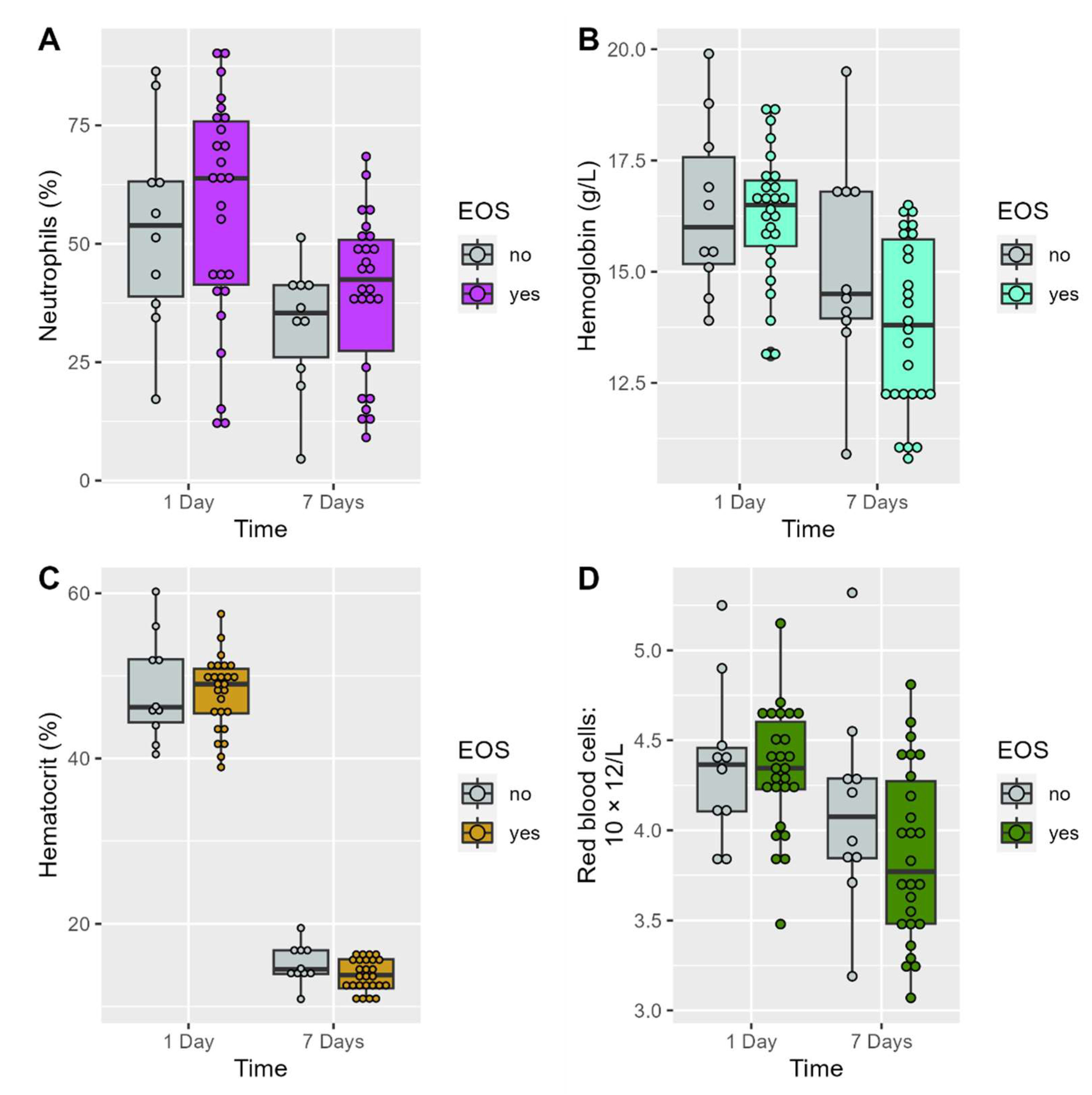
The mean neutrophil count significantly decreased in the EOS preterm neonates (mean difference: 17.3%; 95% CI: [4.0%, 30.5%]; p = 0.0126) and non-EOS group (mean difference: 20.8%; 95% CI: [1.8%, 39.9%]; p = 0.0354) during the study period (Figure 1A). The values of red blood cells, hemoglobin, hematocrit, and C-reactive protein significantly decreased, and platelets significantly increased, in the EOS premature neonates during the study period (Table 3, Figure 1B,D).
Figure 1.
Between and within-subjects variability of laboratory characteristics during the 7-days of follow-up.
Figure 1.
Between and within-subjects variability of laboratory characteristics during the 7-days of follow-up.
3.2. Associations of Studied SNPs' Allele Frequencies with Odds of EOS in Premature Neonates
The genotype distributions of the four studied gene polymorphisms were in line with the Hardy–Weinberg equilibrium in the group without EOS except for IL10 (Table 4). In the allele frequency analysis, the minor allele frequency of the IL10-1082G/A gene polymorphism was higher in the EOS group than in the non-EOS group (50% vs. 25%), with marginal statistical significance (p = 0.0550). The other three SNPs showed no significant differences in allele frequencies between the groups (p > 0.05).
3.3. Association between Studied SNPs' Genotype Frequencies and Odds of EOS in Premature Neonates
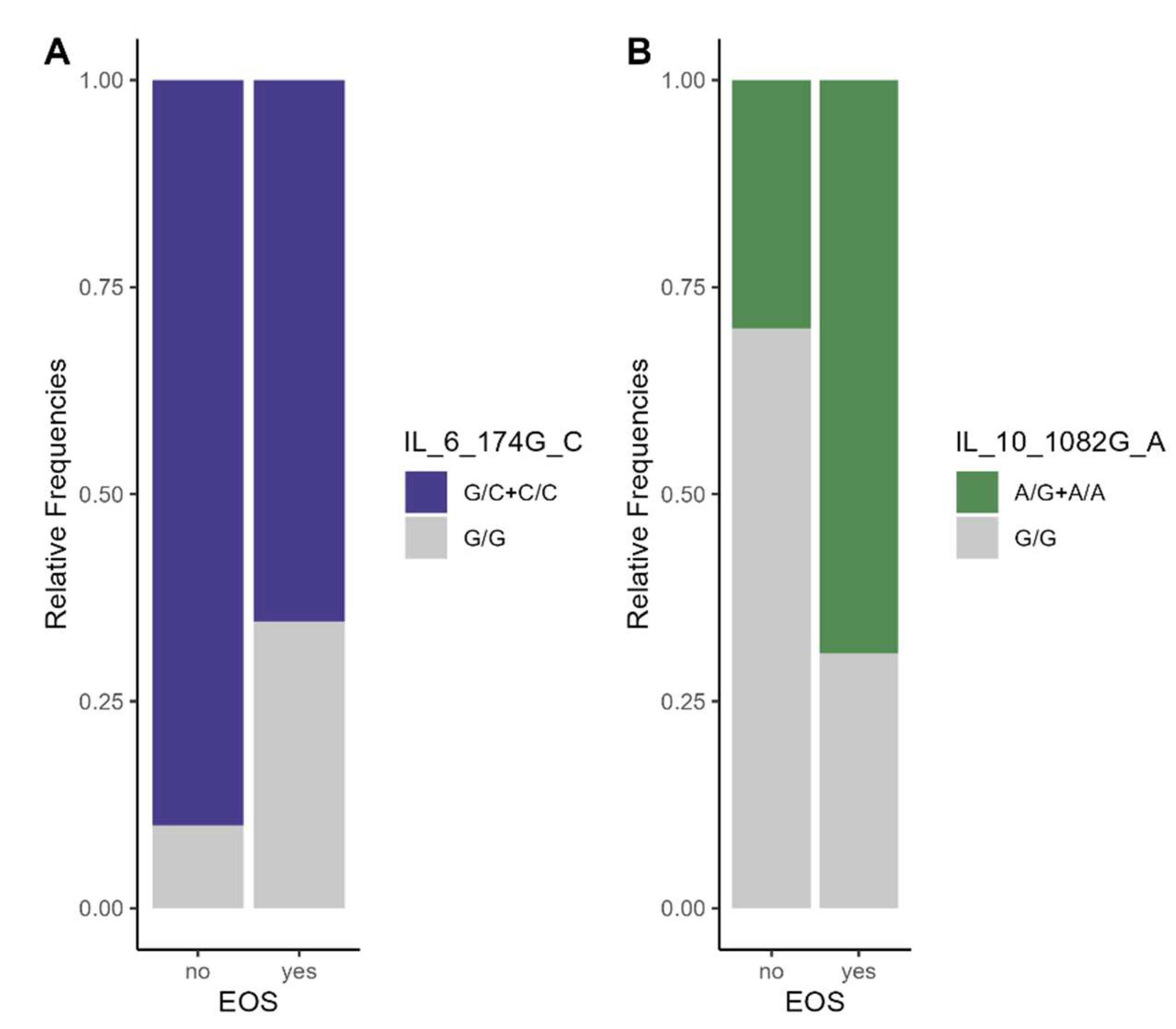
In the dominant model, the A/G + A/A variant genotype of the IL10-1082G/A polymorphism in the EOS group significantly increased the odds of EOS compared with the GG genotype (OR 5.25 [1.07, 25.70]; p = 0.0322). Frequency genotype distributions of IL10-1082G/A polymorphism in the studied groups were presented in Figure 2B. After adjusting for gestational age (<32 weeks vs. ≥32 weeks), the association between the IL10-1082G/A gene polymorphism and EOS tended toward statistical significance (adjusted p = 0.0752). There was no statistical evidence for a significant association of the TLR4-Asp299Gly, TLR2-Arg753Gln, and IL6-174G gene polymorphisms with EOS odds in premature neonates (p > 0.05; Table 5, Figure 2A).
Figure 2.
Frequency Distribution of variant genotype of IL6-174G and IL10-1082G/A gene polymorphisms (dominant model) in the EOS and non-EOS groups.
Figure 2.
Frequency Distribution of variant genotype of IL6-174G and IL10-1082G/A gene polymorphisms (dominant model) in the EOS and non-EOS groups.
4. Discussion
Early-onset sepsis (EOS) remains a serious and often fatal illness among infants born preterm, particularly among newborn infants of the lowest gestational age.
The standard diagnostic methods for EOS can generate false-negative results due to the manifestations of comorbidities associated with prematurity that overlap with the symptoms of sepsis.
The Global Burden of Disease (GBD) Study 2016/2017 estimated 1.3 (95% CI; 0.8 to 2.3) million annual incident cases of neonatal sepsis worldwide, with 3 resulting in 203 000 (95% CI; 178 700 to 267 100) sepsis-attributable deaths [51].
The results of our study performed on preterm newborns with EOS are similar to those performed on adults with EOS or with localized infections.
The current data support the hypothesis that TLRs behave differently in preterm newborns compared with at-term newborns. In at-term newborns, TLRs behave the same as in adults, while their expression is increased in preterm newborns. Differences in cytokine production between adult and neonatal innate mononuclear cells have been reported after the activation of TLRs' bacterial ligands [52].
In our study, we found significant differences in C-reactive protein mean levels between the EOS and non-EOS groups. The mean neutrophil count decreased in non-EOS premature neonates. In addition, mean neutrophils, red blood cells, hemoglobin, hematocrit, and C-reactive protein significantly decreased and platelets significantly increased in the EOS preterm neonates during the study period.
The current study aimed to observe if genotyping and specific SNP detection methods are associated with the early onset of neonatal sepsis in preterm neonates.
According to Nachtigall et al. the presence of two SNPs of TLR2-Arg753Gln and TLR4 -Asp299Gly was associated with a shorter time to onset of severe sepsis or septic shock in medical and surgical adult patients admitted to intensive care units [53]. Behairy et al. found a statistically significant association between the TLR2-Arg753Gln polymorphism and sepsis in the super dominant variant G/G. The same study found an association between the TLR4-Asp299Gly polymorphism A/A-variant and infection with Acinetobacter baumannii (p = 0.001), nonspecific Gram-negative bacilli infection, and sepsis in the adult population [54]. The distribution of the TLR4-Asp299Gly genotype in neonates suggests its association with an increased risk of culture-proven sepsis, according to Sljivancanin Jakovljevic et al. [55]. In the pediatric population, the TLR4-Asp299Gly polymorphism has been detected more frequently in patients with pyelonephritis, is more common in girls, and is always associated with E. coli [56,57].
In the present study, a significant difference in the frequency distributions of the TLR4-Asp299Gly gene polymorphism between the EOS and non-EOS preterm newborn groups was not found. Regarding the TLR2-Arg753Gln polymorphism, the minor allele Gln753 frequency was higher in the EOS group than in the non-EOS group (32.69% vs. 25.00%). Even though the risk of developing EOS increased in the presence of this allele in the studied sample, the results were not statistically significant. Furthermore, the analysis of different genotypic testing models revealed an increased estimated risk of developing EOS in the presence of TLR2-Arg753Gln variant genotypes, but the results failed to show statistical significance for the association.
IL6 is a vital proinflammatory cytokine generated by various cells such as leukocytes, endothelial cells, myocytes, adipocytes, and fibroblasts [35,58]. Several polymorphisms in the IL6 gene's promoter region have been identified. Among these, the common polymorphism-174G/C (rs1800795) contains a DNA-binding site for the nuclear factor IL6, a transcription factor that can interact with estradiol receptor complexes to regulate the expression of the IL6 gene [35,59].
In various genetic studies, IL6 polymorphisms in the promoter region have been shown to increase the risk of sepsis, although with inconsistent results. A meta-analysis of 16 studies performed by Hu P. et al. that systematically examined the correlation of IL6 gene polymorphisms (-174G/C and -572C/G) with susceptibility to sepsis found that the IL6-174G/C gene polymorphism was not statistically associated with the risk of sepsis in adults, neonates, or the pediatric population [35].
Although the studied groups of preterm neonates had different demographic characteristics, our study did not find statistically significant results between the presence of IL6 SNPs and the risk of developing EOS in premature neonates.
Studies have investigated the IL10-1082G/A polymorphism concerning the susceptibility to sepsis in the Asian population and Afro-Colombian patients [60]. A meta-analysis of 11 studies on the adult population concluded that the IL10-1082A/G polymorphism is significantly associated with susceptibility to sepsis in Asian populations [43]. At the same time, the AA genotype of the IL10-1082G/A polymorphism is a risk factor for elevated IL10 production and the development of sepsis due to Gram-negative bacteria, especially in Afro-Colombian patients [60].
In the present study, the results suggest that carriers of the IL10-1082A allele had a 2.91-fold increased risk of developing EOS, with marginal significance (p = 0.055). The results of the univariate logistic regression analysis revealed that the IL10-1082G/A polymorphism increased the odds of EOS in the dominant model (p = 0.032). After controlling for gestational age group, the IL10-1082G/A gene polymorphism remained a risk factor for EOS, with a tendency toward statistical significance.
The strong points of the current study consist in the fact that:
(i)It is an innovative study that directs the spotlight onto the effect of the presence of TLR-2, TLR-4, Interleukin-6 and -10 polymorphisms in the context of early neonatal sepsis in preterm neonates. There are studies in the literature on the involvement of SNPs in sepsis, but not on groups of premature newborns.
(ii)The interesting conclusion is that SNP IL10-A1082G/A increases predisposition to sepsis.
Our study has several limitations: (i) the use of a non-probabilistic sampling method (a convenience sample of premature) might alter the representativeness of the sample; (ii) most of the cases were diagnosed on clinical manifestation due to the small number of blood cultures; (iii) the small number of EOS cases and non-EOS premature neonates included in the SNP analysis might influence the stability of the estimated regression coefficients; (iv) the adjustment for prenatal, perinatal, and postnatal maternal factors of EOS was not performed in the current study due to the small number of neonates, (v) the clinical characteristics of studied sample are unbalanced and (vi) the results with marginal significance should be interpreted with caution (we performed a post-hoc power calculation using “genpwr” R package [61] and after considering a sample size n=36, MAF = 0.4 (mean), proportion of cases in the sample = 0.722, OR = 5, and significance level=0.05, the study power was estimated at 54.89%).
5. Conclusions
Based on our observations, the genotypic testing models revealed that in the codominant model, the IL10-A1082G/A polymorphism increased the odds of early-onset sepsis in premature neonates. Even though there was a trend toward statistical significance regarding the association of the IL10-A1082G/A gene polymorphisms with the odds of EOS in the allelic model, future studies should confirm the impact of individual alleles on early-onset sepsis in premature neonates.
Author Contributions
Melinda Baizat1 – Conceptualization, Investigation, Resources, Methodology, Writing - Original Draft, Data Curation, Visualization, Project administration. Mihaela Iancu2* – Conceptualization, Software, Validation, Formal analysis, Resources, Methodology, Writing - Original Draft, Writing - Review & Editing, Project administration. Gabriela Zaharie1,3 – Conceptualization, Resources, Methodology, Visualization, Validation. Monica Hășmășanu 1,3- Conceptualization, Resources, Methodology, Visualization. Melinda Matyas 1,3- Conceptualization, Resources, Methodology, Visualization, Ioana Rotar1,4- Conceptualization, Resources, Visualization. Lucia M. Procopciuc1,5- Conceptualization, Methodology, Writing - Review & Editing, Project administration, Validation, Supervision. All authors have read and agreed to the published version of the manuscript. All authors have read and agreed to the published version of the manuscript.
Funding
This research received no external funding.
Institutional Review Board Statement
The study was conducted in accordance with the Declaration of Helsinki, and approved by the Ethics Committee of "Iuliu Haţieganu" University of Medicine and Pharmacy, Cluj -Napoca, Romania (Protocol code 183/10.05.2016).
Informed Consent Statement
Informed consent was obtained from all the parents of subjects involved in the study.
Data Availability Statement
The raw data involved in this study can be obtained upon reasonable request ad-dressed to Lucia M. Procopciuc (luciamariaprocopciuc@yahoo.com) and Melinda Baizat (melindabaizat@gmail.com).
Conflicts of Interest
The authors declare no conflict of interest.
References
- Singh M, Alsaleem M, Gray CP. Neonatal sepsis. StatPearls [Internet]: StatPearls Publishing; 2022.
- Levy O. Innate immunity of the newborn: basic mechanisms and clinical correlates. Nature Reviews Immunology. 2007;7(5):379-90. [CrossRef]
- Weiss SL, Peters MJ, Alhazzani W, Agus MSD, Flori HR, Inwald DP, et al. Surviving Sepsis Campaign International Guidelines for the Management of Septic Shock and Sepsis-Associated Organ Dysfunction in Children. Pediatric critical care medicine : a journal of the Society of Critical Care Medicine and the World Federation of Pediatric Intensive and Critical Care Societies. 2020;21(2):e52-e106.
- Emr BM, Alcamo AM, Carcillo JA, Aneja RK, Mollen KP. Pediatric Sepsis Update: How Are Children Different? Surgical infections. 2018;19(2):176-83.
- Iramain R, Ortiz J, Jara A, Bogado N, Morinigo R, Cardozo L, et al. Fluid Resuscitation and Inotropic Support in Patients With Septic Shock Treated in Pediatric Emergency Department: An Open-Label Trial. Cureus. 2022;14(10):e30029. [CrossRef]
- Weiss SL, Balamuth F. Fluid Resuscitation in Children-Better to Be "Normal" or "Balanced"? Pediatric critical care medicine : a journal of the Society of Critical Care Medicine and the World Federation of Pediatric Intensive and Critical Care Societies. 2022;23(3):222-4.
- Kumagai Y, Takeuchi O, Akira S. Pathogen recognition by innate receptors. Journal of Infection and Chemotherapy. 2008;14(2):86-92. [CrossRef]
- Wynn JL, Wong HR. Pathophysiology of neonatal sepsis. Fetal and Neonatal Physiology. 2017:1536. [CrossRef]
- Benitz WE, Gould JB, Druzin ML. Risk factors for early-onset group B streptococcal sepsis: estimation of odds ratios by critical literature review. Pediatrics. 1999;103(6):e77-e. [CrossRef]
- Yancey MK, Duff P, Kubilis P, Clark P, Frentzen BH. Risk factors for neonatal sepsis. Obstetrics & Gynecology. 1996;87(2):188-94. [CrossRef]
- Stoll BJ, Hansen N, Fanaroff AA, Wright LL, Carlo WA, Ehrenkranz RA, et al. Late-onset sepsis in very low birth weight neonates: the experience of the NICHD Neonatal Research Network. Pediatrics. 2002;110(2):285-91. [CrossRef]
- Anderson DC, Rothlein R, Marlin SD, Krater SS, Smith CW. Impaired transendothelial migration by neonatal neutrophils: abnormalities of Mac-1 (CD11b/CD18)-dependent adherence reactions. 1990.
- Sriskandan S, Altmann D. The immunology of sepsis. The Journal of Pathology: A Journal of the Pathological Society of Great Britain and Ireland. 2008;214(2):211-23.
- Xiao W, Mindrinos MN, Seok J, Cuschieri J, Cuenca AG, Gao H, et al. A genomic storm in critically injured humans. Journal of Experimental Medicine. 2011;208(13):2581-90. [CrossRef]
- Hensler E, Petros H, Gray CC, Chung C-S, Ayala A, Fallon EA. The Neonatal Innate Immune Response to Sepsis: Checkpoint Proteins as Novel Mediators of This Response and as Possible Therapeutic/Diagnostic Levers. Frontiers in immunology. 2022;13:940930. [CrossRef]
- Hensler E, Petros H, Gray CC, Chung CS, Ayala A, Fallon EA. The Neonatal Innate Immune Response to Sepsis: Checkpoint Proteins as Novel Mediators of This Response and as Possible Therapeutic/Diagnostic Levers. Frontiers in immunology. 2022;13:940930. [CrossRef]
- Kawai T, Akira S. The roles of TLRs, RLRs and NLRs in pathogen recognition. Int Immunol. 2009;21(4):317-37. [CrossRef]
- Pace E, Yanowitz T, editors. Infections in the NICU: Neonatal sepsis. Seminars in pediatric surgery; 2022: Elsevier.
- Zhang J, Zhou J, Xu B, Chen C, Shi W. Different expressions of TLRs and related factors in peripheral blood of preterm infants. International journal of clinical and experimental medicine. 2015;8(3):4108.
- Sadeghi K, Berger A, Langgartner M, Prusa A-R, Hayde M, Herkner K, et al. Immaturity of infection control in preterm and term newborns is associated with impaired toll-like receptor signaling. The Journal of infectious diseases. 2007;195(2):296-302. [CrossRef]
- Kagan JC, Medzhitov R. Phosphoinositide-mediated adaptor recruitment controls Toll-like receptor signaling. Cell. 2006;125(5):943-55. [CrossRef]
- Angus DC, Burgner D, Wunderink R, Mira J-P, Gerlach H, Wiedermann CJ, et al. The PIRO concept: P is for predisposition. BioMed Central; 2003. [CrossRef]
- Bochud P-Y, Chien JW, Marr KA, Leisenring WM, Upton A, Janer M, et al. Toll-like receptor 4 polymorphisms and aspergillosis in stem-cell transplantation. New England Journal of Medicine. 2008;359(17):1766-77.
- Faber J, Meyer CU, Gemmer C, Russo A, Finn A, Murdoch C, et al. Human toll-like receptor 4 mutations are associated with susceptibility to invasive meningococcal disease in infancy. The Pediatric infectious disease journal. 2006;25(1):80-1. [CrossRef]
- Mockenhaupt FP, Cramer JP, Hamann L, Stegemann MS, Eckert J, Oh N-R, et al. Toll-like receptor (TLR) polymorphisms in African children: common TLR-4 variants predispose to severe malaria. Proceedings of the National Academy of Sciences. 2006;103(1):177-82. [CrossRef]
- Schröder NW, Schumann RR. Single nucleotide polymorphisms of Toll-like receptors and susceptibility to infectious disease. The Lancet infectious diseases. 2005;5(3):156-64.
- Hu L, Tao H, Tao X, Tang X, Xu C. TLR2 Arg753Gln gene polymorphism associated with tuberculosis susceptibility: an updated meta-analysis. BioMed research international. 2019;2019. [CrossRef]
- Xiong Y, Song C, Snyder GA, Sundberg EJ, Medvedev AE. R753Q polymorphism inhibits Toll-like receptor (TLR) 2 tyrosine phosphorylation, dimerization with TLR6, and recruitment of myeloid differentiation primary response protein 88. Journal of Biological Chemistry. 2012;287(45):38327-37. [CrossRef]
- Gao JW, Zhang AQ, Wang X, Li ZY, Yang JH, Zeng L, et al. Association between the TLR2 Arg753Gln polymorphism and the risk of sepsis: a meta-analysis. Critical care (London, England). 2015;19:416. [CrossRef]
- Karananou P, Tramma D, Katafigiotis S, Alataki A, Lambropoulos A, Papadopoulou-Alataki E. The Role of TLR4 Asp299Gly and TLR4 Thr399Ile Polymorphisms in the Pathogenesis of Urinary Tract Infections: First Evaluation in Infants and Children of Greek Origin. J Immunol Res. 2019;2019:6503832. [CrossRef]
- Balistreri CR, Colonna-Romano G, Lio D, Candore G, Caruso C. TLR4 polymorphisms and ageing: implications for the pathophysiology of age-related diseases. Journal of clinical immunology. 2009;29:406-15. [CrossRef]
- Chen R, Gu N, Gao Y, Cen W. TLR4 Asp299Gly (rs4986790) polymorphism and coronary artery disease: a meta-analysis. PeerJ. 2015;3:e1412. [CrossRef]
- Chatzi M, Papanikolaou J, Makris D, Papathanasiou I, Tsezou A, Karvouniaris M, et al. Toll-like receptor 2, 4 and 9 polymorphisms and their association with ICU-acquired infections in Central Greece. Journal of Critical Care. 2018;47:1-8. [CrossRef]
- Palmiere C, Augsburger M. Markers for sepsis diagnosis in the forensic setting: state of the art. Croatian Medical Journal. 2014;55(2):103. [CrossRef]
- Hu P, Chen Y, Pang J, Chen X. Association between IL-6 polymorphisms and sepsis. Innate immunity. 2019;25(8):465-72. [CrossRef]
- Gao J-w, Zhang A-q, Pan W, Yue C-l, Zeng L, Gu W, et al. Association between IL-6-174G/C polymorphism and the risk of sepsis and mortality: a systematic review and meta-analysis. PLoS One. 2015;10(3):e0118843.
- Michalek J, Svetlikova P, Fedora M, Klimovic M, Klapacova L, Bartosova D, et al. Interleukin-6 gene variants and the risk of sepsis development in children. Human immunology. 2007;68(9):756-60. [CrossRef]
- Mosser DM, Zhang X. Interleukin-10: new perspectives on an old cytokine. Immunological reviews. 2008;226(1):205-18. [CrossRef]
- Rea IM, Gibson DS, McGilligan V, McNerlan SE, Alexander HD, Ross OA. Age and age-related diseases: role of inflammation triggers and cytokines. Frontiers in immunology. 2018:586. [CrossRef]
- Eskdale J, Kube D, Tesch H, Gallagher G. Mapping of the human IL10 gene and further characterization of the 5’flanking sequence. Immunogenetics. 1997;46:120-8. [CrossRef]
- Kang X, Kim HJ, Ramirez M, Salameh S, Ma X. The septic shock-associated IL-10 -1082 A > G polymorphism mediates allele-specific transcription via poly(ADP-Ribose) polymerase 1 in macrophages engulfing apoptotic cells. Journal of immunology (Baltimore, Md : 1950). 2010;184(7):3718-24. [CrossRef]
- Lee YH, Kim JH, Song GG. Meta-analysis of associations between interleukin-10 polymorphisms and susceptibility to pre-eclampsia. European journal of obstetrics, gynecology, and reproductive biology. 2014;182:202-7. [CrossRef]
- Ouyang L, Lv Y-D, Hou C, Wu G-B, He Z-H. Quantitative analysis of the association between interleukin-10 1082A/G polymorphism and susceptibility to sepsis. Molecular biology reports. 2013;40:4327-32. [CrossRef]
- Shu Q, Fang X, Chen Q, Stuber F. IL-10 polymorphism is associated with increased incidence of severe sepsis. Chinese medical journal. 2003;116(11):1756-9.
- Mela A, Rdzanek E, Poniatowski Ł A, Jaroszyński J, Furtak-Niczyporuk M, Gałązka-Sobotka M, et al. Economic Costs of Cardiovascular Diseases in Poland Estimates for 2015-2017 Years. Frontiers in pharmacology. 2020;11:1231.
- Mela A, Poniatowski Ł A, Drop B, Furtak-Niczyporuk M, Jaroszyński J, Wrona W, et al. Overview and Analysis of the Cost of Drug Programs in Poland: Public Payer Expenditures and Coverage of Cancer and Non-Neoplastic Diseases Related Drug Therapies from 2015-2018 Years. Frontiers in pharmacology. 2020;11:1123.
- Ben Dhifallah I, Lachheb J, Houman H, Hamzaoui K. Toll-like-receptor gene polymorphisms in a Tunisian population with Behçet's disease. Clinical and experimental rheumatology. 2009;27(2 Suppl 53):S58-62.
- Aker S, Bantis C, Reis P, Kuhr N, Schwandt C, Grabensee B, et al. Influence of interleukin-6 G-174C gene polymorphism on coronary artery disease, cardiovascular complications and mortality in dialysis patients. Nephrology, dialysis, transplantation : official publication of the European Dialysis and Transplant Association - European Renal Association. 2009;24(9):2847-51.
- Cordeiro CA, Moreira PR, Andrade MS, Dutra WO, Campos WR, Oréfice F, et al. Interleukin-10 gene polymorphism (-1082G/A) is associated with toxoplasmic retinochoroiditis. Investigative ophthalmology & visual science. 2008;49(5):1979-82. [CrossRef]
- R Core Team A, Team RC. R: A language and environment for statistical computing. R Foundation for Statistical Computing, Vienna, Austria. 2012. 2022.
- Roth GA, Abate D, Abate KH, Abay SM, Abbafati C, Abbasi N, et al. Global, regional, and national age-sex-specific mortality for 282 causes of death in 195 countries and territories, 1980–2017: a systematic analysis for the Global Burden of Disease Study 2017. The Lancet. 2018;392(10159):1736-88. [CrossRef]
- Baizat IM, Zaharie CG, Hasmasanu M, Matyas M, Procopciuc LM. Is it possible to use the Toll-like receptors as biomarkers for neonatal sepsis? Review of the recent literature. Romanian Journal of Pediatrics. 2022;71(3).
- Nachtigall I, Tamarkin A, Tafelski S, Weimann A, Rothbart A, Heim S, et al. Polymorphisms of the toll-like receptor 2 and 4 genes are associated with faster progression and a more severe course of sepsis in critically ill patients. Journal of international medical research. 2014;42(1):93-110. [CrossRef]
- Behairy MY, Abdelrahman AA, Toraih EA, Ibrahim EE-DA, Azab MM, Sayed AA, et al. Investigation of TLR2 and TLR4 Polymorphisms and Sepsis Susceptibility: Computational and Experimental Approaches. International journal of molecular sciences. 2022;23(18):10982. [CrossRef]
- Sljivancanin Jakovljevic T, Martic J, Jacimovic J, Nikolic N, Milasin J, Mitrović TL. Association between innate immunity gene polymorphisms and neonatal sepsis development: a systematic review and meta-analysis. World Journal of Pediatrics. 2022;18(10):654-70.
- Krakowska A, Cedzyński M, Wosiak A, Swiechowski R, Krygier A, Tkaczyk M, et al. Toll-like receptor (TLR2, TLR4) polymorphisms and their influence on the incidence of urinary tract infections in children with and without urinary tract malformation. Central European Journal of Immunology. 2022;47(1). [CrossRef]
- Karananou P, Tramma D, Katafigiotis S, Alataki A, Lambropoulos A, Papadopoulou-Alataki E. The role of TLR4 Asp299Gly and TLR4 Thr399Ile polymorphisms in the pathogenesis of urinary tract infections: first evaluation in infants and children of Greek origin. Journal of Immunology Research. 2019;2019. [CrossRef]
- Van Snick, J. Interleukin-6: an overview. Annual review of immunology. 1990;8(1):253-78.
- Fishman D, Faulds G, Jeffery R, Mohamed-Ali V, Yudkin JS, Humphries S, et al. The effect of novel polymorphisms in the interleukin-6 (IL-6) gene on IL-6 transcription and plasma IL-6 levels, and an association with systemic-onset juvenile chronic arthritis. The Journal of clinical investigation. 1998;102(7):1369-76. [CrossRef]
- Vivas MC, Villamarín-Guerrero HF, Sanchez CA. Interleukin-10 (IL-10) 1082 promoter Polymorphisms and plasma IL-10 levels in patients with bacterial sepsis. Romanian Journal of Internal Medicine. 2021;59(1):50-7. [CrossRef]
- Moore, C. Jacobson, S. genpwr: Power Calculations under Genetic Model Misspecification. R package version. 2021;1(4).
Figure 1.
(a) TLR2-Arg753Gln polymorphism: lane 1—pBRHaeIIIDigest DNA molecular marker; lanes 2, 6, and 8—Gln/Gln genotype; lanes 3, 4, 7, and 10—Arg/Arg genotype; and lanes 5 and 9—Arg/Gln genotype; (b) TLR4-Asp299Gly polymorphism: lane 1—pBRHAeIII Digest DNA molecular marker, lanes 2, 6, and 10—Asp/Asp genotype, lanes 3, 4, 5, 7, and 9—Asp/Gly genotype, and lane 8—Gly/Gly genotype; (c) IL6-174G/C polymorphism: lane 1—pBRHaeIIIDigest DNA molecular marker, lanes 2 and 6—GG genotype, lanes 3, 5, 8, 9, and 10—GC genotype, and lanes 4 and 7—CC genotype; and (d) IL10-1082G/A polymorphism: lane 1—pBRHaeIIIDigest DNA molecular marker, lane 2—PCR fragment, lane 3—AA genotype, lanes 4, 5, 7, 8, and 10—GA genotype, and lanes 6 and 9—GG genotype. The sequences of specific primers are presented in Table 1.
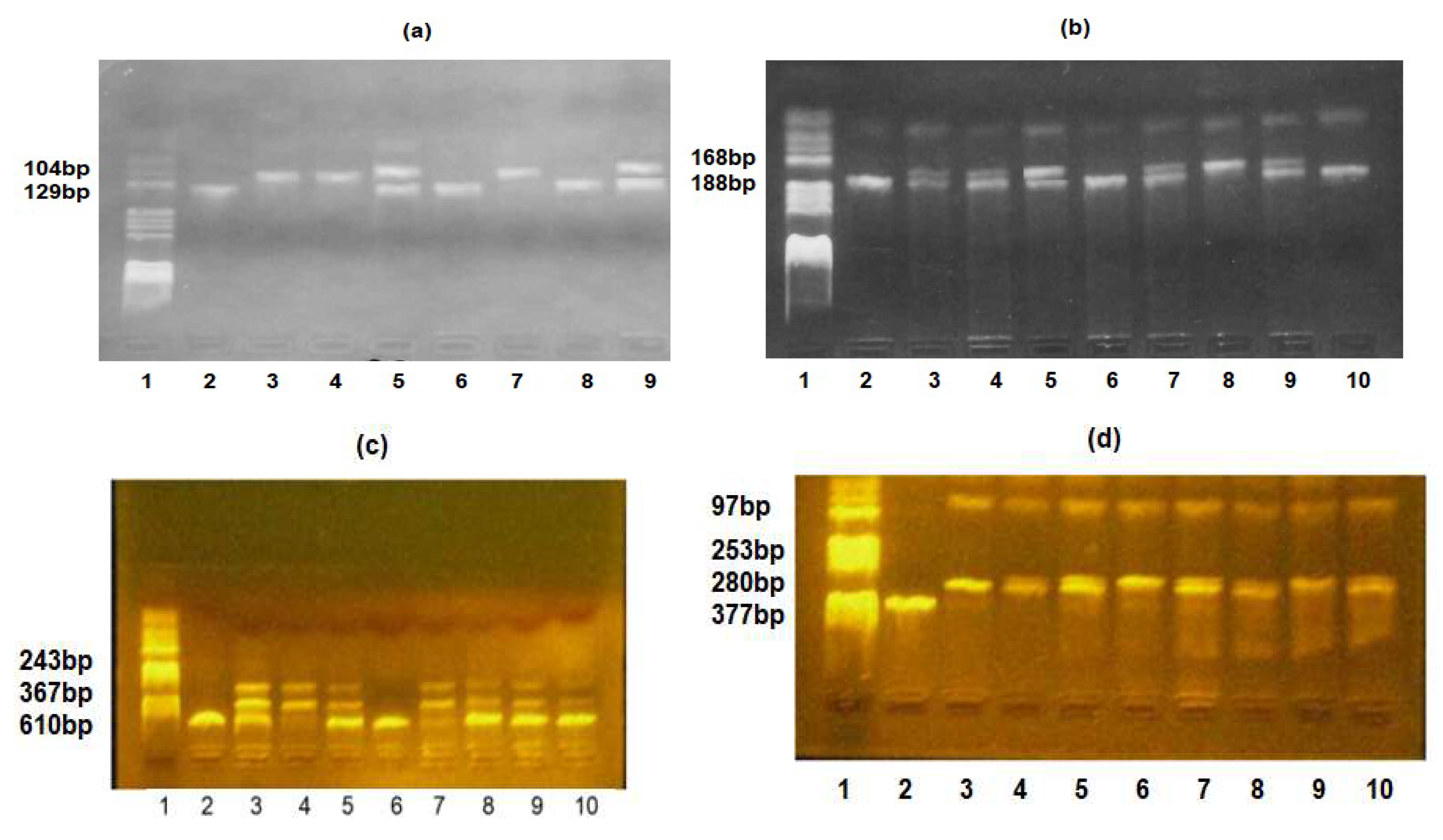

Table 1.
Specific primers were used to identify TLR2-Arg753Gln, TLR4-Asp299Gly, IL6-174G/C, and IL10-10882A/G polymorphisms.
Table 1.
Specific primers were used to identify TLR2-Arg753Gln, TLR4-Asp299Gly, IL6-174G/C, and IL10-10882A/G polymorphisms.
| Genetic Variation | Primers’ Sequences |
|---|---|
| TLR2-Arg753Gln | FW: 5′-CAT TCC CCA GCG CTT CTG CAA GCT CC-3′ |
| RV: 5′-GGA ACC TAG GAC TTT ATC GCA GCT C-3′ | |
| TLR4-Asp299Gly | FW: 5′-GAT TAG CAT ACT TAG ACT ACT ACC TCC ATG-3′ |
| RV: 5′-GAT CAA CTT CTG AAA AAG CAT TCC CAC-3′ | |
| IL6-174G/C | FW: 5′-CAG AAG AAC TCA GAT GAC TGG-3′ |
| RV: 5′-GCT GGG CTC CTG GAG GGG-3′ | |
| IL10-1082G/A | FW: 5′-CCA AGA CAA CAC TAC TAA GGC TCC TTT-3′ |
| RV: 5′-GCT TCT TAT ATG CTA GTC AGG TA-3′ |
FW—forward; RV—reverse.
Table 2.
Demographic and clinical characteristics of preterm neonates stratified by the presence of EOS.
Table 2.
Demographic and clinical characteristics of preterm neonates stratified by the presence of EOS.
| Postnatal Variables | Preterm Neonates without EOS (n1 = 10) | Preterm Neonates with EOS (n2 = 26) | p-Value |
|---|---|---|---|
| Gender, n (%) | 0.836 a | ||
| Female | 5 (50.0) | 14 (53.8) | |
| Male | 5 (50.0) | 12 (46.2) | |
| Gestational age, w | 0.00002 b | ||
| Min.–max. | 29–34 | 25–34 | |
| Mean (SD) | 32.6 (1.1) | 29.4 (2.8) | |
| Delivery way, n (%) | 1.000 a | ||
| Vaginal | 7 (70.0) | 19 (73.1) | |
| Cesarean | 3 (30.0) | 7(26.9) | |
| Birth weight, g | 0.00031 b | ||
| Min.–max. | 1490–2600 | 650–2470 | |
| Mean (SD) | 1984.0 (376.9) | 1342.1 (446.5) | |
| Neonate length (cm) | 0.0005 b | ||
| Min.–max. | 37–47 | 26–48 | |
| Mean (SD) | 43.3 (2.9) | 37.4 (6.4) | |
| Head circumference (cm) | 0.0004 b | ||
| Min.–max. | 27–34 | 20–32 | |
| Mean (SD) | 30.2 (1.8) | 26.5 (2.7) | |
| Apgar 1 min | |||
| Median (IQR) | 9 [7, 9] | 6 [4, 7] | 0.00009 d |
| Apgar 5 min | |||
| Median (IQR) | 9 [8, 9] | 8 [7, 8] | 0.00004 d |
n = absolute frequency; w = week; g = gram; SD = sample standard deviation; min. = minimum value; max. = maximum value; IQR = [Q1, Q3]; Q1 = first quartile; Q3 = third quartile; a Chi-square test; b a two-sided Student’s t-test for two independent samples; c Fisher’s exact test; d exact p-values obtained from Mann–Whitney test.
Table 3.
Comparisons of laboratory characteristics between premature neonates with EOS vs. non-EOS.
| Variables | Groups | On First Day | At 7 Days | p-Value Time Effect |
|---|---|---|---|---|
| WBC (mm3), mean (SD) |
Non-EOS | 12,785 [10,140, 15,292.5] |
10,455 [8742.5, 11,475.0] |
0.1309 |
| EOS | 11,985 [8145, 16,090] |
12,285 [9026.3, 16,942.5] |
0.5009 | |
| p-value between groups | 0.6894 | 0.2411 | ||
| Neutrophils (%), mean (SD) | Non-EOS | 53.6 (21.6) | 32.7 (13.4) | 0.0354 * |
| EOS | 56.8 (23.8) | 39.6 (16.9) | 0.0126 * | |
| p-value between groups | 0.7068 | 0.2588 | ||
| Platelets (mm3), mean (SD) |
Non-EOS | 280.2 [46.2) | 366.5 (70.9) | 0.0031 * |
| EOS | 236.5 (78.2) | 345.6 (155.8) | 0.0004 * | |
| p-value between groups | 0.1077 | 0.5855 | ||
| Red blood cells: 10 × 12/L, mean (SD) |
Non-EOS | 4.4 (0.4) | 4.1 (0.6) | 0.0524 |
| EOS | 4.3 (0.3) | 3.9 (0.5) | 0.00007 * | |
| p-value between groups | 0.8422 | 0.1834 | ||
| Hemoglobin (g/L), | Non-EOS | 16.4 (1.9) | 15.1 (2.4) | 0.0227 * |
| mean (SD) | EOS | 16.3 (1.5) | 13.8 (1.9) | 0.000001 * |
| p-value between groups | 0.7927 | 0.08615 | ||
| Hematocrit (%), mean (SD) |
Non-EOS | 48.4 (6.4) | 44.9 (7.4) | 0.0488 * |
| EOS | 47.9 (4.5) | 41.3 (5.7) | 0.000002 * | |
| p-value between groups | 0.828 | 0.1261 | ||
| C-reactive protein (mg/dL), median (IQR) | Non-EOS | 0.30 [0.09, 0.4] | 0.09 [0.08, 0.11] | 0.0580 |
| EOS | 0.80 [0.3, 1.2] | 0.14 [0.06, 0.38] | 0.0435 * | |
| p-value between groups | 0.0099 | 0.1417 |
SD = sample standard deviation; IQR = [Q1, Q3]; Q1 = first quartile; Q3 = third quartile; * significant result: p-value < 0.05.
Table 4.
Frequency distributions of TLR4, TLR2, IL6, and IL10 alleles and their associations with EOS.
Table 4.
Frequency distributions of TLR4, TLR2, IL6, and IL10 alleles and their associations with EOS.
| SNPs | Position | Locus | MA | fEOS | fnon EOS | HWE p-Value a | OR (95% CI) |
p-Value |
|---|---|---|---|---|---|---|---|---|
| TLR4-Asp299Gly | 9q32-q33 | 299 | Gly | 0.1538 | 0.2000 | 0.3065 | 0.72 [0.19, 3.12] |
0.6379 |
| TLR2- Arg753Gln | 4q32 | 753 | Gln | 0.3269 | 0.2500 | 0.4799 | 1.46 [0.46, 5.11] |
0.5257 |
| IL6-174G/C | 7p21 | −174 | C | 0.3269 | 0.5000 | 0.1998 | 0.49 [0.17, 1.43] |
0.1742 |
| IL10-1082G/A | 1q31-q32 | 1082 | A | 0.5000 | 0.2500 | 0.0464 | 2.91 [0.96, 10.28] |
0.0550 |
SNPs: single-nucleotide polymorphisms; MA: minor allele; f: minor allele frequency; HWE: Hardy–Weinberg equilibrium; a calculated in the non-EOS group; OR: unadjusted odds ratio; 95% CI: 95% confidence interval.
Table 5.
Relationships between SNPs and EOS: results of genotypic model analysis.
| SNP | Model of Inheritance | Genotype | Non-EOS Group n (%) |
EOS Group n (%) |
OR (95% CI) | p-Value a |
|---|---|---|---|---|---|---|
| TLR4 Asp299Gly |
Co-dominant | Asp/Asp | 7 (70.0) | 20 (76.9) | 1 [Reference] | 0.9138 |
| Asp/Gly | 2 (20.0) | 4 (15.4) | 0.70 [0.10, 4.69] | |||
| Gly/Gly | 1 (10.0) | 2 (7.7) | 0.70 [0.05, 8.97] | |||
| Dominant | Asp/Asp | 7 (70.0) | 20 (76.9) | 1 [Reference] | 0.6712 | |
| Asp/Gly + Gly/Gly | 3 (30.0) | 6 (23.1) | 0.70 [0.14, 3.58] | |||
| Recessive | Asp/Asp − Asp/Gly | 9 (90.0) | 24 (92.3) | 1 [Reference] | 0.8254 | |
| Gly/Gly | 1 (10.0) | 2 (7.7) | 0.75 [0.06, 9.32] | |||
| Log-additive | - | - | - | 0.80 [0.26, 2.45] | 0.6955 | |
| TLR2 Arg753Gln |
Co-dominant | Arg/Arg | 6 (60.0) | 13 (50.0) | 1 [Reference] | 0.8430 |
| Arg/Gln | 3 (30.0) | 9 (34.6) | 1.38 [0.27, 7.04] | |||
| Gln/Gln | 1 (10.0) | 4 (15.4) | 1.85 [0.17, 20.26] | |||
| Dominant | Arg/Arg | 6 (60.0) | 13 (50.0) | 1 [Reference] | 0.5892 | |
| Arg/Gln + Gln/Gln | 4 (40.0) | 13 (50.0) | 1.50 [0.34, 6.59] | |||
| Recessive | Arg/Arg + Arg/Gln | 9 (90.0) | 22 (84.6) | 1 [Reference] | 0.6668 | |
| Gln/Gln | 1 (10.0) | 4 (15.4) | 1.64 [0.16, 16.73] | |||
| Log-additive | - | - | - | 1.37 [ 0.47, 3.98] | 0.5591 | |
|
IL6 174G/C |
Co-dominant | G/G | 1 (10.0) | 9 (34.6) | 1 [Reference] | 0.0896 |
| G/C | 8 (80.0) | 17(66.4) | 0.24 [0.03, 2.20] | |||
| C/C | 1 (10.0) | 0 (0.0) | ND | |||
| Dominant | G/G | 1 (10.0) | 9 (34.6) | 1 [Reference] | 0.1140 | |
| G/C + C/C | 9 (90.0) | 17 (65.4) | 0.21 [0.02, 1.93] | |||
| Recessive | G/G + G/C | 9 (90.0) | 26 (100.0) | 1 [Reference] | 0.2778 | |
| C/C | 1 (10.0) | 0 (0.0) | ND | |||
| Log-additive | - | - | - | 0.16 [0.02, 1.36] | 0.0896 | |
|
IL10 1082G/A |
Co-dominant | G/G | 7 (70.0) | 8 (30.8) | 1 [Reference] | 0.0779 |
| A/G | 1 (10.0) | 10(38.5) | 8.75 [0.88, 86.57] | |||
| A/A | 2 (20.0) | 8 (30.8) | 3.50 [0.55, 22.30] | |||
| Dominant | G/G | 7 (70.0) | 8 (30.8) | 1 [Reference] | 0.0322 * | |
| A/G + A/A | 3 (30.0) | 18 (69.2) | 5.25 [1.07, 25.70] | |||
| Recessive | G/G + A/G | 8 (80.0) | 18 (69.2) | 1 [Reference] | 0.5091 | |
| A/A | 2 (20.0) | 8 (30.8) | 1.78 [ 0.31, 10.32] | |||
| Log-additive | - | - | - | 2.27 [0.82, 6.30] | 0.0925 |
SNPs: single-nucleotide polymorphisms; ND = not determined; OR: unadjusted odds ratio; 95% CI: 95% confidence interval; a estimated from unconditional logistic regression (Wald test); * significant result: p-value < 0.05.
Disclaimer/Publisher’s Note: The statements, opinions and data contained in all publications are solely those of the individual author(s) and contributor(s) and not of MDPI and/or the editor(s). MDPI and/or the editor(s) disclaim responsibility for any injury to people or property resulting from any ideas, methods, instructions or products referred to in the content. |
© 2023 by the authors. Licensee MDPI, Basel, Switzerland. This article is an open access article distributed under the terms and conditions of the Creative Commons Attribution (CC BY) license (http://creativecommons.org/licenses/by/4.0/).
Copyright: This open access article is published under a Creative Commons CC BY 4.0 license, which permit the free download, distribution, and reuse, provided that the author and preprint are cited in any reuse.


