
Submitted:
26 August 2023
Posted:
29 August 2023
You are already at the latest version
Abstract
The purpose of this study was to determine the effects of aerobic exercise on carotid to femoral pulse wave velocity (cf-PWV), cell adhesion molecules (intracellular adhesion molecules (ICAM-1), vascular cell adhesion molecules (VCAM-1), and endothelial selectin (E-selectin), and oxidized-LDL in elderly women aged 70–85 years, and to identify the effect of and correlation with vascular stiffness. Forty participants were recruited and divided into three groups; vascular stiffness (VSG, n=14), obesity (OG, n=14), and normal (NG, n=12). All groups were given a 16-week intervention of aerobic exercise, and the data collected before and after exercise were analyzed using SPSS Ver. 23.0. Two-way repeated-measures ANOVA was used to evaluate between-group and time-dependent interaction effects. One-way ANOVA was used to evaluate between-group variations. In addition, the significance was tested using a post-hoc test (Scheffe). The within-group variations by time before and after exercise were examined using a paired t-test, and correlation analysis was performed using Pearson correlation coefficients. Simple regression analysis was performed for variables showing significant differences. The results indicated interaction effects for cf-PWV (p<.001), VCAM-1 (p<.01), E-selectin (p<.05) and oxidized LDL (p<.001). The rate of change of cf-PWV was positively correlated with that of VCAM-1 (r=.352, p<.05) and that of oxidized-LDL (r=.325, p<.05) with statistical significance. To determine the effect of the rate of change of cf-PWV on the rate of change of VCAM-1, the variables were tested, and the coefficient of determination in the regression analysis was .124, indicating that 12.4% of the tested variables fit the standard regression line. The variables for the effect of the rate of change of cf-PWV on the rate of change of oxidized LDL were also tested, and the coefficient of determination in the regression analysis was .106, indicating that 10.6% of the tested variables fit the standard regression line. Thus, the 16-week regular and consistent aerobic exercise program had significant effects on the cf-PWV, ICAM-1, VCAM-1, E-selectin, and oxidized-LDL in elderly Korean women with vascular stiffness, suggesting improvements in vascular stiffness, based on which the intervention is predicted to contribute to the prevention of vascular dysfunction by lowering the risk of cardiovascular disease due to atherosclerosis, as well as having a positive effect in the prevention of impairment of vascular endothelial cells.
Keywords:
aerobic exercise
; elderly Korean women
; arterial stiffness
; cell adhesion molecule
; oxidized-LDL
1. Introduction
Cell adhesion molecules (CAMs) are expressed in vascular endothelial cells as glycoproteins that can receive the signals of IL-1/TNF-α and IL-8 secreted by macrophages to drive leukocytes in peripheral blood to adhere to vascular endothelial cells, allowing them to flow into damaged tissues.
However, in designing the present study, it was presumed that aging in elderly women could lead to vascular dysfunction and impairment of vascular endothelial cells due to the overexpression of CAMs. The previously positive roles of CAMs in normal defense mechanisms of the body could exert a negative effect on vascular and metabolic functions. Nevertheless, a recent study reported that certain types of CAMs, intracellular adhesion molecule-1 (ICAM-1) and vascular cell adhesion molecule-1 (VCAM-1), were overexpressed even in the early stage of atherosclerosis [1], and a high level of endothelial selectin (E-selectin) increased the risk of metabolic syndrome to a greater degree in females than in males, while positive correlations were found for vascular markers, ICAM-1, VCAM-1, body mass index (BMI), and blood pressure [2]. Moreover, considering the reports that the risk of type II diabetes is positively correlated with the expressed serum levels of ICAM-1 and E-selectin and that a high-cholesterol diet could increase the serum concentrations of ICAM-1 and VCAM-1, CAMs should not be overlooked from the perspective of metabolic functions [3,4].
In greater detail, low-density lipoprotein-cholesterol (LDL-C) accumulates in the lumen due to reasons such as hypertension, high blood cholesterol, and dyslipidemia, which flow into the tunica intima via damaged vascular endothelial cells, and are converted through oxidation into oxidized low-density lipoprotein (LDL) by reactive oxygen species (ROS). The excessive flow of LDL-C and the consequent formation of oxidized LDL in the arterial tunica intima lead to the conversion into and accumulation of foam cells, which marks the onset of vascular stiffness [5]. The level of oxidized LDL indicates not only the initial formation of arterial plaques, but also the risk of atherosclerotic cardiovascular disease [6]. Previous studies reporting high serum levels of oxidized LDL in patients with atherosclerosis at an early stage are supportive of the proposed clinical mechanism of vascular stiffness [7]. In addition, as vascular endothelial cells react sensitively in a severe state of inflammation with vascular aging, vascular stiffness in the elderly is accelerated by the induction of CAMs, which increases the adhesion of monocytes [8].
Thereby, vascular stiffness could be accelerated or aggravated when the reactivity of CAMs and oxidized LDL increases inordinately due to aging of vascular endothelial cells and consequently reduced functions. As vascular endothelial cells play key roles in vascular health, it is essential that pathological mechanisms related to vascular aging and stiffness be managed to prevent conversion into a pathological state.
Thus, exercise as an intervention with few side effects on physiological changes in the body is recommended for the elderly to be performed on a regular basis, which highlights the value of determining the intensity of suitable exercise and developing effective exercise programs. Changes in CAMs during exercise are influenced by exercise type or intensity, and when patients with cardiovascular disease are guided to perform aerobic exercise of low-to-moderate intensity, the expression levels of CAMs are reduced [9]. Moreover, regular exercise through a combination of aerobic and anaerobic training of moderate intensity caused positive changes in the oxidized LDL in obese elderly women, while the changes in skeletal muscles and the carotid artery medial thickness and oxidized LDL demonstrated a negative correlation [10]. These results collectively suggest that as a method to delay and conserve the functions of vascular endothelial cells, elderly women should perform moderate to high exercise to improve chronic inflammation caused by excess LDL-C accumulation in the tunica intima, which would downregulate the expression of CAMs and conversion of oxidized LDL into foam cells to improve and prevent vascular stiffness.
Thus, it is necessary to investigate the long-term effect of regular aerobic exercise of moderate to high intensity on the carotid to femoral pulse wave velocity (cf-PWV), CAMs, and oxidized-LDL in elderly Korean women, and to verify the rate of change of vascular stiffness and the effect of and correlations across the variables regarding the rate of change.
2. Materials and Methods
2.1. Participants
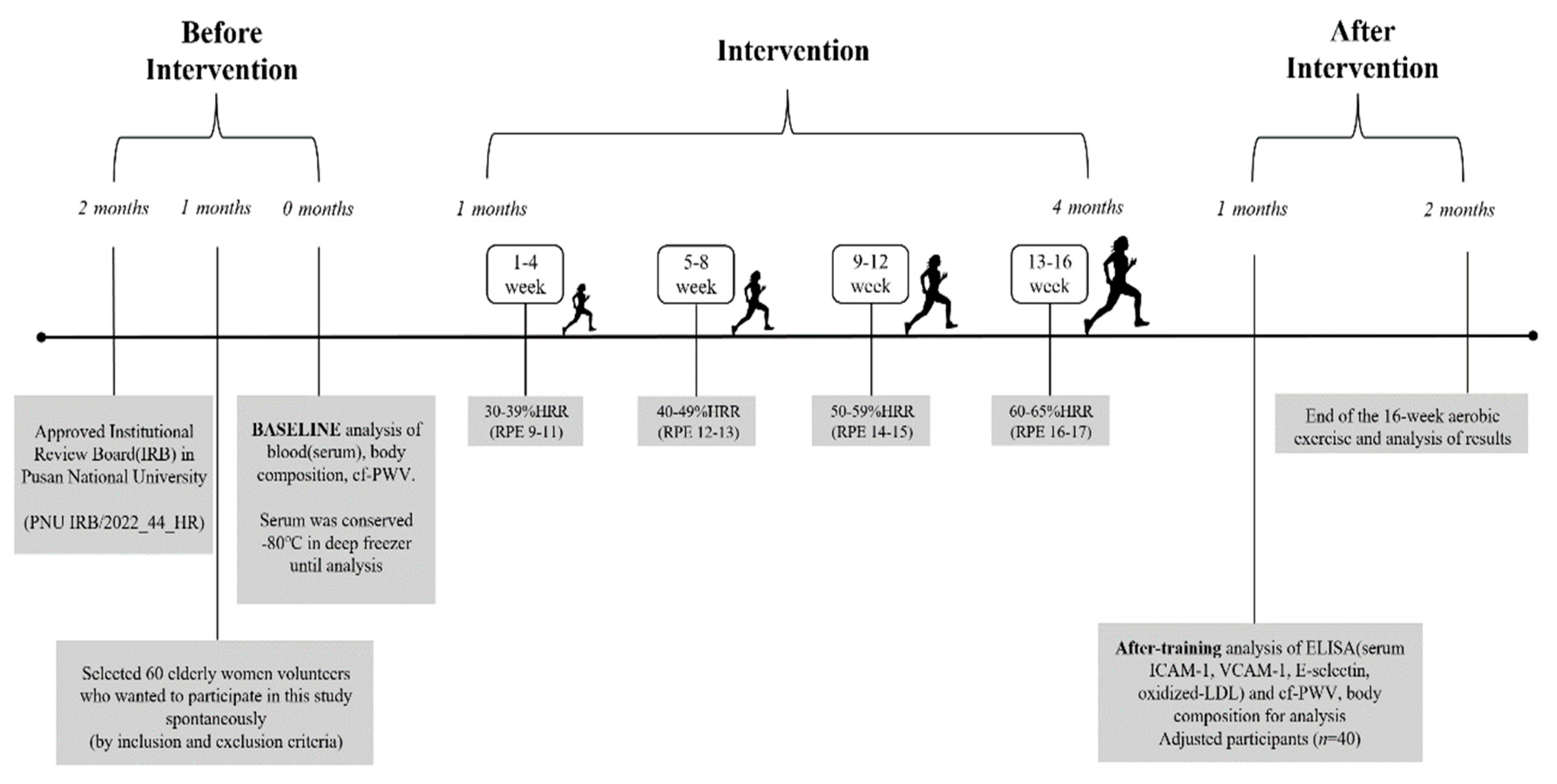
The sample size in this study was estimated to be n=36 using G-Power 3.1, based on the repeated-measures analysis of variance and under the following conditions: effect size f=0.28, group number n=3, repeated measure n=2, testing power at 0.8, and significance level at .05. Considering the drop-out rate, a total of 60 participants were recruited. The inclusion criteria were cf-PWV >12 m/s for the risk of vascular stiffness and BMI ≥25 kg/m2 and %BF ≥30% for obesity [11,12]. After excluding participants who dropped out of the study during the exercise program due to personal reasons and those whose measurements and test results were unreliable, the number of participants was n=40. They were divided into the vascular stiffness group (VSG, n=14), obesity group (OG, n=14), and normal group (NG, n=12), whose measurements and test results were finally analyzed. Prior to the study, approval was obtained from the Pusan National University Institutional Review Board (PNU IRB/2022_44_HR). Detailed explanations of the study purpose and goals were provided to the participants, and signed consent was obtained from those who voluntarily agreed to participate. The study procedures and the physical characteristics of the participants are presented in Table 1 and Figure 1.
2.2. Aerobic Exercise Program
For the aerobic exercise program in this study, the Circuit Exercise on Functional Fitness was revised and modified to develop a 16-week program of aerobic exercise at 60 minutes per session and three sessions per week [13]. The exercise intensity was based on the guidelines of the American College of Sports Medicine (ACSM) as follows [14]: 30-39% HRR (RPE 9-11) for 1– 4 week, 40-49% HRR (RPE 12-13) for 5–8 week, 50-59% HRR (RPE 14-15) for 9–12 week, and 60-65% HRR (RPE 16-17) for the final weeks; 13–16 week [14], while a wireless heart rate sensor (Polar system, Finland) was used. The detailed procedures of aerobic exercise are presented in Figure 2.
Figure 2.
Flowchart of intervention.
3. Data Collection
3.1. Body Composition
The height (cm) of the participants was measured using a digital scale, and the weight (kg), body fat (%), and skeletal muscle mass (kg) were measured using an automatic scale Inbody 430 (Inbody, KOR).
3.2. Blood Collection
For the blood test, the participants were instructed by a clinical pathologist to maintain a fasting state from 8 PM on the day before the test, and between 8 and 10 AM on the day of the test, a vacutainer and needle were used to collect 10 mL of blood from the antebrachial vein. The collected blood was placed in a serum separator tube. After 20 min of centrifugation at 3,000 rpm using Combi-514R (Hanil, KOR), the serum was isolated, and the supernatant was transferred to a 1.5-mL micro tube for storage at –80 °C for subsequent analyses.
3.3. Vascular Stiffness Analysis
To measure central blood pressure and analyze pulse wave and pulse wave velocity (PWV), each participant was instructed to rest in a supine position for 5 min, and vascular stiffness was measured using SphygmoCor XCEL (AtCor Medical Pty Ltd, AUD), a tonometry device. In addition, for PWV, the time variation between measurements in the carotid and femoral arteries and the vascular length were estimated to express the PWV for the aorta as the cf-PWV.
3.4. Blood Analysis
Through enzyme-linked immunosorbent assay (ELISA), CAMs were analyzed using the Human ICAM-1/CD54 Allele-specific, Human VCAM-1/CD106, and Human E-selectin/CD62E of the R&D system at 450-nm absorbance using a microplate reader (Allsheng, CHN). Oxidized-LDL was also analyzed through ELISA by Mercodia oxidized-LDL ELISA kit (Mercodia) and at 450 nm absorbance using a microplate reader (Allsheng, CHN).
3.5. Data Analysis
All data in this study were analyzed using SPSS Ver. 23.0. For each measured item, the mean (M) and standard deviation (SD) were estimated by group and factor. For the group × time interaction, two-way repeated measures analysis of variance (ANOVA) was used. One-way ANOVA was used for between-group variations. For the post hoc test, the Scheffe test was performed. Correlation analysis was based on Pearson correlation coefficients, and a simple regression analysis was performed to verify the effects of the variables. The level of significance was set at .05 for each item.
4. Results
4.1. Changes in cf-PWV
The cf-PWV showed a group × time interaction for VSG, OG, and NG (p<.001). The pre- to post-intervention between-group variation was significant for the VSG, OG, and NG (p<.01). The within-group variation over time was significant for the VSG (p<.01). Table 2 presents the changes in the cf-PWV examined in this study.
4.2. Changes in CAM
4.2.1. ICAM-1
ICAM-1 showed no group × time interaction for VSG, OG, and NG, whereas significant within-group variation by time was found for VSG (p<.01) and NG (p<.05). Table 3 presents the changes in ICAM-1 levels examined in this study.
4.2.2. VCAM-1
VCAM-1 showed a group × time interaction for the VSG, OG, and NG (p<.01). The within-group variation by time was significant for VSG (p<.001), and the rate of change also significantly varied across groups (p.<05). Table 3 presents the changes in VCAM-1 levels examined in this study.
4.2.3. E-selectin
E-selectin showed a group × time interaction for the VSG, OG, and NG (p<.05). The within-group variation by time was significant for VSG (p<.001), and the rate of change also significantly varied across groups (p.<01). Table 3 presents the changes in E-selectin levels examined in this study.
4.3. Changes in Oxidized-LDL
Oxidized LDL in this study showed a group × time interaction for VSG, OG, and NG (p<.001). The within-group variation by time for VSG was significant (p<.01). The rate of change also varied significantly across groups (p.<001). The changes in oxidized LDL levels are presented in Table 4.
4.4. Correlation of the Rate of Change of cf-PWV with VCAM-1 and Oxidized-LDL
The rate of change of cf-PWV in this study had a significant positive correlation with that of VCAM-1 (r=.352, p<.05) and oxidized-LDL (r=.325, p<.05). Figure 3 and Figure 4 show the respective scatter plots.
Figure 2.
Scatter plots for pre-post exercise intervention change ration in cf-PWV and VCAM-1.
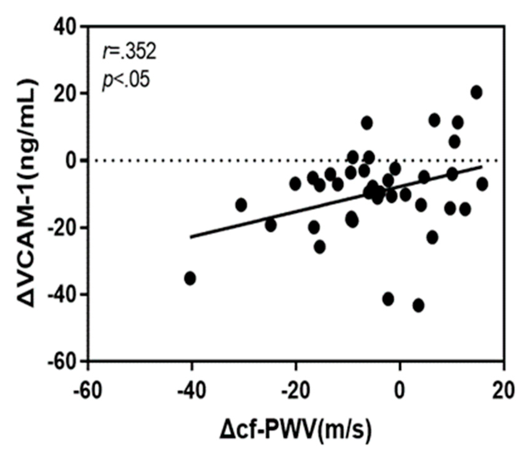
Figure 3.
Figure 1. Scatter plots for pre-post exercise intervention change ration in cf-PWV and oxidized-LDL.
Figure 3.
Figure 1. Scatter plots for pre-post exercise intervention change ration in cf-PWV and oxidized-LDL.
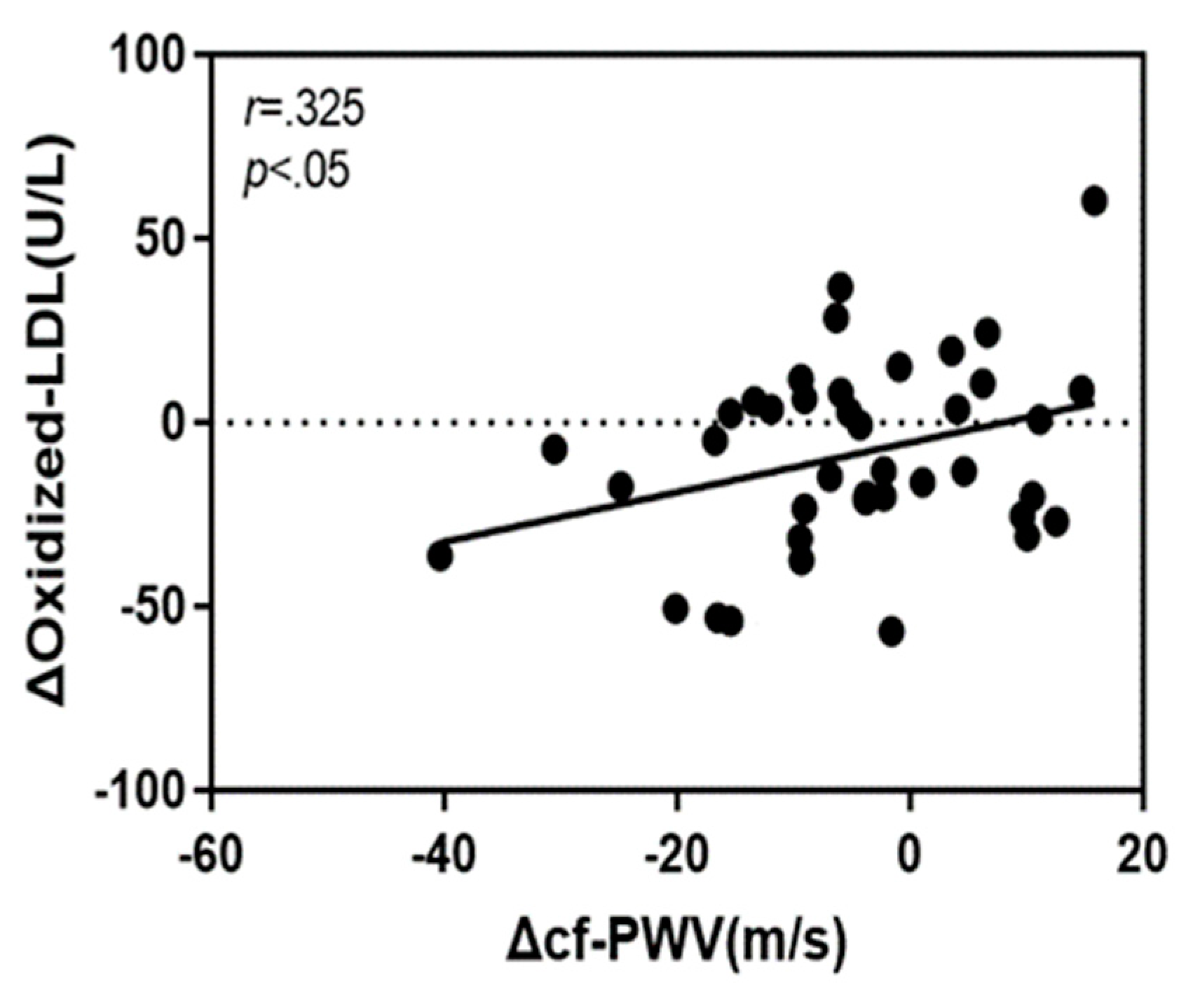
4.5. Analysis of the Effects of cf-PWV, VCAM-1 and Oxidized-LDL
To determine the effect of the rate of change of cf-PWV on the rate of change of VCAM-1, the variables were tested, and a significant effect was observed. The coefficient of determination in the regression analysis was .124, indicating that 12.4% of the tested variables fit the standard regression line.
In addition, the variables for the effect of the rate of change of cf-PWV on the rate of change of oxidized LDL were tested, and the effect was shown to be significant. The coefficient of determination in the regression analysis was .106, indicate that 10.6% of the tested variables fit the standard regression line. Table 5 presents the results.
5. Discussion
In this study, the effects of a 16-week aerobic exercise program on elderly Korean women were evaluated. The intensity of the exercise was gradually increased, and the level of VCAM-1 was shown to be significantly reduced in elderly Korean women in the VSG, suggesting that the intervention of regular aerobic exercise could not only reduce the risk of heart failure, but also serve as an effective noninvasive treatment. In addition, the rate of change of cf-PWV was positively correlated with that of VCAM-1, and following exercise intervention, the reduction rates of cf-PWV and VCAM-1 were both high. In line with this, the results of this study verified that only the rate of change of VCAM-1 across CAMs could have an effect on the rate of change of the cf-PWV. Hence, VCAM-1 is thought to be most closely associated with the risk of vascular stiffness in elderly Korean women, and long-term regular exercise for 16 or more weeks is anticipated to be an effective method to improve vascular stiffness in the elderly.
As shown, the variation between VCAM-1 and ICAM-1 after the intervention could be attributed to the fact that the information conveyed by the quantified expression levels of the two molecules ICAM-1 and VCAM-1 was divided into two. For ICAM-1, the expression is simply a natural part of the inflammatory response, whereas VCAM-1 is expressed in a vascular environment with narrow arteries of low elasticity, which is thought to underlie the interaction effect observed solely for VCAM-1.
The clinical significance of these results is high for VCAM-1 in terms of vascular stiffness. Notably, as the adhesion of monocytes to the cell surface is promoted for their permeation to the endothelium, an increase in their expression could increase the surface permeability of endothelial cells, causing various lipids to accumulate in the extracellular matrix or lead to the dispersion of smooth muscle cells toward the accumulation of vascular plaques [15].
Exercise, however, enhances vascular function and causes the secretion of nitric oxide to increase vascular bioavailability to improve the ratio of the vascular wall to the lumen and the vascular diameter, which results from a positive effect of exercise-related hemodynamic stimulation on the vascular wall [16].
Therefore, when vascular endothelial cells are damaged, the risk of cardiovascular disease in sedentary elderly increases, whereas in the elderly with a habit of regular aerobic exercise, vascular endothelial cells can have enhanced functions. Moreover, cellular aging is prevented so that the functions of vascular endothelial cells could be conserved [17].
In a previous study on aerobic exercise, cardiac afterload was shown to decrease at rest or during exercise, as the increased cardiac output allowed adaptation to the exercise situation regarding vascular conductivity, resistance, blood flow conduction, and microvasculature, which could improve the ventricular functions to contribute to vascular health in the elderly [18].
Similarly, in this study, as the elderly Korean women without regular physical activities in the recent past three months acquired the habit of regular exercise at three times a week for 16 weeks by participating in the study, the functions of various molecules expressed in vascular endothelial cells could recover their functions, and such results are presumed to have had a positive effect on CAMs.
Further, as endothelial-derived particles are activated by high blood glucose concentrations, the expression of E-selectin could be increased by 30%, compared to the normal state in endothelial cells, and this overexpression caused the accumulation of various precipitates including lipids in the vessel, increasing the risk of vascular disease [19].
The pre-test results in this study also indicated an approximately 30% difference in expression when VSG, OG, and NG were compared, which implied that the regulation of E-selectin expression was important in both obesity and vascular stiffness. In addition, the significant changes observed in VSG after the intervention of regular exercise are thought to be significant regarding the functional improvement of endothelial cells, and the effect of reducing E-selectin has been verified further as another mechanism by which regular exercise could manage blood pressure.
By contrast, the OG in this study did not show significant differences in vascular stiffness and CAMs during the time before and after exercise, presumably due to the lack of improvement in vascular stiffness in obese individuals through aerobic exercise. Thus, a low BMI could have a stronger effect on the changes in PWV. In a meta-analysis of a previous study, the effect of aerobic exercise in improving vascular stiffness was also stronger in those with low BMI than in those with high BMI [20].
Similarly, in this study, the OG comprising the obese elderly Korean women with mean BMI ≥25 kg/m2 exhibited no significant effect, in support of a previous study. In other words, individuals with a high BMI, such as those with obesity, are not likely to benefit from regular exercise with respect to the positive effect on vascular stiffness. Hence, it would be more effective for these individuals to lose weight to manage their BMI and prevent vascular stiffness.
Further, a previous study comparing the vascular stiffness between a high-fat diet and a low-fat diet also showed that the stiffness was more severe in the aortic arch than in the descending aorta for a high-fat diet. Vascular endothelial cells in the area of arteries where a state of disturbed flow is created could exhibit a far higher level of stiffness in the process of increasing the absorption capacity of oxidized LDL [21], compared to those in a state of luminal flow. Hence, when shear stress decreases due to disturbed flow, the resulting leak in the area connecting the cells increases LDL permeability, ultimately increasing the probability of atherosclerotic plaques [22].
For this reason, the trend of decrease for VSG with respect to vascular stiffness and oxidized LDL was higher than that for OG, in contrast to the prediction that greater changes in oxidized LDL would be observed for OG as a result of aerobic exercise.
Based on the results, the positive correlation between the rate of change of cf-PWV and that of oxidized-LDL in this study, as well as the effect of the rate of change of cf-PWV on that of oxidized-LDL, could be attributed to the improved luminal flow from the disturbed flow in the elderly. In addition, a reason for the significant reduction in cf-PWV and oxidized-LDL in the VSG after the 16-week aerobic exercise program could be the improvement of vascular stiffness in the aortic arch. It is considered that as vascular elasticity steadily decreases due to aging in the elderly, regular exercise to manage vascular stiffness is critical.
According to Boeno et al. [23], the intensity of aerobic exercise from 60%HRR to 80%HRR could improve the level of oxidized-LDL without weight loss or BMI reduction, and the risk factors of cardiovascular disease were reduced. Similarly, the results in this study indicated that despite the administration of drugs for hyperlipidemia or to control blood pressure in the VSG, the gradually increased intensity of aerobic exercise led to an intensity ≥60%HRR with the beneficial effect on oxidized-LDL.
Thus, long-term regular aerobic exercise with an intensity of 30%–65%HRR improved vascular health in elderly Korean women, and quality of life was shown to increase with increasing exercise duration and decreasing drug burden.
Several limitations of the present study should be acknowledged. First, we could not control the subject’s daily activities. Second, a limitation of the current study is the relatively small sample size. Therefore, the results of this study may be difficult to be generalized to other populations. Third, additional studies are needed to confirm the results of this study, compare the effects of the aerobic exercise on the variables of obese and nonobese or men and women by age or exercise type, and to develop effective aerobic exercise programs with different age groups.
The results in this study thus verified a close association of CAMs and vascular stiffness in elderly Korean women with regular aerobic exercise. In addition to the factors identified in this study, the differences in cf-PWV, CAMs, and oxidized-LDL according to the exercise intensity should be investigated, while a wider array of studies should verify the importance of acute CAM expression.
References
- Habas, K.; Shang, L. Alteration in intercellular adhesion molecule 1(ICAM-1) and vascular cell adhesion molecule 1(VCAM-1) in human endothelial cells. Tissue and Cell 2018, 54, 139–143. [Google Scholar] [CrossRef] [PubMed]
- Lee, C.H.; Kuo, F.C.; Tang, W.H.; Lu, C.H.; Su, S.C.; Liu, J.S.; Hsieh, C.H.; Hung, Y.J.; Lin, F.H. Serum E-selectin concentration is associated with risk of metabolic syndrome in female. Plos One 2019, 14, e0222815. [Google Scholar] [CrossRef] [PubMed]
- Qui, S.; Cai, X.; Liu, J.; Yang, B.; Zugel, M.; Steinacker, J.M.; Sun, Z.; Schumann, U. Association between circulating cell adhesion molecules and risk of type 2 diabetes: A meta-analysis. Atherosclerosis 2019, 287, 147–154. [Google Scholar]
- Saud, A.; Ali, N.A.; Gali, F.; Hadi, N. The role of cytokines, adhesion molecules, and toll-like receptors in atherosclerosis progression: the effect of Atorvastatin. Journal of Medicine and Life 2022, 15, 751–756. [Google Scholar] [CrossRef]
- Maguire, E.M.; Pearce, S.W.; Xiao, Q. Foam cell formation: A new target for fighting atherosclerosis and cardiovascular disease. Vascular Pharmacology 2018, 112, 54–71. [Google Scholar] [CrossRef]
- Gao, S.; Zhao, D.; Wang, M.; Zhao, F.; Han, X.; Qi, Y.; Liu, J. Association between circulating oxidized LDL and atherosclerotic cardiovascular disease: A meta-analysis of observational studies. Canadian Journal of Cardiology 2017, 33, 1624–1632. [Google Scholar] [CrossRef]
- Zhao, X.; Zhang, H.W.; Xu, R.X.; Guo, Y.L.; Zhu, C.G.; Wu, N.Q.; Gao, Y.; Li, J.J. Oxidized-LDL is a useful marker for predicting the very early coronary artery disease and cardiovascular outcomes. Personalized Medicine 2018, 15, 521–529. [Google Scholar] [CrossRef]
- Honda, S.; Ikede, K.; Urata, R.; Yamazaki, E.; Emoto, N.; Matoba, S. Cellular senescence promotes endothelial activation through epigenetic alteration and consequently accelerates atherosclerosis. Scientific Reports 2021, 11, 1–11. [Google Scholar] [CrossRef]
- Koh, Y.; Park, J. Cell adhesion molecules and exercise. Journal of Inflammation Research 2018, 11, 297–306. [Google Scholar] [CrossRef]
- Park, J.H.; Park, H.T.; Lim, S.T.; Park, J.K. Effects of a 12-week healthy-life exercise program on oxidized low-density lipoprotein cholesterol and carotid intima-media thickness in obese elderly women. Journal of Physical Therapy Science 2015, 27, 1435–1439. [Google Scholar] [CrossRef]
- Joint Committee for Guideline Revision. 2018 Chinese guidelines for prevention and treatment of hypertension-A report of the revision committee of Chinese guidelines for prevention and treatment of hypertension. Journal of Geriatric Cardiology 2019, 16, 182–241. [Google Scholar]
- Korean Society for the Study of Obesity (2020). Guideline for the Management of Obesity 2020.
- Ha, S.M.; Kim, D.Y.; Kim, J.H. Effects of Circuit Exercise on Functional Fitness, Estradiol, Serotonin, Depression and Cognitive Function in Elderly Women. Journal of Korean Physical Education Association for Girls and Women 2017, 31, 199–217. [Google Scholar] [CrossRef]
- American College of Sports Medicine (2018). ACSM`s guidelines for exercise testing and prescription. Lippincott Williams & Willkins.
- Wang, T.T.; Wang, X.M.; Zhang, X.L. Circulating vascular cell adhesion molecule-1 (VCAM-1) intercellular adhesion molecule (ICAM-1): relationship with carotid artery elasticity in patients with impaired glucose regulation (IRG). In Annales d`endocrinologies 2019, 80, 72–76. [Google Scholar] [CrossRef] [PubMed]
- Green, D.J.; Smith, K.J. Effects of exercise on vascular function, structure, and health in humans. Cold Spring Harbor Perspectives in Medicine 2018, 8, a029819. [Google Scholar] [CrossRef] [PubMed]
- Rossman, M.J.; Kaplon, R.E.; Hill, S.D.; McNamara, M.N.; Santos-Parker, J.R.; Pierce, G.L.; Seals, D.R.; Donato, A.J. Endothelial cell senescence with aging in healthy humans: prevention by habitual exercise and relation to vascular endothelial function. American Journal of Physiology-Heart and Circulatory Physiology 2017, 313, 890–895. [Google Scholar] [CrossRef]
- Green, D.J.; Hopman, M.T.; Padilla, J.; Laughlin, M.H.; Thijssen, D.H. Vascular adaptation to exercise in human: role of hemodynamic stimuli. Physiology Reviews 2017, 97, 495–528. [Google Scholar] [CrossRef]
- Hijmans, J.G.; Bammert, T.D.; Stockelman, K.A.; Reiakvam, W.R.; Greiner, J.J.; DeSouza, C.A. High glucose-induced endothelial microparticles increase adhesion molecule expression on endothelial cells. Diabetology International 2019, 10, 143–147. [Google Scholar] [CrossRef]
- Li, G.; Lv, Y.; Su, Q.; You, Q.; Yu, L. The effect of aerobic exercise on pulse wave velocity in middle-aged and elderly people: a systematic review and meta-analysis of randomized controlled trials. Frontier Cardiovascular Medicine 2022, 9, e960096. [Google Scholar] [CrossRef]
- LeMaster, E.; Huang, R.T.; Zhang, C.; Bohachkov, Y.; Coles, C.; Shentu, T.P.; Sheng, Y.; Fancher, I.S.; Ng, C.; Christoforidis, T.; Subbaiah, P.V.; Berdyshev, E.; Qain, Z.; Eddington, D.T.; Lee, J.; Cho, M.; Fang, Y.; Minshall, R.D.; Levitan, I. Proatherogenic flow increases endothelial stiffness via enhanced CD36-mediated uptake of oxidized low-density lipoproteins. Atherosclerosis, Thrombosis, and Vascular Biology 2018, 38, 64–75. [Google Scholar] [CrossRef]
- Bertani, F.; Di Francesco, D.; Corrado, M.D.; Talmon, M.; Fresu, L.G.; Boccafoschi, F. Paracrine shear stress dependent signaling from endothelial cells affects downstream endothelial function and inflammation. International Journal of Molecular Science 2021, 22, e13300. [Google Scholar] [CrossRef] [PubMed]
- Boeno, F.P.; Ramis, T.R.; Munhoz, S.V.; Farinha, J.B.; Moritz, C.E.; Leal-Menezes, R.; Ribeiro, J.L.; Christou, D.D.; Reischak-Oliveira, A. Effect of aerobic and resistance exercise training on inflammation, endothelial function and ambulatory blood pressure in middle-aged hypertensive patients. Journal of Hypertension 2020, 38, 2501–2509. [Google Scholar] [CrossRef] [PubMed]
Figure 1.
Procedures of this study.
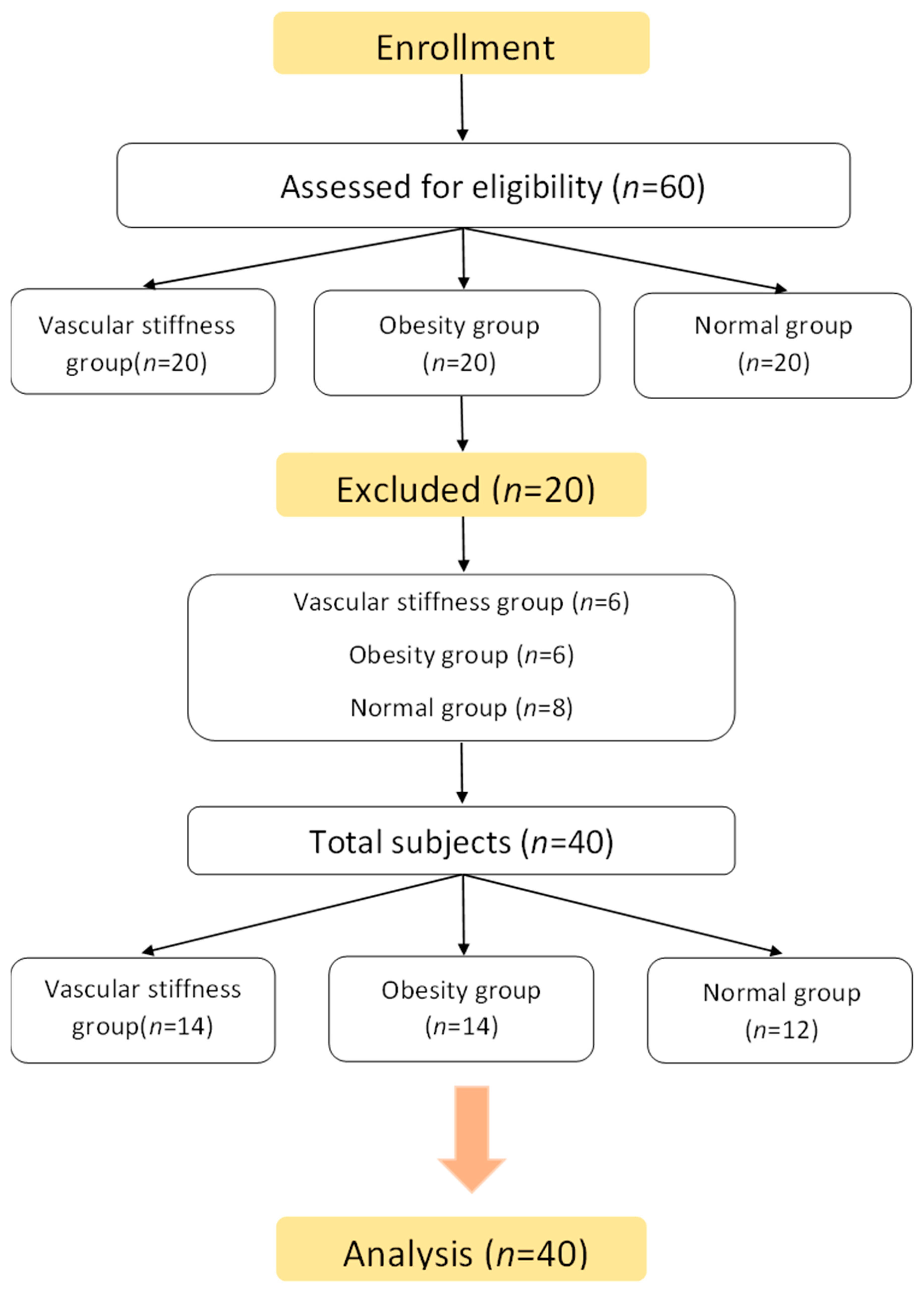

Table 1.
Physical characteristics of subjects in each group.
| Variables Group |
Age (yrs) |
Height (cm) |
Weight (kg) |
BMI (kg/m2) |
%BF (%) |
cf-PWV (m/s) |
|---|---|---|---|---|---|---|
| VSG (n=14) |
78.64 ±4.55 |
152.39 ±4.71 |
51.48 ±6.70 |
22.13 ±2.12 |
29.26 ±5.56 |
14.77 ±1.94 |
| OG (n=14) |
76.50 ±4.82 |
150.14 ±4.22 |
61.97 ±7.43 |
27.40 ±2.35 |
39.04 ±4.78 |
10.89 ±0.98 |
| NG (n=12) |
78.75 ±5.79 |
147.83 ±6.70 |
48.55 ±7.06 |
22.14 ±2.03 |
30.08 ±6.34 |
9.94 ±1.31 |
| F | 13.331*** | 26.535*** | 13.107*** | 40.581*** | ||
| Scheffe | NS | NS | VSG, NG, <OG |
VSG, NG <OG |
VSG, NG <OG |
OG, NG <VSG |
Values are M±SD; BMI: body mass index, %BF: percentage of body fat, cf-PWV: carotid to femoral pulse wave velocity, VSG: vascular stiffness group, OG: obesity group, NG: normal group, ***p<.001, NS: non significant.
Table 2.
Changes in cf-PWV after 16-week aerobic exercise.
| Variable | Group | Pre | Post | diff(%) | T | F | |
|---|---|---|---|---|---|---|---|
| cf-PWV (m/s) |
VSG (n=14) |
14.77 ±1.94 |
12.61 ±1.73 |
-14.62 | 3.712** | Group | 31.127*** |
| OG (n=14) |
10.89 ±0.98 |
10.88 ±1.30 |
-0.09 | 0.058 | Time | 9.422** | |
| NG (n=12) |
9.94 ±1.31 |
9.93 ±1.40 |
-0.10 | 0.052 | G×T | 9.319*** | |
| F | 40.581*** | 10.983*** | 7.730** | ||||
| Scheffe | OG, NG <VSG |
OG, NG <VSG |
NG<OG <VSG |
||||
Values are M±SD, cf-PWV: carotid to femoral pulse wave velocity, VSG: vascular stiffness group, OG: obesity group, NG: normal group, **p<.01, ***p<.001.
Table 3.
Changes in cell adhesion molecules after 16-week aerobic exercise.
| Variable | Group | Pre | Post | diff(%) | t | F | |
|---|---|---|---|---|---|---|---|
| ICAM-1 (ng/mL) |
VSG (n=14) |
10.57 ±2.89 |
9.48 ±2.45 |
-10.31 | 3.493** | Group | 0.804 |
| OG (n=14) |
9.92 ±3.69 |
9.62 ±2.75 |
-3.02 | 0.674 | Time | 13.462*** | |
| NG (n=12) |
11.84 ±4.55 |
10.75 ±3.33 |
-9.20 | 2.740* | G×T | 1.404 | |
| F | 0.873 | 0.760 | 2.898 | ||||
| Scheffe | NS | NS | NS | ||||
| VCAM-1 (ng/mL) |
VSG (n=14) |
33.15 ±6.65 |
26.94 ±4.84 |
-18.73 | 5.241*** | Group | 1.745 |
| OG (n=14) |
28.13 ±5.81 |
25.67 ±4.20 |
-8.75 | 2.144 | Time | 28.254*** | |
| NG (n=12) |
29.29 ±3.67 |
28.11 ±2.41 |
-4.03 | 1.659 | G×T | 6.006** | |
| F | 3.060 | 1.197 | 5.115* | ||||
| Scheffe | NS | NS | NG<OG <VSG |
||||
| E-selectin (ng/mL) |
VSG (n=14) |
4.74 ±1.37 |
3.71 ±1.50 |
-21.73 | 4.236*** | Group | 1.258 |
| OG (n=14) |
4.42 ±2.04 |
4.22 ±1.42 |
-4.52 | 0.816 | Time | 11.157** | |
| NG (n=12) |
3.57 ±1.30 |
3.39 ±1.28 |
-5.04 | 0.771 | G×T | 4.052* | |
| F | 1.766 | 1.143 | 5.389** | ||||
| Scheffe | NS | NS | OG<NG <VSG |
||||
Values are M±SD, ICAM-1: intracellular cell adhesion molecule-1, VCAM-1: vascular cell adhesion molecule-1, E-selectin: endothelial selectin, VSG: vascular stiffness group, OG: obesity group, NG: normal group, *p<.05, **p<.01, ***p<.001, NS: non significant, G×T: between group and time interaction.
Table 4.
Changes in oxidized-LDL after 16-week aerobic exercise.
| Variable | Group | Pre | Post | diff(%) | t | F | |
| Oxidized-LDL(U/L) | VSG(n=14) | 10.40±3.22 | 7.38±2.99 | -29.04 | 4.145** | Group | 0.235 |
| OG(n=14) | 8.69±3.33 | 8.67±2.87 | -0.23 | 0.027 | Time | 11.895*** | |
| NG(n=12) | 8.32±2.98 | 7.96±2.72 | -4.33 | 0.730 | G×T | 8.740*** | |
| F | 1.630 | 0.712 | 8.920*** | ||||
| Scheffe | NS | NS | OG, NG<VSG | ||||
Values are M±SD, Oxidized-LDL: oxidized low density lipoprotein, VSG: vascular stiffness group, OG: obesity group, NG: normal group, **p<.01, ***p<.001, NS: non significant, G×T: between group and time interaction.
Table 5.
Simple regression analysis of cf-PWV change ratio and VCAM-1/oxidized-LDL change ratio following 16-week aerobic exercise.
Table 5.
Simple regression analysis of cf-PWV change ratio and VCAM-1/oxidized-LDL change ratio following 16-week aerobic exercise.
| Dependent Variable |
B | SE | β | t | F | R2 |
|---|---|---|---|---|---|---|
| Constant | -7.723 | 2.110 | -3.660*** | |||
| VCAM-1 | 0.374 | 0.162 | 0.352 | 2.315* | 5.359* | 0.124 |
| Oxidized-LDL | 0.675 | 0.319 | 0.325 | 2.118* | 4.485* | 0.106 |
Disclaimer/Publisher’s Note: The statements, opinions and data contained in all publications are solely those of the individual author(s) and contributor(s) and not of MDPI and/or the editor(s). MDPI and/or the editor(s) disclaim responsibility for any injury to people or property resulting from any ideas, methods, instructions or products referred to in the content. |
© 2023 by the authors. Licensee MDPI, Basel, Switzerland. This article is an open access article distributed under the terms and conditions of the Creative Commons Attribution (CC BY) license (http://creativecommons.org/licenses/by/4.0/).
Copyright: This open access article is published under a Creative Commons CC BY 4.0 license, which permit the free download, distribution, and reuse, provided that the author and preprint are cited in any reuse.


