
Submitted:
10 August 2023
Posted:
11 August 2023
You are already at the latest version
Abstract
Fourteen quinolizidine derivatives, structurally related to the alkaloids lupinine and cytisine and previously studied for other pharmacological aims, were presently tested for antiarrhythmic, inotropic, and chronotropic effects on isolated guinea pig heart tissues, and to assess calcium antagonism in comparison to amiodarone, lidocaine, procainamide, and quinidine. All compounds, but two, compared favorably with the reference drugs. Potent antiarrhythmic activity was observed for compounds (in increasing order) 4, 1, 6 and 5. The last compound N-(3,4,5-trimethoxybenzoyl)aminohomolupinane) was outstanding, exhibiting a nanomolar potency (EC50 = 0.017 µM) for the increase of threshold of ac-arrhythmia, and being devoid of antihypertensive activity against spontaneously hypertensive rats.
Keywords:
Quinolizidine derivatives
; Lupinine
; Cytisine
; Sparteine
; Antiarrhythmic activity
; Inotropic and chronotropic activities
; Vasorelaxant activity
1. Introduction
Arrhythmia is a complex abnormality of cardiac rhythm, affecting an increasing percent of the population with the increase of age. Arrhythmias are commonly induced or accompanied by heart diseases but may occur in patients with other disease and under certain drug therapies, and are, in itself, a danger to the patients. Atrial fibrillation is the most common cardiac arrhythmia, with high risk of thrombo-embolic stroke in older patients.
A large number of drugs are available, which are able to suppress dysrhythmic cardiac activity through different mechanism as it is illustrated in the classification of Vaughan [1,2], recently updated by M. Lei et al. [3]. However, a satisfactory pharmacological therapy has not yet been developed, and the search of new antiarrhythmic agents is still largely pursued, particularly with the aim to obtain compounds with multiple mechanism of action, in order to balance the pro-arrhythmic risk inherent to some, otherwise valuable, type of drugs (as those of class III) [4].
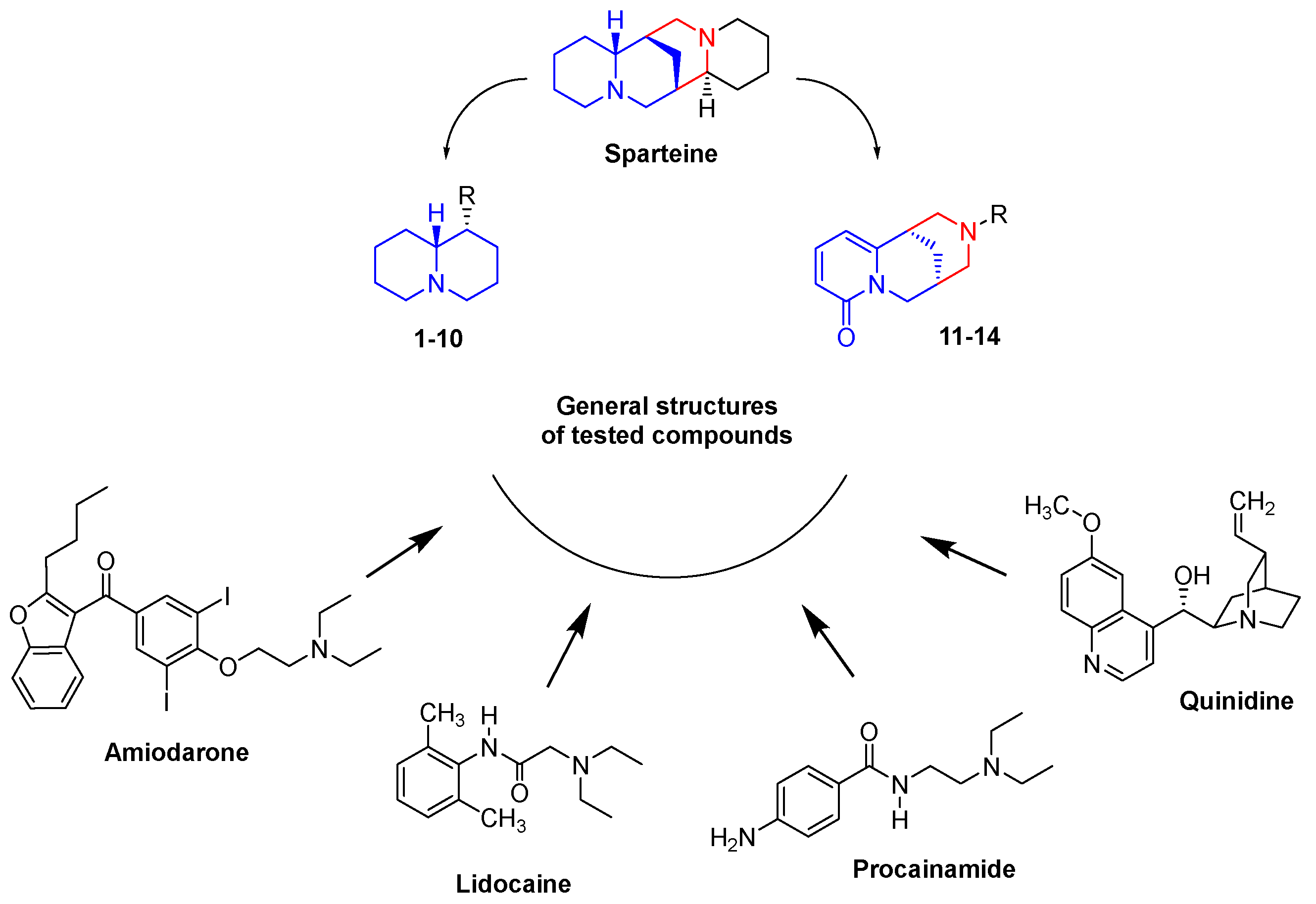
Since long time we are engaged in the investigation of potential antiarrhythmic agents characterized by the presence, in their structures, of the bulky, highly lipophilic and strongly basic quinolizidine moiety [5,6,7,8,9], which characterizes several bi-, tri- and tetracyclic alkaloids, as lupinine, cytisine, sparteine etc.
Indeed, this structural feature is embodied in the molecule of sparteine, which was largely used in the past [10] to slow the heart in tachycardia of various origins, and which has been reappraised as interesting antiarrhythmic agent, as such [11], or in the form of C-substituted derivatives [12,13,14,15], or of molecular truncated analogs [16,17,18,19,20,21,22,23].
Our approach was (and is) to hybridize a truncated portion of sparteine molecule (as a quinolizidinylmethyl- residue) with the aromatic moieties present in other well established antiarrhythmic drugs, like procainamide, lidocaine, amiodarone and quinidine etc.
Figure 1.
Drug design of tested compound.
The previously investigated quinolizidinyl derivative exhibited remarkable antiarrhythmic activity in mice subjected to deep chloroform anesthesia or aconitine infusion [5,6,7], or electrically driven isolated guinea pig (gp) left atria [8,9], often resulting more active and potent than amiodarone, lidocaine, procainamide and quinidine.
In particular, in the in vitro test, many compounds exhibited a EC50 even lower than 1 µM (Figure 2), while quinidine, the most potent of the reference drugs, had EC50 = 10.26 µM. When additionally tested for inotropic, chronotropic and calcium antagonist activity on isolated gp heart tissues, these compounds displayed an interesting profile, competing favorably to reference compounds.
On this ground, we deemed interesting to pursue the investigations of cardio-vascular profile of additional quinolizidine derivatives that were available in our in-house library, and could be allocated into three structural subsets (Figure 3):
- (a)
- N-(Quinolizidinyl-alkyl)-benzamides (1-5), related to the previously studied compounds A and B
- (b)
- (c)
- N-Substituted cytisines (11-14), characterized by the presence of three of the four rings of sparteine’s molecular scaffold. The investigated compounds were selected from a large number of cytisine derivatives previously studied by us as ligands for neuronal nicotine receptor and for various pharmacological activities (as anti-hypertensive and tobacco smoking cessation) [27,28,29,30].
2. Materials and Methods
2.1. Chemistry
2.2.“. In Vitro” Activities
Guinea pigs of either sex (200–400 g) obtained from Charles River (Calco, Como, Italy) were used. The animals were housed according to the ECC Council Directive regarding the protection of animals used for experimental and other scientific purposes (Directive 2010/63/EU of the European Parliament and of the Council) and the WMA Statement on Animal Use in Biomedical Research. All procedures followed the guidelines of animal care and use committee of the University of Bologna (Bologna, Italy). The ethical committee authorization was reported and numbered as “Protocol PR 21.79.14” by the Comitato Etico Scientifico for Animal Research Protocols according to D.L. vo 116/92. Guinea-pigs were sacrificed by cervical dislocation.
Compounds 1-14 were tested for antiarrhythmic activity on isolated guinea pig tissues: left atria driven at 1 Hz, and spontaneously beating right atria to evaluate their inotropic and chronotropic effects respectively. Finally, they were tested on K+-depolarized guinea pig aortic strips to assess calcium antagonist activity (as expression of vasorelaxant activity). In all case compounds were added in a cumulative manner.
In particular, the antiarrhythmic activity was evaluated inducing arrhythmias by application of sinusoidal alternating current (50 Hz) of increasing strength to the isolated left atria driven at 1 Hz, and assessing the “threshold of ac-arrhythmic” (the current strength at which extra beats occur) before and following the compound was added to the tissue bath.
Because arrhythmias by alternating current are mainly due to an increase of Na+ conductance in cardio myocytes, the method is particularly suitable to study antiarrhythmic agents acting as Na+ channel inhibitors (Class I, to which sparteine has been included) [33]. Anyhow, this model avoids the damage, toxicity and drug-drug interactions caused by other chemical methods used to induce arrhythmias [34].
2.2.1. Heart Preparation
After thoracotomy the heart was immediately removed and cleaned. The left and right atria were isolated from ventricle and separately prepared as previously described to test antiarrhythmic, inotropy and chronotropy activities [35].
2.2.2. Aorta Preparation
The thoracic aorta was removed, placed in Tyrode solution, cleaned, and repared as previously described [36].
2.3. Statistical Analysis
Data were analyzed using Student’s test and are presented as the mean (M) ± SEM [37]. Only the significance (P < 0.05) between the control and the experimental value at each concentrations is established. For compounds exhibiting an increase of the threshold of ac-arrhythmia higher than 50%, the EC50 values were calculated.
3. Results and discussion
Compounds 1-14 (Figure 3), together with amiodarone, lidocaine, procainamide, and quinidine as reference drugs, were evaluated in vitro for antiarrhythmic activity on isolated guinea pig left atria driven at 1 Hz, and for influence on the cardiovascular parameters inotropy and chronotropy and on vascular smooth muscle.
While quinidine and lidocaine clearly increased the threshold of ac-arrhytmias, procainamide and amiodarone show weak active (11% at 50 µM and 10 % at 100 µM, respectively).
Concerning the investigated compounds, the antiarrhythmic activity has been found, once more, in all subsets a) – c) of quinolizidine derivatives, despite the large structural diversity of the moieties, to which the quinolizidine ring is joined. Thus, the claimed link [8,9] between this structural feature (as truncated sparteine) and antiarrhythmic activity is further strengthened.
Indeed, with the only exception of compound 2 and 12, the tested quinolizidine derivatives showed rather high activity, resulting comparable or more active than lidocaine and procainamide. In some cases (1 and 4-6), the activity and potency of quinidine (the best reference drug) was reached and even largely exceeded. Compounds 4, 1 and 6 (in the order) were equipotent or up to 15-fold more potent than quinidine, while an outstanding potency (EC50 = 0.017 µM) was observed for compound 5, that resulted six hundred times more potent than quinidine and 8.8 times more potent than our previously described [9] most potent quinolizidine derivative C of Figure 2.
Compound 5 is the (one-carbon)-homolog of the previously described [9] N-(3,4,5-trimethoxybenzoyl)aminolupinane, which exhibited a quite lower antiarrhythmic potency (EC50 = 2.62 µM). The elongation of the linker between the quinolizidine ring and the aromatic nucleus produced a 150-fold increase in the potency.
A favorable effect of the linker length on the antiarrhythmic potency was already observed in the past (compare compounds D with the N-homolupinanoyl-2,6-aniline [8], and compounds F with E), but an opposite effect is also observed in the present study, by comparing the EC50 of compounds 1 with 2, as well as 6 with 8 (Table 1).
The outstanding potency of compound 5 (in itself and in comparison, to its lower homolog) could be related to the combination in the molecule of two pharmacophoric substructures, linked each other in the appropriate distance.
Indeed, some intrinsic anti-arrhythmic properties should be attributed to the trimethoxybenzoyl residue. On the base of the known activities of reserpine (including the antiarrhythmic one [40,41]), a number of basic esters and amides of the 3,4,5-trimethoxybenzoic acid were investigated and found endowed with different degrees of various cardiovascular activities. Particularly, the 5-(3,4,5-trimethoxy)benzamido-2-methyl-trans-decahydroisoquinoline (M32) displayed an anti-arrhythmic activity (i.p. in mice) five fold superior to that of quinidine [42,43].
Comparing the structures of 5 and M32, it is observed that the common aromatic moiety is linked to the respective basic nitrogen through a chain with the same number (6) of atoms, and that the two bicyclic systems (quinolizidine and decahydroisoquinoline) may adequately (even if not completely) overlap each other. Therefore, the two compounds could hit the same molecular target.
Importantly, also non-basic derivatives of trimethoxybenzoic acid may display valuable antiarrhythmic activity: in capobenic acid, where the trimethoxybenzoic acid is linked to ε-aminocaproic acid, the typical anti-fibrinolytic action of the latter is abolished, but a potent antiarrhythmic activity emerges [44].
Figure 4.
Comparison of the structures of compounds 5 and M32.
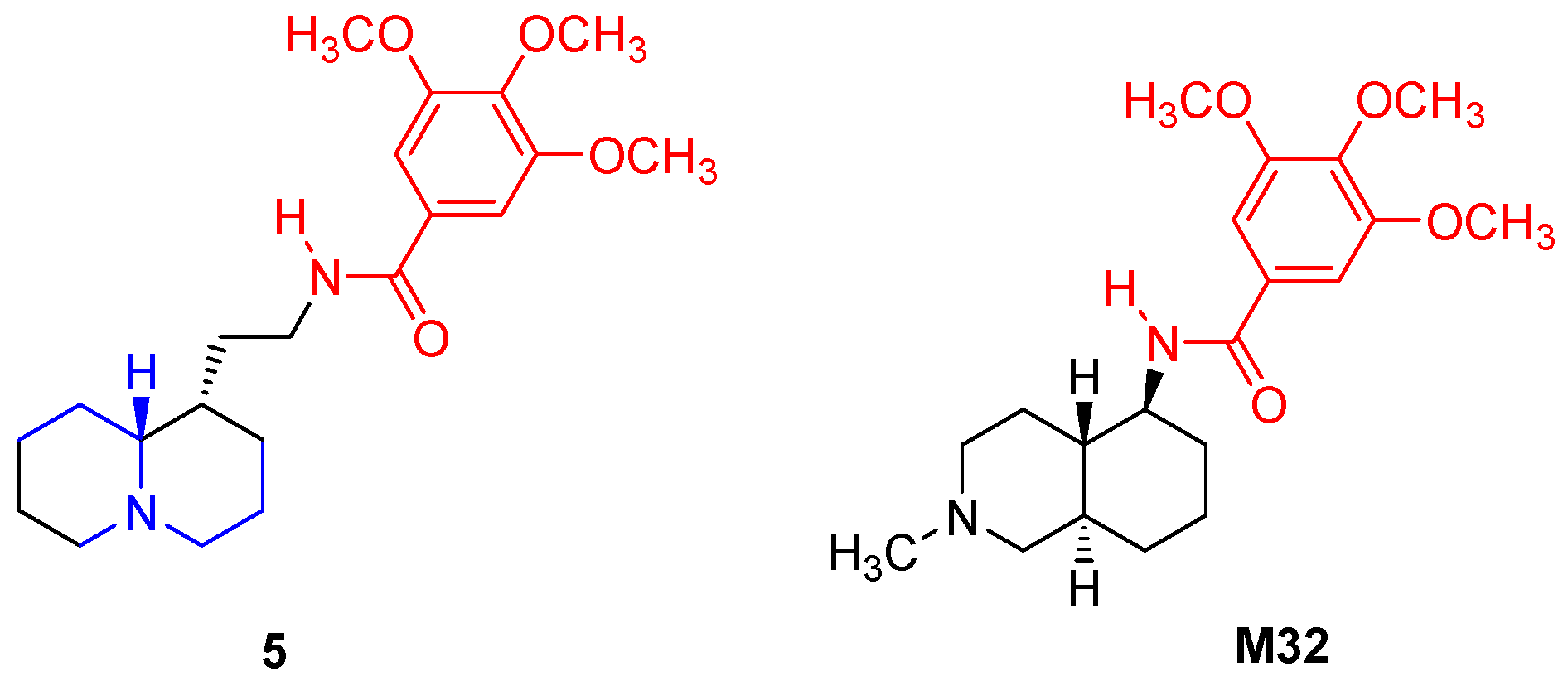
In contrast to the observed positive effect on antiarrhythmic activity, the lengthening of the linker produced an opposite effect on antihypertensive action: actually, compound 5 was found endowed with negligeable activity in spontaneously hypertensive rats [32], while its lower homolog displayed potent and long-lasting hypotensive activity in normal rabbits [45].
Compound 1, still displaying a moderate antiarrhythmic activity (EC50 = 3.66 µM), is a structural analog of sulpiride, whose (1-ethylpyrrolidin-2-yl)methyl moiety is replaced with the lupinyl ((quinolizidine-1-yl)methyl) residue.
Sulpiride displays a potent and selective antagonism versus D2 receptor, to which are related its psycholeptic, antiemetic and gastro-entero prokinetic activities, but it shows neither antiarrhythmic, nor arrhythmogenic potentials and exhibits a very low liability to prolong the QT interval in ECG (IC50 for hERG blockade > 100 µM) [46,47].
Compared to sulpiride, compound 1 displays a 1000-fold lower affinity to D2 receptor [7], and is also devoid [31] of affinity to 5-HT4 receptor that is determinative for the prokinetic activity of cisapride; nevertheless 1 maintains an appreciable activity on intestine transit rate in mice, with a mechanism still undefined (it increases the length of the small intestine colored by charcoal for 8.8%, versus 14% for sulpiride and 6.7% for metoclopramide [7]).
The N-lupinylbenzamido derivative 4, which is endowed with an antiarrhythmic activity potency (EC50 = 10.67 µM) comparable to that quinidine, is structurally related to the 6-methoxysalicylamides, described as particularly potent antagonists of D2 receptor [48,49]. As observed for compound 1, also in this case, the introduction of a lupinyl in place of the (1-ethyl-pyrrolidin-2-yl methyl moiety reduced the affinity to D2 receptor, even if for a minor extent, due to the presence of the free hydroxy group [48,49]. Anyhow, the absence of any causal relation between antiarrhythmic activity and affinity to D2 receptor is still observed.
Among the 2-(benzotriazol-2-yl)methyl benzimidazoles (subset b), compound 6 displays a potent antiarrhythmic activity with EC50 = 0.68 µM, confirming the activity previously observed [25] against ouabain induced arrhythmia in dogs. Structural modifications of compound 6, as epimerization (7) or elongation (8) of the basic side chain decreased both activity and potency; particularly deleterious was the introduction of substituent in position 5 of the benzimidazole ring (9 and 10).
Finally, the cytisine derivatives (11, 13 and 14) displayed moderate antiarrhythmic activity, approaching that of lidocaine. It is worth noting that N-(hydroxyethyl)cytisine (11) was already found active by Russian authors against aconitine induced arrhythmia in anesthetized rats, with a potency close to that of lappaconitine (allapinine) [50,51,52].
To better evaluate the pharmacological profile of the investigated quinolizidine derivatives 1-14, was deemed useful the comparison of their influence on additional cardiovascular parameters with that elicited by the reference drug (Table 2).
With one exception, all compounds strongly decreased the developed tension on driven g.p. left atria with EC50 in the range 0.009 – 0.083 µM, thus being comparable to lidocaine (EC50 = 0.017 µM). Compound 11, the least potent as negative inotropic agent, displayed a EC50 = 0.14 µM, that is still 24 times lower than that of quinidine.
Anyway, such general high negative inotropic activity does not invalidate the importance of several compounds, in particular of compound 5, whose EC50 for the increase of threshold of ac-arrhythmia remains lower than that for negative inotropism (0.017 µM versus 0.050 µM, respectively).
Interestingly, in preliminar investigation [27], it was observed that compound 14 (N-phenethylcytisine) at concentrations above 1 µM displayed positive inotropism, that reaches a maximum at 31 µM, with a 48% increase of force, which was comparable to that exerted by trequinsin (an ultra-potent PDE inhibitor) at 25 µM concentration [27].
At concentration above 100 µM, a negative inotropism was again observed. This alternating effect on the developed tension was not observed for the other cytisine derivatives.
Concerning the chronotropic activity (Table 2), detected on spontaneously beating right atria, amiodarone, and quinidine (at 100 and 50 µM, respectively) exerted a strong negative effect (72% - 86%), while procainamide and lidocaine showed a moderately positive effect (9% and 29%, at 100 µM and 5 µM, respectively).
All, but one (3), tested compound displayed negative chronotropic activity, that was, generally, rather modest; thus, for only three compounds (6, 7 and 8) was possible to calculate the EC50 values. For compound 6, the negative chronotropic activity was comparable (EC50 = 11.15 µM) to that of amiodarone (EC50 = 14.95 µM), but for the corresponding homolog 8 a remarkable (507-fold) increase of negative activity was observed (EC50 = 0.019 µM).
Also, the vasorelaxant activity, as expressed by the inhibition of calcium induced contraction on K+-depolarized (80 µM) g.p. aorta strips, was rather modest. Compounds 1, 2, 3 and 11 were practically inactive, similarly to amiodarone and procainamide; all the other inhibited the aorta strip contraction for 11-36% at 50-100 µM, as lidocaine and quinidine.
4. Conclusions
Fourteen quinolizidine derivatives (or emboding this ring in their structure) were tested for antiarrhythmic, inotropic, and chronotropic effects on isolated guinea pig heart tissues, and to assess calcium antagonism (vasorelaxant activity), in comparison to amiodarone, lidocaine, procainamide and quinidine.
The tested compounds were grouped in three subsets, in relation to the different molecular moieties to which the quinolizidine ring is linked.
Various degree of antiarrhythmic activity was observed in all the studied subsets of quinolizidine derivatives, as already found for other groups of derivatives [5,6,7,8,9], further supporting the existence of a rather peculiar interaction between the rigid and bulky quinolizidine ring and the cellular structures involved in the regulation of heart activity.
Excluding compounds 2 and 12, all compounds compared favorably with the reference drugs. In particular, potent antiarrhythmic activity was observed for compounds (in order of increasing potency) 4, 1, 6, and 5; the last of which was very outstanding with a EC50 = 17 nM. It is suggested that the unusual potency of compound 5 might be related to the combination in the same molecule, at the appropriate distance, of two distinct pharmacophores, as the bulky and lipophilic quinolizidine ring and the 3,4,5-trimethoxybenzoyl residue.
On the whole, compound 5 exhibits an interesting cardiovascular profile, being endowed with high antiarrhythmic activity and potency and negligible vasorelaxant activity and devoid of anti-hypertensive activity on spontaneously hypertensive rats.
Also appreciable is the benzimidazole derivative 6, with submicromolar potency (EC50 = 0.68 µM) for antiarrhythmic activity, while the negative chronotropism (EC50 = 6.36 µM), was minor than that of amiodarone (EC50 = 5.57 µM).
Worth of note is compound 1, whose valuable cardiovascular profile is associated to a gastro-enteric prokinetic activity, which might be profitable in the case of concomitant (and reciprocally worsening) cardiac and gastric pathologies [51].
The cytisine derivative 14, even if quite less potent than the foregoing compounds, still increased for 16% the threshold of ac-arrhythmic at 1 µM concentration, at which it started to display an unusual positive inotropic activity, very useful in arrhythmia associated to heart failure. Interestingly, this compound at the dose of 30 mg/kg (p.os and i.p.) did not exhibit any sign of toxicity in mice, and produced a 50% reduction of stress induced ulcers in rats [27].
Concluding, the mentioned compounds deserve further investigation to define more completely their pharmacological profile and mechanism of action, allowing the development of leads for better antiarrhythmic agents.
Author Contributions
Conceptualization, F.S.; methodology, B.T; R.B.; M.T.; V.B.; L.B.M.; investigation, F.S.; R.B.; B.T.; M.T.; L.B.M.; V.B.; writing—original draft preparation, F.S.; R.B.; writing—review and editing, F.S.; B.T.; R.B.; A.C.; All authors have read and agreed to the published version of the manuscript.
Funding
This research received no external funding.
Institutional Review Board Statement
The animal study protocol was approved by the Comitato Etico Scientifico for Animal Research Protocols according to D.L. vo 116/92 (protocol code PR 21.79.14).
Informed Consent Statement
Not applicable
Data Availability Statement
Not applicable
Conflicts of Interest
The authors declare no conflict of interest.
References
- Vaughan Williams, E.M. Classification of antidysrhythmic drugs. Pharmacol Ther. B 1975, 1, 115–138. [Google Scholar] [CrossRef]
- Vaughan Williams, E.M. Classifying antiarrhythmic actions: by facts or speculation. J. Clin. Pharmacol., 1992, 32, 964–977. [Google Scholar] [CrossRef]
- Lei, M.; Wu, L.; Terrar, D.A.; Huang, C.L.-H. Modernized classification of cardiac antiarrhythmic drugs. Circulation, 2018, 138, 1879–1896. [Google Scholar] [CrossRef]
- a) Matyus, P.; Varro, A.; Papp, J.G.; Wamhoff, H.; Vargas, I.; Virag, L. Antiarrhythmic agents: current status and perspectives. Med Res. Rev., 1997, 17, 427-451. b) Geng, M.; Lin, A.; Nguyen, T.P. Revisiting antiarrhythmic drug therapy for atrial fibrillation: reviewing lessons learned and redefining therapeutic paradigms. Front. Pharmacol. 2020, 11, 581837.
- Sparatore, A.; Sparatore, F. Preparation and pharmacological activities of 10-homolupinanoyl-2-R-phenothiazines. Farmaco, 1994, 49, 5–17. [Google Scholar] [PubMed]
- Sparatore, A.; Sparatore, F. Preparation and pharmacological activities of homolupinanoyl anilides. Farmaco, 1995, 50, 153–166. [Google Scholar]
- Iusco, G.; Boido, V.; Sparatore, F. Synthesis and preliminary pharmacological investigation of N-lupinyl-2-methoxybenzamides. Farmaco, 1996, 51, 159–174. [Google Scholar] [PubMed]
- Vazzana, I.; Budriesi, R.; Terranova, E.; Joan, P.; Ugenti, M.P.; Tasso, B.; Chiarini, A.; Sparatore, F. Novel quinolizidinyl derivatives as antiarrhythmic agents, J. Med. Chem. 2007, 50, 334–343. [Google Scholar] [CrossRef]
- Tasso, B.; Budriesi, R.; Vazzana, I.; Joan, P.; Micucci, M.; Novelli, F.; Tonelli, M.; Sparatore, A.; Chiarini, A.; Sparatore, F. Novel quinolizidinyl derivatives as antiarrhythmic agents. 2. Further investigation. J. Med. Chem. 2010, 53, 4668–4677. [Google Scholar] [CrossRef]
- E.L. McCawley, Cardioactive alkaloids. In: R.H.F. Manske Ed. “The alkaloids. Chemistry and Physiology. Vol. 5. Academic Press, New York, 1955, 93-97.
- Philipsborn, G.V.; Wilhelm, E.; Homburger, H. Untersuchungen zur Wirkung von Spartein am isolierten Vorhofmyokard von Maerschweinchen. Naunyn-Scmiedeberg’ Arch. Pharmacol. 1973, 277, 281–290. [Google Scholar] [CrossRef] [PubMed]
- Raschaek, M. Wirking von Spartein und Spartein derivate auf Herz und Kreislauf. Azneim. Forsch. 1974, 24, 753–759. [Google Scholar]
- Engelmann, K.; Radke, W.; Petter, A. Die Bedentung hydrophober Gruppen fur die antiarrhythmische eigenschaft alkylierte Sparteine. Arzneim. Forsch. 1974, 24, 759–764. [Google Scholar]
- Zetler, G.; Strubelt, O. Antifibrillatory, cardiovascular and toxic effects of sparteine, butylsparteine and pentylsparteine. Arznreim. Forsch. 1980, 30, 1497–1502. [Google Scholar]
- Gawall, V.S.; Simeonov, S.; Drescher, M.; Knott, T.; Scheel, O.; Kudolo, J.; Kahlig, H.; Hochenegg, U.; Roller, A.; Todt, H.; Maulide, N. C2-Modified sparteine derivatives are a new class of potentially long-acting sodium channel blockers. ChemMedChem Comm. 2017, 12, 1819–1822. [Google Scholar] [CrossRef]
- Ruenitz, P.C.; Mokler, C.M. Analogs of sparteine. 5. Antiarrhythmic activity of selected N,N’-disubstituted bispidines. J. Med. Chem. 1977, 20, 1668–1671. [Google Scholar] [CrossRef]
- Ruenitz, P.C.; Mokler, C.M. Anthiarrhythmic activity of some N-alkylbispidinebenzamides. J. Med. Chem. 1979, 22, 1142–1147. [Google Scholar] [CrossRef]
- Hiraoka, M.; Sunami, A.; Tajima, K. Bisaramil, a new class I antiarrhythmic agent. Cardiovasc. Drug Rev. 1993, 11, 516–524. [Google Scholar] [CrossRef]
- Schoen, U.; Antel, J.; Bruckner, R.; Messinger, J.; Franke, R.; Gruska, A. Synthesis, pharmacological characterization, and quantitative structure-activity relationship analyses of 3,7,9,9-tetraalkylbispidines: Derivatives with specific bradycardic activity. J. Med. Chem. 1998, 41, 318–331. [Google Scholar] [CrossRef]
- Takanaka, C.; Sarma, J.S.; Singh, B.N. Electrophysiological effects of ambasilide (LU 47110), a novel class II antiarrhythmic agent, on the properties of isolated rabbit and canine cardiac muscle. J. Cardiovasc. Pharmacol. 1992, 19, 290–298. [Google Scholar] [CrossRef]
- Pugsley, M.; Walker, M.J.A.; Garrison, G.B.; Howard, P.G.; Lazzara, R.; Patterson, E.; Penz, W.P.; Scherlag, B.J.; Berlin, K.D. The cardiovascular and antiarrhythmic properties of a series of novel sparteine analogs. Proc. West. Pharmacol. Soc. 1992, 35, 87–91. [Google Scholar] [PubMed]
- Pugsley, M.K.; Saint, D.A.; Hayes, E.; Berlin, K.D.; Walker, M.J. The cardiac electrophysiological effects of sparteine and its analog BRD-1-28 in the rat. Eur. Pharmacol. 1995, 294, 319–327. [Google Scholar] [CrossRef] [PubMed]
- Tomassoli, J.; Gundish, D. Bispidine as a priviliged scaffold. Curr. Topics in Med. Chem. 2016, 16, 1314–1342. [Google Scholar] [CrossRef] [PubMed]
- Pagani, F.; Sparatore, F. Benzotriazolylalkyl-benzimidazoles and their dialkylaminoalkyl derivatives. Boll. Chim. Far. 1965, 104, 427–431. [Google Scholar]
- Paglietti, G.; Boido, V.; Sparatore, F. Dialkylaminoalkylbenzimidazoles of pharmacological interest. Farmaco, Ed.Sci. 1975, 30, 505–511. [Google Scholar]
- Tonelli, M.; Paglietti, G.; Boido, V.; Sparatore, F.; Marongiu, F.; Marongiu, E.; La Colla, P.; Loddo, R. Antiviral activity of benzimidazole derivatives. I. Antiviral activity of 1-substituted-2-[(benzotriazol-1/2-yl)methyl]benzimidazoles. Chem. Biodiversity 2008, 5, 2386–2401. [Google Scholar] [CrossRef] [PubMed]
- Canu Boido, C.; Sparatore, F. Synthesis and preliminary pharmacological evaluation of some cytisine derivative. Farmaco 1999, 54, 438–451. [Google Scholar] [CrossRef]
- Canu Boido, C.; Tasso, B.; Boido, V.; Sparatore, F. Cytisine derivatives as ligands for neuronal nicotinic receptors and with various pharmacological activities. Farmaco 2003, 58, 265–277. [Google Scholar] [CrossRef]
- Tasso, B.; Canu Boido, C.; Terranova, E.; Gotti, C.; Riganti, L.; Clementi, F.; Artali, R.; Bombieri, G.; Meneghetti, F.; Sparatore, F. Synthesis, binding and modeling studies of new cytisine derivatives, as ligand for neuronal nicotinic acetylcholine receptor subtypes. J. Med. Chem. 2009, 52, 4345–4357. [Google Scholar] [CrossRef]
- Sala, M.; Braida, D.; Pucci, L.; Manfredi, I.; Marks, M.J.; Wageman, C.R.; Grady, S.R.; Loi, B.; Fucile, S.; Fasoli, F.; Zoli, M.; Tasso, B.; Sparatore, F.; Clementi, F.; Gotti, C. CC4, a dimer of cytisine, is a selecctive partial agonist at alpha4beta2/alpha6beta2 nAChR with improved selectivity for tobacco smoking cessation. Brit. J. Pharmacol. 2013, 168, 835–849. [Google Scholar] [CrossRef] [PubMed]
- Iusco, G.; Boido, V.; Sparatore, F.; Colombo, G.; Saba, P.L.; Rossetti, Z.; Vaccari, A. New benzamide-derived 5-HT3 receptor antagonists which prevent the effects of ethanol on extracellular dopamine and fail to reduce voluntary alcohol intake in rats. Farmaco 1997, 52, 141–146. [Google Scholar] [CrossRef]
- Boido, V.; Boido, A.; Boido Canu, C.; Sparatore, F. Quinolizidinylalkylamines with antihypertensive activity. Farmaco Ed.Sci. 1979, 34, 2–16. [Google Scholar]
- Borchard, U.; Bosken, R.; Greeff, K. Characterization of antiarrhythmic drugs by alternating current induced arrhythmias in isolated heart tissue. Arch. Int. Pharmacodyn. 1982, 256, 253–268. [Google Scholar] [PubMed]
- Bhatt, L.K.; Naudakumar, K.; Bodhankar, L.S. Experimental animal models to induce cardiac arrhythmia. Indian J. Pharmacol. 2005, 37, 348–357. [Google Scholar] [CrossRef]
- Roselli, M.; Carrocci, A.; Budriesi, R.; Micucci, M.; Toma, M.; Di Cesare Mannelli, L.; Lovece, A.; Catalano, A.; Cavalluzzi, M.M.; Bruno, C.; De Palma, A.; Contino, M.; Perrone, M.G.; Colabufo, N.A.; Chiarini, A.; Franchini, C.; Ghelardini, C.; Habtemarian, S.; Lwentini, G. Synthesis, antiarrhythmic activity, and toxicological evaluation of mexiletine analogues. Eur. J. Med. Chem. 2016, 121, 300–307. [Google Scholar] [CrossRef] [PubMed]
- Zeka, K.; Marrazzo, P.; Micucci, M.; Ruparelia, K.C.; Arroo, R.R.J.; Macchiarelli, G.; Nottola, S.A.; Continenza, M.A.; Chiarini, A.; Angeloni, C.; Hrelia, S.; Budriesi, R. Activity af antioxidants from Crocus sativus L. petals. Preventive effects towards cardiovascular system. Antioxidants (Basel) 2020, 9(11), 1102. [Google Scholar] [CrossRef]
- R.J. Tallarida, R.B. Murray, Manual of pharmacologic calculations with computer programs, 2nd Ed. Springer Verlag: New York 1987.
- GraphPad Prism 4.03; Gaphpad Software Inc., San Diego, CA, http://www.graphpad.com.
- GraphPad Prism 3.02; Gaphpad Software Inc., San Diego, CA, http://www.graphpad.com.
- Ciofalo, E.; Levitt, B.; Roberts, J. Some aspects of the antiarrhythmic activity of reserpine. Brit J. Pharmacol. Chemother. 1966, 28, 44–50. [Google Scholar] [CrossRef]
- Lawson, J.W. Antiarrhythmic activity of some isoquinoline derivatives determined by a rapid screening procedure in the mouse. J. Pharmacol. Expl. Therap. 1968, 22–31. [Google Scholar]
- Mathison, J.W.; Gueldner, R.C.; Lawson, J.W.; Fawler, S.J.; Peters, E.R. The stereochemistry of 5-substituted decahydroisoquinolines and their antiarrhythmic activity. J. Med. Chem. 1968, 11, 997–1000. [Google Scholar] [CrossRef]
- Mathison, S.W.; Pennington, R.J. Synthesis and antiarrhythmic properties of some 5-benzamido-2-methyl-trans-decahydroisoquinolines. J. Med. Chem. 1980, 23, 206–209. [Google Scholar] [CrossRef]
- a) Garzia, A. Pharmaceutical omega-(trimethoxybenzamido)fatty acids. DE 2034192 (1971), Chem. Abstr. 1971,75, 55230c. b) Istituto Chemioterapico Italiano, L’infarto del miocardio: terapia e profilassi delle complicanze con C-TRE (3,4,5-trimethoxybenzoyl-epsilon-aminocaproic acid). Lediberg s.n.c- Bergamo, 1971.
- Boido, V.; Sparatore, F. Derivatives of natural aminoalcohols and diamines of pharmacological interest. 5. Novel derivatives of lupinine and aminolupinane. Preliminary observations on their pharmacological activity. Ann. Chim. (Rome), 1969, 59, 526–538. [Google Scholar]
- O’Connor, S.E.; Brown, R.A. The pharmacology of sulpiride – a dopamine receptor antagonist, Gen. Pharmacol. 1982, 13, 185–193. [Google Scholar] [CrossRef]
- Silvestre, J.S.; Prous, J. Comparative evaluation of hERG potassium channel blockade by antipsychotics. Methods Find Exp. Clin. Pharmacol. 2007, 29, 457–465. [Google Scholar] [CrossRef] [PubMed]
- De Paulis, T.; Kumar, Y.; Johansson, L.; Raemsby, S.; Florvell, L.; Hall, H.; Aengeby-Muller, K.; Ogren, S.O. Potential neuroleptic agents. 3. Chemistry and antidopaminergic properties of 6-methoxysalicylamides. J. Med. Chem. 1985, 28, 1263–1269. [Google Scholar] [CrossRef]
- De Paulis, T.; Kumar, Y.; Johansson, L.; Raemsby, S.; Hall, H.; Saellemark, M.; Aengeby-Muller, K.; Ogren, S.O. Potential neuroleptic agents. 4. Chemistry, behavioral pharmacology and inhibition of [3H]spiperone binding of 3,5-disubstituted N-[(1-ethyl-2-pyrrolidinyl)methyl]-6-methoxysalicylamides. J. Med. Chem. 1986, 29, 61–69. [Google Scholar] [CrossRef] [PubMed]
- Khisamatdinova, R.Y.; Yarmukhamedov, N.N.; Gabdrakhmanova, S.F.; Karachurina, L.T.; Sapozhnikova, T.A.; Baibulatova, N.Z.; Baschenko, N.Z.; Zarudi, F.S. Synthesis and antiarrhythmic activity of N-(2-hydroxyethyl)cytisine hydrochloride and 3-(2-hydroxyethyl)-1,5-dinitro-3-azabicyclo-[3.3.1]non-3-ene hydrochloride. Pharmaceutical Chem. J. 2004, 38, 311–313. [Google Scholar] [CrossRef]
- Shishkin, D.V.; Shaimuratova, A.R.; Lobov, A.N.; Baibulatova, N.Z.; Spirikhin, L.; Yunusov, M.S.; Makara, N.S.; Baschenko, N.Z.; Dokichev, V.A. Synthesis and biological activity of N-(2-hydroxyethyl)cytisine derivatives. Chem. Nat. Comp. 2007, 43, 190–196. [Google Scholar] [CrossRef]
- Tsipisheva, J.P.; Kovolskaya, A.V.; Khalilova, I.U.; Bakhtina, Y.Y.; Khisamutdinova, R.; Gabdrakhmanova, S.F.; Lobov, A.N.; Zarudi, F.S.; Yunusov, S.Y. New 12-N-β-Hydroxyethylcytisine derivatives with potential antiarrhythmic activity. Chem Nat. Comp. 2014, 56, 333–338. [Google Scholar] [CrossRef]
- Saeed, M.; Bhandohal J.S.; Visco, F.; Pekler, G.; Mushiyev, S. Gastrocardiac syndrome A forgotten entity. Am. J. Emergency Med. 2018, 1525e5-1525e7.Author 1, A.; Author 2, B. Title of the chapter. In Book Title, 2nd ed.; Editor 1, A., Editor 2, B., Eds.; Publisher: Publisher Location, Country, 2007; Volume 3, pp. 154–196. [CrossRef]
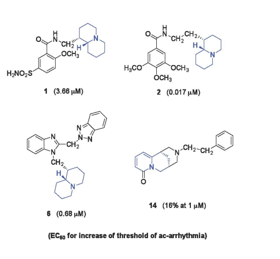
Figure 2.
Structures (A-H) of the most potent anti-arrhythmic quinolizidine derivatives previously studied: for A, G, H [8]; for B-F [9]; (EC50: μM).
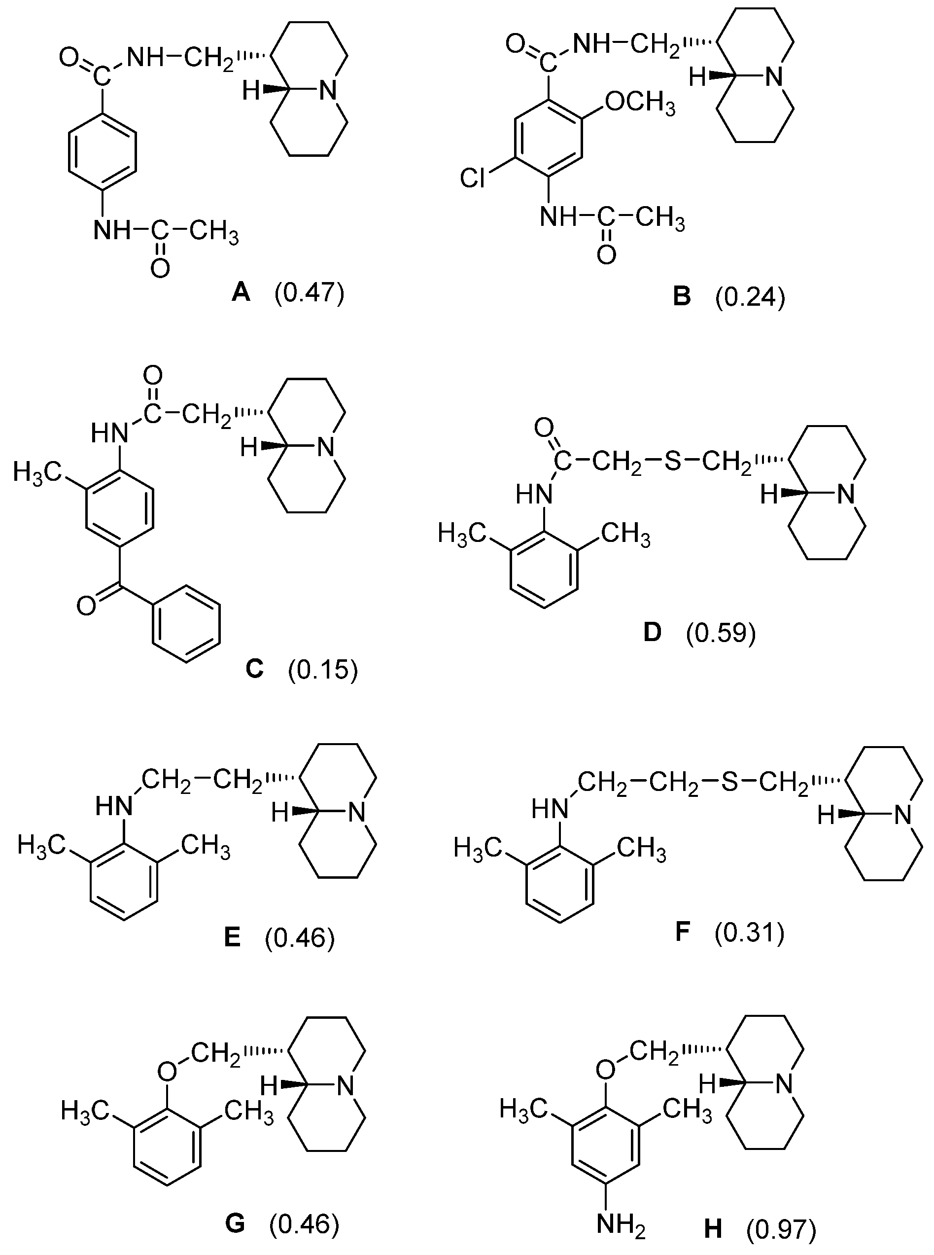
Figure 3.
Structures of the presently investigated quinolizidine derivatives (1-14).
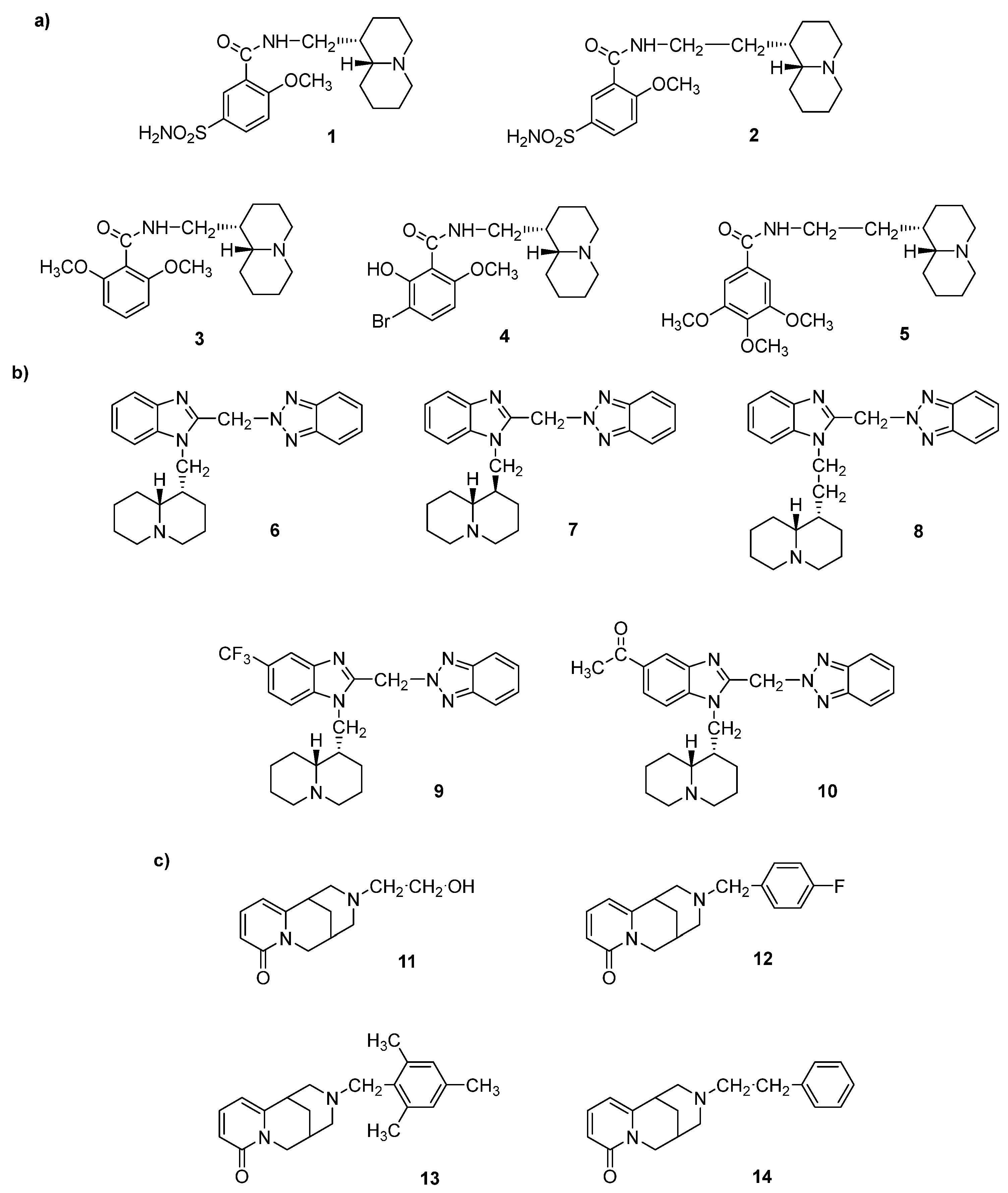

Table 1.
Anti-arrhythmic activity of compounds 1-14 (Figure 3).
Table 1.
Anti-arrhythmic activity of compounds 1-14 (Figure 3).
|
Compd |
Max% increase of threshold of ac-arrhythmia after pretreatment with compounda (M ± SEM) |
EC50b (μM) |
95% conf lim (μM) |
|---|---|---|---|
| Amiodarone | 10 ± 0.5c | ||
| Lidocaine | 34 ± 2.6 | ||
| Procainamide | 11 ± 0.4 | ||
| Quinidine | 69 ± 0.4 | 10.26 | 8.44 – 12.46 |
| N-(Quinolizidinyl-alkyl)-benzamides related compds | |||
| 1 | 59 ± 1.7 | 3.66 | 2.10 – 6.38 |
| 2 | 9 ± 0.6d | ||
| 3 | 17 ± 0.4e | ||
| 4 | 92 ± 1.3 | 10.67 | 7.56 – 14.91 |
| 5 | 104 ± 3.4 | 0.017 | 0.0068 – 0.046 |
| 1-(Quinolizidinyl)alkyl-2-(benzotriazol-2-yl)methyl benzimidazoles related compds | |||
| 6 | 75 ± 1.8d | 0.68 | 0.47 – 0.98 |
| 7 | 37 ± 3.4f | ||
| 8 | 39 ± 1.7d | ||
| 9 | 16 ± 0.9 | ||
| 10 | 20 ± 0.4 | ||
| N-Substituted cytisines related compds | |||
| 11 | 26 ± 0.3c | ||
| 12 | 4 ± 0.2 | ||
| 13 | 27 ± 1.3c | ||
| 14 | 16 ± 0.7g | ||
a Increase of threshold of ac-arrhythmia: increase in current strenght of 50 Hz alternating current required to produce arrhythmia in guinea pig left atria driven at 1 Hz in presence of each tested compounds at 5 x 10⁻⁵ M (unless otherwise stated). For all data P < 0.05, with the exception of compd 12. b Calculated from log concentration – response curves (Probit analysis according to Litchfield and Wilcoxon [38] with n = 6-8). When maximum effect was < 50%, the EC50 values were not calculated. c At 10-4 M. d At 10-5 M. e At 10-7 M. f At 5 x 10⁻⁶ M. g At 10-6 M.
Table 2.
Influences of compounds 1-14 on cardiovascular parameters.
| Heart | Aorta | ||||||
|---|---|---|---|---|---|---|---|
| Left atria | Right atria | ||||||
| Negative inotropy | Negative chronotropy | Vasorelaxant | |||||
| Compd | IAa (M ± SEM) |
EC50b (μM) |
95% conf lim (μM) |
IAc (M ± SEM) |
EC50b (μM) |
95% conf lim (μM) |
IAd (M ± SEM) |
| Amiodarone | 30 ± 2.6e | 72 ± 4.5e | 14.95 | 11.07 – 20.16 | 3 ± 0.1g | ||
| Lidocaine | 88 ± 3.0 | 0.017 | 0.012 – 0.024 | 29 ± 0.9#,j | 14 ± 0.9 | ||
| Procainamide | 92 ± 1.4f | 0.014 | 0.011 – 0.017 | 9 ± 0.6#,e | 3 ± 0.2 | ||
| Quinidine | 71 ± 3.6g | 3.38 | 2.69 – 4.25 | 86 ± 0.5 | 25.31 | 14.45 – 44.32 | 30 ± 1.6g |
| N-(Quinolizidinyl-alkyl)-benzamides related compds | |||||||
| 1 | 92 ± 1.4h | 0.037 | 0.027 – 0.051 | 24 ± 1.3k | 5 ± 0.2 | ||
| 2 | 93 ± 1.4i | 0.0091 | 0.002 – 0.021 | 2 ± 0.1h | 3 ± 0.2 | ||
| 3 | 75 ± 2.3i | 0.011 | 0.0079 – 0.014 | 25 ± 1.6# | 2 ± 0.1 | ||
| 4 | 98 ± 1.3 | 0.021 | 0.016 – 0.027 | 46 ± 2.2 | 36 ± 1.3 | ||
| 5 | 85 ± 2.2 | 0.050 | 0.035 – 0.071 | 25 ± 0.9h | 22 ± 1.6 | ||
| 1-(Quinolizidinyl)alkyl-2-(benzotriazol-2-yl)methyl benzimidazoles related compds | |||||||
| 6 | 91 ± 2.4h | 0.046 | 0.035 – 0.061 | 67 ± 0.7 | 11.15 | 9.05 – 13.74 | 25 ± 1.7g |
| 7 | 93 ± 2.7j | 0.083 | 0.064 – 0.11 | 69 ± 1.3h | 0.49 | 0.43 – 0.65 | 16 ± 1.1 |
| 8 | 92 ± 1.3 | 0.022 | 0.015 – 0.031 | 83 ± 2.4 | 0.019 | 0.014 – 0.026 | 19 ± 1.2 |
| 9 | 87 ± 1.1j | 0.056 | 0.042 – 0.076 | 44 ± 1.5h | 24 ± 1.6 | ||
| 10 | 94 ± 3.4 | 0.021 | 0.014 – 0.032 | 22 ± 1.2e | 11 ± 1.0 | ||
| N-Substituted cytisines related compds | |||||||
| 11 | 86 ± 2.2h | 0.14 | 0.095 – 0.20 | 35 ± 1.4k | 0.3 ± 00.1 | ||
| 12 | 92 ± 1.8h | 0.018 | 0.013 – 0.026 | 26 ± 1.9 | 25 ± 1.4 | ||
| 13 | 87 ± 1.4h | 0.044 | 0.028 – 0.068 | 20 ± 0.3h | 32 ± 2.2 | ||
| 14 | 71 ± 0.7i | 0.016 | 0.0081 – 0.023 | 47 ± 1.1e | 20 ± 1.6 | ||
a IA (Intrinsic Activity): decrease in developed tension on isolated guinea-pig left atrium driven at 1 Hz at 10-6 M (unless otherwise stated), expressed as percent changes from the control (n = 4-6). For all data P < 0.05. b Calculated from log concentration – response curves (Probit analysis according to Litchfield and Wilcoxon [38] with n = 6-8). When the maximum effect was < 50% the EC50 (inotropy), the EC50 (chronotropy) and IC50 (vasorelaxant) values were not calculated. c IA (Intrinsic Activity): decrease in atrial rate on guinea-pig spontaneously beating isolated right atria at 5 x 10⁻⁵ M (unless otherwise stated), expressed as percent changes from the control (n = 6-8). Pretreatment heart rate ranged from 170-195 beats/min. For all data P < 0.05, with the exception of compd 2. d IA (Intrinsic Activity): percent inhibition of calcium-induced contraction on K+-depolarized guinea–pig aortic strips at 10-4 M (unless otherwise stated). The 10-4 M concentration gave the maximum effect for all but one (6, at 5 x 10⁻⁵ M) compounds. P < 0.05 for amiodarone, procainamide and compds 1, 2, 3, and 11. e At 10-4 M. f At 5 x 10⁻⁷ M. g At 5 x 10⁻⁵ M. h At 10-5 M. i At 10-7 M. j At 5 x 10⁻⁶ M. # Positive chronotropic effect. k At 10-6 M.
Disclaimer/Publisher’s Note: The statements, opinions and data contained in all publications are solely those of the individual author(s) and contributor(s) and not of MDPI and/or the editor(s). MDPI and/or the editor(s) disclaim responsibility for any injury to people or property resulting from any ideas, methods, instructions or products referred to in the content. |
© 2023 by the authors. Licensee MDPI, Basel, Switzerland. This article is an open access article distributed under the terms and conditions of the Creative Commons Attribution (CC BY) license (http://creativecommons.org/licenses/by/4.0/).
Copyright: This open access article is published under a Creative Commons CC BY 4.0 license, which permit the free download, distribution, and reuse, provided that the author and preprint are cited in any reuse.


