Submitted:
31 July 2023
Posted:
02 August 2023
You are already at the latest version
Abstract
Purpose: To assess clinical outcomes of corneal cross-linking (CXL) intervention in a population diagnosed with progressive keratoconus.
Methods: This single-center retrospective cohort study included consecutive patients who underwent standard CXL or accelerated CXL for progressive keratoconus at a major teaching hospital in south Israel between January 2015 and December 2019. Patient medical files were reviewed to extract demographic, clinical, and tomographic data from outpatient clinic reports before treatment and one year post-operatively.
Results: This study included 166 patients (representing 198 eyes), out of which 98 patients (123 eyes) were ethnically Bedouin, and 68 patients (75 eyes) were ethnically Jewish. Overall, 126 patients (144 eyes) had a follow-up of at least 12 months (16.84±5.76). The mean patient age was 20.62 ± 7.1 years old. There were significant baseline differences between the two ethnical groups in best-corrected visual acuity (BCVA; p < 0.001), uncorrected visual acuity (UCVA; p < 0.001), mean keratometry (p = 0.028), and corneal thickness (p < 0.001). Significant changes in BCVA, UCVA, and pachymetry parameters within each group were found after 12 months. Negative binomial regression analysis showed a maximal keratometry below 55D (RR=1.247, p < 0.001) and a standard CXL procedure (RR=1.147, p = 0.041) are significantly related to the stability of KC after 12 months. However, the effect size of the origin of patients is negligible (RR=1.047, p = 0.47).
Conclusions: In this study, the Bedouin population suffered from more progressive keratoconus when compared to the Jewish population. CXL was significantly effective in improving BCVA and UCVA in both groups after 12 months of follow-up.
Keywords:
epidemiology
; ectasia
; Bedouin
; Jewish
; Kmax
1. Introduction
Keratoconus (KC) is a common bilateral non-inflammatory ectatic corneal disorder characterized by stromal thinning, irregular astigmatism of the cornea, and reduced visual acuity (VA) [1]. The disease is multifactorial with many risk factors, including familial, environmental, and regional factors as well as atopy and eye rubbing. Incipient stages of the disease can be asymptomatic and are often diagnosed only by revealing the changes of the corneal parameters by corneal tomography [2]. This method is considered the gold standard for diagnosis and monitoring the disease. Tomographic parameters reveal steeping of the anterior and posterior surface and the corneal thickness (pachymetry) [3,4]. As the disease progresses, cone-shaped cornea may be visible at the slit lamp [3], VA deteriorates, and patients may require penetrating keratoplasty. Hence, stabilization of the disease in its early stages is crucial.
Corneal cross-linking (CXL) has improved the treatment of ectatic corneal disorders, particularly KC [5]. CXL forms chemical bonds among collagen fibrils based on an interaction between ultra-violet A irradiation at a wavelength of 370 nm and riboflavin [6,7]. Case-control studies showed a significant decrease in the disease progression after CXL treatment, particularly presented by improvements in keratometry values and in VA in progressive disease cases [8].
Soroka University Medical Center (SUMC) is a major teaching hospital in the Negev area in south Israel and serves both Jewish (approximately 60%) and Bedouin (approximately 36%) populations [9]. The prevalence of KC in the Middle East has been found to be about 2%-5% [10,11,12]. In Israel, KC has been associated with eye rubbing, a family history of KC, and lower socio-economic status [13]. Additionally, Chorney and colleagues have found the socioeconomically challenged Bedouin minority [14] to be at risk for health complications such as diabetic retinopathy [15]. In this study, we focused on the corneal differences between SUMC’s ethnically diverse KC patients.
A previous meta-analysis population study on the natural progression of KC has found that ethnicity significantly affects corneal characteristics such as topography, VA, and pachymetry [16]. Maximum keratometry (Kmax) for example, has been found to naturally increase significantly greater among Middle Eastern populations when compared to European and East-Asian populations [16]. This study seeks to compare these corneal characteristics between the populations we encounter in SUMC after an intervention such as a CXL procedure. We expect that assessment of the differences between two ethnical groups with different approaches towards health and diseases [17] in terms of the consequences of KC treatment will elucidate the gravity of the disease and will help us understand treatment efficiency for each population.
2. Materials and Methods
2.1. Participants
This single-center retrospective cohort study included consecutive patients who underwent CXL or accelerated CXL (A-CXL) for progressive KC at the Department of Ophthalmology at a major teaching hospital in south Israel (SUMC) between January 2015 and December 2019. We included 166 patients (representing 198 eyes), of which 98 patients (123 eyes) belonged ethnically to the Bedouin group and 68 patients (75 eyes) belonged ethnically to the Jewish group. In total, 126 patients (144 eyes) had a follow up of at least 12 months (16.84±5.76). The mean age ± std. deviation of patients was 20.62 ± 7.1 years old, and 62.6% had been identified as male in their patient files. Table 1 shows patient demographic and clinical characteristics stratified by ethnic groups.
Progression was defined as an increase of 1.50 diopters (D) in mean keratometric value, a 1.00 D increase in Kmax, or a decrease of 5.0% in central corneal thickness at two consecutive evaluations by Scheimpflug-based corneal tomography (Pentacam HR; Oculus Optikgeräte, Wetzlar, Germany) [18,19,20,21,22]. In cases of suspected progression, a repeat exam including imaging was performed to confirm diagnosis. Exclusion criteria were a follow-up of less than 12 months, a history of previous ocular surgery, autoimmune diseases, diabetes mellitus, pregnancy or lactation, and sensitivity to riboflavin or any other substance used in CXL procedure. Patients were instructed to avoid eye rubbing prior to and during every follow-up. Patients with signs or symptoms of vernal kerato-conjunctivitis were treated appropriately to reduce the chances of eye rubbing.
2.2. Data Collection
Medical files of all patients were reviewed, and the following data were extracted: age, gender, ethnicity, date of CXL procedure, minimal corneal thickness (MCT), anterior/posterior mean keratometric power, anterior/posterior flat keratometric power, anterior/posterior steep keratometric power, maximum keratometric power, and uncorrected/corrected distance visual acuity. Keratometry and total corneal thickness were measured with Pentacam. Data regarding corneal thickness, keratometric power, and VA were measured before and after CXL was performed.
2.3. Main Outcomes Measures
The primary outcome in this study was the Δ of the Kmax in the two study populations prior to CXL procedure and the most recent visit (12 to 34 months after CXL). Secondary outcomes in this study were VA measures (uncorrected/corrected distance visual acuity) and corneal topography data of both anterior and posterior segments of the cornea: average keratometry value (Kmean), steep keratometry (Ksteep) and flat keratometry (Kflat), corneal astigmatism, and corneal pachymetry prior to CXL and the most recent visit (12 to 34 months after CXL), as well as the occurrence of any adverse events throughout the study period. Progression after CXL treatment, as defined by a maximal corneal curvature reading at the corneal apex (Kmax), increase of 1.5D within a year.
2.4. Surgical Technique
Following topical anesthesia with 0.4% benoxinate hydrochloride drops, MCT was confirmed by ultrasound pachymeter (PachPen; Accutome, Malvern, PA). Following removal of the central 8 mm of epithelium, the MCT was re-measured to ensure MCT above 400 μm. Randomly, either standard CXL (S-CXL) or A-CXL was then performed. S-CXL was performed similarly to the Dresden Protocol.
Briefly, iso-osmolar 0.1% riboflavin solution (Medio-Cross 0.1%; Peschke Meditrade, Huenenberg, Switzerland) was instilled every 2 min for 20 min. Adequate riboflavin penetration was confirmed by appropriate flare in the anterior chamber. The cornea was then continuously irradiated at 365 nm with either an intensity of 3 mW/cm2 for 30 min (S-CXL, total fluence 5.4 J/cm2) or an intensity of 9 mW/cm2 for 10 min (A-CXL, total fluence 5.4 J/cm2) using a commercially available device (LightLink-CXL; LightMed, San Clemente, CA). The patient was instructed to fixate on the light source and adequate centration was constantly monitored by one of the two surgeons (BK or RMK). In cases where MCT was less than 400 μm, contact lens assisted CXL modification was performed using a method similar to Matlov Kormas and colleagues [23].
2.4. Statistical Analysis
Clinical parameters were tabulated and analyzed using SPSS (version 23; IBM, Armonk, NY). For the analysis of demographical and clinical characteristics, t-test was used for normally distributed variables and Mann-Whitney test - continuous variables departing from normal distribution. To compare continuous parameters before and after treatment, we used paired t-test and Wilcoxon test normally distributed variables and other factors, respectively. A negative binomial regression model was performed to estimate an independent effect of various factors while adjusting to other factors. We used generalized estimating equations to adjust for clusters created by patients who were treated for both eyes.
3. Results
There were significant baseline differences between the two ethnical groups in best-corrected visual acuity (BCVA), uncorrected visual acuity (UCVA), mean keratometry and corneal thickness. Deeper analysis of the corneal thickness thinnest point showed a significant difference between the groups in different levels, while the thickness tends to be significantly higher in the Jewish group (see Table 1).
Intra-group comparison showed no significant difference in Kmax and Kmean after 12 months when compared to the baseline. However, there was a significant difference in BCVA, UCVA, and corneal thickness (central, apical, and minimal point) after 12 months.
We found a significant difference between the Jewish and Bedouin population in the Negev area of south Israel in corneal thickness prior to CXL interventional procedure (see Table 1). The differences in corneal thickness were also significant between each group before and after CXL was performed (see Table 2).
Moreover, we found that corneal thickness was significantly higher among the Jewish group than the Bedouin group (see Table 1).
CXL procedure has been found significantly effective in improving UCVA and BCVA in both groups after 12 months of follow-up (see Table 2). However, no significant findings were found in comparison between the groups in the mean change of corneal characteristics (see Table 3).
Stability was defined as an increase in less than 1.5D or a decrease in Kmax after 12 months after CXL procedure was performed. This study has found that the stability was 77.2% in the Jewish group and 88.2% in the Bedouin group (see Figure 1).
A negative binomial regression analysis (see Table 4) showed Kmax below 55D (RR=1.247, P<0.001) and a S-CXL procedure (RR=1.147, P=0.041) are significantly related to the stability of KC after 12 months of follow up. However, the effect size of the origin of patients is negligible (RR=1.047, P=0.472 for Jewish origin).
4. Discussion
KC is a relatively common multifactorial disease. Even though previous studies suggested both genetic and environmental risk factors [24,25], the etiology of KC disease remains unknown. KC has been associated with eye rubbing, atopy, floppy eyelid syndrome, pregnancy, and thyroid hormone disturbances [25]. Studies regarding eye rubbing, for example, showed a reduction of 18.4% in epithelial thickness immediately after rubbing [26]; and some even found eye-rubbing as the most significant risk factor for KC [27]. Atopy has also been associated with KC [28], though some authors suggested that the association is indirect only via itch which leads to eye-rubbing [27]. Kaya and colleagues showed that KC patients with atopy had a steeper and thinner ectatic cornea [29]. Studies evaluating the effect of pregnancy showed that KC progressed during the pregnancy period and continued to progress during the post-partum period [30].
The tendency of corneal thickness to be higher among the Jewish group than the Bedouin group (see Table 1) can be viewed through the risk factors associated to KC.
Data regarding eye rubbing can be complicated to compare between groups; however, its association to KC development and progression can be compared indirectly through atopy [27]. Regarding atopy, asthma, for example, has been found to be more prevalent among low socioeconomic populations in 63.0% of studies reviewing the issue [31]. Being a socioeconomically challenged population in south Israel means also significant exposure to dust and sand. Thus, some corneal characteristics of the Bedouin group can be explained by these risk factors. Since KC has been found to progress during pregnancy, extremely high birth rate among the Bedouin population might also contribute to the progression of KC among this population [30,32,33]. This finding could possibly explain the higher number of Bedouin women (46.0%) in comparison to Jewish women (22.1%) in our study group (see Table 1).
In addition to corneal thickness, other baseline corneal and clinical parameters were significantly inferior in the Bedouin group than in the Jewish group (e.g., UCVA, BCVA, and Kmean). These parameters can emphasize the fact that Bedouin patients have more progressed levels of KC. Studies regarding genetic factors and consanguinity, a prevalent phenomenon among the Bedouin population in the Negev [34] have found that children of consanguineous parents have a four-fold risk of KC when compared to children of unrelated parents [35]. In our study, positive family history of KC has been found significantly higher among the Bedouin group (see Table 1), a factor considered to increase risk for KC [11] can help explain the inferior corneal condition of this group.
Studies regarding compliance among the Bedouin population in a variety of diseases and disorders (e.g., cardiovascular disorders [36], diabetes mellitus [37], and genetic screening tests [38]) have concluded that compliance in this population is lower than in the Jewish population. Lower mean levels of corneal characteristics among the Bedouin group (see Table 2) can be explained via this fact also.
CXL procedure has been found significantly effective in improving UCVA and BCVA in both groups after 12 months of follow-up with no significant difference between the groups. Our multivariate analysis showed the effect size of the origin of the patients is negligible. These findings implicate that CXL procedure is effective in halting the progression of KC regardless of ethnic group or level of KC. Moreover, stabilization rates which were found to be similar in both groups, confirming this conclusion.
The corneal differences we report in this study arise questions regarding steps which possibly can be taken towards the Bedouin minority for prevention, diagnosis and treatment of KC (e.g., screening tests). First, rising the awareness for such pathology and its ramification may lead to better collaboration and early diagnosis. A clear and plain explanation of the disease causes, prevention, treatment, and routine follow-up, both clinically and tomographically, may increase both awareness and response to routine check-up. Secondly, improving the availability of ophthalmology services for Bedouin population and performing routine examinations may identify patients with KC in early stages. For example, retinoscopy is a simple screening test that can implicate the existence of KC. Al-Mahrouqi and colleagues have found that retinoscopy can serve as sensitive and reliable test for detection of KC even in early stages [39]. Another example of a screening tool is imaging. Corneal and epithelial thickness map and patterns, in addition to corneal topography or tomography, can be used to improve the screening of KC disease with a sensitivity of 97.8% [40].
Regarding stability, our multivariate analysis has found connection between preoperative maximal keratometry less than 55D to stabilization of CXL after 12 months of follow up (see Table 4). Koller and colleagues also found higher Kmax a significant risk factor for failure of CXL (Kmax>58D) [41]. Another study showed that patients with Kmax>60D were at higher risk for failure. S-CXL procedure has also been found positively related to stability [42] (see Table 4). This finding is supported by other studies which have found S-CXL associated with higher stabilization rates than A-CXL [43,44]. Regarding differences between the Jewish and Bedouin populations, our multivariate study has found the differences to be negligible (see Table 4), suggesting that the procedure results do not depend on the ethnical origin of the patients but on the preoperative severity of the disease.
A delay in diagnosis among the Bedouin population and the rapid deterioration emphasis that early intervention should be considered. Performance of CXL procedure under the age of 17 years old, was not found significantly different than among patients over 17 years old [45], implicating that early intervention is effective for early stabilization and better progress of corneal parameters [46]. Therefore, we suggest in order to achieve better rates of stability and halt the disease in earlier stages among the Bedouin population, intervention should follow diagnosis as soon as possible.
We have found CXL to be effective in halting the progression of KC for at least one year regardless of ethnicity and stage at diagnosis. We have also found significant tomographic and clinical differences between the Jewish and Bedouin populations in south Israel. These differences can be explained by a delayed diagnosis, cultural differences (e.g., consanguinity), negative influences of some environmental factors (allergy, atopy) and poor compliance to treatment and follow up. We suggest that health promotion regarding the consequences of untreated KC, screening tests (e.g., refraction, retinoscopy and corneal topography) and early intervention should take place in the community clinics of the minority population in the Negev.
Considering the Bedouin population in the Negev is a socioeconomically challenged minority with limited access and compliance to healthcare, our conclusions might be relevant to similar minority groups around the world.
5. Study Limitations and Future Research
This study does have limitations. As a retrospective study, the conclusions are limited to available data. A future prospective study would allow more control, which could lead to more specific conclusions for each population. In addition, only 72.0% of patients who had undergone CXL procedure had a follow up of a least 12 months. Thus, our conclusions could bias the comparison between the groups towards only compliant patients. Future work could ensure a more stringent follow-up protocol.
Author Contributions
Conceptualization, B.K. and R.M.K.; Methodology, B.K.; Validation, B.M., M.B., R.T., and B.K.; Formal Analysis, J.A.Y.; Investigation, J.A.Y., R.M.K. and B.K.; Data Curation, J.A.Y.; Writing – Original Draft Preparation, J.A.Y and R.M.K.; Writing – Review & Editing, R.M.K., B.M, M.B., R.T. and B.K; Supervision, B.K. and R.M.K.
Funding
This research received no external funding.
Institutional Review Board Statement
The study was conducted according to the guidelines of the Declaration of Helsinki, and approved by the Institutional Ethics Committee of Soroka University Medical Center (protocol code #0246-17-SOR, date of approval: November 1st, 2017).
Acknowledgments
The authors wish to thank the Soroka Clinical Research Center for their support of the study.
Conflicts of Interest
The authors declare no conflict of interest.
References
- Rabinowitz, Y.S. Keratoconus. Survey of Ophthalmology 1998, 42, 297–319. [Google Scholar] [CrossRef]
- Romero-Jiménez M, Santodomingo-Rubido J, Wolffsohn JS. Keratoconus: A review. Contact Lens and Anterior Eye 2010, 33, 157–166. [Google Scholar] [CrossRef] [PubMed]
- Gomes JA, Tan D, Rapuano CJ, Belin MW, Ambrósio Jr R, Guell JL, et al. Global consensus on keratoconus and ectatic diseases. Cornea 2015, 34, 359–369. [Google Scholar] [CrossRef] [PubMed]
- Wollensak G, Spoerl E, Seiler T. Riboflavin/ultraviolet-a–induced collagen crosslinking for the treatment of keratoconus. American Journal of Ophthalmology 2003, 135, 620–627. [Google Scholar] [CrossRef] [PubMed]
- Jhanji, V.; Sharma, N.; Vajpayee, R.B. Management of keratoconus: current scenario. Br. J. Ophthalmol. 2010, 95, 1044–1050. [Google Scholar] [CrossRef] [PubMed]
- Mohammadpour M, Heidari Z, Hashemi H. Updates on Managements for Keratoconus. Journal of Current Ophthalmology 2018, 30, 110–124. [Google Scholar] [CrossRef] [PubMed]
- Ashwin, P.T.; McDonnell, P.J. Collagen cross-linkage: a comprehensive review and directions for future research. Br. J. Ophthalmol. 2009, 94, 965–970. [Google Scholar] [CrossRef] [PubMed]
- Hersh PS, Stulting RD, Muller D, Durrie DS, Rajpal RK, Binder PS, Donnenfeld ED, Durrie D, Hardten D, Hersh P, Price Jr F. United States multicenter clinical trial of corneal collagen crosslinking for keratoconus treatment. Ophthalmology 2017, 124, 1259–1270. [Google Scholar] [CrossRef]
- Statistical Abstract of Israel 2019, No. 70. Jerusalem: Central Bureau of Statistics.
- Shneor, E.; Frucht-Pery, J.; Granit, E.; Gordon-Shaag, A. The prevalence of corneal abnormalities in first-degree relatives of patients with keratoconus: a prospective case-control study. Ophthalmic Physiol. Opt. 2020, 40, 442–451. [Google Scholar] [CrossRef]
- Millodot, M.; Shneor, E.; Albou, S.; Atlani, E.; Gordon-Shaag, A. Prevalence and Associated Factors of Keratoconus in Jerusalem: A Cross-sectional Study. Ophthalmic Epidemiology 2011, 18, 91–97. [Google Scholar] [CrossRef] [PubMed]
- Netto, E.A.T.; Al-Otaibi, W.M.; Hafezi, N.L.; Kling, S.; Al-Farhan, H.M.; Randleman, J.B.; Hafezi, F. Prevalence of keratoconus in paediatric patients in Riyadh, Saudi Arabia. Br. J. Ophthalmol. 2018, 102, 1436–1441. [Google Scholar] [CrossRef]
- Gordon-Shaag, A.; Millodot, M.; Kaiserman, I.; Sela, T.; Itzhaki, G.B.; Zerbib, Y.; Matityahu, E.; Shkedi, S.; Miroshnichenko, S.; Shneor, E. Risk factors for keratoconus in Israel: a case-control study. Ophthalmic Physiol. Opt. 2015, 35, 673–681. [Google Scholar] [CrossRef]
- Rudnitzky A, Ras TA. The Bedouin population in the Negev.: Abraham Fund Inititatives; 2012.
- Chorny, A.; Lifshits, T.; Kratz, A.; Levy, J.; Golfarb, D.; Zlotnik, A.; Knyazer, B. Prevalence and risk factors for diabetic retinopathy in type 2 diabetes patients in Jewish and Bedouin populations in southern Israel. Harefuah 2011, 150, 906–910. [Google Scholar] [PubMed]
- Ferdi AC, Nguyen V, Gore DM, Allan BD, Rozena JJ, Watson SL. Keratoconus Natural Progression: A Systematic Review and Meta-analysis of 11,529 eyes. Ophthalmology 2019, 126, 935–945. [Google Scholar] [CrossRef]
- Nickens, H.W. Health Promotion and Disease Prevention Among Minorities. Heal. Aff. 1990, 9, 133–143. [Google Scholar] [CrossRef] [PubMed]
- Jacob, S.; Kumar, D.A.; Agarwal, A.; Basu, S.; Sinha, P.; Agarwal, A. Contact Lens-Assisted Collagen Cross-Linking (CACXL): A New Technique for Cross-Linking Thin Corneas. J. Refract. Surg. 2014, 30, 366–372. [Google Scholar] [CrossRef] [PubMed]
- Hersh PS, Greenstein SA, Fry KL. Corneal collagen crosslinking for keratoconus and corneal ectasia: One-year results. J Cataract Refract Surg 2011, 37, 149–160. [Google Scholar] [CrossRef]
- Shetty, R.; Pahuja, N.K.; Nuijts, R.M.; Ajani, A.; Jayadev, C.; Sharma, C.; Nagaraja, H. Current Protocols of Corneal Collagen Cross-Linking: Visual, Refractive, and Tomographic Outcomes. Arch. Ophthalmol. 2015, 160, 243–249. [Google Scholar] [CrossRef]
- Vinciguerra, P.; Albè, E.; Trazza, S.; Rosetta, P.; Vinciguerra, R.; Seiler, T.; Epstein, D. Refractive, Topographic, Tomographic, and Aberrometric Analysis of Keratoconic Eyes Undergoing Corneal Cross-Linking. Ophthalmology 2009, 116, 369–378. [Google Scholar] [CrossRef]
- Randleman, J.B.; Santhiago, M.R.; Kymionis, G.D.; Hafezi, F. Corneal Cross-Linking (CXL): Standardizing Terminology and Protocol Nomenclature. J. Refract. Surg. 2017, 33, 727–729. [Google Scholar] [CrossRef] [PubMed]
- Kormas, R.M.; Abu Tailakh, M.; Chorny, A.; Jacob, S.; Knyazer, B. Accelerated CXL Versus Accelerated Contact Lens–Assisted CXL for Progressive Keratoconus in Adults. J. Refract. Surg. 2021, 37, 623–630. [Google Scholar] [CrossRef] [PubMed]
- Sugar J, Macsai MS. What Causes Keratoconus? Cornea 2012, 31. [Google Scholar]
- Crawford AZ, Zhang J, Gokul A, McGhee CNJ, Ormonde SE. The Enigma of Environmental Factors in Keratoconus. The Asia-Pacific Journal of Ophthalmology 2020, 9. [Google Scholar]
- McMonnies, C.W.; Alharbi, A.; Boneham, G.C. Epithelial Responses to Rubbing-Related Mechanical Forces. Cornea 2010, 29, 1223–1231. [Google Scholar] [CrossRef] [PubMed]
- Bawazeer, A.M.; Hodge, W.G.; Lorimer, B. Atopy and keratoconus: a multivariate analysis. Br. J. Ophthalmol. 2000, 84, 834–836. [Google Scholar] [CrossRef]
- Nemet, A.Y.; Vinker, S.; Bahar, I.; Kaiserman, I. The Association of Keratoconus With Immune Disorders. Cornea 2010, 29, 1261–1264. [Google Scholar] [CrossRef] [PubMed]
- Kaya, V.; Karakaya, M.; Utine, C.A.; Albayrak, S.; Oge, O.F.; Yilmaz, O.F. Evaluation of the Corneal Topographic Characteristics of Keratoconus With Orbscan II in Patients With and Without Atopy. Cornea 2007, 26, 945–948. [Google Scholar] [CrossRef] [PubMed]
- Naderan, M.; Jahanrad, A. Topographic, tomographic and biomechanical corneal changes during pregnancy in patients with keratoconus: a cohort study. Acta Ophthalmol. 2016, 95, e291–e296. [Google Scholar] [CrossRef] [PubMed]
- Uphoff, E.; Cabieses, B.; Pinart, M.; Valdés, M.; Antó, J.M.; Wright, J. A systematic review of socioeconomic position in relation to asthma and allergic diseases. Eur. Respir. J. 2014, 46, 364–374. [Google Scholar] [CrossRef] [PubMed]
- Singer, S.; Davidovitch, N.; Abu Fraiha, Y.; Abu Freha, N. Consanguinity and genetic diseases among the Bedouin population in the Negev. J. Community Genet. 2019, 11, 13–19. [Google Scholar] [CrossRef] [PubMed]
- Jani, D.; McKelvie, J.; Misra, S.L. Progressive corneal ectatic disease in pregnancy. Clin. Exp. Optom. 2021, 104, 815–825. [Google Scholar] [CrossRef]
- Vardi-Saliternik, R.; Friedlander, Y.; Cohen, T. Consanguinity in a population sample of Israeli Muslim Arabs, Christian Arabs and Druze. Ann. Hum. Biol. 2002, 29, 422–431. [Google Scholar] [CrossRef] [PubMed]
- Gordon-Shaag A, Millodot M, Essa M, Garth J, Ghara M, Shneor E. Is Consanguinity a Risk Factor for Keratoconus? Optometry Vision Sci 2013, 90. [Google Scholar]
- Tamir, O.; Peleg, R.; Dreiher, J.; Abu-Hammad, T.; Abu Rabia, Y.; Abu Rashid, M.; Eisenberg, A.; Sibersky, D.; Kazanovich, A.; Khalil, E.; et al. Cardiovascular risk factors in the Bedouin population: management and compliance. Isr. Med Assoc. J. IMAJ 2007, 9, 652–655. [Google Scholar] [PubMed]
- Galil A, Carmel S, Lubetzky H, Vered S, Heiman N. Compliance with home rehabilitation therapy by parents of children with disabilities in Jews and Bedouin in Israel. Dev Med Child Neurol 2001, 43, 261–268. [Google Scholar] [CrossRef] [PubMed]
- Sukenik-Halevy, R.; Abu Leil-Zoabi, U.; Peled-Perez, L.; Zlotogora, J.; Allon-Shalev, S. Compliance for genetic screening in the Arab population in Israel. Isr. Med Assoc. J. IMAJ 2012, 14, 538–542. [Google Scholar] [PubMed]
- Al-Mahrouqi, H.; Oraba, S.B.; Al-Habsi, S.; Mundemkattil, N.; Babu, J.; Panchatcharam, S.M.; Al-Saidi, R.; Al-Raisi, A. Retinoscopy as a Screening Tool for Keratoconus. Cornea 2019, 38, 442–445. [Google Scholar] [CrossRef] [PubMed]
- Yang, Y.; Pavlatos, E.; Chamberlain, W.; Huang, D.; Li, Y. Keratoconus detection using OCT corneal and epithelial thickness map parameters and patterns. J. Cataract. Refract. Surg. 2021, 47, 759–766. [Google Scholar] [CrossRef] [PubMed]
- Koller, T.; Mrochen, M.; Seiler, T. Complication and failure rates after corneal crosslinking. J. Cataract Refract. Surg. 2009, 35, 1358–1362. [Google Scholar] [CrossRef] [PubMed]
- Janine L, Robert H, Christiane O, Eberhard S, Pillunat Lutz E, Frederik R. Risk Factors for Progression of Keratoconus and Failure Rate After Corneal Cross-linking. Journal of Refractive Surgery 2021, 37, 816–823. [Google Scholar] [CrossRef]
- Ng ALK, Chan TCY, Cheng ACK. Conventional versus accelerated corneal collagen cross-linking in the treatment of keratoconus. Clin Exp Ophthalmol 2016, 44, 8–14. [Google Scholar] [CrossRef] [PubMed]
- Beloshevski, B.; Shashar, S.; Mimouni, M.; Novack, V.; E Malyugin, B.; Boiko, M.; Knyazer, B. Comparison between three protocols of corneal collagen crosslinking in adults with progressive keratoconus: Standard versus accelerated CXL for keratoconus. Eur. J. Ophthalmol. 2020, 31, 2200–2205. [Google Scholar] [CrossRef] [PubMed]
- Caporossi, A.; Mazzotta, C.; Baiocchi, S.; Caporossi, T.; Denaro, R. Age-Related Long-Term Functional Results after Riboflavin UV A Corneal Cross-Linking. J. Ophthalmol. 2011, 2011, 1–6. [Google Scholar] [CrossRef] [PubMed]
- Barbisan, P.R.T.; Pinto, R.D.P.; Gusmão, C.C.; de Castro, R.S.; Arieta, C.E.L. Corneal Collagen Cross-Linking in Young Patients for Progressive Keratoconus. Cornea 2019, 39, 186–191. [Google Scholar] [CrossRef] [PubMed]
Figure 1.
Percentage of patients with a Kmax decreased or increased in less than 1.5D in 12 months after CXL procedure by ethnicity. Note. Kmax = maximum keratometry; CXL = corneal cross-linking.
Figure 1.
Percentage of patients with a Kmax decreased or increased in less than 1.5D in 12 months after CXL procedure by ethnicity. Note. Kmax = maximum keratometry; CXL = corneal cross-linking.
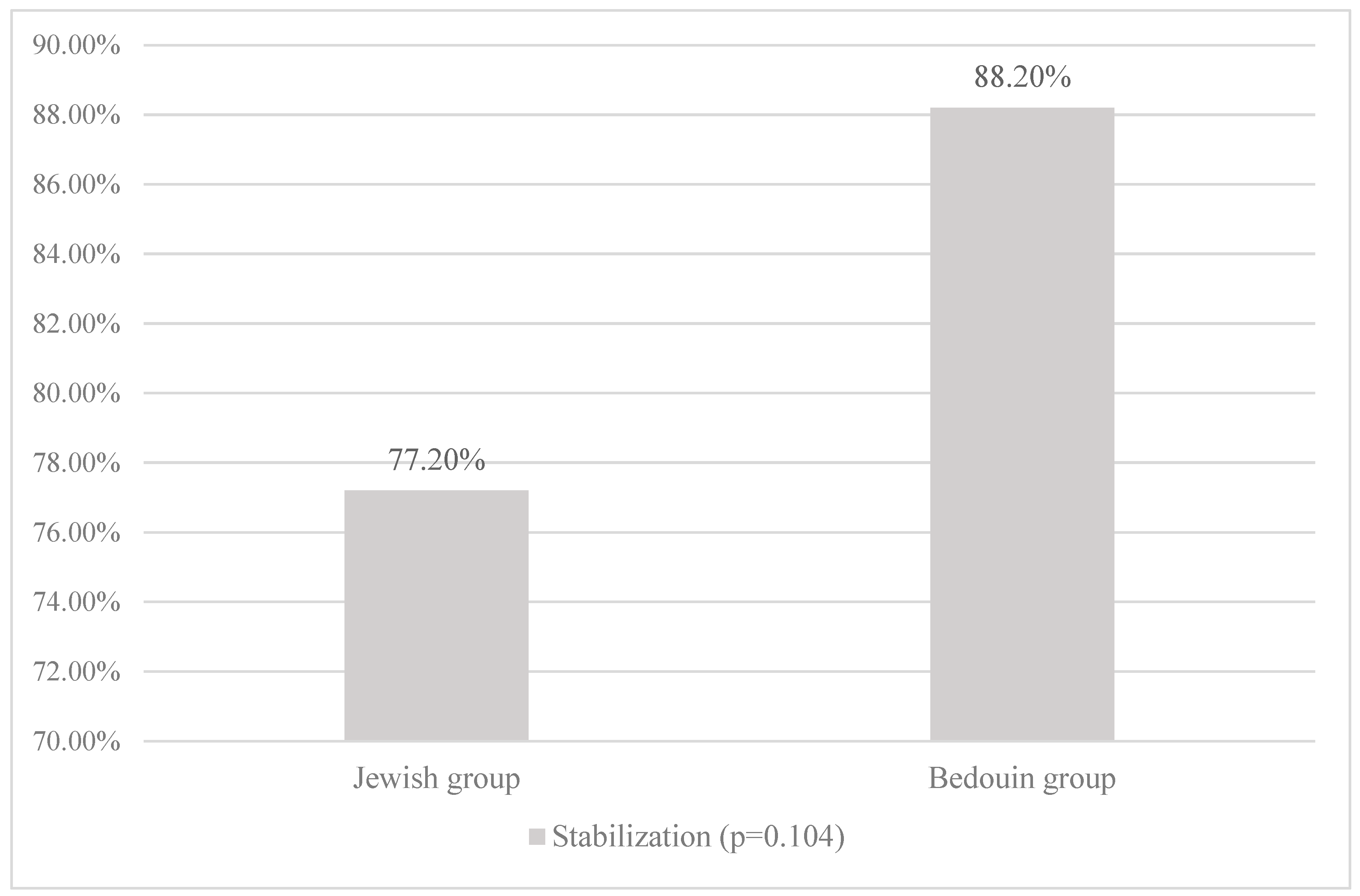

Table 1.
Demographic and clinical characteristics.
| Patient Characteristics | Jewish group (68 patients, 75 eyes ( | Bedouin group (98 patients, 123 eyes) | p-Value |
|---|---|---|---|
| Demographical characteristics | |||
| Time until follow-up, months Mean ± SD (n) Median Min; Max |
17.55 ± 6.58 (57/75) 15.00 12.00;40.00 |
16.38 ± 5.14 (87/123) 14.00 12.00;34.00 |
0.256 |
| Age, years Mean ± SD (n) Median Min; Max |
23.64 ± 6.90 (75) 22.00 9.10;45.00 |
18.68 ± 6.60 (123) 18.00 7.60;41.00 |
<0.001 |
| Gender, % (n/N) Male |
77.9% (53/68) | 54.0% (53/98) | 0.002 |
| Treated eye, % (n/N) Right eye |
46.7% (35/75) | 49.6% (61/123) | 0.770 |
| Family History of KC, % (n/N) Yes |
12.1% (8/66) | 39.1% (36/92) | <0.001 |
| Vernal Keratoconjunctivitis per history, % (n/N) Yes |
18.4% (12/65) | 29.4% (28/95) | 0.138 |
| Wearing of Glasses, % (n/N) Yes |
37.1% (23/62) | 37.0% (30/81) | 1.000 |
| Clinical characteristics | |||
| Type of CXL, % (n/N) Standard Accelerated |
32.0% (24/75) 68.0% (51/75) |
16.3% (20/123) 83.7% (103/123) |
0.013 |
| UCVA, logMAR Mean ± SD (n) Median Min; Max |
0.59±0.45 (73) 0.47 0.00; 2.00 |
0.89±0.59 (106) 0.70 0.00; 3.00 |
<0.001 |
| BCVA, logMAR Mean ± SD (n) Median Min; Max |
0.30±0.21 (73) 0.22 0.00; 1.00 |
0.45±0.24 (107) 0.47 0.00; 1.00 |
<0.001 |
| K Max, D Mean ± SD (n) Median Min; Max |
56.28±6.62 (75) 54.10 45.00; 81.40 |
58.15±8.94 (119) 56.50 44.60; 88.90 |
0.229 |
| K Mean, D Mean ± SD (n) Median Min; Max |
48.34±4.28 (75) 47.40 42.20; 62.70 |
50.24±5.87 (122) 49.40 38.70; 69.20 |
0.028 |
| MCT, μm Mean ± SD (n) Median Min; Max |
465.18±46.42 (75) 464.00 338.00; 560.00 |
434.85±49.52 (122) 436.00 244.00; 531.00 |
<0.001 |
| MCT, % (n/N) <400μm 400μm <Thickness<450μm 450μm <Thickness<500μm >500μm |
8.0% (6/75) 30.7% (23/75) 30.7% (23/75) 30.7% (23/75) |
8.0% (22/122) 41.8% (51/122) 32.0% (39/122) 8.2% (10/122) |
<0.001 |
Note. N = number of patients; SD = standard deviation; D = diopter; UDVA = uncorrected distance visual acuity; BCVA = best corrected visual acuity; K = keratometry; MCT = minimum corneal thickness.
Table 2.
Preoperative and 12 months postoperative clinical parameters by ethnic group.
| Clinical Parameters | Jewish group (52 Patients, 57 Eyes) | Bedouin group (71 Patients, 87 Eyes) | ||||
|---|---|---|---|---|---|---|
| Baseline | >12 months | P value | Baseline | >12 months | P value | |
| (N=57) | (N=57) | (N=87) | (N=87) | |||
| UCVA (logMAR) | 0.003 | <0.001 | ||||
| Mean+sd (n) | 0.57±0.44 (55) | 0.44±0.39 (52) | 0.90±0.59 (76) | 0.62±0.46 (69) | ||
| Median | 0.4 | 0.26 | 0.74 | 0.5 | ||
| Min; Max | 0.00; 2.00 | 0.00; 2.00 | 0.00; 3.00 | 0.00; 2.00 | ||
| BCVA (logMAR) | 0.008 | 0.003 | ||||
| Mean+sd (n) | 0.32±0.22 (55) | 0.23±0.21 (52) | 0.45±0.25 (79) | 0.39±0.36 (79) | ||
| Median | 0.22 | 0.2 | 0.48 | 0.3 | ||
| Min; Max | 0.00; 1.00 | 0.00; 1.00 | 0.00; 1.00 | 0.00; 2.00 | ||
| Kmax (D) | 0.981 | 0.624 | ||||
| Mean+sd (n) | 55.80±6.02 (57) | 55.66±6.22 (57) | 57.15±7.24 (85) | 57.74±7.93 (87) | ||
| Median | 54.1 | 52.7 | 56.1 | 56.6 | ||
| Min; Max | 47.40; 72.00 | 45.60; 70.30 | 44.60; 79.60 | 46.20; 85.90 | ||
| K1 Flat front (D) | 0.257 | 0.566 | ||||
| Mean+sd (n) | 45.87±3.42 (57) | 46.06±3.73 (57) | 48.05±5.17 (87) | 48.30±5.24 (87) | ||
| Median | 45.3 | 46 | 47.1 | 47.3 | ||
| Min; Max | 38.20; 55.70 | 37.70; 56.60 | 36.90; 66.50 | 38.80; 65.00 | ||
| K2 steep front (D) | 0.238 | 0.979 | ||||
| Mean+sd (n) | 50.07±4.31 (57) | 49.61±4.26 (57) | 52.37±5.77 (87) | 52.65±6.11 (87) | ||
| Median | 50.4 | 49.1 | 52.5 | 51.9 | ||
| Min; Max | 42.80; 63.30 | 42.20; 65.20 | 40.50; 72.10 | 43.50; 70.80 | ||
| K mean front (D) | 0.711 | 0.391 | ||||
| Mean+sd (n) | 47.76±3.62 (57) | 47.74±3.82 (57) | 50.00±5.31 (87) | 50.40±5.43 (86) | ||
| Median | 47.1 | 47.3 | 49.6 | 49.5 | ||
| Min; Max | 42.20; 59.20 | 40.10; 60.60 | 38.70; 69.20 | 41.40; 67.20 | ||
| K mean back (D) | 0.479 | 0.194 | ||||
| Mean+sd (n) | -7.41±0.82 (57) | -7.00±0.73 (57) | -7.46±0.92 (85) | -7.42±1.39 (85) | ||
| Median | -7.2 | -6.9 | -7.4 | -7.5 | ||
| Min; Max | -9.90; -6.00 | -9.60; -5.90 | -11.00; -5.60 | -10.70; 0.60 | ||
| Astigmatism front (D) | 0.125 | 0.859 | ||||
| Mean+sd (n) | 4.18±2.57 (57) | 3.56±2.17 (57) | 4.31±2.00 (87) | 4.28±2.05 (87) | ||
| Median | 3.5 | 2.9 | 4 | 4.2 | ||
| Min; Max | 1.00; 13.70 | 0.40; 9.80 | 0.10; 11.30 | 0.00; 10.20 | ||
| Astigmatism back (D) | 0.166 | 0.104 | ||||
| Mean+sd (n) | 0.83±0.45 (57) | 0.74±0.43 (57) | 0.89±0.38 (86) | 0.91±0.82 (87) | ||
| Median | 0.8 | 0.7 | 0.8 | 0.8 | ||
| Min; Max | 0.00; 2.10 | 0.10; 2.00 | 0.20; 1.80 | 0.00; 6.40 | ||
| CCT (μm) | 0.003 | <0.001 | ||||
| Mean+sd (n) | 488.49±44.36 (57) | 480.64±41.09 (57) | 454.77±41.30 (85) | 440.75±56.84 (85) | ||
| Median | 484 | 483 | 454 | 442 | ||
| Min; Max | 384.00; 574.00 | 385.00; 560.00 | 356.00; 547.00 | 119.00; 609.00 | ||
| ACT (μm) | <0.001 | <0.001 | ||||
| Mean+sd (n) | 482.19±44.36 (57) | 470.45±46.42 (57) | 448.01±41.80 (87) | 430.47±59.21 (85) | ||
| Median | 480 | 470 | 444 | 435 | ||
| Min; Max | 346.00; 542.00 | 355.00; 563.00 | 346.00; 542.00 | 108; 599.00 | ||
| MCT (μm) | <0.001 | <0.001 | ||||
| Mean+sd (n) | 466.86±45.96 (57) | 453.19±50.12 (57) | 434.70±44.04 (87) | 418.54±45.69 (86) | ||
| Median | 463 | 460 | 432 | 423.5 | ||
| Min; Max | 338.00; 560.00 | 277.00; 541.00 | 339.00; 531.00 | 297.00; 517.00 | ||
| Corneal volume (mm3) | 0.003 | 0.004 | ||||
| Mean+sd (n) | 57.32±4.84 (57) | 56.68±4.74 (57) | 55.98±3.96(85) | 55.11±4.68 (87) | ||
| Median | 57.4 | 56.6 | 55.3 | 55.2 | ||
| Min; Max | 35.60; 68.70 | 34.60; 66.70 | 48.30; 68.20 | 30.50; 66.00 | ||
Note. N = number of patients; SD = standard deviation; D = diopter; UDVA = uncorrected distance visual acuity; BCVA = best corrected visual acuity; K = keratometry; CCT = central corneal thickness; ACT = apical corneal thickness; MCT = minimum corneal thickness.
Table 3.
Mean change in outcomes 12 months post CXL by ethnicity.
| Change (calculated as 12+ months post procedure minus at baseline) | Jewish group (52 Patients, 57 Eyes) | Bedouin group (71 Patients, 87 Eyes) | P-Value |
|---|---|---|---|
| UCVA (logMAR) | 0.56 | ||
| Mean+sd (n) | -0.12±0.35 (52) | -0.23±0.53 (66) | |
| Median | -0.17 | -0.11 | |
| Min; Max | -1.00; 1.40 | -2.00; 0.90 | |
| BCVA (logMAR) | 0.864 | ||
| Mean+sd (n) | -0.07±0.21 (50) | -0.08±0.29 (73) | |
| Median | -0.02 | -0.02 | |
| Min; Max | -0.80; 0.60 | -0.78; 1.00 | |
| Kmax (D) | 0.973 | ||
| Mean+sd (n) | -0.16±2.45 (56) | 0.09±2.14 (84) | |
| Median | 0 | 0.1 | |
| Min; Max | -4.40; 8.60 | - 8.60; 7.00 | |
| K1 Flat front (D) | 0.588 | ||
| Mean+sd (n) | 0.18±2.02 (57) | 0.25±1.9 (87) | |
| Median | 0 | 0 | |
| Min; Max | -10.40; 4.70 | -3.20; 11.70 | |
| K2 steep front (D) | 0.35 | ||
| Mean+sd (n) | -0.45±3.01 (57) | 0.28±3.10 (87) | |
| Median | -0.2 | 0 | |
| Min; Max | -19.00; 7.60 | -5.10; 22.90 | |
| K mean front (D) | 0.776 | ||
| Mean+sd (n) | -0.02±2.40 (57) | 0.33±2.24 (86) | |
| Median | 0 | 0.1 | |
| Min; Max | -14.00; 6.60 | -3.60; 12.80 | |
| K mean back (D) | 0.747 | ||
| Mean+sd (n) | -0.02±0.31 (57) | 0.08±8.65 (83) | |
| Median | 0 | 0 | |
| Min; Max | -1.30; 1.10 | -1.70; 6.95 | |
| Astigmatism front (D) | 0.327 | ||
| Mean+sd (n) | -0.61±2.18 (57) | -0.02±1.65 (87) | |
| Median | -0.3 | 0 | |
| Min; Max | -8.60; 4.50 | -7.10; 4.90 | |
| Astigmatism back (D) | 0.932 | ||
| Mean+sd (n) | -0.08±0.37 (57) | 0.03±0.8 (86) | |
| Median | -0.1 | -0.05 | |
| Min; Max | -1.50; 0.60 | -0.90; 5.00 | |
| CCT (μm) | 0.639 | ||
| Mean+sd (n) | -7.84±19.24 (57) | -14.92±45.77 (83) | |
| Median | -11 | -10 | |
| Min; Max | -57.00; 40.00 | -344.00; 93.00 | |
| ACT (μm) | 0.733 | ||
| Mean+sd (n) | -11.73±18.52 (57) | -18.04±44.02 (85) | |
| Median | -12 | -11 | |
| Min; Max | -60.00; 35.00 | -333.00; 92.00 | |
| MCT (μm) | 0.686 | ||
| Mean+sd (n) | -13.67±19.78 (57) | -16.6±24.94 (86) | |
| Median | -13 | -15 | |
| Min; Max | -83.00; 39.00 | -98.00; 27.00 | |
| Corneal volume (mm3) | 0.896 | ||
| Mean+sd (n) | -0.63±1.63 (57) | -1.02±3.63 (85) | |
| Median | -1 | -0.6 | |
| Min; Max | -4.60; 3.40 | -27.50; 3.90 |
Note. N = number of patients; SD = standard deviation; D = diopter; UDVA = uncorrected distance visual acuity; BCVA = best corrected visual acuity; K = keratometry; CCT = central corneal thickness; ACT = apical corneal thickness; MCT = minimum corneal thickness.
Table 4.
Factors related to KC stability within 12 months after CXL (multivariate analysis results).
Table 4.
Factors related to KC stability within 12 months after CXL (multivariate analysis results).
| Patient Characteristics | RR | p-Value | 95% Confidence Interval (RR) | |
|---|---|---|---|---|
| Lower | Upper | |||
| Kmax < 55 at baseline | 1.24 | <0.001 | 1.11 | 1.39 |
| MCT > 450 at baseline | 1.08 | 0.171 | 0.96 | 1.23 |
| Standard CXL procedure | 1.14 | 0.041 | 1.01 | 1.31 |
| Jewish origin | 1.05 | 0.472 | 0.92 | 1.19 |
| Vernal Keratoconjunctivitis per history | 1.06 | 0.486 | 0.91 | 1.23 |
Note. RR = relative risk; K = keratometry; MCT = minimum corneal thickness; CXL = corneal cross-linking.
Disclaimer/Publisher’s Note: The statements, opinions and data contained in all publications are solely those of the individual author(s) and contributor(s) and not of MDPI and/or the editor(s). MDPI and/or the editor(s) disclaim responsibility for any injury to people or property resulting from any ideas, methods, instructions or products referred to in the content. |
© 2023 by the authors. Licensee MDPI, Basel, Switzerland. This article is an open access article distributed under the terms and conditions of the Creative Commons Attribution (CC BY) license (http://creativecommons.org/licenses/by/4.0/).
Copyright: This open access article is published under a Creative Commons CC BY 4.0 license, which permit the free download, distribution, and reuse, provided that the author and preprint are cited in any reuse.


