Submitted:
19 July 2023
Posted:
20 July 2023
You are already at the latest version
Abstract
Biosensors are analytical devices that utilize biological interactions to detect and quantify clinical biomarkers, contaminants, allergens, and microorganisms. They combine different disciplines including analytical chemistry, molecular biology, and electrical engineering. Biosensors operate by coupling a bioreceptor, such as nucleic acid or proteins, with a transducer that converts the biological interaction into an electrical signal. Electrochemical and optical transduction are commonly used approaches due to their high detection capability and compatibility with miniaturization. Biosensors provide both high specificity and sensitivity and can be integrated into low-cost microfluidic platforms for rapid and point-of-care applications. These attributes make these devices valuable tools in analytical chemistry, particularly for early diagnostic applications. However, conventional biosensors face challenges related to the immobilization of biorecognition elements on the transducer surface, leading to issues like lost of sensitivity and selectivity. To address these problems, the introduction of nanomaterials, particularly magnetic nanoparticles (MNPs) and magnetic beads (MBs), has been implemented. MNPs combine their magnetic properties with other interesting characteristics such as small size, high surface-to-volume ratio, and excellent biocompatibility. They can be tailored for specific applications and have been extensively used in various fields, including biosensing and clinical diagnosis. Furthermore, MNPs simplify sample preparation by isolating target analytes through magnetic separation, thus improving sensitivity, and reducing analysis time and interference phenomena. The synthesis and modification of MNPs play a crucial role in adjusting their properties for different applications. This review presents an overview of the synthesis and surface modifications of magnetic nanoparticles, their role in the development of biosensors and bioassays, and their applications across various scientific areas.
Keywords:
Magnetic nanoparticles
; biosensor
; bioassay
; functionalization
; nanomaterial synthesis
; electrochemical detection
; optical detection
1. Introduction
In recent decades, emerging analytical devices (biosensors) have gained the attention of the research community and have improved the early diagnosis of clinical biomarkers [1], contaminants [2], allergens [3] and microorganisms [4] in diverse fields of applications. Additionally, biosensors are a good example of interdisciplinarity collaboration where many fields of science and engineering converge (e.g., analytical chemistry, surface and material sciences, molecular biology, biochemistry, electrochemistry, electrical and electronic engineering). A biosensor is an electronic device used to transform a biological interaction into an electrical signal [5]. This device is based on the direct spatial coupling of the immobilized biologically active element, the so-called “bioreceptor”, with a transducer that acts as a detector and electronic amplifier. Different types of bioreceptors (e.g. enzymes, antibodies, aptamers, oligonucleotides, affinity proteins) combined with electrochemical, optical, or mechanical transductions have been used for the fabrication of biosensors with excellent results [6]. Among these approaches, electrochemical transduction offers the advantages of high sensitivity, which can be enhanced by attaching (bio)catalytic labels to the bioreceptor-target to amplify the detection signal [7]. Other benefits include their potential for miniaturization, the low cost of production and that they do not require expensive instrumentation for read-out [8]. There are two basic properties of these devices that make them interesting in analytical chemistry: (1) high specificity generated by the biological component that selectively interacts with the target analyte and (2) high sensitivity determined by the transducer properties. Finally, multiplexed detection of several biomarkers integrated into low-cost biosensing platforms opens a wide range of opportunities for their use as point-of-care (PoC) test in early diagnosis applications [9,10]. As a result of these advantages, today such technology is gradually supplanting other more sophisticated techniques and they are set to become a vital tool in healthcare and other areas of applications. Undoubtedly, the most important biosensor, with a high impact in the health and the control of diabetes, is the glucometer [11]. Such device enables the detection of discrete glucose concentration by pricking the finger to obtain a blood sample (finger stick monitors). Such methodology may result tedious in some patients, such as children, as it requires adult supervision for obtaining blood samples multiple times a day and may result painful and time-consuming. However, recent advancements in commercial devices have enabled the measurement of glucose from interstitial fluids just underneath the skin in continuous mode, offering automatic monitoring without patient interaction.
Another example where biosensor will have an important impact is in the diagnosis of infectious diseases (Dengue, Malaria, Chikungunya Fever) [12]. According to the WHO guidelines, the recommended diagnostic tests of such infections include microbiological isolation, serological tests and molecular techniques such as polymerase chain reaction (PCR) [13,14]. However, although PCR is the most convenient method due to its high sensitivity and relatively short analysis time, it has an elevated cost of equipment and supplies. On the other hand, it requires specialized skills from laboratory personnel for the execution and interpretation of results, which makes its use in routine analysis difficult. Moreover, they are prone to generating false positives due to non-specific amplifications, and its integration into PoC systems is complicated.
To overcome these problems, user-friendly, low-cost, and fast methods are still needed. In this context, the development of more efficient bioassays, including biosensors, has the potential to significantly contribute to the development of enhanced and more effective analytical tools for biomedical diagnosis [15,16].
The main drawback of conventional non-structured biosensors is associated with the proper immobilization of the biocatalytic or biorecognition element on the surface of the transducer. Non-oriented immobilization of the biomolecules may produce reusability and reproducibility problems, and low sensitivity and selectivity due to loss of catalytic activity or biorecognition reaction, which in turn hinders the electron transfer reaction and reduces the electrochemical signal. To address these issues, different approaches have been introduced such as nano-structuration of the transducer and the utilization of magnetic nanoparticles (MNPs) and magnetic beads (MBs) as solid phases for the recognition reaction.
Nanomaterials, with dimensions ranging from 1 to 100 nm, have played a crucial role for science, technology, and medicine over the past two decades [17,18]. Their unique characteristics (small size, high surface-to-volume ratio, excellent biocompatibility, biodistribution, outstanding catalytic properties, etc.) offer great possibilities for its integration into new and better devices, analytical methods, diagnosis tools, among others. [18]. One of the main properties of such materials is that their chemical and physical properties are drastically different from those of their bulk materials and can be tailor-made for a specific task [19,20]. In this sense, MNPs have been receiving remarkable attention in multiple areas such as data storage, biosensing, catalyst, bioremediation, neural stimulation, pharmacological liberation, and clinical diagnosis [21]. The use of magnetic sorbents significantly simplifies the sample manipulation by isolating the target (after the extraction process) applying an external magnetic field. Similar strategies may be used in other analytical applications (protein purification, remediation, chromatography applications) improving the selectivity, sensitivity, and time for results [22]. Even the correct design and modification of the MNPs offer a plethora of possibilities to design biocompatible and biomimetic surfaces for biosensing applications in the detection of different analytes [23,24,25,26].
MNPs may be synthesized using different methods, grouped in top-down and bottom-up approaches [27,28]. The correct selection of the synthetic route and their later biofunctionalization are one of the most important steps to adjust the magnetic properties, phase composition, biodistribution and biocompatibility, stability, aggregation effects, degradation, toxicity, and size distribution of the MNPs. On the other hand, the combination of MNPs and electrochemical read-out methods has allowed the development of new analytical techniques such as Enzyme Linked Immuno Magnetic Electrochemical (ELIME) methods [29]. Here, MNPs act as nanosized supports for the immobilization of biomolecules (antibodies, aptamers, oligonucleotides) and are used for the isolation of the target from complex matrices and its concentration before detection. The utilization of MNPs enhances the effectiveness of analyte isolation and concentration, minimizes matrix effects due to simplified washing and separation procedures, allows faster assay kinetics, and improves the sensitivity, limits of detection (LOD) and reduces the time for analysis [30]. Usually, ELIME bioassays are developed as sandwich-type format, where two specific antibodies are used (capture-Ab and labeled-Ab). The read-out is done using differential pulse voltammetry (DPV), constant potential amperometry (CPA), linear sweep voltammetry (LSV), or similar electrochemical techniques with screen-printed electrodes (SPEs) as transducers [31]. Moreover, the use of magnetic beads in combination with biosensing strategies can avoid individual electrode surface modifications with biological elements, which simplify storage and ensures proper washing and preconcentration of the sample on the working electrode area, and finally, improves the reproducibility, sensitivity, and the limit of detection (LOD) of the strategies.
This review highlights the latest advancements in (1) the synthesis of magnetic nanosupports (MNPs and MBs) and (2) the most common surface modifications and materials with improved characteristics for its incorporation in different areas of applications such as biomedicine, veterinary, bioanalytical and contaminant applications, allergens detection, and quality control in agro-food and pharmacy industries, among others.
2. Synthesis and modification of MNPs
2.1. Synthesis
Depending on their applications, different synthetic and modification protocols can be used [32,33]. The choice of such methods depends on the specific requirements of the bioassay/application and the desired properties of the MNPs, such as their shape, size, biocompatibility, stability and magnetic properties.
In the following sections, we describe the most employed chemical methods within the bottom-up approach [19]. Other protocols under top-down approach or physical methods can be found in the literature [34].
2.1.1. Co-precipitation
This procedure involves the addition of an aqueous solution of metal salts to a basic medium of an alkaline agent in the presence of a reductant, resulting in the formation of MNPs (see Eq. 1, described for iron-based magnetic particles). This approach is simple and cost-effective. Due to its scalability, low-cost and user-friendly reaction, these methods are widely used in biosensing applications. Nevertheless, such method can lead to the formation of large particles and agglomerates, which can affect their further application.
2FeSO4 + N2H4 (reducing agent) + 4NaOH -> 2Fe3O4 + N2 + 4H2O
Another frequently employed synthetic route for magnetite or maghemite (Fe3O4 and Fe2O3, respectively) nanoparticles involves using an aqueous solution of Fe(II) and Fe(III) salts that is added dropwise to an alkaline (pH ̴ 9-12) solution (NaOH, KOH or NH4OH) under vigorous stirring. Following the reaction (see Equation (2)), the resulting precipitate is subsequently magnetically decanted and washed with deionized water until neutrality and dried overnight at 60-80°C.
2Fe2+ + Fe3+ + 8OH- → Fe3O4 + 4H2O
The co-precipitation technique has been used in several works during the last ten years [35,36]. This method was employed to synthesize Zinc-substituted Fe3O4 nanoparticles (ZnxFe3-xO4) with a spinel structure to create a novel electrochemical non-enzymatic glucose sensing system [37]. For this purpose, a two-step procedure followed by hydrothermal treatment in a microwave field was used to develop modified carbon paste electrodes. The results showed linear detection range of 0.1 to 2 mM with a LOD of 0.03 mM, and a short response time of less than 3 seconds, making this NPs a potential nanomaterial for biosensing and magnetic hyperthermia applications. In the same way, the co-precipitation method was used to synthesize Fe3O4 nanoparticles and coat them with varying concentrations of polyethylene glycol (PEG), resulting in smaller particle size and reducing agglomeration phenomenon [38].
Furthermore, the synthesis by chemical co-precipitation of MnFe2O4@chitosan/MWCNTs/PDMS composite film has also been reported (Figure 1) [39]. The sensing platform was constructed to detect alpha2-macroglobulin (α2-M), which is critical in the diabetic nephropathy diagnosis. This synthesis approach yielded numerous sites for highly efficient binding of the α2-M antibody. The results revealed a detection limit of 0.13 ng/mL and a linear range from 10 ng/mL to 100 μg/mL, which is significantly lower than the limit required for clinical assessment.
Finally, cerium doped magnetite nanoparticles were synthesized by co-precipitation method and then used as a co-reactant in a luminol-K3Fe(CN)6 chemiluminescence system [40]. The effective quenching of metronidazole resulted in a linear detection range of 3.47 to 93.7 mol/L and a LOD of 0.391 mol/L. The simple and fast synthesis route combined with the highly sensitive and selective detection of α2-M demonstrates great potential for use in the PoC device.
2.1.2. Solvothermal synthesis
This method involves the use of a high-pressure autoclave to carry out the synthesis reaction in a high-boiling-point organic solvent. The high temperature and pressure conditions facilitate the formation of MNPs with uniform characteristics. This method is suitable for the synthesis of MNPs with controlled properties, narrow size distribution and bioimaging and biodistribution use. A common precursor under this approach is the iron pentacarbonyl (Fe(CO)5). Usually, such reactive is dissolved in a high-boiling-point organic solvent, such as oleylamine or oleic acid. Then, the solution is heated under high-pressure conditions in an autoclave at a temperature between 200-300°C. In the presence of a reducing agent, such as sodium hydroxide (NaOH), the iron pentacarbonyl is reduced to form magnetite (Fe3O4) nanoparticles (see Equation (3)). The time reaction varies from several hours to days. However, excessively long reaction times can lead to the formation of large particles or agglomerates, which can be detrimental to their application in bioassays. Therefore, the reaction time should be optimized to achieve the desired particle size and size distribution.
5Fe(CO)5 + 20NaOH → Fe3O4 + 25CO2 + 10Na2CO3 + 10H2O
The solvothermal method was employed by Wang et al. [41] to produce Fe3O4@Cu@Cu2O nanocomposite. This nanocomposite demonstrated peroxidase-like activity and was able to facilitate the oxidation of common peroxidase substrates such as o-phenylenediamine and 3,3’,5,5’-tetramethylbenzidine in the presence of hydrogen peroxide (H2O2) and it also revealed excellent recyclability and reusability without significant loss of catalytic activity.
Likewise, the same solvothermal method was employed to obtain Au@Fe3O4 nanoparticles with the aim of enhancing peroxidase-like activity through a synergistic effect between Fe3O4 NPs and gold nanoparticles (Au@Fe3O4 NPs) [41]. These nanoparticles were used to create an aptasensor by attaching aptamers to glass beads and then labeling capture DNA for detecting ochratoxin A (OTA). The designed aptasensor exhibited high specificity and sensitivity, detecting concentrations as low as 30 pg/mL OTA. Furthermore, a hybrid nanocomposite, Fe3O4@ZIF-8/RGO, has been created by a facile one-step solvothermal approach, followed by fabrication of MOFs (ZIF-8) on the surface of Fe3O4 [42]. The developed biosensor displayed exceptional properties for the determination of dopamine in phosphate buffer solution, including a broad linear range of 2.0 nM to 10 M, and a very low limit of detection of 0.667 nM. Along with this, the ability to selectively detect dopamine even in the presence of interferents such as ascorbic acid and uric acid, highlights its potential for the detection of dopamine in biological fluids.
2.1.3. Thermal decomposition
This method involves the thermal decomposition of a metal precursor in a high-boiling-point organic solvent in the presence of a stabilizing agent. This technique requires heating the precursor material (iron salts, such as iron chlorides, iron carboxylates, or iron acetylacetonate (Fe(acac)3) to a high temperature in the presence of a solvent. Typically, benzyl alcohol, trioctylamine or dodecanethiol (C12H25SH), which serves as reducing agents and a medium for the thermal decomposition reaction, are used as solvent (see Equation (4)). The solution is heated to a high temperature, usually in the range of 200-400°C under controlled atmosphere (e.g. argon or nitrogen) for several hours. During the heating process, the precursor decomposes to form iron oxide nanoparticles, which are stabilized by the surfactant or stabilizing agents present in the solution.
Fe(acac)3 + 3C12H25SH → Fe3O4 + 3C12H25SCH3 + 3CH3COCH3
The resulting magnetite nanoparticles, with high-quality and narrow size distributions, are typically coated with the surfactant or stabilizing agent, which can be removed by washing or thermal treatment. As other methods described above, a precise control over the size and shape of the MNPs may be obtained adjusting the main reaction parameters, such as temperature, precursor concentration, and reaction time.
The synthesis of Fe3O4 nanoparticles by thermal decomposition for the development of a sensor composed of MNPs and cetyltrimethylammonium bromide was reported by Guivar et al. [43]. This sensor operates as a peroxidase mimetic system and could achieve an amperometric detection limit of H2O2 of 103 μmol/L and a linear response in the range from 100 μmol / L to 1.8 mmol/L (R2 = 0.994) with a sensitivity of 16 nA/mol L. Moreover, a narrow and monodisperse size distribution (diameter of 4.8±0.6 nm and polydispersity index: 13%) was achieved by this synthesis. The absence of agglomeration indicated high stability in inorganic media and the tested interfering agents (K+, Na+, Cl-, Mg2+, Ca2+, and uric acid) verified the selectivity towards H2O2, being comparable with some enzyme-based biosensors.
2.1.4. Microemulsion method
This method involves the use of a microemulsion, which is a dispersion of two immiscible liquids (usually oil and water) stabilized by a surfactant. The microemulsion method can be used to synthesize a variety of MNPs, including iron oxide, cobalt oxide, and nickel ferrite nanoparticles. The precursors of the MNPs are dissolved in one of the phases of the microemulsion, typically the oil phase. The surfactant stabilizes the resulting droplets of the oil phase in the water phase, creating a highly homogeneous reaction environment for NPs synthesis. Since the reaction takes place in the confined space of the droplets, the resulting MNPs presents a uniform size.
Overall, the microemulsion method is a versatile and effective technique for synthesizing MNPs with controlled size and properties [44]. The resulting particles can be coated with various organic and inorganic materials to enhance their stability and biocompatibility making it a valuable tool in various fields, including biomedicine, electronics, and catalysis.
The microemulsion method has been utilized by Rivas et al. [45] to produce stable core-shell Fe@Au NPs. This one-pot successive reaction method in microemulsions can easily obtain nanoparticles with a diameter of approximately 6 nm and a 3 nm Fe core, with a saturation magnetization of 1.13 emu/g. The resulting Fe@Au nanoparticles exhibit excellent stability after magnetic separation from the solution, indicating a properly coating of the iron core by the Au shell. Furthermore, the same technique was employed to develop a new type of multifunctional nanomaterial, FePt/Fe3O4-CdSe heteronanostructures coated in silica, which displayed both luminescent and magnetic properties [46]. Reverse micelle microemulsion technique was used to coat the heteronanostructures with silica, making them water dispersible, highly colloidally stable and capable of emitting photoluminescence in the blue-green region. All these properties confirm that this unique luminomagnetic system has great capacity for (bio)sensing applications.
2.1.5. Green synthesis.
This method involves the use of natural resources, such as plants, bacteria, and fungi to synthesize MNPs [47]. This synthesis pathway is eco-friendly and avoids the use of toxic solvents and hazardous reagents, making it a sustainable alternative to traditional synthesis methods. Nevertheless, it requires parameter optimizations (pH, temperature, reaction time) to improve the reproducibility and stability of the particles. In addition, the use of natural resources can lead to batch-to-batch variation in the properties of the synthesized MNPs, which can be a challenge for large-scale production. In the near future, the advancement and refinement of more efficient, scalable, and reproducible green synthesis protocols is expected to significantly contribute to the sustainable production of nanoparticles for a wide range of applications, including biomedical imaging, drug delivery, and environmental remediation [48]. These advances will reduce the environmental impact associated with traditional synthesis methods.
A green and ultrafast technique was developed for synthesizing iron oxides MNPs using high energy sonochemical approach, considering the amplitude (energy) of the ultrasound probe and sonication time [49]. These MNPs were evaluated for mercury detection strategy in water and as a carrier on which to anchor polyclonal antibodies against TRIB2 protein (Tribbles Pseudokinase 2) as part of an immunoprecipitation assay. Additionally, a green approach was also applied for enhance electrode electron transfer by the deposition of carbon and silver on the Fe3O4 NPs core, resulting in the formation of core-shell Fe3O4@C@Ag NPs on the electrode surface [50]. The biosensor exhibited high sensitivity with a value of 0.0346 μA/mM cm and a LOD of 0.5 mM. The excellent sensitivity of the biosensor was attributed to the large surface area, which allows effective loading of HRP and high electron communication capacity. Lastly, Ahmadian-Fard-Fini et al. [51], aimed to develop a novel photoluminescence nanocomposite for the detection of bacterial pathogens based on magnetite-carbon. Carbon dots and magnetite NPs were obtained using extracts of lemon, grapefruit, and turmeric. These nanoparticles were employed as a non-toxic sensor for the detection of E. coli bacteria, and it was observed that the photoluminescence of the nanocomposite was quenched by increasing the quantity of bacteria detection confirming its effectiveness for biosensor applications.
2.2. Surface modifications
The chemical stability, hydrophobicity, catalytic properties and biocompatibility of the NPs can be improved by conjugating different organic and inorganic chemicals compounds such as silica, gold, platinum, ceria, chitosan, polyethylene glycol, polyvinyl alcohol, poly(lactic-co-glycolic acid), and polyethylenimine), during or after the synthesis of the NPs.
2.2.1. Organic coatings/ligands
MNPs synthesis based on organic coatings/ligands may confer hydrophobic properties to the nanomaterial. Therefore, additional modifications are needed to improve its dispersion in water-based medium, such as ligand exchange method or core-shell structures. Additionally, organic ligands can provide new functional groups for conjugation with bioreceptors like antibodies or peptides, enabling specific targeting of cells or biomolecules [52]. Within the current approach, the biorecognition element can be modified with fluorescent probes (e.g. fluorescein, rhodamine and cyanine) [53] to enable specific targeting of cells or (bio)molecules. These fluorescent dyes emit light when they are excited by a specific wavelength, allowing the detection of biomolecules in biological samples with high sensitivity and specificity. Particles, synthesized by this technique have been applied in a wide range of applications, including disease diagnosis, drug discovery, and biomarker detection.
A very interesting approach for core-shell methodology is the use of organic polymers such us polydopamine (PDA), polypyrrole (PPy), polyaniline (PANI). MNPs coated with conductive polymers (e.g. PANI and PPy) can be used for biosensing applications [54]. For example, DNA or aptamers probes immobilized on polypyrrole or polyaniline-modified transducers/nanoparticles [55,56,57,58] may offer a conductive coating with high stability and biocompatibility for the biorecognition.
2.2.1.1. Chitosan
Recent MNPs functionalization with chitosan (CS) have been reported, including the creation of a chitosan-functionalized graphene (CG) material through the combination of carboxylic chitosan and graphite using ball milling technique [59]. The incorporation of nitrogen, derived from chitosan, significantly enhances the catalytic activity, improving its sensing performance and creating a favorable environment for enzyme immobilization. Fe3O4 MNPs were added to the CG to create multifunctional nanocomposites with potential use as magnetic resonance imaging agents and in vivo biosensors. Then, glucose oxidase was immobilized on the CG by covalent binding, resulting in a biosensor with high sensitivity (5.658 mA mM-1cm-2), a low detection limit of 16 μM, and a linear detection range up to 26 mM.
Similarly, Peng et al. [60] also reported a chitosan-functionalized nanocomposite for glucose determination. This nanocomposite consists of magnetic nanoparticles, chitosan, β-cyclodextrin (Fe3O4-CS-CD) and multi-walled carbon nanotubes (MWCNTs). The nanoparticle synthesis and the electrode modification are represented in Figure 2. The results showed a direct electron transfer and offered a low detection limit (19.30 μM), broad linear range (40 μM to 1.04 mM), high sensitivity (23.59 μA/mM cm ), excellent selectivity, and long-term stability. This biosensor has been successfully tested on human serum samples, indicating its practical application in biomedical diagnostics.
Moreover, it was reported a chitosan-coated magnetic glassy carbon electrode biosensor (CNP-L/CuONP/MWCNT/Pe/GC) to measure hemoglobin directly through enzyme-like catalysis of oxygen reduction [61]. Briefly, a glassy carbon electrode (GCE) was polished and modified with chitosan and hemoglobin (Hb) to create a CS-modified GCE for electrochemical measurement. A magnetic core was added into the GCE to obtain the working electrode. The electrode was immersed in aqueous solution containing Hb on it. The results revealed high sensing response against Hb, with a detection limit of 0.01 μg/mL under alkaline conditions and increased response with an external magnetic field. Another chitosan-functionalized electrochemical biosensor for the determination of triglycerides (TGs) was reported by Di Tocco et al. [62]. This biosensor was based on lipase immobilized on chitosan coated MNPs on a dispersion of multiwalled carbon nanotubes/pectin (MWCNT/Pe) modified with copper oxide NPs (CuONP) on a GCE. It involved the oxidation of the glycerol produced through an enzymatic reaction between TGs and the immobilized lipase. According to theoretical studies, the detection and quantification limits were from 3.2-3.6 mg/L, and from 9.6-11 mg/L, respectively, confirming its viability for determining TGs in human serum clinical samples.
2.2.1.2. Poly (ethylene glycol)
A recent study reported the functionalization of Fe3O4@Au nanoparticles with PEG and hyaluronic acid (HA) [63]. Fe3O4@Au@PEG@HA NPs demonstrated high selectivity, sensitivity, and high capacity to prevent biofouling. The immunosensor displayed a wide linear response range to brucellosis antibody in serum (10 fg/mL to 10 pg/mL) and low LOD (0.36 fg/mL), making it a highly promising device for health application. Similarly, Shin et al. [64] has developed a colorimetric assay for the detection of H2O2 and glucose using Fe3O4 magnetic nanoparticles functionalized with PEG ligands. The PEG-functionalization led to a significant increase in the catalytic activity of the MNPs, which act as enzyme mimetics with peroxidase-like activity. Furthermore, PEG ligands prevent self-aggregation of the nanoparticles and facilitate the transfer of H2O2 and diammonium salt substrate towards the MNPs through hydrophilic interaction. The glucose bioassay using this functionalized system exhibit a limit of detection of 3 μM, demonstrating high potential for practical applications.
In addition to this, PEG was also utilized as a mediator to prepare novel PEG-mediated APBA (m-aminophenylboronic acid) functionalized MPs (APBA-PEG-MPs) for detecting Staphylococcus aureus by using magnetic separation and fluorescence analysis (Figure 3) [65]. The LOD in pure culture, spinach, fruit juice, and pool water were 270 CFU/mL within 80 min. APBA-PEG-MNs confirmed good stability and could maintain good capture efficiency at 4 ºC and different pH values. The collected results indicate that the magnetic separation-fluorescence sensor system can achieve fast, accurate, and specific detection of S. aureus.
2.2.1.3. Polypyrrole (PPy)
Polypyrrole polymer has been studied because of its high conductivity and potential use as a support for immobilizing enzymes and as a substrate for cell adhesion. Therefore, iron oxide cores and Ppy shells were created by using micelles as templates producing iron oxide@Ppy core-shell nanoparticles [66]. The thickness of the shell can be easily adjusted, obtaining NPs with magnetic core and conductive shell properties that can be promising for their use as photothermal agents and electrochemical biosensors. Moreover, Ppy and chitosan-coated Fe3O4 were electrochemically polymerized onto pencil graphite electrodes and used as a platform to immobilize glucose-6-phosphate dehydrogenase for chronopotentiometric detection of glucose-6-phosphate (G6P) [67]. The advantages of Ppy are that can be used in a neutral pH region, and its stability to be polymerized onto different substrate materials. The results exhibited a good linear response (0.0025–0.05 mM), high selectivity and recovery, making this biosensor suitable for G6P measurements. Additionally, it was also reported a photothermal biosensor based on polypyrrole NPs for C-reactive protein detection [68]. This biosensor allowed for dual temperature and pressure readout for the detection of protein biomarkers. The Ppy study confirmed the easy polymerization using pyrrole and Fe3+, efficient light-to-heat conversion due to strong absorption in the near-infrared region, and high photothermal stability and good reproducibility. These properties demonstrated this dual-mode biosensor has significant potential for PoC testing and biomarker detection.
2.2.1.4. Polyaniline (PANI)
Polyaniline was recently utilized for the synthesis of polyaniline-iron oxide magnetic nanohybrid for the amperometric detection of catechol [69]. The conjugated structure of PANI, along with its electrical and proton conductivity in acidic media, provides efficient electron transfer for catechol oxidation and stability. This sensor demonstrated high sensitivity, low detection limit (312 μA/μL), and successful recovery of catechol from tap water samples. In addition, a new sensing platform employing Fe3O4@PANI NPs has been synthesized for the determination of creatinine in biological fluids [70]. The Fe3O4@PANI NPs are hydrophilic and stable, revealing a detection limit of 0.35 nmol/L. This nanomaterial provides many functionalized sites where creatinine molecules can bind by hydrogen bonding with the amino groups of the PANI matrix. Furthermore, PANI/magnetic graphene (MG) composites were also synthesized with the aim of immobilizing laccase, a multi-copper polyphenol oxidase used to determine phenols in food, environment, and biological fluids (Figure 4) [71].
The obtained biosensing platform showed superior electrical properties, high sensitivity between the linear range of 0.4-337.2 μM and demonstrated a detection limit of 2.94 μM. These results hold great potential for the prepared biosensor as phenolic biosensor in real water samples.
2.2.1.5. Polydopamine (pDA)
The use of polydopamine coating on nanoparticles for biosensing has several advantages. Firstly, polydopamine is a biocompatible material that does not induce an immune response, making it safe for its use in biological applications. Secondly, the polydopamine coating increases the stability of the nanoparticles, reducing the possibility of aggregation and being easily deposited on virtually all types of organic and inorganic materials [72]. Thirdly, the coating enhances the ability of nanoparticles to bind to biological molecules, improving the sensitivity and selectivity of biosensors. Finally, the coating can be easily modified to introduce functional groups or biomolecules for specific targeting and detection. The most common method for producing pDA is the oxidation method under slight alkaline conditions (aqueous solution around pH 8.5). The self-polymerization reaction is mild and does not require complicated instrumentation or harsh conditions. The thickness of the polymer can be easily controlled by adjusting several factors, such as pH, concentration of monomer, presence of copper ions, and reaction time. Many examples of its applications in biomedical, biosensing, separation methods, and remediation are reported in the literature.
For instance, core-shell glucose oxidase-Au-polydopamine-Fe3O4 magnetic bionanoparticles (GOx-Au-pDA-Fe3O4 MBNPs) for glucose detection were synthesized by using a one-pot chemical polymerization method [73]. In this system, MNPs permit the easy manipulation, PDA provides biocompatibility to sustain the native structure of GOx and AuNPs facilitate direct electron transfer of GOx. The amperometric results showed good linear response from 0.02 to 1.87 mM confirming its great potential for application in biocatalysis and biosensing. Moreover, it has been reported a magnetoimmunoassay in which core-shell MNPs were modified with polydopamine (MNPs@pDA-Ab) for the detection of Legionella pneumophila SG1, a human pathogen that can be found in natural and artificial freshwater systems [74]. To accomplish this, a specific capture antibody was attached to MNPs@pDA and incubated with bacteria. The bacteria were captured and sandwiched between an antibody labeled with horseradish peroxidase (Ab-HRP) and the modified MNPs@pDA. Finally, the MNPs@pDA-Ab-Legionella pneumophila-Ab-HRP complex was held by a magnetic field onto the electrode surface. Electrochemical detection was performed on disposable SPCEs achieving a LOD of 10 CFU/mL, which is suitable for the analysis of moderate to severe contaminated samples. In addition, other PDA-modified MNPs for the enzymatic biosensing of H2O2 in human plasma samples were reported [75]. The proposed strategy, shown in Figure 5, consists of the immobilization of HRP on the MNPs@pDA and the electrochemical polymerization of L-arginine and toluidine blue (Tb) onto the GCE surface obtaining HRP/ MNPs@pDA /(L-Arg/Tb).
The obtained hybrid thin film provides an efficient grafting of the MNPs@pDA and facilitates the covalent immobilization of HRP due to the presence of plenty active functionalization sites. The biosensor performance revealed that it was able to reduce H2O2 in a range of 0.5 to 30 μM with a limit of detection of 0.23 μM confirming its high potential for H2O2 analysis.
2.2.3. Inorganic coatings and modifications
Inorganic coatings (silica, carbonaceous materials, gold, platinum) also provide better catalytic properties, stability and biocompatibility to magnetic nanoparticles, while offering unique magnetic, electrical and optical properties for imaging and sensing applications. For example, iron oxide nanoparticles can be used for magnetic resonance imaging of cells and tissues, while gold-coated nanoparticles can be used for surface-enhanced Raman scattering sensing of biomolecules. The combination of the magnetic properties of the NPs with the large surface-to-volume ratio of the graphene have been also used to improve the electrocatalytic properties of many magnetic nanocomposite, especially, when they are also modified with catalytic materials such as Pt, Co, Ni, Au. The use of Au-modified MNPs in electrochemical biosensing applications offers the advantage of further modifications with many biomolecules such as proteins, enzymes, DNAs, aptamers that may be used for the biorecognition or the catalytic conversion of the target analyte.
2.2.3.1. Silica
Silica coating has been utilized to obtain manganese ferrite nanoparticles for efficient anti-prostate specific membrane antigen (PSMA) immobilization [76]. The MnFe2O4 nanoparticles were prepared by co-precipitation and then suspended in an ethanol-water solution with tetraethyl orthosilicate to form core-shell structures (MnFe2O4@SiO2). Subsequently, the NPs were linked to an specific antibody to PSMA, validating their potential for improving ELISA-based assays. Another study related to MNPs silica coating is the synthesis of ZnO-capped mesoporous silica nanoparticles for the construction of a microfluidic biosensor for detecting Salmonella Typhimurium (S. Typhimurium) [77].
Samples with higher bacterial concentration revealed a more pronounced color change and higher fluorescence, as can be seen in Figure 6a,b, respectively. Figure 6c,d show the absorbance and fluorescence measurements, respectively, for different bacterial concentrations. In addition, quantitative detection of S. Typhimurium was possible in a wide range from 102 to 107 CFU/mL, with a detection limit of 63 CFU/mL for the colorimetric measurement and 40 CFU/mL for the fluorescent read-out (see Figure 6e,f). The use of mesoporous silica provided wide surface area which facilitated the loading of a larger number of (bio)molecules increasing the sensitivity [77].
Lastly, silica coating of MNPs was performed to create an effective sensing platform for the immobilization of hemoglobin for multiplex detection of dopamine, uric acid and folic acid [78]. The procedure consisted in the modification of a carbon paste electrode with Hb immobilized on silica-coated MNPs and MWCNTs. This system was successfully evaluated for dopamine, uric acid and folic acid showing detection limits of 12, 14 and 18 nM and a linear range of 1-30.6, 1-286 and 1-369 μM, respectively. However, the applicability of the biosensor in serum samples was only verified for dopamine analyte.
2.2.3.2. Gold (Au)
Among the latest research, the modification of NPs surface with gold NPs was described. Innovative hierarchically porous tridimensional magnetic molybdenum trioxide–pDA-gold functionalized nanospheres (3D mag-MoO3–PDA@Au nanospheres) have been reported as a multifunctional hybrid composed of plasmonic, semiconductor, and magnetic NPs [79]. This structure has been used to develop a magnetically induced nanogap-enhanced Raman scattering (MINERS) sensing platform for ultrasensitive detection of SARS-CoV-2 (Figure 7.a). By utilizing a magnetic actuation process, the MINERS system enhances Raman signal stability and reproducibility, facilitating the highly sensitive detection of the SARS-CoV-2 spike protein. The sandwich-type immunoreaction carried out resulted in a highly reliable biosensing system, showing that the detection of SARS-CoV-2 spike protein is viable in the range of fg/mL with a broad linear dynamic detection range spanning from 10 fg/mL to 1 ng/mL (Figure 7b,c).
Another platform based on gold coating consists of a biosensor platform using AuNP-coated magnetic beads for the determination of alkaline phosphatase [80]. Its performance was tested with extracellular vesicles detection showing its potential as a biosensor in biological environments. Moreover, an aptamer-based electrochemical biosensor was prepared for the detection of leukemic cancer cells [81]. For this purpose, a thiolated specific aptamer was immobilized on gold-coated magnetic Fe3O4 NPs acting as substrate, and nitrogen-doped graphene as the detection electrode. It is considered that the gold coating significantly increased the sensitivity, and the results showed a linear response from 10 to 1 × 10⁶ cells/mL, which implies a wide dynamic range for leukemic cancer cells, confirming its use for detection in human blood plasma.
Besides, a sandwich-type biosensor with magnetic Fe3O4 particles and dithiobis(sulfosuccinimidylpropionate)-modified gold nanoparticles (DTSSP-AuNPs) was developed for dopamine analysis [82]. The method was based on the color change of the DTSSP-AuNPs and the UV/Vis signal measurement obtaining a detection limit of 10 nM for dopamine. A novel biosensor was also created by using gold nanoparticles-loaded magnetic reduced graphene oxide (MrGO@AuNPs) for detecting the endocrine disruptor Bisphenol A (BPA). The biosensor displayed good sensitivity under optimal conditions, with a detection limit as low as 0.141 pg/mL. The design consisted of a combination of BPA aptamer-MrGO@AuNPs and methylene blue-loaded gold nanoparticle to form a stable complex for synergistic signal amplification [83].
Another interesting recent report is a simple method to synthesize AuNPs/Bovine Serum Albumin/Fe3O4 composite nanoparticles for electrochemical glucose sensing. High reproducibility, sensitivity and stability were obtained, with short response time (0.8 s), linear dynamic range from 0.25 to 7.0 mM and a low detection limit of 3.54 μM. All this characteristics make this composite a great material for amperometric biosensor design [84]. Alternatively, the specificity of AuNPs in biosensors has been used to increase the sensitivity of the quartz-crystal microbalances and as optical transducers for colorimetric biosensors. For this purpose, gold-coated core-shell magnetic NPs were functionalized with photochemical immobilization technique in order to bind antibodies vertically on the gold surface which could improve the detection limit of colorimetric biosensors [85]. Finally, Fe3O4@Au NPs were used for Pb2+ detection obtaining a detection limit of 15 pM. The design of these NPs allows their magnetic control and interferences reduction with an increase of the specific surface area, also providing the possibility to detect other heavy metal ions [86].
2.2.3.3. Platinum (Pt)
Among the platinum-modified magnetic materials that have been reported in recent years, it can be found a novel biosensor design based on 1,1′-oxalyldiimidazole chemiluminescent enzyme immune assay (ODI CLEIA) [87]. The system consisted of using thyroid stimulant hormone (TSH) capture antibody attached on MB and TSH detection antibody-conjugated HRP immobilized on Pt nanoparticles. A ldetection limit of 0.004 mU/L and a wide dynamic range from 0.013 to 12 mU/L was achieved confirming that it can effectively quantify TSH in human serum for the early diagnosis of thyroid cancer. In addition, Fe3O4-Pt/core-shell nanoparticles (MPt/CS NPs) in aqueous phase were synthesized using citrate as a surface-stabilizing and reducing agent for their integration in lateral flow immunoassay (LFIA) strips [88]. The results show a two-fold increase in sensitivity compared to commercially available Au NPs-LFIA (LOD: 3.7 ng/mL) versus the low detection limit of 0.039 ng/mL with the synthesized nanoparticles. It was proved that MPt/CS NPs exhibit high catalytic efficiency, high affinity for the colorimetric substrate and great potential for PoC testing.
Lastly, a layer-by-layer biosensor for the amperometric detection of xanthine was constructed. Its design was based on pDA-modified MNPs coated with 4-generation ethylenediamine core polyamidoamine G-4 dendrimers that were decorated with Pt-NPs. The material was deposited on GCE coated with a graphene oxide-carboxymethylcellulose nanomaterial, and xanthine oxidase was successfully immobilized on the scaffold. Xanthine could be detected in the range of 50 nM to 12 μM and a LOD of 13 nM was obtained with good reproducibility and repeatability results in real fish samples [89].
2.2.3.4. Quantum dots (QDs)
Quantum dots are semiconductor nanoparticles (such as CdSe, InAs, among others) that presents unique optical and electrical properties depending of their size. They are brighter, more stable and have a longer fluorescence lifetime. The emission wavelength of QDs can be tuned by changing their size, enabling multicolor imaging and detection of biomolecules or cells in complex biological samples. QDs have several advantages over traditional organic dyes and other electrochemical labels for bioimaging and biodetection. Significantly, QDs can enhance the performance of immunological sensors by improving the immobilization of biomolecules and labels, facilitating the electron transfer, and amplifying the electrochemical signals [90]. A very interesting advance on the use of QDs is the multiplexing where multiple types of QDs, with different emission wavelengths/redox response can be used simultaneously for multiplexed imaging or detection, allowing for the detection of multiple biomolecules or cells at once.
Likewise, QDs have also been used for the construction of a prototype biosensor for the detection of E. coli in water samples [91]. For this purpose, superparamagnetic Fe3O4 NPs were conjugated with E. coli-specific aptamer, which facilitates the separation of E. coli cells thanks to magnetic separation obtaining values of fluorescence intensity from 100 to 400 μg/mL. The ATmega 328P prototype biosensor developed with this system successfully detected low bacterial counts in water samples, also enabling the possibility of its application to food samples.
Finally, quantum dots were also used to develop an optical sensor platform for the fluorescence determination of histamine [92]. Histamine can be found in contaminated food and is used as a signal of food safety. The system consists of a cysteine-containing peptide incorporated into gold-coated magnetic nanoparticles (MNP@Au NPs) and can be used for purification and inspection of fish samples. The synthesis of the QDs was performed by the hydrothermal one-pot method allowing their fluorescence to be efficiently quenched by peptides due to electron transfer exchanges but recovering it with the addition of histamine. The biosensor was investigated on real fish samples demonstrating a detection range of 0.1 to 100 ppm and a detection limit of 13.0 ppb and showing great potential as a histamine sensor for food protection.
3. Conclusions
In recent years several strategies have been developed for the synthesis of magnetic nanoparticles finding that the most popular approaches are co-precipitation, solvothermal, microemulsion and green synthesis methods. Here, we presented most widely employed synthesis methods, including the adequate modification or development of new magnetic nanomaterials to overcome the main difficulties and add novel properties to the MNPs. In this regard, functionalizing nanomaterials enables the improvement of sensitivity, chemical stability, catalytic efficiency, and biocompatibility of the biosensing platforms. Briefly, the most widespread functionalizing agents, introduced in this review, are chitosan, polyethylene glycol and silica, among others. In addition, various organic (polyaniline, polydopamine and polypyrrole) and inorganic coatings (gold, silica or platinum) are also broadly employed. Despite the challenges, advances in MNPs research have demonstrated their great potential for use in biomedical, environmental, and food safety applications, among others. Finally, in each section of this review, a wide variety of works were introduced illustrating the synthesis and functionalization methods and their application in different areas of science, mainly focused on analytical and biomedical aspects.
Acknowledgments
This work was funded by Interreg MAC 2014–2020 program (project: MacBioidi 2). Soledad Carinelli gratefully acknowledges the financial support of the “Juan de la Cierva Programme” (FJC2020-043734-I) financed by the “Ministerio de Ciencia e Innovación” and the “Agencia Estatal de Investigación” of the Spanish Government.
References
- Prabowo, B.A., et al. The Challenges of Developing Biosensors for Clinical Assessment: A Review. Chemosensors, 2021. 9. [CrossRef]
- Bhavadharini, B., et al., Recent Advances in Biosensors for Detection of Chemical Contaminants in Food — a Review. Food Analytical Methods, 2022. 15(6): p. 1545-1564. [CrossRef]
- Aquino, A. and C.A. Conte-Junior A Systematic Review of Food Allergy: Nanobiosensor and Food Allergen Detection. Biosensors, 2020. 10. [CrossRef]
- Péter, B., et al. Review of Label-Free Monitoring of Bacteria: From Challenging Practical Applications to Basic Research Perspectives. Biosensors, 2022. 12. [CrossRef]
- Nagel, B., H. Dellweg, and L.M. Gierasch, Glossary for chemists of terms used in biotechnology (IUPAC Recommendations 1992). 1992. 64(1): p. 143-168. [CrossRef]
- biosensor. 2014.
- Sumitha, M.S. and T.S. Xavier, Recent advances in electrochemical biosensors – A brief review. Hybrid Advances, 2023. 2: p. 100023. [CrossRef]
- Ronkainen, N.J., H.B. Halsall, and W.R. Heineman, Electrochemical biosensors. Chem Soc Rev, 2010. 39(5): p. 1747-63.
- Biswas, G.C., et al. A Review on Potential Electrochemical Point-of-Care Tests Targeting Pandemic Infectious Disease Detection: COVID-19 as a Reference. Chemosensors, 2022. 10,. [CrossRef]
- Kulkarni, M.B., N.H. Ayachit, and T.M. Aminabhavi Recent Advances in Microfluidics-Based Electrochemical Sensors for Foodborne Pathogen Detection. Biosensors, 2023. 13. [CrossRef]
- Zafar, H., et al. Comprehensive Review on Wearable Sweat-Glucose Sensors for Continuous Glucose Monitoring. Sensors, 2022. 22,. [CrossRef]
- Cheon, J., et al., Advances in Biosensor Technologies for Infection Diagnostics. Accounts of Chemical Research, 2022. 55(2): p. 121-122. [CrossRef]
- Martín, M.-S., et al., First bioelectronic immunoplatform for quantitative secretomic analysis of total and metastasis-driven glycosylated haptoglobin. Analytical and bioanalytical chemistry, 2023. 415(11): p. 2045-2057. [CrossRef]
- Brady, N., et al., An immunoturbidimetric assay for bovine haptoglobin. Comparative Clinical Pathology, 2019. 28: p. 21-27. [CrossRef]
- Lou, D., et al., Advances in nanoparticle-based lateral flow immunoassay for point-of-care testing. View, 2022. 3(1): p. 20200125.
- Jin, Y., et al., Multifunctional nanoparticles as coupled contrast agents. Nat Commun, 2010. 1: p. 41. [CrossRef]
- Ramos, A.P., et al., Biomedical applications of nanotechnology. Biophys Rev, 2017. 9(2): p. 79-89. [CrossRef]
- Diez-Pascual, A.M. and A. Rahdar, Functional Nanomaterials in Biomedicine: Current Uses and Potential Applications. ChemMedChem, 2022. 17(16): p. e202200142. [CrossRef]
- Baig, N., I. Kammakakam, and W. Falath, Nanomaterials: a review of synthesis methods, properties, recent progress, and challenges. Materials Advances, 2021. 2(6): p. 1821-1871. [CrossRef]
- Zhong, Z., et al., Recent Advances in Magnetic Nanoparticles-Assisted Microfluidic Bioanalysis. Chemosensors, 2023. 11(3): p. 173. [CrossRef]
- Khan, I., K. Saeed, and I. Khan, Nanoparticles: Properties, applications and toxicities. Arabian Journal of Chemistry, 2019. 12(7): p. 908-931.
- Scigalski, P. and P. Kosobucki, Recent Materials Developed for Dispersive Solid Phase Extraction. Molecules, 2020. 25(21). https://doi:10.3390/molecules25214869. [CrossRef]
- Predoi, D., et al., Biocompatible Layers Obtained from Functionalized Iron Oxide Nanoparticles in Suspension. Coatings, 2019. 9(12): p. 773. [CrossRef]
- Campuzano, S., et al., Cutting-Edge Advances in Electrochemical Affinity Biosensing at Different Molecular Level of Emerging Food Allergens and Adulterants. Biosensors (Basel), 2020. 10(2). [CrossRef]
- Mohsin, A., et al., Recent Advances of Magnetic Nanomaterials for Bioimaging, Drug Delivery, and Cell Therapy. ACS Applied Nano Materials, 2022. 5(8): p. 10118-10136. [CrossRef]
- Mukhtar, M., et al., Nanomaterials for diagnosis and treatment of brain cancer: Recent updates. Chemosensors, 2020. 8(4): p. 117. https://doi:10.3390/chemosensors8040117. [CrossRef]
- Ma, K., et al., Magnetosome-inspired synthesis of soft ferrimagnetic nanoparticles for magnetic tumor targeting. Proc Natl Acad Sci U S A, 2022. 119(45): p. e2211228119. [CrossRef]
- Ali, A., et al., Review on Recent Progress in Magnetic Nanoparticles: Synthesis, Characterization, and Diverse Applications. Front Chem, 2021. 9: p. 629054. [CrossRef]
- Fernández, I., et al., Electrochemical bioassay based on l-lysine-modified magnetic nanoparticles for Escherichia coli detection: Descriptive results and comparison with other commercial magnetic beads. Food Control, 2023. 145: p. 109492. [CrossRef]
- Campuzano, S., et al., Beyond Sensitive and Selective Electrochemical Biosensors: Towards Continuous, Real-Time, Antibiofouling and Calibration-Free Devices. Sensors (Basel), 2020. 20(12). [CrossRef]
- Campuzano, S., et al., Electrochemical biosensors for autoantibodies in autoimmune and cancer diseases. Analytical Methods, 2019. 11(7): p. 871-887. [CrossRef]
- Tripathy, A., M.J. Nine, and F.S. Silva, Biosensing platform on ferrite magnetic nanoparticles: Synthesis, functionalization, mechanism and applications. Adv Colloid Interface Sci, 2021. 290: p. 102380. [CrossRef]
- Schladt, T.D., et al., Synthesis and bio-functionalization of magnetic nanoparticles for medical diagnosis and treatment. Dalton Trans, 2011. 40(24): p. 6315-43. [CrossRef]
- Abid, N., et al., Synthesis of nanomaterials using various top-down and bottom-up approaches, influencing factors, advantages, and disadvantages: A review. Advances in Colloid and Interface Science, 2022. 300: p. 102597. [CrossRef]
- Alromi, D.A., S.Y. Madani, and A. Seifalian, Emerging Application of Magnetic Nanoparticles for Diagnosis and Treatment of Cancer. Polymers (Basel), 2021. 13(23). [CrossRef]
- Bustamante-Torres, M., et al., Polymeric Composite of Magnetite Iron Oxide Nanoparticles and Their Application in Biomedicine: A Review. Polymers (Basel), 2022. 14(4). [CrossRef]
- Ognjanović, M., et al., Bifunctional (Zn,Fe)3O4 nanoparticles: Tuning their efficiency for potential application in reagentless glucose biosensors and magnetic hyperthermia. Journal of Alloys and Compounds, 2019. 777: p. 454-462. [CrossRef]
- Antarnusa, G. and E. Suharyadi, A synthesis of polyethylene glycol (PEG)-coated magnetite Fe3O4 nanoparticles and their characteristics for enhancement of biosensor. Materials Research Express, 2020. 7(5): p. 056103. [CrossRef]
- Xing Guo, J.H., Yang Ge, Dong Zhao, Shengbo Sang and Jianlong Ji, Highly Sensitive Magnetoelastic Biosensor for Alpha2-Macroglobulin Detection Based on MnFe2O4@chitosan. Micromachines, 2023. 14(401).
- Orooji, Y., et al., Cerium doped magnetite nanoparticles for highly sensitive detection of metronidazole via chemiluminescence assay. Spectrochim Acta A Mol Biomol Spectrosc, 2020. 234: p. 118272. [CrossRef]
- Wang, Z., et al., One-step solvothermal synthesis of Fe3O4@Cu@Cu2O nanocomposite as magnetically recyclable mimetic peroxidase. Journal of Alloys and Compounds, 2016. 682: p. 432-440. [CrossRef]
- Wang, Y., et al., Magnetic Fe3O4@MOFs decorated graphene nanocomposites as novel electrochemical sensor for ultrasensitive detection of dopamine. RSC Advances, 2015. 5(119): p. 98260-98268. [CrossRef]
- Guivar, J.A., E.G. Fernandes, and V. Zucolotto, A peroxidase biomimetic system based on Fe3O4 nanoparticles in non-enzymatic sensors. Talanta, 2015. 141: p. 307-14. [CrossRef]
- Okoli, C., et al., Comparison and functionalization study of microemulsion-prepared magnetic iron oxide nanoparticles. Langmuir, 2012. 28(22): p. 8479-85. [CrossRef]
- Rivas, J., et al., Air-stable Fe@Au nanoparticles synthesized by the microemulsion’s methods. Journal of the Korean Physical Society, 2013. 62(10): p. 1376-1381. [CrossRef]
- Souza, C.G.S.d., et al., Luminomagnetic Silica-Coated Heterodimers of Core/Shell FePt/Fe3O4 and CdSe Quantum Dots as Potential Biomedical Sensor. Journal of Nanomaterials, 2017. 2017: p. 1-9.
- Ying, S., et al., Green synthesis of nanoparticles: Current developments and limitations. Environmental Technology & Innovation, 2022. 26: p. 102336. [CrossRef]
- Samuel, M.S., et al. A Review on Green Synthesis of Nanoparticles and Their Diverse Biomedical and Environmental Applications. Catalysts, 2022. 12,. [CrossRef]
- Perez-Beltran, C.H., et al., One-minute and green synthesis of magnetic iron oxide nanoparticles assisted by design of experiments and high energy ultrasound: Application to biosensing and immunoprecipitation. Mater Sci Eng C Mater Biol Appl, 2021. 123: p. 112023. [CrossRef]
- Satvekar, R.K. and S.H. Pawar, Multienzymatic Cholesterol Nanobiosensor Using Core–Shell Nanoparticles Incorporated Silica Nanocomposite. Journal of Medical and Biological Engineering, 2018. 38(5): p. 735-743. [CrossRef]
- Ahmadian-Fard-Fini, S., M. Salavati-Niasari, and D. Ghanbari, Hydrothermal green synthesis of magnetic Fe(3)O(4)-carbon dots by lemon and grape fruit extracts and as a photoluminescence sensor for detecting of E. coli bacteria. Spectrochim Acta A Mol Biomol Spectrosc, 2018. 203: p. 481-493. [CrossRef]
- Yang, H.Y., Y. Li, and D.S. Lee, Functionalization of Magnetic Nanoparticles with Organic Ligands toward Biomedical Applications. Advanced NanoBiomed Research, 2021. 1(5): p. 2000043. [CrossRef]
- Serrano García, R., S. Stafford, and Y.K. Gun’ko Recent Progress in Synthesis and Functionalization of Multimodal Fluorescent-Magnetic Nanoparticles for Biological Applications. Applied Sciences, 2018. 8,. [CrossRef]
- Bustamante-Torres, M., et al. Polymeric Composite of Magnetite Iron Oxide Nanoparticles and Their Application in Biomedicine: A Review. Polymers, 2022. 14,. [CrossRef]
- Kulikova, T., et al., Electrochemical DNA Sensors with Layered Polyaniline-DNA Coating for Detection of Specific DNA Interactions. Sensors (Basel), 2019. 19(3). [CrossRef]
- Gong, Q., et al., Sensitive electrochemical DNA sensor for the detection of HIV based on a polyaniline/graphene nanocomposite. Journal of Materiomics, 2019. 5(2): p. 313-319. [CrossRef]
- Zhang, X., et al. Application of Polypyrrole-Based Electrochemical Biosensor for the Early Diagnosis of Colorectal Cancer. Nanomaterials, 2023. 13. [CrossRef]
- Tertiş, M., et al., Label-free electrochemical aptasensor based on gold and polypyrrole nanoparticles for interleukin 6 detection. Electrochimica Acta, 2017. 258: p. 1208-1218. [CrossRef]
- Zhang, W., et al., Multifunctional glucose biosensors from Fe(3)O(4) nanoparticles modified chitosan/graphene nanocomposites. Sci Rep, 2015. 5: p. 11129. [CrossRef]
- Peng, L., et al., A Novel Amperometric Glucose Biosensor Based on Fe3O4-Chitosan-β-Cyclodextrin/MWCNTs Nanobiocomposite. Electroanalysis, 2020. 33(3): p. 723-732. [CrossRef]
- Yuan, Y., X. Ni, and Y. Cao, Electrochemical determination of hemoglobin on a magnetic electrode modified with chitosan based on electrocatalysis of oxygen. Journal of Electroanalytical Chemistry, 2019. 837: p. 219-225.
- Di Tocco, A., et al., Development of an electrochemical biosensor for the determination of triglycerides in serum samples based on a lipase/magnetite-chitosan/copper oxide nanoparticles/multiwalled carbon nanotubes/pectin composite. Talanta, 2018. 190: p. 30-37. [CrossRef]
- Lv, S., et al., The detection of brucellosis antibody in whole serum based on the low-fouling electrochemical immunosensor fabricated with magnetic Fe(3)O(4)@Au@PEG@HA nanoparticles. Biosens Bioelectron, 2018. 117: p. 138-144. [CrossRef]
- Shin, H.Y., et al., Visual determination of hydrogen peroxide and glucose by exploiting the peroxidase-like activity of magnetic nanoparticles functionalized with a poly(ethylene glycol) derivative. Microchimica Acta, 2017. 184(7): p. 2115-2122. [CrossRef]
- Wang, Z., et al., A novel PEG-mediated boric acid functionalized magnetic nanomaterials based fluorescence biosensor for the detection of Staphylococcus aureus. Microchemical Journal, 2022. 178: p. 107379. [CrossRef]
- Cha, B.G., et al., Iron Oxide@Polypyrrole Core–Shell Nanoparticles as the Platform for Photothermal Agent and Electrochemical Biosensor. Journal of Nanoscience and Nanotechnology, 2016. 16(7): p. 6942-6948. [CrossRef]
- Sahin, S., et al., Development of Voltammetric Glucose-6-phosphate Biosensors Based on the Immobilization of Glucose-6-phosphate Dehydrogenase on Polypyrrole- and Chitosan-Coated Fe(3)O(4) Nanoparticles/Polypyrrole Nanocomposite Films. Appl Biochem Biotechnol, 2019. 188(4): p. 1145-1157. [CrossRef]
- Song, E., et al., A polypyrrole-mediated photothermal biosensor with a temperature and pressure dual readout for the detection of protein biomarkers. Analyst, 2022. 147(12): p. 2671-2677. [CrossRef]
- Chandra, S., H. Lang, and D. Bahadur, Polyaniline-iron oxide nanohybrid film as multi-functional label-free electrochemical and biomagnetic sensor for catechol. Anal Chim Acta, 2013. 795: p. 8-14. [CrossRef]
- Wen, T., et al., Novel electrochemical sensing platform based on magnetic field-induced self-assembly of Fe3O4@Polyaniline nanoparticles for clinical detection of creatinine. Biosens Bioelectron, 2014. 56: p. 180-5. [CrossRef]
- Lou, C., et al., Laccase immobilized polyaniline/magnetic graphene composite electrode for detecting hydroquinone. Int J Biol Macromol, 2020. 149: p. 1130-1138. [CrossRef]
- Hsine, Z., et al., Recent progress in graphene based modified electrodes for electrochemical detection of dopamine. Chemosensors, 2022. 10(7): p. 249. [CrossRef]
- Peng, H.P., et al., Facile preparation of novel core-shell enzyme-Au-polydopamine-Fe(3)O(4) magnetic bionanoparticles for glucosesensor. Biosens Bioelectron, 2013. 42: p. 293-9.
- Martin, M., et al., Rapid Legionella pneumophila determination based on a disposable core-shell Fe(3)O(4)@poly(dopamine) magnetic nanoparticles immunoplatform. Anal Chim Acta, 2015. 887: p. 51-58. [CrossRef]
- Sardaremelli, S., M. Hasanzadeh, and F. Seidi, Enzymatic recognition of hydrogen peroxide (H(2) O(2) ) in human plasma samples using HRP immobilized on the surface of poly(arginine-toluidine blue)- Fe(3) O(4) nanoparticles modified polydopamine; A novel biosensor. J Mol Recognit, 2021. 34(11): p. e2928.
- Rashid, Z., et al., Effective surface modification of MnFe 2 O 4 @SiO 2 @PMIDA magnetic nanoparticles for rapid and high-density antibody immobilization. Applied Surface Science, 2017. 426: p. 1023-1029. [CrossRef]
- Huang, F., et al., An Acid-Responsive Microfluidic Salmonella Biosensor Using Curcumin as Signal Reporter and ZnO-Capped Mesoporous Silica Nanoparticles for Signal Amplification. Sensors and Actuators B: Chemical, 2020. 312: p. 127958.
- Javad Ghodsi, A.H., Amir Abbas Rafati, Yalda Shoja, Olena Yurchenko and a.G. Urban, Electrostatically immobilized hemoglobin on silica-coated magnetic nanoparticles for simultaneous determination of dopamine, uric acid, and folic acid. Journal of The Electrochemical Society, 2016. 163(13). [CrossRef]
- Achadu, O.J., et al., 3D hierarchically porous magnetic molybdenum trioxide@gold nanospheres as a nanogap-enhanced Raman scattering biosensor for SARS-CoV-2. Nanoscale Adv, 2022. 4(3): p. 871-883.
- Seo-Eun Lee, S.-E.J., Jae-Sang Hong, Hyungsoon Im, Sei-Young Hwang, Jun Kyun Oh and Seong-Eun Kim, Gold-Nanoparticle-Coated Magnetic Beads for ALP-Enzyme-Based Electrochemical Immunosensing in Human Plasma. Materials, 2022. 15(19). [CrossRef]
- Khoshfetrat, S.M. and M.A. Mehrgardi, Amplified detection of leukemia cancer cells using an aptamer-conjugated gold-coated magnetic nanoparticles on a nitrogen-doped graphene modified electrode. Bioelectrochemistry, 2017. 114: p. 24-32. [CrossRef]
- Wang, Z., et al., Magnetic Fe(3)O(4)-Based Sandwich-Type Biosensor Using Modified Gold Nanoparticles as Colorimetric Probes for the Detection of Dopamine. Materials (Basel), 2013. 6(12): p. 5690-5699. [CrossRef]
- Hu, L., et al., An ultrasensitive electrochemical biosensor for bisphenol A based on aptamer-modified MrGO@AuNPs and ssDNA-functionalized AuNP@MBs synergistic amplification. Chemosphere, 2022. 311(Pt 2): p. 137154.
- He, C., et al., A Highly Sensitive Glucose Biosensor Based on Gold Nanoparticles/Bovine Serum Albumin/Fe3O4 Biocomposite Nanoparticles. Electrochimica Acta, 2016. 222: p. 1709-1715. [CrossRef]
- Della Ventura, B., et al., Gold Coated Nanoparticles Functionalized by Photochemical Immobilization Technique for Immunosensing. 2021. 753: p. 113-118. [CrossRef]
- Weng, C., et al., A label-free electrochemical biosensor based on magnetic biocomposites with DNAzyme and hybridization chain reaction dual signal amplification for the determination of Pb(2). Mikrochim Acta, 2020. 187(10): p. 575. [CrossRef]
- Choi, G., et al., A cost-effective chemiluminescent biosensor capable of early diagnosing cancer using a combination of magnetic beads and platinum nanoparticles. Talanta, 2017. 162: p. 38-45. [CrossRef]
- Kim, M.S., et al., Pt-Decorated Magnetic Nanozymes for Facile and Sensitive Point-of-Care Bioassay. ACS Appl Mater Interfaces, 2017. 9(40): p. 35133-35140.
- Borisova, B., et al., Reduced graphene oxide-carboxymethylcellulose layered with platinum nanoparticles/PAMAM dendrimer/magnetic nanoparticles hybrids. Application to the preparation of enzyme electrochemical biosensors. Sensors and Actuators B: Chemical, 2016. 232: p. 84-90. [CrossRef]
- Šafranko, S., et al., An Overview of the Recent Developments in Carbon Quantum Dots—Promising Nanomaterials for Metal Ion Detection and (Bio) Molecule Sensing. Chemosensors, 2021. 9(6): p. 138. [CrossRef]
- Pandit, C., et al., Development of magnetic nanoparticle assisted aptamer-quantum dot based biosensor for the detection of Escherichia coli in water samples. Sci Total Environ, 2022. 831: p. 154857. [CrossRef]
- Shi, R., et al., Fluorescence detection of histamine based on specific binding bioreceptors and carbon quantum dots. Biosens Bioelectron, 2020. 167: p. 112519. [CrossRef]
Figure 1.
Schematic diagram for the synthesis of MnFe2O4@chitosan/MWCNTs/PDMS composite film and it integration in a -M sensing platform. Adapted with permission from Ref. [39]. Copyright 2023. Creative Commons license.
Figure 1.
Schematic diagram for the synthesis of MnFe2O4@chitosan/MWCNTs/PDMS composite film and it integration in a -M sensing platform. Adapted with permission from Ref. [39]. Copyright 2023. Creative Commons license.
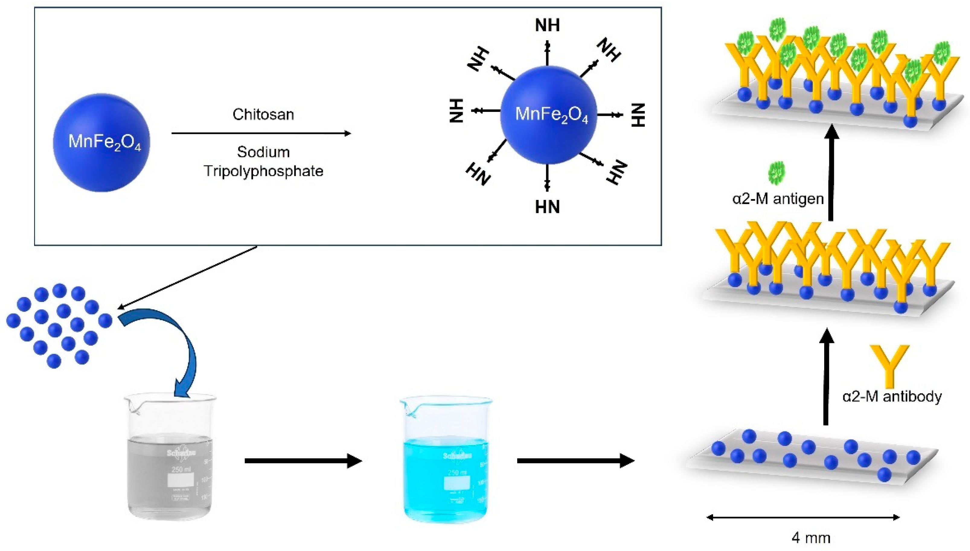
Figure 2.
Schematic illustration of the fabrication of the glucose biosensor. With permission from Ref. [60] Copyright 2020. Electroanalysis.
Figure 2.
Schematic illustration of the fabrication of the glucose biosensor. With permission from Ref. [60] Copyright 2020. Electroanalysis.
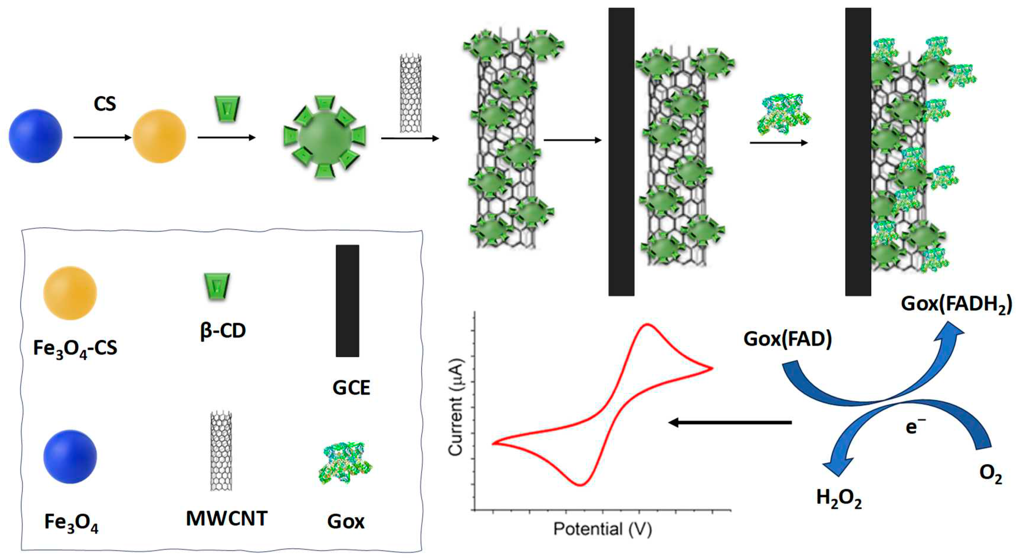
Figure 3.
Schematic diagram of fluorescence assays combined with APBA-PEG-MNs for S. aureus detection. Adapted with permission from Ref. Copyright 2022. Microchemical Journal.
Figure 3.
Schematic diagram of fluorescence assays combined with APBA-PEG-MNs for S. aureus detection. Adapted with permission from Ref. Copyright 2022. Microchemical Journal.
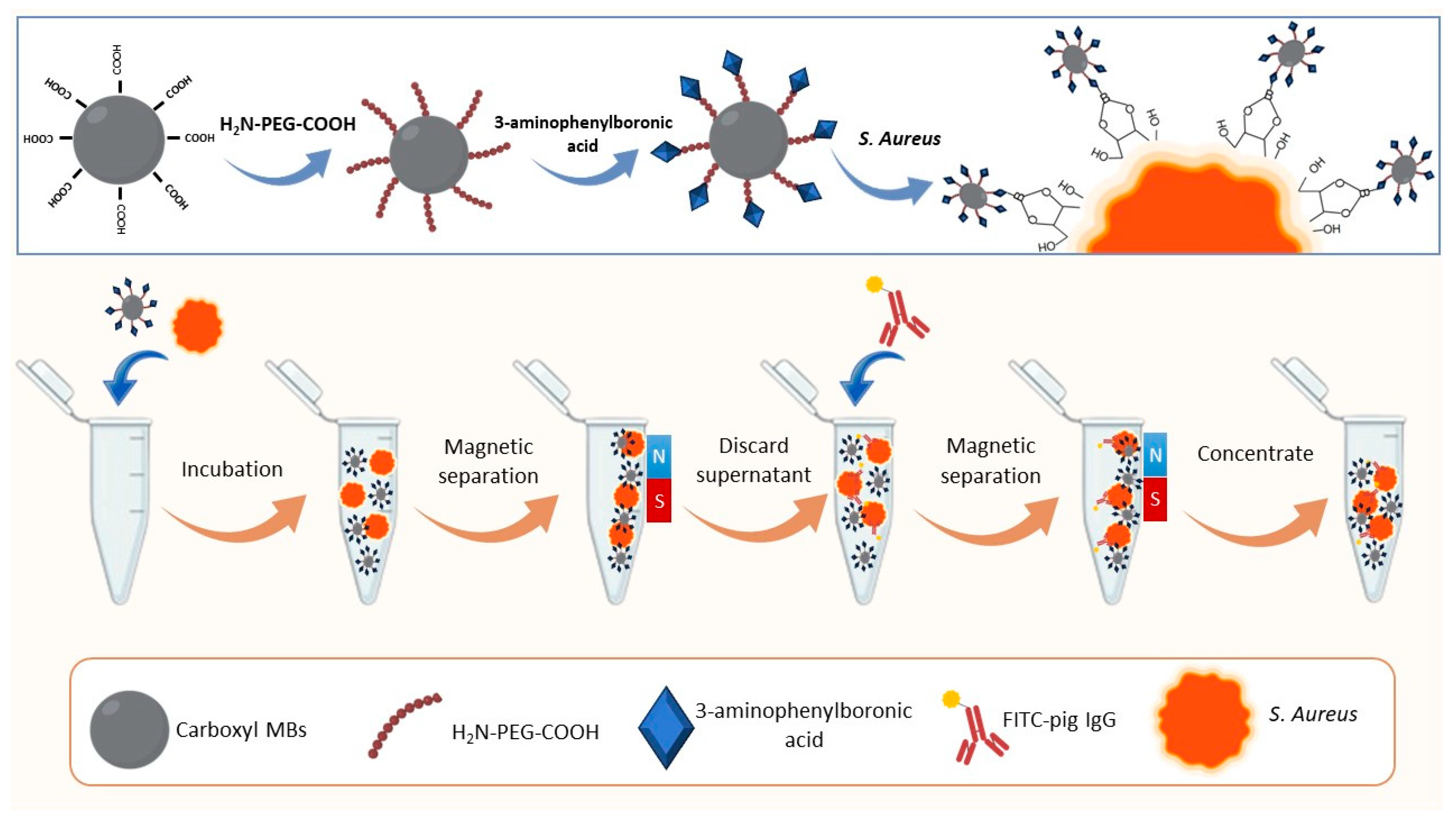
Figure 4.
Schematic illustration of the synthesis of polyaniline/magnetic graphene-Lac-GCE, along with the enzymatic oxidation of hydroquinone using laccase and its subsequent electrochemical reduction on the GCE. Adapted with permission from Ref [71]. Copyright 2020. Int J Biol Macromol.
Figure 4.
Schematic illustration of the synthesis of polyaniline/magnetic graphene-Lac-GCE, along with the enzymatic oxidation of hydroquinone using laccase and its subsequent electrochemical reduction on the GCE. Adapted with permission from Ref [71]. Copyright 2020. Int J Biol Macromol.
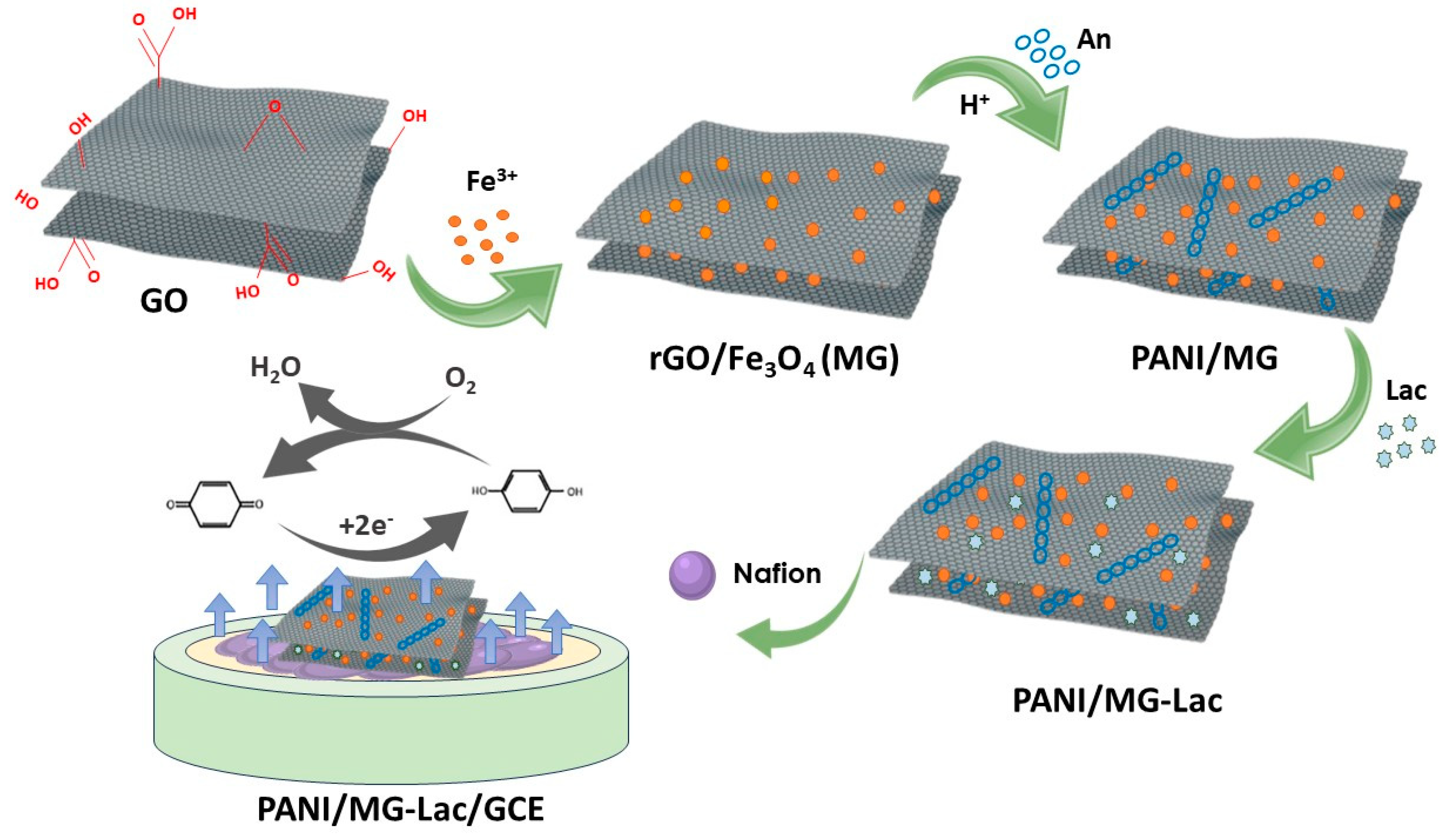
Figure 5.
Synthesis procedure of HRP/PDA-MNPs/(L-Arg/Tb) hybrid thin film for H2O2 detection. Taked with permission from Ref. [75]. Copyright 2021. Journal of Molecular recognition.
Figure 5.
Synthesis procedure of HRP/PDA-MNPs/(L-Arg/Tb) hybrid thin film for H2O2 detection. Taked with permission from Ref. [75]. Copyright 2021. Journal of Molecular recognition.
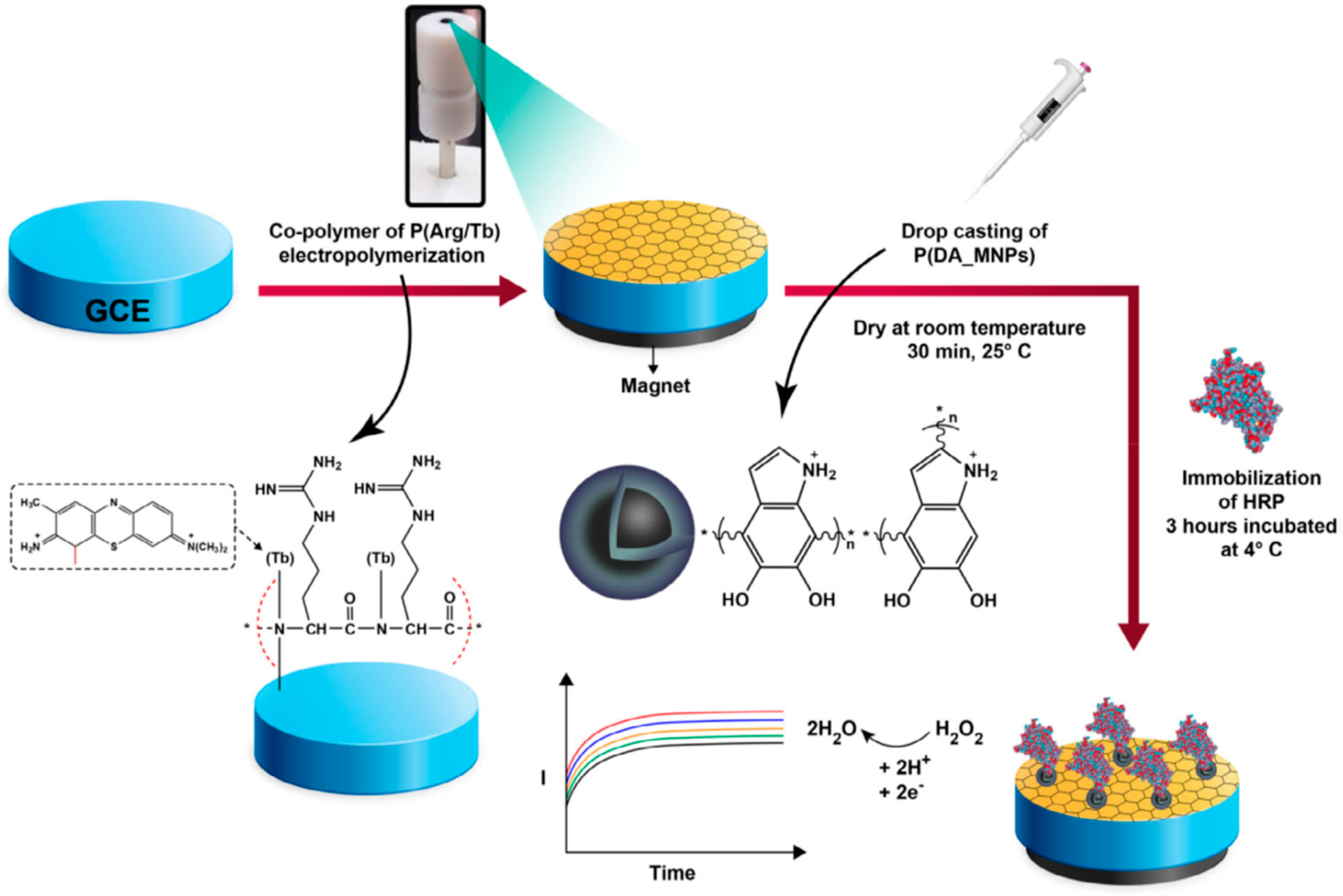
Figure 6.
Results of ZnO-capped mesoporous silica NPs as biosensor platform for S. Typhimurium detection: the (a) absorbance and (b) fluorescence photographs; the (c) absorbance and (d) fluorescence spectra; calibration curve of (e) the absorbance and (f) fluorescence intensity. Taked with permission from Ref. [77]. Copyright 202. Sensors and Actuators B: Chemical.
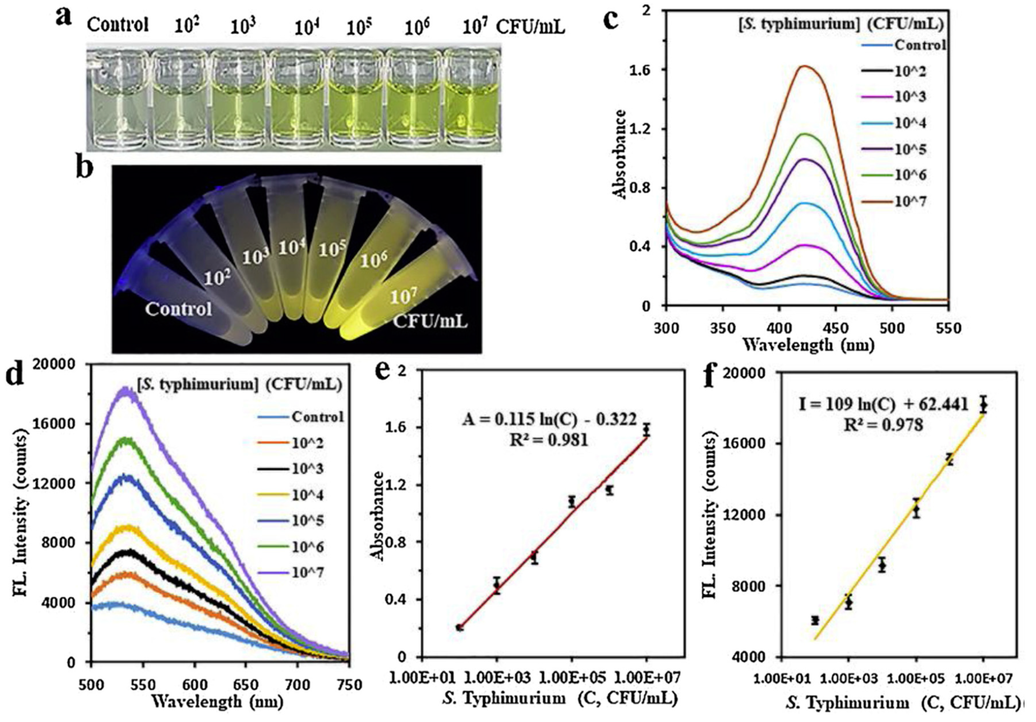
Figure 7.
Magnetic nanogap-enhanced Raman scattering (MINERS) bioassay for detection of SARS-CoV-2 spike protein. (a) Scheme of the functionalization of the 3D mag-MoO3–PDA@Au nanostructure with ACE-2, followed by the capture/immunoreaction with spike protein on the MINERS substrate. (b) MINERS spectra obtained by varying concentration of SARS-CoV-2 spike protein. (c) The calibration plots corresponding to the MINERS bioassay of SARS-CoV-2 spike protein in both PBS and whole cell lysate media. Taked with permission from Ref. [79]. Copyright 2022. Nanoscale Advances Journal.

Disclaimer/Publisher’s Note: The statements, opinions and data contained in all publications are solely those of the individual author(s) and contributor(s) and not of MDPI and/or the editor(s). MDPI and/or the editor(s) disclaim responsibility for any injury to people or property resulting from any ideas, methods, instructions or products referred to in the content. |
© 2023 by the authors. Licensee MDPI, Basel, Switzerland. This article is an open access article distributed under the terms and conditions of the Creative Commons Attribution (CC BY) license (http://creativecommons.org/licenses/by/4.0/).
Copyright: This open access article is published under a Creative Commons CC BY 4.0 license, which permit the free download, distribution, and reuse, provided that the author and preprint are cited in any reuse.


