
Submitted:
25 June 2023
Posted:
26 June 2023
You are already at the latest version
Abstract
Exosomes have shown promising potential as a therapeutic approach for wound healing. Nevertheless, the translation from experimental studies to commercially available treatments is still lacking. To assess the current state of research in this field, a systematic review was performed to examine the methodological heterogeneity among studies conducted over the past five years. Additionally, the review analyzed the suitability of animal models used and their relevance to human medicine. A PubMed search was performed for english-language, full-text available papers published from 2018 to June 2023, focusing on exosomes derived from mammalian sources and their application in wound healing, particularly those involving in vivo assays. Out of 531 results, 148 papers were selected for analysis. The findings revealed significant variability in terms of cell sources and types, biomaterials, and administration routes under investigation, indicating the need for further research in this field. Additionally, a comparative examination encompassing diverse cellular origins, types, administration pathways, or biomaterials is imperative. Furthermore, the predominance of rodent-based animal models raises concerns, as there have been limited advancements towards more complex in vivo models and scale-up assays. These constraints underscore the substantial efforts that remain necessary before attaining commercially viable and extensively applicable therapeutic approaches using exosomes.
Keywords:
animal models
; exosomes
; skin regeneration
; wound healing
; systematic review.
1. Introduction
The skin serves as the body’s external protection against harmful agents, regulating the internal temperature and integrity, while maintaining homeostasis. Under normal conditions, the skin can regenerate itself, through a complex process, that comprehends four distinct phases: hemostasis, inflammation, proliferation, and remodeling. However, when this process fails or is disrupted, it can culminate in impaired tissue regeneration or prolonged wound healing, leading to the formation of chronic wounds [1,2,3].
Chronic wounds are characterized by a prolonged inflammation (lasting from 4 to 12 weeks), often associated with infections, microbial biofilms, and impaired response from epithelial cells [4]. These wounds are multifactorial and frequently occur in individuals with several diseases, including diabetes, infections and arterial/venous insufficiency [5].
The prevalence of chronic non-healing wounds is increasing due to factors such as population aging and aging-associated diseases, concomitant diseases, tumors and congenital defects, negatively impacting the quality of life of millions of people worldwide. Therefore, the socioeconomical and health care burden is also increasing [1,2,3].
Current therapies such as debridement, antibiotherapy and dressings remain insufficient, as they are not efficient and there is still a need for new treatments. In the last few years, regenerative medicine has gained popularity, and extensive research has been conducted on mesenchymal stem cells (MSCs) and their derivates in several fields, including wound healing and skin regeneration.
MSCs are defined by the International Society of Cellular Therapy as similar to fibroblasts, adherent to plastic and with the ability to differentiate into three different cell lines, in vitro (chondrocytes, osteoblasts, and adipocytes). These cells should express the surface markers CD73, CD90 and CD105, while not expressing hematopoietic markers (CD14, CD45, CD34, CD19/HLA-DR and CD11b/CD79). These undifferentiated cells have the potential to repair different tissues as they undergo differentiation. Furthermore, MSCs can be obtained from different sources and species. In recent years, there has been a notable rise in the use of MSCs for wound healing purposes, with numerous studies showcasing promising outcomes utilizing cells obtained from diverse sources [1,2].
The use of MSC-derived products, such as secretome and exosomes, when compared to MSCs offers some advantages, including a reduced risk of tumorigenesis and minimal immune rejection [4], being therefore steadily rising. Exosomes are nano-vesicles secreted from the endosomal system (30-150nm) and represent one of the three major subpopulations of extracellular vesicles [6]. The other subpopulations include apoptotic bodies (>100nm) and microvesicles (100-1000nm). Exosomes are produced by several different cell types from different origins [7].
Several studies have shown that exosomes derived from MSCs have similar therapeutic properties, angiogenic ability and immune modulation as the cells from which they originate [3,8,9,10].
The aim of this systematic review was to access the methodological heterogeneity in studies and furnish the scientific community with a comprehensive overview of the advancements made in this field over the past five years. Additionally, the review encompassed an analysis of the animal models used to evaluate the translational potential of the data to human medicine.
2. Data and Methods
This systematic review was performed according to PRISMA (Preferred Reporting Items for Systematic Reviews and Meta-Analyses) guidelines.
The main goal of this study was to assess the methodology heterogeneity and provide insights into the progress of exosome research in wound healing over the past five years.
The research involved using the PubMed database, covering the period from 2018 to June 2023. The search query used the following keywords: “wound healing” [Title/Abstract] AND “exosomes” [Title/Abstract] NOT “review” [Title/Abstract], which initially retrieved 531 results.
All 531 publications underwent titles, abstracts and full-text articles examination.
The eligibility criteria included: 1) English language, 2) Full access to the publication, 3) Exosomes from mammalian sources, 4) Exosomes applications in wound healing, and 5) Use of animal models (in vivo studies).
Exclusion criteria were applied to filter out the studies that did not meet the research goals, such as: 1) in vitro studies, 2) review articles, 3) non-english language publication, 4) studies unrelated to wound healing, 5) no full access to the publication, and 6) studies not involving exosomes.
To ensure rigorous study selection, all authors participated in the process and conducted double-checks. Any discrepancies or disagreements were resolved through discussion and consensus. Duplicates were searched through Endnote software and data analysis was performed using an Excel form specifically designed by the authors.
Among the initial 531 studies, 383 were excluded based on the exclusion criteria mentioned above, resulting in 148 papers. These remaining papers underwent a thorough double-check by all authors.
The extracted information from the selected studies included PMID (PubMed Identification), paper title, publication date, corresponding author’s country, cell species, cell type, biomaterial usage, administration route, animal models and exclusion criteria (when applicable).
GraphPad Prism version 8.0.1 was used to elaborate the graphical representations of the collected data.
Bias assessment was evaluated for each study regarding the adherence to MSCs minimal criteria and animal models were examined to determine external validity.
The selection process is summarized in Figure 1.
Through this systematic review, the study aimed to provide valuable insights and contribute to the understanding of exosome-derived treatments in wound healing, with potential implications for potential translation into human medicine.
3. Results
3.1. Retrieved Data
The following table summarizes the retrieved data from the selected 148 papers (Table 1).
3.2. Scientific data production and publication distribution between 2018 and June 2023:
All 148 papers selected were comprehensively analyzed to assess the temporal distribution of its publication across the five-year timeframe (Figure 2). In 2018, a total of 7 papers were published (4,7%). The subsequent year 2019, witnessed a notable increase in publications, with 16 papers accounting for 10.8% of the selection. The publication rate continued to rise in 2020, reaching 30 papers (20,3%). In 2021, 29 papers were published (19,6%). The most significant publication rate occurred in 2022, with 42 papers published (28,4%). Up until June 2023, 24 additional papers have already been published, indicating that the year will probably surpass previous records (16,2%). These data demonstrate that there has been a continuous and stable increase in scientific investment in this research area, with the number of works carried out and published results increasing continuously.
The geographical distribution of scientific publications in the field of exosome application in wound healing was examined, focusing on the corresponding author’s country over a 5-year timeframe (Figure 3). China emerged as the country with the highest scientific publication rate in this field, with 122 publications (82,4%). The United States of America (USA) followed with 8 publications (5,4%), South Korea with 5 publications (3,4%) and Portugal with 3 publications (2,0%). Other countries have lower publication rates: Japan recorded 2 publications (1,4%), as did Iran (1,4%) and India (1,4%). Similarly, Egypt and Taiwan each had 1 publication (0,7%). Additionally, a collaboration between China and Finland resulted in 2 publications (1,4%).
3.3. Cell source and type
Cell source was analyzed as seen in Figure 4. Among the papers analyzed, human tissues were the preferred cell source for exosome extraction (73,6%). Then, rodent-derived cells corresponded to 23%, with 15,5% from mice and 7,4% from rat. The other sources, although less frequent, consisted of dog (0,7%) and macaque (0,7%). Furthermore, 1 study compared mice and rat-derived exosomes (0,7%), while 2 independent studies compared both mice and human-derived exosomes (1,4%).
According to the data presented in Table 2, the most commonly used to extract exosomes in humans is the adipose tissue (ADSC) with 26,6%, followed by the UCMSCs (umbilical cord mesenchymal stem cells) with 22,9%, and the BMSCs (bone marrow derived mesenchymal stem cells) with 10,1%. Other tissues still present considerable percentages of use, such as placenta (2,8%), peripheral blood (3,7%), epidermal (3,7%), DP (dental pulp derived mesenchymal stem cells) (2,8%) and umbilical vein (7,3%). Regarding mice, adipose tissue emerges as the preferred source (30,4%), followed by bone marrow (26,1%). In rats, both adipose tissue and bone marrow (36,4%) are favored as primary sources for exosome extraction. These results are in agreement with the general scientific literature related to the use of cell-based therapies, where adipose tissue, bone marrow and umbilical cord are the most explored tissues and the respective MSCs are the most studied and characterized cells both for their direct use and of their secretion products.
3.4. Biomaterials and Administration Route
As seen in Figure 5, the analysis of biomaterials used in all 148 papers revealed that the majority of studies chose to use exosomes without any biomaterial (73,6%). However, when a biomaterial was selected, hydrogels were the most commonly used (18,2%). Other biomaterials were also employed in several studies, such as scaffolds (2,7%), patches (1,4%), sponges (2,0%), nanoparticles (0,7%) and dressings (1,4%).
In the analysis of these 148 papers, the administration route of exosomes was analyzed and compared to the selected biomaterials, as illustrated in Figure 6. The preferred method, regardless of the presence of biomaterials, was via subcutaneous (SC) injection at the wound margins (66,2%). Within this route, subcutaneous injection without a biomaterial (57,8%) was the most common approach, while the association with a biomaterial was only 8,8%.
Topical administration was used 25,7%, with 16,9% involving the use of a biomaterial and 8,8% without one. Other administration routes included intradermal (ID) injection (2,7%), intraperitoneal (IP) injection (1,4%) and endovenous (EV) injection (2,0%). There were also 2 papers that compared topical and subcutaneous injections (1,4%), while 1 study compared topical and intradermal injections (0,7%).
3.5. Animal Models
Among the studies included in the analysis, rodents were used in the majority, accounting for 96,6% of the total (143 studies), with mice comprising 66,9% (99 studies) and rats 29,7% (44 studies). Also, two studies used both mice and rats (1,4%), while one study used a non-human primate model (0,7%) and the last used a canine model (0,7%). Figure 7 provides a visual representation of the distribution of in vivo models used in the selected studies.
4. Discussion
4.1. Scientific data production and publication distribution between 2018 and June 2023:
The publication rate regarding the use of exosomes for wound healing has shown a significant increase in the last five years, with 2022 marking the highest publication rate to date. Based on the count of 24 publications as of June, 2023 is expected to surpass previous records.
This notable increase in scientific publications reflects the recent emergence and promising outcomes of exosome-based therapies in wound healing. It is expected that even more valuable data will be published in the next years.
In addition, China stands out as the leading country regarding publication rate, which demonstrates the high importance this topic in this country. However, there remains a gap in research development and publication in this field, in other countries, particularly in Europe and America. It is essential to encourage research and publication in these regions, in order to promote advancements in the field of exosome-based therapy worldwide.
4.2. Cell source and type
Among the preferred tissue source for exosome production, human-derived cells accounted for 73,6% of the studies, followed by rodent-derived cells at 23%, with 15,5% from mice and 7,4% from rat.
Within the human-derived cells, the most commonly used tissue is the adipose tissue (26,6%), followed by the umbilical cord (22,9%) and the bone marrow (10,1%). In mice, adipose tissue emerges as the preferred source (30,4%), followed by bone marrow (26,1%). In rats, both adipose tissue and bone marrow (36,4%) are favored as primary sources for exosome extraction.
Given that the ultimate objective of most studies is the development of exosome-based therapies for human medicine, the retrieval of exosomes from human tissues appears to be a logical approach. However, the significant heterogeneity within the tissues, poses challenges in comparing results, as researchers have not reached an understanding of the most efficacious treatment option.
The preferred use of ADSCs (adipose tissue derived mesenchymal stem cells) is probably due to their low ethical issues, easy extraction, and cost-effectiveness. ADSCs have shown potential in wound healing, by increasing vascularization, fibroblasts migration and differentiation, and upregulating macrophages chemotaxis [2,159,160].
BMSCs have also demonstrated great potential in wound healing, increasing angiogenesis and reducing wound contraction [160]. Additionally, BM-MSCs have garnered significant attention as the most extensively investigated subset of MSCs and have been recognized for their relatively low immunogenicity [2].
UCMSCs have also shown their wound healing potential, as they can differentiate into epidermal tissue and are easier to harvest than BM-MSCs [1,2].
The variation observed in exosomes derived from different species and tissues still needs further research and understanding. The goal should be to identify the most effective sources and optimize and standardize the isolation processes to ensure consistent and reliable outcomes. By doing so, the scientific community can more accurately make comparisons between studies and advance towards the development of efficacious therapeutic approaches.
In addition, it is valuable to explore alternative sources of exosomes beyond those currently described, as it may uncover potential benefits and characteristics, broadening the range of therapeutic options.
Overall, while human-derived exosomes remain the preferred choice due to their ultimate clinical relevance, efforts should be made to refine the current methodologies and promote collaborations between research groups to better understand the most effective exosome-base treatments.
4.3. Biomaterials and Administration Route:
In most studies, exosomes were administered via SC injection in the wound margins, either without a biomaterial (57,8%) or in combination with a biomaterial (8,8%). This delivery method offers ease, speed, and localized treatment administration. Considering that most skin wounds are created on the animal’s dorsum, incorporating biomaterials can be difficult, requiring prior development and testing. SC injection of MSCs has also demonstrated great results regarding wound closure, angiogenesis and re-epithelization. Alternatively, topical administration is also used (25,7%), as it is less invasive and less painful than the injection methods [161].
However, when a biomaterial was selected it was predominantly a hydrogel (18,2%). Other biomaterials were also applied in several studies, including scaffolds (2,7%), patches (1,4%), sponges (2,0%), nanoparticles (0,7%) and dressings (1,4%).
The combination of biomaterials aims to improve the therapeutic functionality of exosomes by stabilizing them and prolonging their release at the wound site, thereby preventing rapid entry into blood circulation and systemic dilution. Hydrogels, specifically, offer several advantages in wound healing, such as antibacterial activity, facilitation of tissue adhesion, protection against UV radiation, hemostatic capacity, promotion of spontaneous regeneration and easy injectability. They can also provide a 3D environment and mimic the extracellular matrix, while maintaining proper moister levels at the wound site. Therefore, the use of exosomes associated with hydrogels has shown to improve wound healing, enhancing re-epithelization and vascularization [4,162].
Hydrogels based on chitosan or methylcellulose are considered great options for diabetic wound treatment and have been used in some of the selected studies. These polymers have good biodegradability, biocompatibility and are nontoxic. Geng et al, developed a loaded carboxyethyl chitosan hydrogel loaded with bone marrow derived exosomes to improve chronic diabetic wound healing. It increased angiogenesis and neovascularization, reduced local inflammation and improved wound healing in diabetic rats [26].
Pluronic F-12 hydrogels have also been used in selected studies, as they are injectable, biocompatible and thermosensitive. Zhou et al used a pluronic F-12 hydrogel combined with adipose tissue-derived exosomes to improve re-epithelization, angiogenesis, collagen synthesis and enhanced wound healing and cellular proliferation in mice [16]. Yang et al, also used a similar hydrogel combined with human umbilical cord-derived exosomes with increased wound closure rate and granulation tissue in rats [12].
Gelatin methacryloyl (GelMA) hydrogels were also chosen in different studies, due to their mechanical properties and ability to retain exosomes for a prolonged time. Zhao et al, used Human Umbilical Vein Endothelial Cells (HUVECs) derived exosomes in association with a GelMA hydrogel and demonstrated an improvement of angiogenesis and collagen maturity in rats [55]. Hu et al, used a similar hydrogel with ADSCs-derived exosomes and showed an improvement of wound healing with an increased blood vessel regeneration, proliferation and migration, in mice [42].
Several studies have combined hydrogels and MSCs, with promising results in skin regeneration. The use of BMSCs seeded into hydrogels improved angiogenesis and accelerated wound healing, in mice [163]. Another study demonstrated reduced scar formation, improved angiogenesis, collagen, granulation and re-epithelization in rabbits, using ADSCs combined with an hydrogel [164].
These findings emphasize the importance of carefully selecting the biomaterials and administration route to optimize the therapeutic effects of exosomes in wound healing.
4.4. Animal Models
The results showed that rodents are the main animal models in studies involving exosome-based therapies in wound healing (96,6%). These findings are consistent with Al-Masawa et al previous findings up until March 2021 [3].
The use of small animal models has several advantages such as researchers’ familiarity, easy handling, affordability, and availability. However, there are also limitations associated with these models, including skin thickness, fast hair growth cycles, follicular pattern and wound size [165]. Rodents exhibit a thin epidermis and loose skin adherence, along with dense hair that has been suggested to potentially enhance the wound healing rate. In addition, these animals lack apocrine and eccrine glands, but possess a subcutaneous panniculus carnosus muscle, that enhances rapid wound contraction. Moreover, they also have stronger immune systems and have endogenous sources of vitamin C, which plays a significant role in wound healing [166,167].
Although rodent models are frequently used in the initial stages of new therapies approaches, it is necessary to scale-up to more complex animal models to better reflect the similarities between such models and the human species. The main goal of most researchers is to develop new treatments options for non-healing chronic wounds and make them commercially available to the human population. Therefore, the consistent use of rodents in 96,6% of studies over the last five years limits the broader application of this data.
Although the use of exosome-based therapies has been showing promising results over last few years, the inclusion of larger animal models, such as ovine, swine, dog and non-human primates is crucial. In addition, it is important to fulfill the 3Rs principle (replace, reduce and refine) regarding animal use, implying that research data should evolve until commercialization becomes possible [168,169].
However, these more complex models present challenges, as they are more expensive, more difficult to handle and require large set-ups. Pigs, for instance, are regarded as standard models for wound healing research due to the resemblance of their skin to that of humans. They also present physiological and anatomical similarity to the human species. Nonetheless, to date, no studies have been conducted on this particular species. Non-human primates, although sharing greater similarity with humans, are rarely used mainly due to ethical concerns [165,166].
The porcine model has been used in wound healing research, with promising results. In particular, the administration of BMSC and ADSC intradermally into partial-thickness wounds enhanced local epithelization and improved wound appearance, when compared to the control [170,171]. This suggests that the use of this cells can accelerate the wound healing process.
Martinello et al, used the ovine model in wound healing and achieved great results. The local injection of peripheral blood MSCs in the wound margins revealed improved re-epithelization, proliferation, neovascularization and contraction, with higher wound closer rate [172].
In dogs, the use of MSCs to treat chronic wounds has also demonstrated great potential. UCMSCs have been used in association with a PVA hydrogel showed significant progress in wound regeneration and decreased local ulceration [173]. Other study, using ADSc also improved re-epithelization, reduced local inflammation, promoted epidermal and dermal regeneration in both acute and chronic wounds [174]. In the selected study using a dog model, Bahr et al used BM-MSCs derived exosomes in association with a carboxymethylcellulose hydrogel. The results were promising, as the treatment enhanced wound healing with no scaring, with organized collagen deposition and increased dermal fibroblasts [157].
Lu et al, used autologous and allogeneic iPSCs derived exosomes to improve wound healing in macaques. It demonstrated an increased angiogenesis, collagen deposition, epithelial coverage and wound closure rate [55].
While acknowledging the differences among animal models, current approaches in wound healing remain highly relevant. However, it is crucial to increase the use of diverse and more complex animal models to bridge the gap between current findings and their practical application in the human species [175].
5. Conclusions
This comprehensive systematic review highlights the great potential of exosomes as therapeutic options for non-healing chronic wounds. Despite their promising role, the methodology associated with the use of exosomes-based therapies in wound healing remains highly heterogeneous. Further research endeavors are imperative to facilitate the commercial availability and clinical application of these treatments.
The considerable variability in cell sources, types, biomaterials, and administration routes under investigation shows the urgent need of further research in this field. Moreover, the lack of comparative studies exploring different cell sources/types, administration routes or even biomaterials is a critical gap that must be addressed. Furthermore, the predominant use of rodent-based animal models raises concerns, as limited progress has been made in advancing toward more complex in vivo models that closely resemble human physiology.
This study also has certain limitations, primarily due to the potential bias associated with study design and methodology of the included studies. To address these limitations, it is crucial that future research incorporates measures to mitigate bias, such as randomization, blinding and standardized protocols.
To achieve a commercially viable and widely accessible range of therapeutic options, several key objectives must be pursued in the future. Standardizing methodologies is paramount to ensure more reliable and comparable results. In addition, the inclusion of more complex animal models that closely mimic the human species will enable effective translation of research outcomes. These collective efforts will drive the field closer to its ultimate goal of achieving large-scale production and widespread availability of exosome-based therapeutic option for wound healing.
Author Contributions
Conceptualization: P.S., B.L., A.C.S., R.A, A.M., A.C., N.A., S.G. A.C.M.; methodology: P.S., B.L., A.C.S., R.A, A.M., A.C., N.A., S.G. A.C.M.; software, P.S., B.L., A.C.S., R.A, A.M., A.C., N.A., S.G. A.C.M.; validation, P.S., B.L., A.C.S., R.A, A.M., A.C., N.A., S.G. A.C.M.; formal analysis, P.S., B.L., A.C.S., R.A, A.M., A.C., N.A., S.G. A.C.M.; investigation, P.S., B.L., A.C.S., R.A, A.M., A.C., N.A., S.G. A.C.M; resources, P.S., B.L., A.C.S., R.A, A.M., A.C., N.A., S.G. A.C.M; data curation, P.S., B.L., A.C.S., R.A, A.M., A.C., N.A., S.G. A.C.M; writing—original draft preparation, P.S., B.L., A.C.S., R.A, A.M., A.C., N.A., S.G. A.C.M ; writing—review and editing, P.S., B.L., A.C.S., R.A, A.M., A.C., N.A., S.G. A.C.M; visualization, P.S., B.L., A.C.S., R.A, A.M., A.C., N.A., S.G. A.C.M; supervision, R.A., N.A., A.C.M.; project administration, N.A., A.C.M.; funding acquisition, N.A., A.C.M. All authors have read and agreed to the published version of the manuscript.
Funding
Patrícia Sousa acknowledges University of Porto (UP) and Centro de Estudos de Ciêcia Animal (CECA), Instituto de Ciências, Tecnologias e Agroambiente (ICETA) for the funding and availability of all resources needed for this work. Ana Catarina Sousa (SFRH/BD/ 146689/2019), and Bruna Lopes (2021.05265.BD) acknowledge Fundaçao para a Ciência e Tecnologia (FCT) for financial support. Rui Damásio Alvites acknowledges the Animal Science Studies Centre (CECA), Agroenvironment, Technologies and Sciences Institute (ICETA), Porto University (UP), and FCT for the funding and availability of all technical, structural, and human resources necessary for the development of this work. The work was supported through the project UIDB/00211/2020 funded by FCT/MCTES national funds. This research was funded by Projects PEst-OE/AGR/UI0211/2011 from FCT, and COMPETE 2020, from ANI–Projetos ID&T Empresas em Copromoçao, and by the project “H2Cure—Desenvolvimento de formulações de géis e pensos de Mel, Goma Gelana e Ácido Hialurónico para tratamento de feridas”( POCI-01-0247-FEDER-047032).
Institutional Review Board Statement
Not applicable.
Informed Consent Statement
Not applicable.
Data Availability Statement
Further data on the reported results are available from the corresponding author on request.
Conflicts of Interest
The authors declare no conflict of interest.
Registration and protocol
Not registered and no protocol was prepared.
Abbreviation
| ADSC | Adipose Tissue derived Mesenchymal Stem Cells |
| BMSC | Bone Marrow derived Mesenchymal Stem Cells |
| DPs | Dental Pulp derived Mesenchymal Stem Cells |
| EV | Endovenous |
| ID | Intradermal |
| IP | Intraperitoneal |
| iPSCs | Induced Pluripotent Stem Cells |
| MSCs | Mesenchymal Stem Cells |
| PMID | PubMed Identification |
| PRISMA | Preferred Reporting Items for Systematic Reviews and Meta-Analyses |
| SC | Subcutaneous |
| UCMSC | Umbilical Cord Mesenchymal Stem Cells |
| UVEC | Umbilical Vein Endothelial Cells |
References
- Carolina, M.; et al. Application of Cell-Based Therapies in Veterinary Dermatology, in Wound Healing—Recent Advances and Future Opportunities, M. Prof. Ana Colette, A. Dr. Rui Damásio, and G. Dr. Müzeyyen, Editors. 2023, IntechOpen: Rijeka. p. Ch. 1.
- Lopes, B.; et al. The Application of Mesenchymal Stem Cells on Wound Repair and Regeneration. Applied Sciences 2021, 11, 3000. [Google Scholar] [CrossRef]
- Al-Masawa, M.E.; et al. Efficacy and safety of small extracellular vesicle interventions in wound healing and skin regeneration: A systematic review and meta-analysis of animal studies. Theranostics 2022, 12, 6455–6508. [Google Scholar] [CrossRef] [PubMed]
- Zhao, H.; et al. Bioengineered MSC-derived exosomes in skin wound repair and regeneration. Front Cell Dev Biol 2023, 11, 1029671. [Google Scholar] [CrossRef] [PubMed]
- Wu, J.; et al. Mesenchymal stem cell-derived exosomes: The dawn of diabetic wound healing. World J Diabetes 2022, 13, 1066–1095. [Google Scholar] [CrossRef] [PubMed]
- Bailey, A.J.M.; et al. MSC-Derived Extracellular Vesicles to Heal Diabetic Wounds: a Systematic Review and Meta-Analysis of Preclinical Animal Studies. Stem Cell Rev Rep 2022, 18, 968–979. [Google Scholar] [CrossRef]
- Nikfarjam, S.; et al. Mesenchymal stem cell derived-exosomes: a modern approach in translational medicine. Journal of Translational Medicine 2020, 18, 449. [Google Scholar] [CrossRef]
- An, Y.; et al. Exosomes from adipose-derived stem cells and application to skin wound healing. Cell Proliferation 2021, 54, e12993. [Google Scholar] [CrossRef]
- Prasai, A.; et al. Role of Exosomes in Dermal Wound Healing: A Systematic Review. Journal of Investigative Dermatology 2022, 142, 662–678. [Google Scholar] [CrossRef]
- Hu, J.C.; et al. Mesenchymal stem cell-derived exosomes: A novel and potential remedy for cutaneous wound healing and regeneration. World J Stem Cells 2022, 14, 318–329. [Google Scholar] [CrossRef]
- Wang, C.; et al. Engineering Bioactive Self-Healing Antibacterial Exosomes Hydrogel for Promoting Chronic Diabetic Wound Healing and Complete Skin Regeneration. Theranostics 2019, 9, 65–76. [Google Scholar] [CrossRef]
- Yang, J.; et al. Umbilical Cord-Derived Mesenchymal Stem Cell-Derived Exosomes Combined Pluronic F127 Hydrogel Promote Chronic Diabetic Wound Healing and Complete Skin Regeneration. Int J Nanomedicine 2020, 15, 5911–5926. [Google Scholar] [CrossRef]
- Hu, Y.; et al. Exosomes from human umbilical cord blood accelerate cutaneous wound healing through miR-21-3p-mediated promotion of angiogenesis and fibroblast function. Theranostics 2018, 8, 169–184. [Google Scholar] [CrossRef] [PubMed]
- Liu, W.; et al. Melatonin-stimulated MSC-derived exosomes improve diabetic wound healing through regulating macrophage M1 and M2 polarization by targeting the PTEN/AKT pathway. Stem Cell Res Ther 2020, 11, 259. [Google Scholar] [CrossRef] [PubMed]
- Yuan, M.; et al. GelMA/PEGDA microneedles patch loaded with HUVECs-derived exosomes and Tazarotene promote diabetic wound healing. Journal of Nanobiotechnology 2022, 20, 147. [Google Scholar] [CrossRef] [PubMed]
- Zhou, Y.; et al. Human adipose-derived mesenchymal stem cells-derived exosomes encapsulated in pluronic F127 hydrogel promote wound healing and regeneration. Stem Cell Res Ther 2022, 13, 407. [Google Scholar] [CrossRef]
- Wu, D.; et al. Exosomes Derived from Bone Mesenchymal Stem Cells with the Stimulation of Fe(3)O(4) Nanoparticles and Static Magnetic Field Enhance Wound Healing Through Upregulated miR-21-5p. Int J Nanomedicine 2020, 15, 7979–7993. [Google Scholar] [CrossRef]
- Hu, Y.; et al. Exosomes derived from pioglitazone-pretreated MSCs accelerate diabetic wound healing through enhancing angiogenesis. J Nanobiotechnology 2021, 19, 150. [Google Scholar] [CrossRef]
- Wang, J.; et al. Hypoxia adipose stem cell-derived exosomes promote high-quality healing of diabetic wound involves activation of PI3K/Akt pathways. J Nanobiotechnology 2021, 19, 202. [Google Scholar] [CrossRef]
- Li, X.; et al. Exosomes from adipose-derived stem cells overexpressing Nrf2 accelerate cutaneous wound healing by promoting vascularization in a diabetic foot ulcer rat model. Exp Mol Med 2018, 50, 1–14. [Google Scholar] [CrossRef]
- Xiong, Y.; et al. All-in-One: Multifunctional Hydrogel Accelerates Oxidative Diabetic Wound Healing through Timed-Release of Exosome and Fibroblast Growth Factor. Small 2022, 18, e2104229. [Google Scholar] [CrossRef]
- Wang, P.; et al. Exosomes Derived from Epidermal Stem Cells Improve Diabetic Wound Healing. J Invest Dermatol 2022, 142, 2508–2517. [Google Scholar] [CrossRef]
- Zhou, X.; et al. Exosome-Mediated Crosstalk between Keratinocytes and Macrophages in Cutaneous Wound Healing. ACS Nano 2020, 14, 12732–12748. [Google Scholar] [CrossRef]
- Shi, R.; et al. Hypoxic ADSC-derived exosomes enhance wound healing in diabetic mice via delivery of circ-Snhg11 and induction of M2-like macrophage polarization. Biomed Pharmacother 2022, 153, 113463. [Google Scholar] [CrossRef]
- Yu, M.; et al. Exosomes derived from atorvastatin-pretreated MSC accelerate diabetic wound repair by enhancing angiogenesis via AKT/eNOS pathway. Stem Cell Res Ther 2020, 11, 350. [Google Scholar] [CrossRef] [PubMed]
- Geng, X.; et al. A multifunctional antibacterial and self-healing hydrogel laden with bone marrow mesenchymal stem cell-derived exosomes for accelerating diabetic wound healing. Biomater Adv 2022, 133, 112613. [Google Scholar] [CrossRef]
- Xiong, Y.; et al. Circulating Exosomal miR-20b-5p Inhibition Restores Wnt9b Signaling and Reverses Diabetes-Associated Impaired Wound Healing. Small 2020, 16, e1904044. [Google Scholar] [CrossRef]
- Shiekh, P.A.; Singh, A.; Kumar, A. Exosome laden oxygen releasing antioxidant and antibacterial cryogel wound dressing OxOBand alleviate diabetic and infectious wound healing. Biomaterials 2020, 249, 120020. [Google Scholar] [CrossRef]
- Xie, Y.; et al. SHED-derived exosomes promote LPS-induced wound healing with less itching by stimulating macrophage autophagy. J Nanobiotechnology 2022, 20, 239. [Google Scholar] [CrossRef]
- Wang, M.; et al. Efficient Angiogenesis-Based Diabetic Wound Healing/Skin Reconstruction through Bioactive Antibacterial Adhesive Ultraviolet Shielding Nanodressing with Exosome Release. ACS Nano 2019, 13, 10279–10293. [Google Scholar] [CrossRef] [PubMed]
- Zhang, Y.; et al. Exosomes derived from human umbilical cord blood mesenchymal stem cells stimulate regenerative wound healing via transforming growth factor-β receptor inhibition. Stem Cell Res Ther 2021, 12, 434. [Google Scholar] [CrossRef] [PubMed]
- He, X.; et al. MSC-Derived Exosome Promotes M2 Polarization and Enhances Cutaneous Wound Healing. Stem Cells Int 2019 2019, 7132708. [Google Scholar] [CrossRef] [PubMed]
- Kwak, G.; et al. Sustained Exosome-Guided Macrophage Polarization Using Hydrolytically Degradable PEG Hydrogels for Cutaneous Wound Healing: Identification of Key Proteins and MiRNAs, and Sustained Release Formulation. Small 2022, 18, e2200060. [Google Scholar] [CrossRef]
- Shi, R.; et al. Exosomes derived from mmu_circ_0000250-modified adipose-derived mesenchymal stem cells promote wound healing in diabetic mice by inducing miR-128-3p/SIRT1-mediated autophagy. Am J Physiol Cell Physiol 2020, 318, C848–c856. [Google Scholar] [CrossRef] [PubMed]
- Qiu, X.; et al. Exosomes released from educated mesenchymal stem cells accelerate cutaneous wound healing via promoting angiogenesis. Cell Prolif 2020, 53, e12830. [Google Scholar] [CrossRef] [PubMed]
- Qian, L.; et al. Adipose mesenchymal stem cell-derived exosomes accelerate skin wound healing via the lncRNA H19/miR-19b/SOX9 axis. Lab Invest 2021, 101, 1254–1266. [Google Scholar] [CrossRef] [PubMed]
- Wang, Y.; et al. VH298-loaded extracellular vesicles released from gelatin methacryloyl hydrogel facilitate diabetic wound healing by HIF-1α-mediated enhancement of angiogenesis. Acta Biomater 2022, 147, 342–355. [Google Scholar] [CrossRef] [PubMed]
- Heo, J.S. , Selenium-Stimulated Exosomes Enhance Wound Healing by Modulating Inflammation and Angiogenesis. Int J Mol Sci 2022, 23. [Google Scholar] [CrossRef]
- Ren, S.; et al. Exosomes from Adipose Stem Cells Promote Diabetic Wound Healing through the eHSP90/LRP1/AKT Axis. Cells 2022, 11. [Google Scholar] [CrossRef]
- Kim, H.; et al. Exosome-Guided Phenotypic Switch of M1 to M2 Macrophages for Cutaneous Wound Healing. Adv Sci (Weinh) 2019, 6, 1900513. [Google Scholar] [CrossRef] [PubMed]
- Li, B.; et al. The MSC-Derived Exosomal lncRNA H19 Promotes Wound Healing in Diabetic Foot Ulcers by Upregulating PTEN via MicroRNA-152-3p. Mol Ther Nucleic Acids 2020, 19, 814–826. [Google Scholar] [CrossRef]
- Hu, N.; et al. Hypoxia-pretreated ADSC-derived exosome-embedded hydrogels promote angiogenesis and accelerate diabetic wound healing. Acta Biomater 2023, 157, 175–186. [Google Scholar] [CrossRef]
- Xiao, S.; et al. Human acellular amniotic membrane incorporating exosomes from adipose-derived mesenchymal stem cells promotes diabetic wound healing. Stem Cell Res Ther 2021, 12, 255. [Google Scholar] [CrossRef]
- Chen, L.; et al. Serum exosomes accelerate diabetic wound healing by promoting angiogenesis and ECM formation. Cell Biol Int 2021, 45, 1976–1985. [Google Scholar] [CrossRef] [PubMed]
- Xia, W.; et al. Young fibroblast-derived exosomal microRNA-125b transfers beneficial effects on aged cutaneous wound healing. J Nanobiotechnology 2022, 20, 144. [Google Scholar] [CrossRef]
- Zhang, W.; et al. Cell-free therapy based on adipose tissue stem cell-derived exosomes promotes wound healing via the PI3K/Akt signaling pathway. Exp Cell Res 2018, 370, 333–342. [Google Scholar] [CrossRef] [PubMed]
- Ren, S.; et al. Microvesicles from human adipose stem cells promote wound healing by optimizing cellular functions via AKT and ERK signaling pathways. Stem Cell Res Ther 2019, 10, 47. [Google Scholar] [CrossRef] [PubMed]
- Li, Q.; et al. MiR146a-loaded engineered exosomes released from silk fibroin patch promote diabetic wound healing by targeting IRAK1. Signal Transduct Target Ther 2023, 8, 62. [Google Scholar] [CrossRef]
- Chen, B.; et al. Human embryonic stem cell-derived exosomes promote pressure ulcer healing in aged mice by rejuvenating senescent endothelial cells. Stem Cell Res Ther 2019, 10, 142. [Google Scholar] [CrossRef]
- Zhang, Z.; et al. Comprehensive proteomic analysis of exosome mimetic vesicles and exosomes derived from human umbilical cord mesenchymal stem cells. Stem Cell Res Ther 2022, 13, 312. [Google Scholar] [CrossRef]
- Liang, Z.H.; et al. Exosomes from mmu_circ_0001052-modified adipose-derived stem cells promote angiogenesis of DFU via miR-106a-5p and FGF4/p38MAPK pathway. Stem Cell Res Ther 2022, 13, 336. [Google Scholar] [CrossRef]
- Li, Y.; et al. Serum-derived exosomes accelerate scald wound healing in mice by optimizing cellular functions and promoting Akt phosphorylation. Biotechnol Lett 2021, 43, 1675–1684. [Google Scholar] [CrossRef] [PubMed]
- Chen, J.; et al. Blockade of lncRNA-ASLNCS5088-enriched exosome generation in M2 macrophages by GW4869 dampens the effect of M2 macrophages on orchestrating fibroblast activation. Faseb j 2019, 33, 12200–12212. [Google Scholar] [CrossRef] [PubMed]
- Zhu, Z.; et al. Exosomes Derived From Umbilical Cord Mesenchymal Stem Cells Treat Cutaneous Nerve Damage and Promote Wound Healing. Front Cell Neurosci 2022, 16, 913009. [Google Scholar] [CrossRef]
- Lu, M.; et al. Enhanced wound healing promotion by immune response-free monkey autologous iPSCs and exosomes vs. their allogeneic counterparts. EBioMedicine 2019, 42, 443–457. [Google Scholar] [CrossRef] [PubMed]
- Zhu, J.; Quan, H. Adipose-derived stem cells-derived exosomes facilitate cutaneous wound healing by delivering XIST and restoring discoidin domain receptor 2. Cytokine 2022, 158, 155981. [Google Scholar] [CrossRef] [PubMed]
- Yan, C.; et al. Human Umbilical Cord Mesenchymal Stem Cell-Derived Exosomes Accelerate Diabetic Wound Healing via Ameliorating Oxidative Stress and Promoting Angiogenesis. Front Bioeng Biotechnol 2022, 10, 829868. [Google Scholar] [CrossRef] [PubMed]
- Zhang, Y.; et al. Adipose mesenchymal stem cell exosomes promote wound healing through accelerated keratinocyte migration and proliferation by activating the AKT/HIF-1α axis. J Mol Histol 2020, 51, 375–383. [Google Scholar] [CrossRef]
- Teng, L.; et al. Exosomes Derived from Human Umbilical Cord Mesenchymal Stem Cells Accelerate Diabetic Wound Healing via Promoting M2 Macrophage Polarization, Angiogenesis, and Collagen Deposition. Int J Mol Sci 2022, 23. [Google Scholar] [CrossRef] [PubMed]
- Zhao, G.; et al. MSC-derived exosomes attenuate cell death through suppressing AIF nucleus translocation and enhance cutaneous wound healing. Stem Cell Res Ther 2020, 11, 174. [Google Scholar] [CrossRef]
- Zhao, D.; et al. GelMA combined with sustained release of HUVECs derived exosomes for promoting cutaneous wound healing and facilitating skin regeneration. J Mol Histol 2020, 51, 251–263. [Google Scholar] [CrossRef]
- Li, J.; et al. Enhancing Cutaneous Wound Healing Based on Human Induced Neural Stem Cell-derived Exosomes. Int J Nanomedicine 2022, 17, 5991–6006. [Google Scholar] [CrossRef]
- Ge, L.; et al. Engineered exosomes derived from miR-132-overexpresssing adipose stem cells promoted diabetic wound healing and skin reconstruction. Front Bioeng Biotechnol 2023, 11, 1129538. [Google Scholar] [CrossRef] [PubMed]
- Wei, P.; et al. Exosomes derived from human amniotic epithelial cells accelerate diabetic wound healing via PI3K-AKT-mTOR-mediated promotion in angiogenesis and fibroblast function. Burns Trauma 2020, 8, tkaa020. [Google Scholar] [CrossRef] [PubMed]
- Lyu, L.; et al. Exosomes derived from M2 macrophages induce angiogenesis to promote wound healing. Front Mol Biosci 2022, 9, 1008802. [Google Scholar] [CrossRef] [PubMed]
- Zhou, Y.; et al. Combined topical and systemic administration with human adipose-derived mesenchymal stem cells (hADSC) and hADSC-derived exosomes markedly promoted cutaneous wound healing and regeneration. Stem Cell Res Ther 2021, 12, 257. [Google Scholar] [CrossRef] [PubMed]
- Han, C.; et al. Human Umbilical Cord Mesenchymal Stem Cell Derived Exosomes Delivered Using Silk Fibroin and Sericin Composite Hydrogel Promote Wound Healing. Front Cardiovasc Med 2021, 8, 713021. [Google Scholar] [CrossRef]
- Li, M.; et al. Macrophage-derived exosomes accelerate wound healing through their anti-inflammation effects in a diabetic rat model. Artif Cells Nanomed Biotechnol 2019, 47, 3793–3803. [Google Scholar] [CrossRef] [PubMed]
- Zhang, X.; et al. Bioinspired Adaptable Indwelling Microneedles for Treatment of Diabetic Ulcers. Adv Mater 2023, 35, e2210903. [Google Scholar] [CrossRef] [PubMed]
- Zhang, Y.; et al. Preparation of exosomes encapsulated nanohydrogel for accelerating wound healing of diabetic rats by promoting angiogenesis. Mater Sci Eng C Mater Biol Appl 2021, 120, 111671. [Google Scholar] [CrossRef]
- Shen, Y.F.; et al. PTH Derivative promotes wound healing via synergistic multicellular stimulating and exosomal activities. Cell Commun Signal 2020, 18, 40. [Google Scholar] [CrossRef]
- Wang, L.; et al. Pharmaceutical Activation of Nrf2 Accelerates Diabetic Wound Healing by Exosomes from Bone Marrow Mesenchymal Stem Cells. Int J Stem Cells 2022, 15, 164–172. [Google Scholar] [CrossRef] [PubMed]
- Li, C.; et al. Adipose Mesenchymal Stem Cell-Derived Exosomes Promote Wound Healing Through the WNT/β-catenin Signaling Pathway in Dermal Fibroblasts. Stem Cell Rev Rep 2022, 18, 2059–2073. [Google Scholar] [CrossRef] [PubMed]
- Liu, Y.; et al. Human umbilical cord mesenchymal stem cell-derived exosomes promote murine skin wound healing by neutrophil and macrophage modulations revealed by single-cell RNA sequencing. Front Immunol 2023, 14, 1142088. [Google Scholar] [CrossRef]
- Liu, Y.; et al. Application of adipose mesenchymal stem cell-derived exosomes-loaded β-chitin nanofiber hydrogel for wound healing. Folia Histochem Cytobiol 2022, 60, 167–178. [Google Scholar] [CrossRef] [PubMed]
- Zhang, Y.; et al. Exosomes Derived from Adipose Mesenchymal Stem Cells Promote Diabetic Chronic Wound Healing through SIRT3/SOD2. Cells 2022, 11. [Google Scholar] [CrossRef]
- Sjöqvist, S.; et al. Exosomes derived from clinical-grade oral mucosal epithelial cell sheets promote wound healing. J Extracell Vesicles 2019, 8, 1565264. [Google Scholar] [CrossRef]
- Zhou, Z.; et al. Exosomes derived from dental pulp stem cells accelerate cutaneous wound healing by enhancing angiogenesis via the Cdc42/p38 MAPK pathway. Int J Mol Med 2022, 50. [Google Scholar] [CrossRef]
- Han, X.; et al. Exosomes derived from autologous dermal fibroblasts promote diabetic cutaneous wound healing through the Akt/β-catenin pathway. Cell Cycle 2021, 20, 616–629. [Google Scholar] [CrossRef]
- Camões, S.P.; et al. 3D-MSCs A151 ODN-loaded exosomes are immunomodulatory and reveal a proteomic cargo that sustains wound resolution. Journal of Advanced Research 2022, 41, 113–128. [Google Scholar] [CrossRef]
- Zhang, Y.; et al. Placental stem cells-derived exosomes stimulate cutaneous wound regeneration via engrailed-1 inhibition. Front Bioeng Biotechnol 2022, 10, 1044773. [Google Scholar] [CrossRef]
- Zhu, J.; et al. Exosome Mimetics-Loaded Hydrogel Accelerates Wound Repair by Transferring Functional Mitochondrial Proteins. Front Bioeng Biotechnol 2022, 10, 866505. [Google Scholar] [CrossRef] [PubMed]
- Chen Md, G.; et al. Effect of MicroRNA-146a Modified Adipose-Derived Stem Cell Exosomes on Rat Back Wound Healing. Int J Low Extrem Wounds 2021, 15347346211038092. [Google Scholar] [CrossRef] [PubMed]
- Yu, Q.; et al. MiR-221-3p targets HIPK2 to promote diabetic wound healing. Microvasc Res 2022, 140, 104306. [Google Scholar] [CrossRef]
- Wang, X.; et al. Fetal Dermal Mesenchymal Stem Cell-Derived Exosomes Accelerate Cutaneous Wound Healing by Activating Notch Signaling. Stem Cells Int 2019, 2019, 2402916. [Google Scholar] [CrossRef]
- Xu, J.; et al. miRNA-221-3p in Endothelial Progenitor Cell-Derived Exosomes Accelerates Skin Wound Healing in Diabetic Mice. Diabetes Metab Syndr Obes 2020, 13, 1259–1270. [Google Scholar] [CrossRef]
- Ahmadpour, F.; et al. Effects of exosomes derived from fibroblast cells on skin wound healing in Wistar rats. Burns 2023. [Google Scholar] [CrossRef]
- Hsu, H.H.; et al. Therapeutic Potential of Exosomes Derived from Diabetic Adipose Stem Cells in Cutaneous Wound Healing of db/db Mice. Pharmaceutics 2022, 14. [Google Scholar] [CrossRef]
- Parvanian, S.; et al. Exosomal vimentin from adipocyte progenitors accelerates wound healing. Cytoskeleton (Hoboken) 2020, 77, 399–413. [Google Scholar] [CrossRef] [PubMed]
- Born, L.J.; et al. HOTAIR-Loaded Mesenchymal Stem/Stromal Cell Extracellular Vesicles Enhance Angiogenesis and Wound Healing. Adv Healthc Mater 2022, 11, e2002070. [Google Scholar] [CrossRef]
- Jiang, L.; et al. Exosomes derived from TSG-6 modified mesenchymal stromal cells attenuate scar formation during wound healing. Biochimie 2020, 177, 40–49. [Google Scholar] [CrossRef]
- Liu, J.; et al. Exosomes Derived from Human Umbilical Cord Mesenchymal Stem Cells Accelerate Cutaneous Wound Healing by Enhancing Angiogenesis through Delivering Angiopoietin-2. Stem Cell Reviews and Reports 2021, 17, 305–317. [Google Scholar] [CrossRef]
- Sun, Y., Y. Ju, and B. Fang, Exosomes from human adipose-derived mesenchymal stromal/stem cells accelerate angiogenesis in wound healing: implication of the EGR-1/lncRNA-SENCR/DKC1/VEGF-A axis. Hum Cell 2022, 35, 1375–1390. [Google Scholar] [CrossRef]
- Shiekh, P.A., A. Singh, and A. Kumar, Data supporting exosome laden oxygen releasing antioxidant and antibacterial cryogel wound dressing OxOBand alleviate diabetic and infectious wound healing. Data Brief 2020, 31, 105671. [Google Scholar] [CrossRef] [PubMed]
- Wang, C.; et al. The fabrication of a highly efficient self-healing hydrogel from natural biopolymers loaded with exosomes for the synergistic promotion of severe wound healing. Biomater Sci 2019, 8, 313–324. [Google Scholar] [CrossRef] [PubMed]
- Jiang, T., Z. Wang, and J. Sun, Human bone marrow mesenchymal stem cell-derived exosomes stimulate cutaneous wound healing mediates through TGF-β/Smad signaling pathway. Stem Cell Res Ther 2020, 11, 198. [Google Scholar] [CrossRef] [PubMed]
- Bakadia, B.M.; et al. Engineering homologous platelet-rich plasma, platelet-rich plasma-derived exosomes, and mesenchymal stem cell-derived exosomes-based dual-crosslinked hydrogels as bioactive diabetic wound dressings. Bioact Mater 2023, 28, 74–94. [Google Scholar] [CrossRef]
- Parvanian, S.; et al. Exosomal Vimentin from Adipocyte Progenitors Protects Fibroblasts against Osmotic Stress and Inhibits Apoptosis to Enhance Wound Healing. Int J Mol Sci 2021, 22. [Google Scholar] [CrossRef] [PubMed]
- Zhang, X.F.; et al. Hypoxic ucMSC-secreted exosomal miR-125b promotes endothelial cell survival and migration during wound healing by targeting TP53INP1. Mol Ther Nucleic Acids 2021, 26, 347–359. [Google Scholar] [CrossRef] [PubMed]
- Duan, M.; et al. Epidermal stem cell-derived exosomes promote skin regeneration by downregulating transforming growth factor-β1 in wound healing. Stem Cell Res Ther 2020, 11, 452. [Google Scholar] [CrossRef]
- Lv, Q.; et al. Engineered Human Adipose Stem-Cell-Derived Exosomes Loaded with miR-21-5p to Promote Diabetic Cutaneous Wound Healing. Mol Pharm 2020, 17, 1723–1733. [Google Scholar] [CrossRef]
- Xu, N.; et al. Wound healing effects of a Curcuma zedoaria polysaccharide with platelet-rich plasma exosomes assembled on chitosan/silk hydrogel sponge in a diabetic rat model. Int J Biol Macromol 2018, 117, 102–107. [Google Scholar] [CrossRef] [PubMed]
- Xu, M.; et al. Hydrogen Peroxide-Induced Senescence Reduces the Wound Healing-Promoting Effects of Mesenchymal Stem Cell-Derived Exosomes Partially via miR-146a. Aging Dis 2021, 12, 102–115. [Google Scholar] [CrossRef] [PubMed]
- Dalirfardouei, R.; et al. Promising effects of exosomes isolated from menstrual blood-derived mesenchymal stem cell on wound-healing process in diabetic mouse model. J Tissue Eng Regen Med 2019, 13, 555–568. [Google Scholar] [CrossRef] [PubMed]
- Mi, B.; et al. Saliva exosomes-derived UBE2O mRNA promotes angiogenesis in cutaneous wounds by targeting SMAD6. J Nanobiotechnology 2020, 18, 68. [Google Scholar] [CrossRef]
- Zhang, Y.; et al. Exosome/metformin-loaded self-healing conductive hydrogel rescues microvascular dysfunction and promotes chronic diabetic wound healing by inhibiting mitochondrial fission. Bioact Mater 2023, 26, 323–336. [Google Scholar] [CrossRef]
- Chen, K., T. Yu, and X. Wang, Inhibition of Circulating Exosomal miRNA-20b-5p Accelerates Diabetic Wound Repair. Int J Nanomedicine 2021, 16, 371–381. [Google Scholar] [CrossRef]
- Xu, Y.; et al. Platelet-Rich Plasma-Derived Exosomal USP15 Promotes Cutaneous Wound Healing via Deubiquitinating EIF4A1. Oxid Med Cell Longev 2021, 2021, 9674809. [Google Scholar] [CrossRef]
- Kobayashi, H.; et al. <Editors’ Choice> Effects of exosomes derived from the induced pluripotent stem cells on skin wound healing. Nagoya J Med Sci 2018, 80, 141–153. [Google Scholar]
- Abdelsaid, K.; et al. Exercise improves angiogenic function of circulating exosomes in type 2 diabetes: Role of exosomal SOD3. Faseb j 2022, 36, e22177. [Google Scholar] [CrossRef]
- Li, P.; et al. Endothelial progenitor cell derived exosomes mediated miR-182-5p delivery accelerate diabetic wound healing via down-regulating PPARG. Int J Med Sci 2023, 20, 468–481. [Google Scholar] [CrossRef]
- Gao, S.; et al. Exosomal miR-135a derived from human amnion mesenchymal stem cells promotes cutaneous wound healing in rats and fibroblast migration by directly inhibiting LATS2 expression. Stem Cell Res Ther 2020, 11, 56. [Google Scholar] [CrossRef] [PubMed]
- Fu, W.; et al. Long noncoding RNA LINC01435 impedes diabetic wound healing by facilitating YY1-mediated HDAC8 expression. iScience 2022, 25, 104006. [Google Scholar] [CrossRef] [PubMed]
- Liu, Y.; et al. Adipose-derived mesenchymal stem cell-loaded β-chitin nanofiber hydrogel promote wound healing in rats. J Mater Sci Mater Med 2022, 33, 12. [Google Scholar] [CrossRef] [PubMed]
- Jin, M.H.; et al. Peroxiredoxin II with dermal mesenchymal stem cells accelerates wound healing. Aging (Albany NY) 2021, 13, 13926–13940. [Google Scholar] [CrossRef]
- Park, D.J.; et al. Serpin-loaded extracellular vesicles promote tissue repair in a mouse model of impaired wound healing. J Nanobiotechnology 2022, 20, 474. [Google Scholar] [CrossRef] [PubMed]
- Li, Q.; et al. Exosomes derived from Nr-CWS pretreated MSCs facilitate diabetic wound healing by promoting angiogenesis via the circIARS1/miR-4782-5p/VEGFA axis. Chin J Nat Med 2023, 21, 172–184. [Google Scholar] [CrossRef]
- Ding, J.; et al. Exosomes Derived from Human Bone Marrow Mesenchymal Stem Cells Stimulated by Deferoxamine Accelerate Cutaneous Wound Healing by Promoting Angiogenesis. Biomed Res Int 2019, 2019, 9742765. [Google Scholar] [CrossRef]
- Zhang, Y.; et al. Human Amniotic Fluid Stem Cell-Derived Exosomes as a Novel Cell-Free Therapy for Cutaneous Regeneration. Front Cell Dev Biol 2021, 9, 685873. [Google Scholar] [CrossRef]
- Pi, L.; et al. Exosomal microRNA-125a-3p from human adipose-derived mesenchymal stem cells promotes angiogenesis of wound healing through inhibiting PTEN. Mol Cell Biochem 2022, 477, 115–127. [Google Scholar] [CrossRef]
- Zhao, B.; et al. Exosomal MicroRNAs Derived from Human Amniotic Epithelial Cells Accelerate Wound Healing by Promoting the Proliferation and Migration of Fibroblasts. Stem Cells Int 2018, 2018, 5420463. [Google Scholar] [CrossRef]
- Qiu, J.; et al. Exosomes from linc00511-overexpressing ADSCs accelerates angiogenesis in diabetic foot ulcers healing by suppressing PAQR3-induced Twist1 degradation. Diabetes Res Clin Pract 2021, 180, 109032. [Google Scholar] [CrossRef]
- Xiong, Y.; et al. Inhibition of circulating exosomal microRNA-15a-3p accelerates diabetic wound repair. Aging (Albany NY) 2020, 12, 8968–8986. [Google Scholar] [CrossRef]
- Shi, M.; et al. Adaptive Gelatin Microspheres Enhanced Stem Cell Delivery and Integration With Diabetic Wounds to Activate Skin Tissue Regeneration. Front Bioeng Biotechnol 2022, 10, 813805. [Google Scholar] [CrossRef]
- Li, X.; et al. Magnetic targeting enhances the cutaneous wound healing effects of human mesenchymal stem cell-derived iron oxide exosomes. J Nanobiotechnology 2020, 18, 113. [Google Scholar] [CrossRef]
- Kim, J.; et al. Clinical-Scale Mesenchymal Stem Cell-Derived Extracellular Vesicle Therapy for Wound Healing. Int J Mol Sci 2023, 24. [Google Scholar] [CrossRef]
- Liu, Z.; et al. Local transplantation of GMSC-derived exosomes to promote vascularized diabetic wound healing by regulating the Wnt/β-catenin pathways. Nanoscale Adv 2023, 5, 916–926. [Google Scholar] [CrossRef]
- Li, Q.; et al. Exosome loaded genipin crosslinked hydrogel facilitates full thickness cutaneous wound healing in rat animal model. Drug Deliv 2021, 28, 884–893. [Google Scholar] [CrossRef] [PubMed]
- Han, Z.F.; et al. Exosomal lncRNA KLF3-AS1 derived from bone marrow mesenchymal stem cells stimulates angiogenesis to promote diabetic cutaneous wound healing. Diabetes Res Clin Pract 2022, 183, 109126. [Google Scholar] [CrossRef] [PubMed]
- Kuang, L.; et al. Human Keratinocyte-Derived Exosomal MALAT1 Promotes Diabetic Wound Healing by Upregulating MFGE8 via microRNA-1914-3p. Int J Nanomedicine 2023, 18, 949–970. [Google Scholar] [CrossRef] [PubMed]
- Liu, Y.; et al. Exosomes derived from stem cells from apical papilla promote craniofacial soft tissue regeneration by enhancing Cdc42-mediated vascularization. Stem Cell Res Ther 2021, 12, 76. [Google Scholar] [CrossRef]
- Qian, Z.; et al. A moisturizing chitosan-silk fibroin dressing with silver nanoparticles-adsorbed exosomes for repairing infected wounds. J Mater Chem B 2020, 8, 7197–7212. [Google Scholar] [CrossRef] [PubMed]
- Nie, W.; et al. Exosomal miR-17-92 derived from human mesenchymal stem cells promotes wound healing by enhancing angiogenesis and inhibiting endothelial cell ferroptosis. Tissue Cell 2023, 83, 102124. [Google Scholar] [CrossRef] [PubMed]
- Huang, J.; et al. Development of a novel RNAi therapy: Engineered miR-31 exosomes promoted the healing of diabetic wounds. Bioact Mater 2021, 6, 2841–2853. [Google Scholar] [CrossRef] [PubMed]
- Liu, M.; et al. Dendritic epidermal T cells secreting exosomes promote the proliferation of epidermal stem cells to enhance wound re-epithelialization. Stem Cell Res Ther 2022, 13, 121. [Google Scholar] [CrossRef]
- Qian, J.; et al. Genetic Background and Kinetics Define Wound Bed Extracellular Vesicles in a Mouse Model of Cutaneous Injury. Int J Mol Sci 2021, 22. [Google Scholar] [CrossRef]
- Yang, H.; et al. Analysis of miR-203a-3p/SOCS3-mediated induction of M2 macrophage polarization to promote diabetic wound healing based on epidermal stem cell-derived exosomes. Diabetes Res Clin Pract 2023, 197, 110573. [Google Scholar] [CrossRef]
- Wang, S.; et al. Circulating Exosomal miR-181b-5p Promoted Cell Senescence and Inhibited Angiogenesis to Impair Diabetic Foot Ulcer via the Nuclear Factor Erythroid 2-Related Factor 2/Heme Oxygenase-1 Pathway. Front Cardiovasc Med 2022, 9, 844047. [Google Scholar] [CrossRef]
- Henriques-Antunes, H.; et al. The Kinetics of Small Extracellular Vesicle Delivery Impacts Skin Tissue Regeneration. ACS Nano 2019, 13, 8694–8707. [Google Scholar] [CrossRef]
- Zeng, J.; et al. M2 macrophage-derived exosome-encapsulated microneedles with mild photothermal therapy for accelerated diabetic wound healing. Mater Today Bio 2023, 20, 100649. [Google Scholar] [CrossRef]
- Cardoso, R.M.S.; et al. Development of an optimized and scalable method for isolation of umbilical cord blood-derived small extracellular vesicles for future clinical use. Stem Cells Transl Med 2021, 10, 910–921. [Google Scholar] [CrossRef]
- Zhao, X.; et al. Optogenetic engineered umbilical cord MSC-derived exosomes for remodeling of the immune microenvironment in diabetic wounds and the promotion of tissue repair. J Nanobiotechnology 2023, 21, 176. [Google Scholar] [CrossRef]
- Zhang, L.; et al. Exosomes from microRNA-126 overexpressing mesenchymal stem cells promote angiogenesis by targeting the PIK3R2-mediated PI3K/Akt signalling pathway. J Cell Mol Med 2021, 25, 2148–2162. [Google Scholar] [CrossRef]
- Kou, X.; et al. The Fas/Fap-1/Cav-1 complex regulates IL-1RA secretion in mesenchymal stem cells to accelerate wound healing. Sci Transl Med 2018, 10. [Google Scholar] [CrossRef]
- Xia, W.; et al. Lean adipose tissue macrophage derived exosome confers immunoregulation to improve wound healing in diabetes. J Nanobiotechnology 2023, 21, 128. [Google Scholar] [CrossRef]
- Chen, T.; et al. Sphingosine-1-phosphate derived from PRP-Exos promotes angiogenesis in diabetic wound healing via the S1PR1/AKT/FN1 signalling pathway. Burns Trauma 2023, 11, tkad003. [Google Scholar] [CrossRef] [PubMed]
- Yang, H.; et al. Hair follicle mesenchymal stem cell exosomal lncRNA H19 inhibited NLRP3 pyroptosis to promote diabetic mouse skin wound healing. Aging (Albany NY) 2023, 15, 791–809. [Google Scholar] [CrossRef] [PubMed]
- Cao, G.; et al. Human Adipose-Derived Mesenchymal Stem Cells-Derived Exosomal microRNA-19b Promotes the Healing of Skin Wounds Through Modulation of the CCL1/TGF-β Signaling Axis. Clin Cosmet Investig Dermatol 2020, 13, 957–971. [Google Scholar] [CrossRef] [PubMed]
- Wu, M.; et al. Exosomal IRF1-loaded rat adipose-derived stem cell sheet contributes to wound healing in the diabetic foot ulcers. Mol Med 2023, 29, 60. [Google Scholar] [CrossRef]
- Zhang, Y.; et al. Sprayable alginate hydrogel dressings with oxygen production and exosome loading for the treatment of diabetic wounds. Int J Biol Macromol 2023, 242 Pt 3, 125081. [Google Scholar] [CrossRef]
- Xu, Y.; et al. Inhibition of exosomal miR-24-3p in diabetes restores angiogenesis and facilitates wound repair via targeting PIK3R3. J Cell Mol Med 2020, 24, 13789–13803. [Google Scholar] [CrossRef]
- Deng, D.; et al. Biotin-Avidin System-Based Delivery Enhances the Therapeutic Performance of MSC-Derived Exosomes. ACS Nano 2023, 17, 8530–8550. [Google Scholar] [CrossRef] [PubMed]
- Bae, Y.U.; et al. Embryonic Stem Cell-Derived mmu-miR-291a-3p Inhibits Cellular Senescence in Human Dermal Fibroblasts Through the TGF-β Receptor 2 Pathway. J Gerontol A Biol Sci Med Sci 2019, 74, 1359–1367. [Google Scholar] [CrossRef] [PubMed]
- Yang, C.; et al. Highly-expressed micoRNA-21 in adipose derived stem cell exosomes can enhance the migration and proliferation of the HaCaT cells by increasing the MMP-9 expression through the PI3K/AKT pathway. Arch Biochem Biophys 2020, 681, 108259. [Google Scholar] [CrossRef] [PubMed]
- Sanders, M.C.; et al. Protecting human amnion and chorion matrices (HACM) during processing: Performance enhancement in a Diabetic Mouse Model and Human Co-culture System. Wound Repair Regen 2023. [Google Scholar] [CrossRef]
- Li, B.; et al. A therapeutic role of exosomal lncRNA H19 from adipose mesenchymal stem cells in cutaneous wound healing by triggering macrophage M2 polarization. Cytokine 2023, 165, 156175. [Google Scholar] [CrossRef]
- Bahr, M.M.; et al. Proficiency of Carboxymethylcellulose as a Cryoprotectant. Clinical and Histological Evaluation of Cryopreserved Heterogenous Mesenchymal Stem Cell-Exosomal Hydrogel on Critical Size Skin Wounds in Dogs. Int J Hematol Oncol Stem Cell Res 2021, 15, 178–191. [Google Scholar]
- Li, Y.; et al. Exosomes derived from human adipose mesenchymal stem cells attenuate hypertrophic scar fibrosis by miR-192-5p/IL-17RA/Smad axis. Stem Cell Res Ther 2021, 12, 221. [Google Scholar] [CrossRef]
- Huayllani, M.T.; et al. Adipose-derived stem cells in wound healing of full-thickness skin defects: a review of the literature. Journal of Plastic Surgery and Hand Surgery 2020, 54, 263–279. [Google Scholar] [CrossRef]
- Pang, C.; et al. An overview of the therapeutic potential of regenerative medicine in cutaneous wound healing. International Wound Journal 2017, 14, 450–459. [Google Scholar] [CrossRef]
- Shojaei, F., S. Rahmati, and M. Banitalebi Dehkordi, A review on different methods to increase the efficiency of mesenchymal stem cell-based wound therapy. Wound Repair and Regeneration 2019, 27, 661–671. [Google Scholar] [CrossRef]
- Golchin, A.; et al. Combination Therapy of Stem Cell-derived Exosomes and Biomaterials in the Wound Healing. Stem Cell Reviews and Reports 2022, 18, 1892–1911. [Google Scholar] [CrossRef]
- Rustad, K.C.; et al. Enhancement of mesenchymal stem cell angiogenic capacity and stemness by a biomimetic hydrogel scaffold. Biomaterials 2012, 33, 80–90. [Google Scholar] [CrossRef]
- Nilforoushzadeh, M.A.; et al. Mesenchymal Stem Cell Spheroids Embedded in an Injectable Thermosensitive Hydrogel: An In Situ Drug Formation Platform for Accelerated Wound Healing. ACS Biomaterials Science & Engineering 2020, 6, 5096–5109. [Google Scholar]
- Grada, A.; Mervis, J.; Falanga, V. Research Techniques Made Simple: Animal Models of Wound Healing. Journal of Investigative Dermatology 2018, 138, 2095–2105. [Google Scholar] [CrossRef] [PubMed]
- Sami, D.G.; Heiba, H.H.; Abdellatif, A. Wound healing models: A systematic review of animal and non-animal models. Wound Medicine 2019, 24, 8–17. [Google Scholar] [CrossRef]
- Saeed, S.; Martins-Green, M. Animal models for the study of acute cutaneous wound healing. Wound Repair and Regeneration 2023, 31, 6–16. [Google Scholar] [CrossRef]
- Peric, M.; et al. The rational use of animal models in the evaluation of novel bone regenerative therapies. Bone 2015, 70, 73–86. [Google Scholar] [CrossRef]
- de Vries, R.B.M.; et al. The Usefulness of Systematic Reviews of Animal Experiments for the Design of Preclinical and Clinical Studies. ILAR Journal 2014, 55, 427–437. [Google Scholar] [CrossRef]
- Hanson, S.E.; et al. Local delivery of allogeneic bone marrow and adipose tissue-derived mesenchymal stromal cells for cutaneous wound healing in a porcine model. Journal of Tissue Engineering and Regenerative Medicine 2016, 10, E90–E100. [Google Scholar] [CrossRef]
- Ochiai, H.; et al. Transplanted mesenchymal stem cells are effective for skin regeneration in acute cutaneous wounds of pigs. Regenerative Therapy 2017, 7, 8–16. [Google Scholar] [CrossRef]
- Martinello, T.; et al. Allogeneic mesenchymal stem cells improve the wound healing process of sheep skin. BMC Veterinary Research 2018, 14, 202. [Google Scholar] [CrossRef]
- Ribeiro, J.; et al. Cell therapy with human MSCs isolated from the umbilical cord Wharton jelly associated to a PVA membrane in the treatment of chronic skin wounds. Int J Med Sci 2014, 11, 979–87. [Google Scholar] [CrossRef] [PubMed]
- Enciso, N.; et al. Cutaneous wound healing: canine allogeneic ASC therapy. Stem Cell Research & Therapy 2020, 11, 261. [Google Scholar]
- Cibelli, J.; et al. Strategies for Improving Animal Models for Regenerative Medicine. Cell Stem Cell 2013, 12, 271–274. [Google Scholar] [CrossRef] [PubMed]
Figure 1.
PRISMA Flow Diagram that summarizes the selection process.
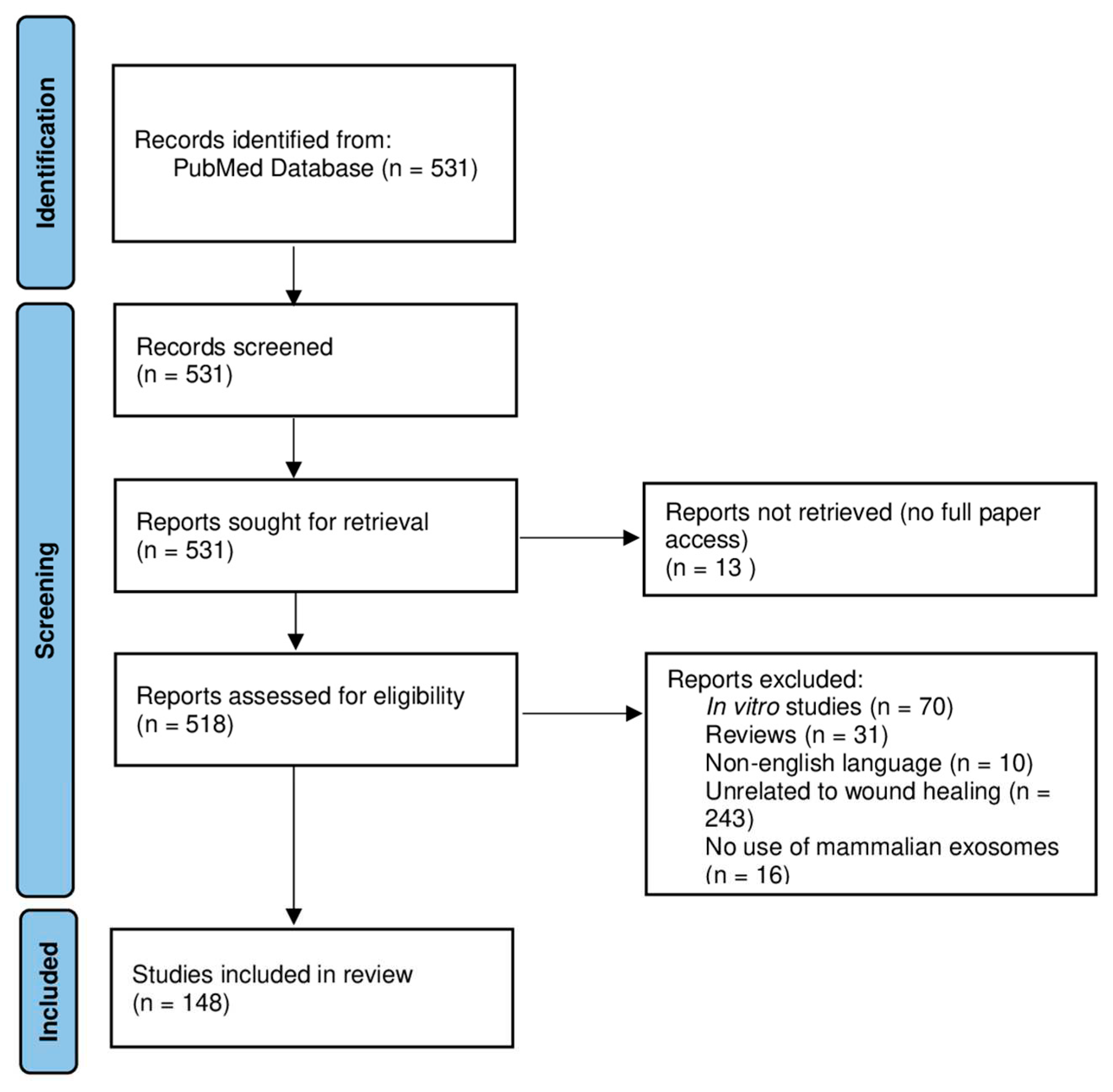
Figure 2.
Graphical representation of publication distribution per year, from 2018 to June 2023.
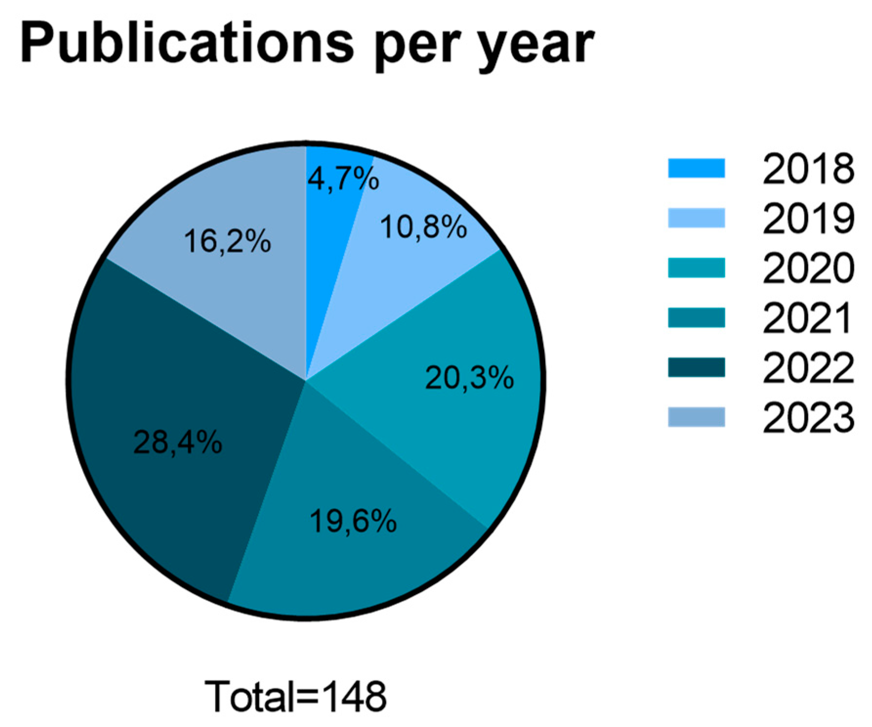
Figure 3.
Graphical representation of publication distribution per corresponding authors country (2018- June 2023).
Figure 3.
Graphical representation of publication distribution per corresponding authors country (2018- June 2023).
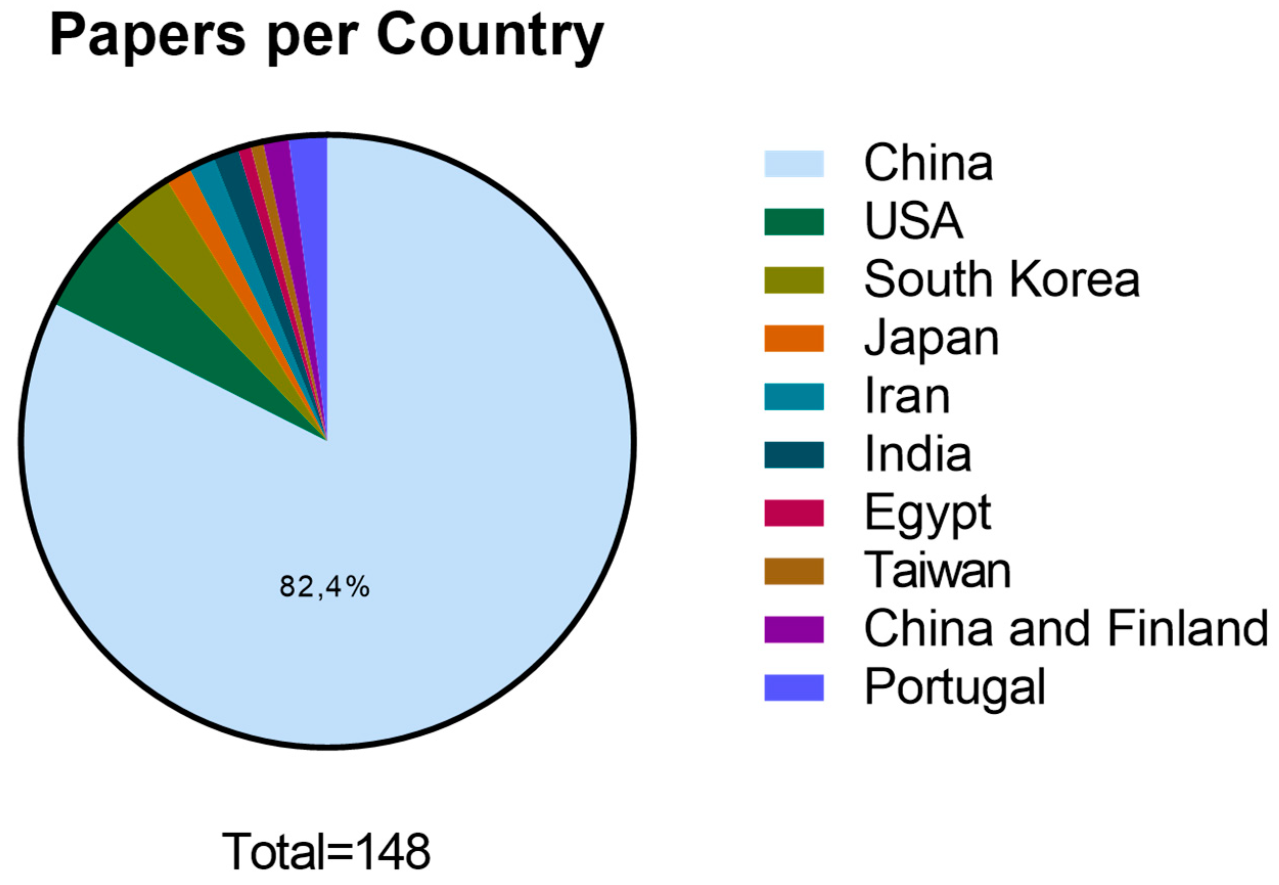
Figure 4.
Graphical representation of cell tissue source distribution in scientific literature between 2018 and June 2023.
Figure 4.
Graphical representation of cell tissue source distribution in scientific literature between 2018 and June 2023.
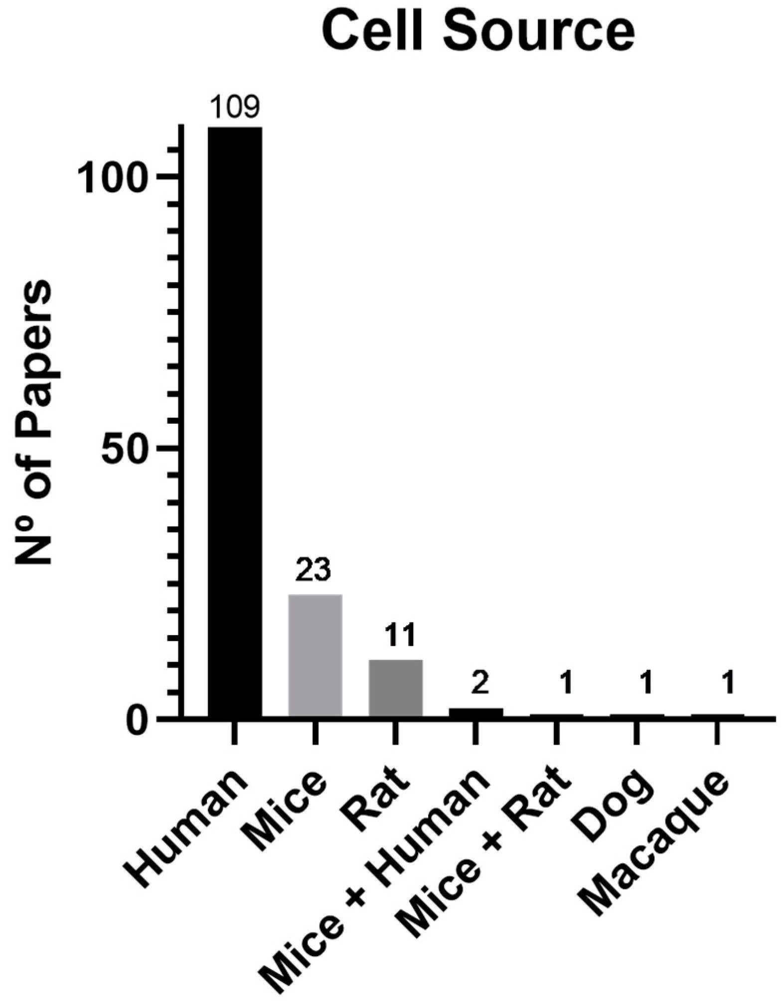
Figure 5.
Graphical representation of the biomaterials used in publications related to wound healing between 2018 and June 2023.
Figure 5.
Graphical representation of the biomaterials used in publications related to wound healing between 2018 and June 2023.
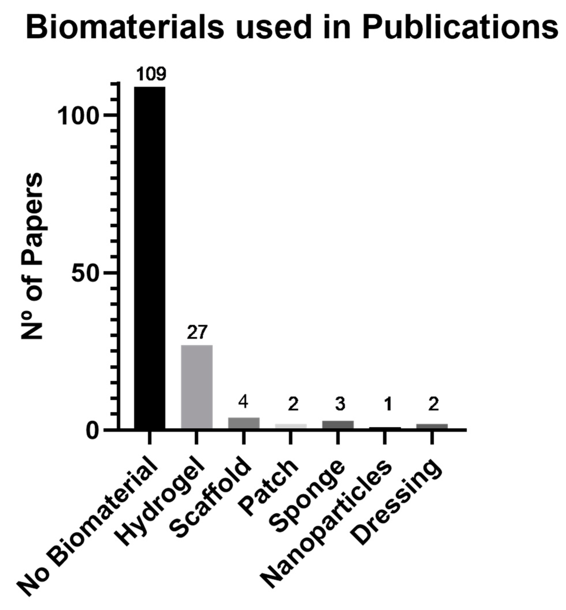
Figure 6.
Graphical representation of the administration route of exosomes used in wound healing between 2018 and June 2023. EV – Endovenous, ID – Intradermal, IP – Intraperitoneal, SC -Subcutaneous.
Figure 6.
Graphical representation of the administration route of exosomes used in wound healing between 2018 and June 2023. EV – Endovenous, ID – Intradermal, IP – Intraperitoneal, SC -Subcutaneous.
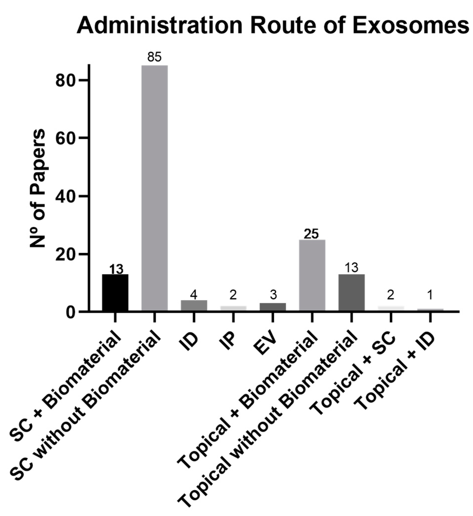
Figure 7.
Graphical representation of the animal models used in wound healing between 2018 and June 2023.
Figure 7.
Graphical representation of the animal models used in wound healing between 2018 and June 2023.
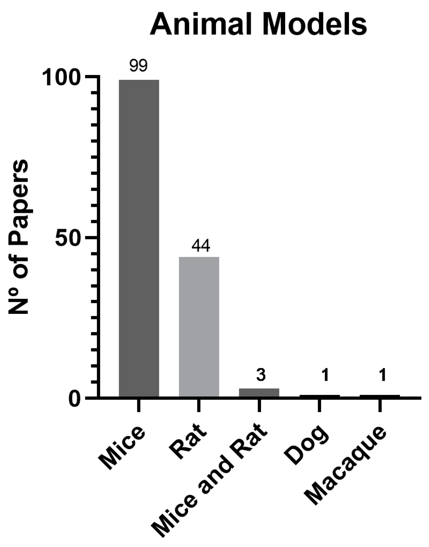

Table 1.
Summary of the retrieved data from the 148 papers.
| Ref | Year | Country | Cell Source | Cell Type | Biomaterial | Administration Route | Animal Models |
|---|---|---|---|---|---|---|---|
| [11] | 2019 | China | Human | ADSC | Hydrogel | SC Injection | Mice |
| [12] | 2020 | China | Human | UCMSC | Hydrogel | SC Injection | Rat |
| [13] | 2018 | China | Human | UCMSC | x | SC Injection | Mice |
| [14] | 2020 | China | Human | BMSC | x | SC Injection | Rat |
| [15] | 2022 | China | Human | UVEC | Patch | Patch | Mice |
| [16] | 2022 | China | Human | ADSC | Hydrogel | Topical | Mice |
| [17] | 2020 | China | Human | BMSC | x | SC Injection | Rat |
| [18] | 2021 | China | Rat | BMSC | x | SC Injection | Rat |
| [19] | 2021 | China | Human | ADSC | x | SC Injection | Mice |
| [20] | 2018 | China | Human | ADSC | x | SC Injection | Rat |
| [21] | 2022 | China | Mice | BMSC | Hydrogel | SC Injection | Mice |
| [22] | 2022 | USA | Human | Epidermal | x | SC Injection | Mice |
| [23] | 2020 | USA | Mice | Keratinocyte | x | SC Injection | Mice |
| [24] | 2022 | China | Human | ADSC | x | SC Injection | Mice |
| [25] | 2020 | China | Human | BMSC | x | SC Injection | Rat |
| [26] | 2022 | China | Rat | BMSC | Hydrogel | Topical | Rat |
| [27] | 2020 | China | Human | Peripheral Blood | x | SC Injection | Mice |
| [28] | 2020 | India | Rat | ADSC | Scaffold | Scaffold | Rat |
| [29] | 2022 | China | Human | DPs | x | SC Injection | Mice |
| [30] | 2019 | China | Human | ADSC | Scaffold | Scaffold | Mice |
| [31] | 2021 | China | Human | UCMSC | x | EV Injection | Rat |
| [32] | 2019 | China | Human | BMSC | x | SC Injection | Mice |
| [33] | 2022 | Korea | Mice | BMSC | Hydrogel | SC Injection | Mice |
| [34] | 2020 | China | Human | ADSC | x | SC Injection | Rat |
| [35] | 2020 | China | Mice | BMSC | x | ID Injection | Mice |
| [36] | 2021 | China | Human | ADSC | x | SC Injection | Mice |
| [37] | 2022 | China | Human | Epidermal | Hydrogel | SC Injection | Mice |
| [38] | 2022 | Korea | Human | ADSC | x | SC Injection | Mice |
| [39] | 2022 | China | Human | ADSC | x | SC Injection | Mice |
| [40] | 2019 | Korea | Mice | BMSC | x | SC Injection | Mice |
| [41] | 2020 | China | Human | BMSC | x | SC Injection | Mice |
| [42] | 2023 | China | Mice | ADSC | Hydrogel | SC Injection | Mice |
| [43] | 2021 | China | Human | ADSC | Scaffold | Scaffold | Mice |
| [44] | 2021 | China | Mice | Serum | x | SC Injection | Mice |
| [45] | 2022 | China | Mice | Fibroblast | x | ID Injection | Mice |
| [46] | 2018 | China | Human | ADSC | x | SC and ID Injection | Mice |
| [47] | 2019 | China | Human | ADSC | x | SC Injection | Mice |
| [48] | 2023 | China | Human | Placenta | Patch | Patch | Mice |
| [49] | 2019 | China | Human | Embryonic | x | Topical | Mice |
| [50] | 2022 | China | Human | UCMSC | x | SC Injection | Mice |
| [51] | 2022 | China | Mice | ADSC | x | SC Injection | Mice |
| [52] | 2021 | China | Rat and Mice | Serum | x | SC Injection | Mice |
| [53] | 2019 | China | Human | Macrophage | x | SC Injection | Mice |
| [54] | 2022 | China | Human | UCMSC | x | Topical | Mice |
| [55] | 2019 | China | Macaque | iPSCs | x | Topical | Macaque |
| [56] | 2022 | China | Mice | ADSC | x | SC Injection | Mice |
| [57] | 2022 | China | Human | UCMSC | x | SC Injection | Mice |
| [58] | 2020 | China | Human | ADSC | x | SC Injection | Mice |
| [59] | 2022 | China | Human | UCMSC | x | SC Injection | Rat |
| [60] | 2020 | China | Human | UCMSC | x | SC Injection | Mice |
| [61] | 2020 | China | Human | UVEC | Hydrogel | Topical | Rat |
| [62] | 2022 | China | Human | iPSCs | Hydrogel | Topical | Mice |
| [63] | 2023 | China | Mice | ADSC | x | SC Injection | Mice |
| [64] | 2020 | China | Human | Amniotic Membrane | x | SC Injection | Mice |
| [65] | 2022 | China | Human | UVEC | x | SC Injection | Mice |
| [66] | 2021 | China | Human | ADSC | x | Injection and Topical | Mice |
| [67] | 2021 | China | Human | UCMSC | Hydrogel | Topical | Mice |
| [68] | 2019 | China | Human | UVEC | x | SC Injection | Rat |
| [69] | 2023 | China | Human | UVEC | Hydrogel | Microneedle | Rat |
| [70] | 2021 | China | Human | UCMSC | Hydrogel | SC Injection | Rat |
| [71] | 2020 | China | Human | UVEC | Hydrogel | SC Injection | Rat |
| [72] | 2022 | China | Rat | BMSC | x | SC Injection | Rat |
| [73] | 2022 | China | Human | ADSC | x | SC Injection | Rat |
| [74] | 2023 | China | Human | UCMSC | x | SC Injection | Mice |
| [75] | 2022 | China | Mice | ADSC | Hydrogel | Topical | Rat |
| [76] | 2022 | China | Human | ADSC | x | SC Injection | Mice |
| [77] | 2019 | Japan | Human | Epithelial | x | Topical | Rat |
| [78] | 2022 | China | Human | DPs | x | SC Injection | Mice |
| [79] | 2021 | China | Rat | Dermal | x | SC Injection | Rat |
| [80] | 2022 | Portugal | Human | UCMSC | x | SC Injection | Rat |
| [81] | 2022 | China | Rat | Placenta and ADSC | x | SC Injection | Rat |
| [82] | 2022 | China | Human | UCMSC | Hydrogel | SC Injection | Mice |
| [83] | 2021 | China | Human | ADSC | x | SC Injection | Rat |
| [84] | 2022 | China | Human | UVEC | x | SC Injection | Mice |
| [85] | 2019 | China | Human | Fetal dermal | x | SC Injection | Mice |
| [86] | 2020 | China | Mice | BMSC | x | Topical | Mice |
| [87] | 2023 | Iran | Human | Fetal dermal | x | Topical | Rat |
| [88] | 2022 | Taiwan | Mice | ADSC and dermal | x | Topical | Mice |
| [89] | 2020 | China and Finland | Human | ADSC | x | IP Injection | Mice |
| [90] | 2022 | USA | Human | BMSC | x | SC Injection | Mice and Rat |
| [91] | 2020 | China | Human | BMSC | x | SC Injection | Mice |
| [92] | 2021 | China | Human | UCMSC | x | SC Injection | Rat |
| [93] | 2022 | China | Human | ADSC | x | SC Injection | Mice |
| [94] | 2020 | India | Rat | ADSC | Scaffold | Scaffold | Rat |
| [95] | 2019 | China | Human | Placenta | Hydrogel | SC Injection | Mice |
| [96] | 2020 | China | Human | BMSC | x | SC Injection | Rat |
| [97] | 2023 | China | Rat | BMSC and plasma | Hydrogel | Topical | Rat |
| [98] | 2021 | China and Finland | Human | ADSC | x | IP Injection | Mice |
| [99] | 2021 | China | Human | UCMSC | x | SC Injection | Mice |
| [100] | 2020 | China | Human | Epidermal | x | SC Injection | Rat |
| [101] | 2020 | China | Human | ADSC | Hydrogel | Topical | Rat |
| [102] | 2018 | China | Human | Plasma | Sponge | Sponge | Rat |
| [103] | 2021 | China | Human | UCMSC and ADSC | x | SC Injection | Mice |
| [104] | 2019 | Iran | Human | Menstrual Blood | x | ID Injection | Mice |
| [105] | 2020 | China | Human | Saliva | x | SC Injection | Mice |
| [106] | 2023 | China | Human | ADSC | Hydrogel | SC Injection | Mice |
| [107] | 2021 | China | Human | Peripheral Blood | x | SC Injection | Mice |
| [108] | 2021 | China | Mice | Plasma | x | SC Injection | Mice |
| [109] | 2018 | Japan | Human | iPSCs | x | SC Injection | Mice |
| [110] | 2022 | USA | Mice and Human | Plasma | x | Topical | Mice |
| [111] | 2023 | China | Human | UCMSC | x | Topical | Mice |
| [112] | 2020 | China | Human | Amnion | x | SC Injection | Rat |
| [113] | 2022 | China | Human | Keratinocyte | x | SC Injection | Mice |
| [114] | 2022 | China | Mice | ADSC | Hydrogel | SC Injection | Rat |
| [115] | 2021 | China | Mice | Dermal | x | ID Injection | Mice |
| [116] | 2022 | USA | Mice | Skin | Sponge | Sponge | Mice |
| [117] | 2023 | China | Human | UCMSC | x | SC Injection | Mice |
| [118] | 2019 | China | Human | BMSC | x | SC Injection | Rat |
| [119] | 2021 | China | Human | Amniotic Fluid | x | SC Injection | Rat |
| [120] | 2022 | China | Human | ADSC | x | SC Injection | Mice |
| [121] | 2018 | China | Human | Amniotic Fluid | x | SC Injection | Mice |
| [122] | 2021 | China | Human | ADSC | x | SC Injection | Rat |
| [123] | 2020 | China | Human | Peripheral Blood | x | Injection | Mice |
| [124] | 2022 | China | Rat | ADSC | Hydrogel | Topical | Rat |
| [125] | 2020 | China | Human | UCMSC | Nanoparticles | EV Injection | Rat |
| [126] | 2023 | Korea | Human | UCMSC | x | Injection and Topical | Mice and Rat |
| [127] | 2022 | China | Human | Gingival | x | SC Injection | Mice |
| [128] | 2021 | China | Human | UCMSC | Hydrogel | Topical | Rat |
| [129] | 2022 | China | Human | BMSC | x | EV Injection | Mice |
| [130] | 2023 | China | Human | Keratinocyte | x | SC Injection | Mice |
| [131] | 2021 | China | Human | DPs | x | Topical | Mice |
| [132] | 2020 | China | Human | UCMSC | Dressing | Topical | Mice |
| [133] | 2023 | China | Human | UCMSC | x | SC Injection | Mice |
| [134] | 2021 | China | Human | Embryonic | x | SC Injection | Rat |
| [135] | 2022 | China | Mice | Dendritic epidermal T cells | x | SC Injection | Mice |
| [136] | 2021 | USA | Mice | Skin | Sponge | Topical | Mice |
| [137] | 2023 | China | Human | Epidermal | x | SC Injection | Mice |
| [138] | 2022 | China | Human | UVEC | x | SC Injection | Mice |
| [139] | 2019 | Portugal | Human | UCMSC | Hydrogel | Topical | Mice |
| [140] | 2023 | China | Mice | Macrophage | x | Topical | Rat |
| [141] | 2021 | Portugal | Human | UCMSC | Hydrogel | Topical | Mice |
| [142] | 2023 | China | Human | UCMSC | x | SC Injection | Mice |
| [143] | 2021 | China | Human | BMSC | x | SC Injection | Mice |
| [144] | 2018 | USA | Mice and Human | BMSCs, Skin and Gingiva | x | SC Injection | Mice |
| [145] | 2023 | China | Mice | ADSC | x | SC Injection | Mice |
| [146] | 2023 | China | Human | Plasma | x | SC Injection | Mice |
| [147] | 2023 | China | Human | Hair follicle | x | SC Injection | Mice |
| [148] | 2020 | China | Human | ADSC | x | SC Injection | Mice |
| [149] | 2023 | China | Rat | ADSC | x | SC Injection | Rat |
| [150] | 2023 | China | Mice | BMSC | Dressing | Topical | Rat |
| [151] | 2020 | China | Human | Peripheral Blood | x | SC Injection | Mice |
| [152] | 2023 | China | Rat | BMSC | Hydrogel | SC Injection | Mice and Rat |
| [153] | 2019 | Korea | Human | Fibroblast | x | Topical | Mice |
| [154] | 2020 | China | Human | ADSC | x | SC Injection | Mice |
| [155] | 2023 | USA | Human | Placenta | x | Topical | Mice |
| [156] | 2023 | China | Human | ADSC | x | SC Injection | Mice |
| [157] | 2021 | Egypt | Dog | BMSC | Hydrogel | Topical | Dog |
| [158] | 2021 | China | Human | ADSC | x | SC Injection | Mice |
Table 2.
Summary of the most used cell types of the most common species.
| Human (109 studies) | Mice (23 studies) | Rat (11 studies) | |
| BMSC | 11 (10,1%) | 6 (26,1%) | 4 (36,4%) |
| ADSC | 29 (26,6%) | 7 (30,4%) | 4 (36,4%) |
| UCMSC | 25 (22,9%) | X | X |
| UVECS | 8 (7,3%) | X | X |
| DP | 3 (2,8%) | X | X |
| Epidermal | 4 (3,7%) | X | X |
| Peripherical Blood | 4 (3,7%) | X | X |
| Placenta | 3 (2,8%) | X | X |
| Others | 22 (20,2%) | 10 (43,5%) | 3 (27,3%) |
Disclaimer/Publisher’s Note: The statements, opinions and data contained in all publications are solely those of the individual author(s) and contributor(s) and not of MDPI and/or the editor(s). MDPI and/or the editor(s) disclaim responsibility for any injury to people or property resulting from any ideas, methods, instructions or products referred to in the content. |
© 2023 by the authors. Licensee MDPI, Basel, Switzerland. This article is an open access article distributed under the terms and conditions of the Creative Commons Attribution (CC BY) license (http://creativecommons.org/licenses/by/4.0/).
Copyright: This open access article is published under a Creative Commons CC BY 4.0 license, which permit the free download, distribution, and reuse, provided that the author and preprint are cited in any reuse.


