
Submitted:
06 June 2023
Posted:
07 June 2023
You are already at the latest version
Abstract
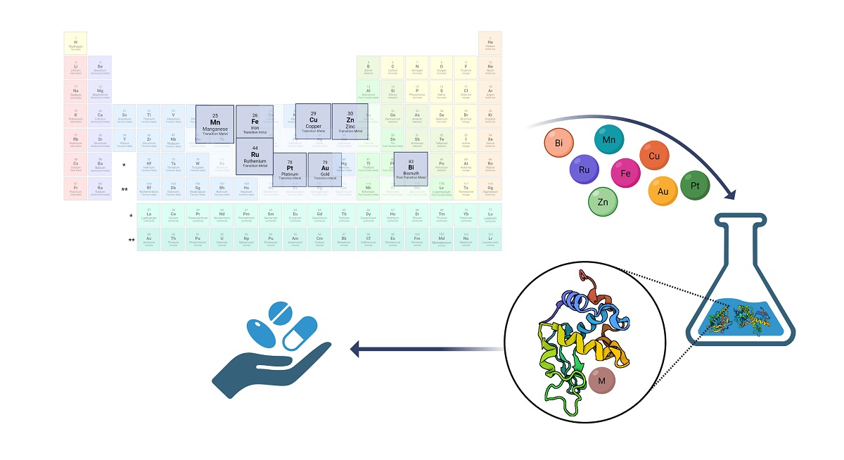
Metals are indispensable in the life of all organisms, in fact, their dysregulation causes various disorders caused by an interruption of their homeostasis. Nowadays different transition metals are incorporated into pharmaceutical products as diagnostic and therapeutic agents because of their electronic structure which gives them versatility in terms of tuning molecule properties, differently from organic molecules. Hence the interest in the study of metal-drug complexes from different points of view was born and many approaches were developed for the characterization, activation, delivery and molecular mechanisms clarification. The integration of these different approaches starting from chemoproteomics passing through nanoparticle systems to different activation strategies allows the understanding of the cellular responses of metallic drugs, which can be the basis for the design of new drugs and/or modification of drugs currently in use. This review aims to briefly summarize the recent advances in this area by describing the technological platforms and their possible applications to identify protein targets for discovering the mechanisms of action of metallodrugs, and to improve their efficiency in terms of administration.
Keywords:
metallodrugs
; medicinal chemistry
; chemoproteomics
; thermal proteome profiling
; pro-metallodrugs
; delivery systems
; nanoparticles
1. Introduction
The involvement of metals in many cellular and subcellular functions and their role in numerous vital processes is known. For example, zinc (Zn) is a mineral involved in numerous functions of cellular metabolism by supporting the catalytic activity of over 100 enzymes [1]. Calcium (Ca) is essential for cellular physiology and its transfer across membranes serves as a signal for many cellular processes, such as muscle contraction and the transmission of nerve impulses [2]. Copper (Cu) plays a key role in the central nervous system (CNS) [3] and in hematopoiesis [4], the process of making blood cells. Furthermore, Cu and Cu-dependent enzymes are cofactors of numerous redox reactions and are involved, for example, in neurotransmission [5]. Evolution has made metals indispensable in the life of all organisms, in fact their dysregulation causes various disorders, sometimes even serious ones, such as a series of neurodegenerative diseases caused by an interruption of their homeostasis [6,7]. It is ancient knowledge that metals may serve as therapeutic agents too, for example as antimicrobial and anti-viral weapons [8,9,10]. More recently, many metals are incorporated into pharmaceutical products as diagnostic and therapeutic agents. In fact, the electronic structure of transition metals has the advantage of great versatility in terms of tuning molecule properties, differently from organic molecules [11] The use of Pt coordination compounds in cancer therapy has certainly been [12] one of the most unexpected developments in the field of medicines in the last 50 years. This new class of chemotherapy was discovered by chance by Barnett Rosemberg (1927 – 2009) in 1965. The clinical success of cisplatin as an anticancer drug has accelerated the search for metals in medicine chemistry [13]. Subsequently, several other Pt drugs, including carboplatin and oxaliplatin, have been developed to improve therapeutic efficacy and reduce systemic toxicity [14],[15],[16].
Clinical studies gave controversial indications: while some patients showed very positive results, others highlighted the high toxicity of the compound [17]. The spectacular results obtained in patients suffering from tumors of the genital tract, especially testicular cancer where cisplatin proved to be practically curative, led to its approval by the FDA (Food and Drug Administration) in 1978. Since then, cisplatin has been one of the most used drugs in the world in anticancer therapy and is by far one of the most successful drugs (a so-called Blockbuster drug) [18],[19]. The resistance of tumors to cisplatin, spontaneous or induced, is an extremely serious problem, as it considerably limits the use of the drug. The main cellular processes through which cisplatin attacks tumor cells are uptake and transport, formation of adducts with DNA and their recognition by specific proteins (damage-response proteins), the “translation” of the DNA damage signal which inhibits its replication and transcription and activates numerous signal transduction pathways which, through a wide range of proteins that control cell growth, its differentiation and response to stress, can lead to cell cycle arrest and repair of damage or to cell death [20]. The research was soon also directed towards other metals with the aim of finding complexes that have activity against tumors resistant to platinum compounds and which, possibly, also present lower systemic toxicity. For this reason, other metals have been also tested as potential anticancer compounds such as those containing ruthenium (Ru) [21], arsenic (As) [22], gold (Au) [23], and osmium (Os) [24], and they are currently under development [25,26].
Among them, Ru compounds were the first to be studied and still represent a very active research sector today, followed by As compounds, at the time, the only other non-radioactive metal approved by the FDA in 2000 for the treatment of tumors [27]. It was introduced into clinical use in the 1970s, for the treatment of numerous leukaemias and especially acute promyelocytic leukemia [28]. To date, gold compounds are getting more and more attention because they have displayed fewer tolerability limits and appear to be target-specific [29]. Recent studies show that Au(I) complexes have antitumor activity through the induction of apoptosis in different cancer cell lines in vitro, comparing the effect such compounds have on resistant cells and analyzing the mechanism of action [30]. In addition, gold compounds have been proposed as potential anti-infective and anti-tubercular agents [31].
Despite all the potentialities of these metallo-compounds, they have several target “sites”, so drugs that target a single protein or enzyme can easily develop resistance. For this reason, critical points are the identification of the molecular targets of these drugs and the elucidation of the mechanisms of action as well as their "delivery" to the correct target and their activation.
Functional proteomics aims at the identification of protein-protein (PPI) [32,33], protein-DNA/RNA [34] interactions in vivo in order to define protein complexes and, therefore, cellular pathways involved in biological processes of interest as extensively described in multiple works and reviews [35,36]. There are many proteomic approaches used for the study of such interactions based on classical biochemical protocols adapted to the study of the so-called "interactomics" investigation thanks also to the coupling with advanced mass spectrometry instruments. This approach can also be used for the definition and identification of the molecular partners of metal compounds, inside the chemical proteomics branch. On the other hand, drug delivery, i.e. the vehicle of a substance in a targeted manner towards the tissue or cell where its consequent controlled release will guarantee greater efficiency, represents an important field of study. Application in the pharmacological field allows a molecule to be conveyed within our body, making it selectively reach the target tissue and releasing it in a controlled way [37,38], [39]. This methodology allows to decrease the doses of the drug administered, consequently decreasing its possible side effects, and making it more bioavailable. Nanoparticles (NPs) currently represent a great step forward in this area [40].
In this mini-review, we aim to give an overview of purification and identification strategies of protein targets of metal compounds, from affinity purification approaches, including labeling and cross-linking (photo-affinity) strategies for the identification of transient interactions. Furthermore, we intend to describe the different methodologies used nowadays to improve the efficiency of metallodrugs in terms of activation and delivery in vivo from the pro-drug approach to the different delivery systems. In fact, we are convinced that the study and the combination of the two points (i.e, target identification and delivery) are milestones towards the clinical route of a metallodrug.
2. Protein targets identification through proteomics approaches
The investigation of protein interactions with different classes of biomolecules has ancient roots in the proteomics field, but developing and advancing many different biochemical strategies coupled with mass spectrometry techniques [35,36]. In recent years, several proteomics strategies coupled with mass spectrometry (MS) have emerged as a powerful and systematic approach for the large-scale identification of drug-protein interactions and the elucidation of their associated mechanisms also.
In fact, chemical proteomics, or chemoproteomics, is mostly dedicated to the investigation of protein-small molecule interaction getting more and more attention in drug target discovery [41,42]. Chemoproteomics is a lead tool in the field of drug discovery, relying on affinity-based or label-free approaches to identify protein targets.
2.1. Affinity-based strategies
The affinity-based chemical proteomics approach aims of immobilizing the drug on a solid support and of isolating the protein targets. This strategy is also known as drug pulldown [43,44]. Pull-down strategies and, more in general, affinity purification-mass spectrometry approaches (AP-MS) are proteomic techniques that allow isolating multiprotein complexes starting from a known molecule to establish hypotheses about the intracellular processes in which it is involved in [45,46,47].
Drug pull-down requires four main steps: 1) pharmacophore immobilization on resin beads through affinity chromatography; 2) targets isolation; 3) targets identification using liquid chromatography-tandem mass spectrometry (LC-MS/MS) techniques; and 4) bioinformatics analysis for the protein identification and quantification (Figure 1).
Figure 1.
Drug pull-down is composed of four steps: 1) pharmacophore immobilization on beads, which can be performed by different approaches (streptavidin beads and biotinylated metallodrugs or human serum albumin (HSA)-antiHSA antibody interaction); 2) target isolation that can be preceded by a stabilization step with a UV-crosslinking between the proteic target and the drug; 3) mass spectrometry analysis of the target; 4) bioinformatic analysis with Mascot, Maxquant and Perseus software. Legend: M: metallodrug; B: biotin. Created with BioRender.com.
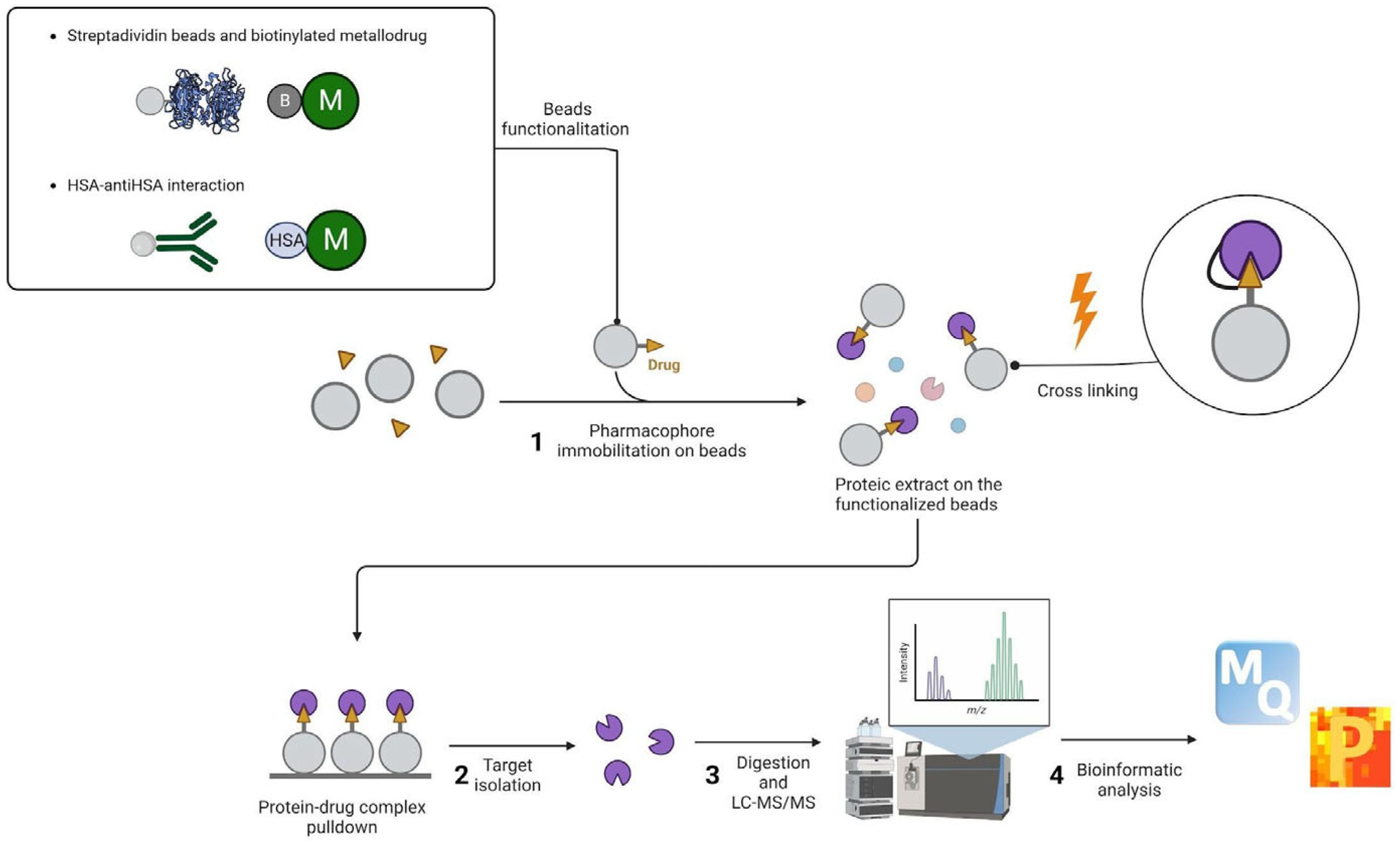
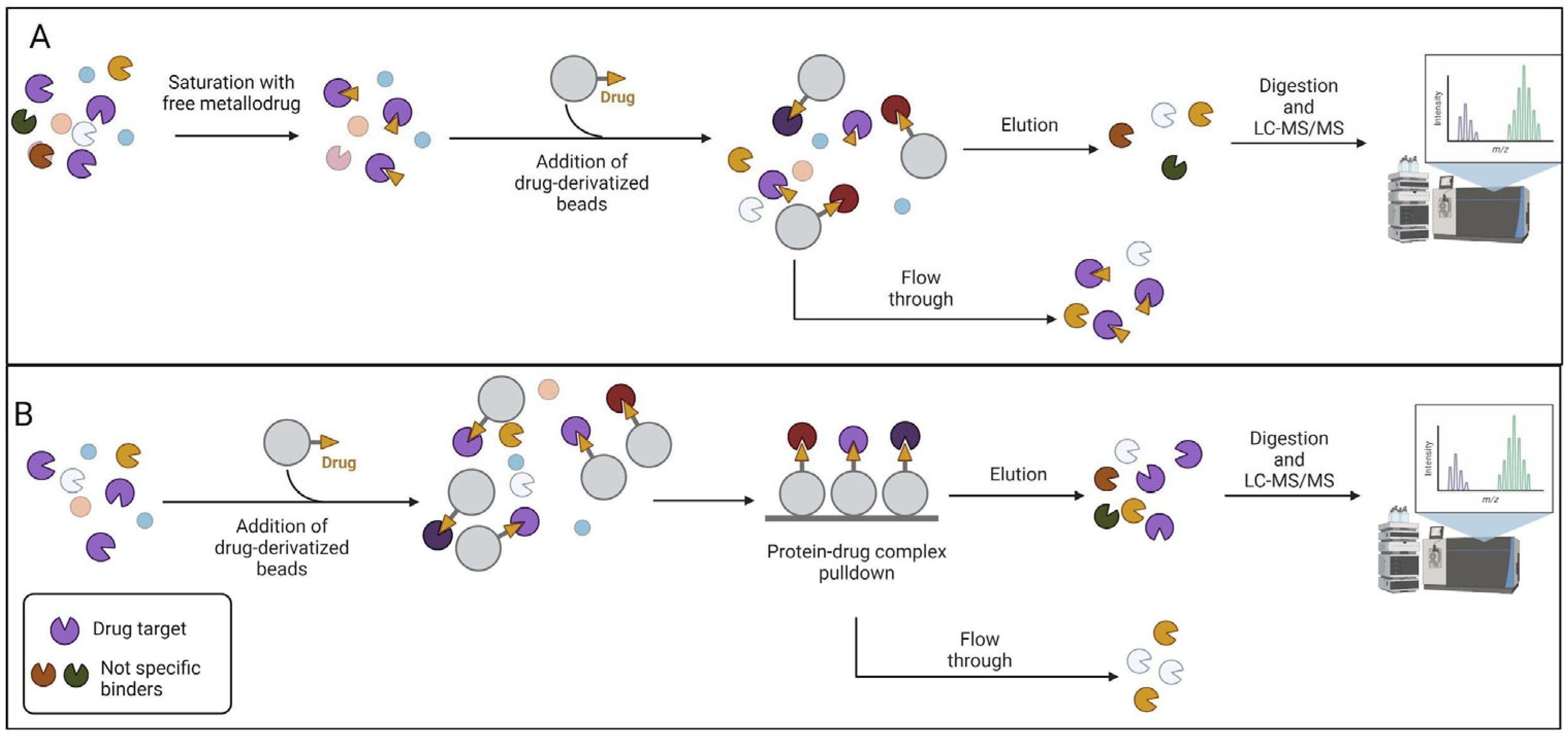
The advantage of using pulldown techniques lies in preserving the physiological state of the proteins, concentration levels (i.e., abundance), post-translational modifications, and natural binding partners, simply using non-denaturing lysis buffers. In particular, for step 1 of the drug-pulldown workflow (Figure 1), different methods can be used to immobilize the active pharmacophore also depending on the stability of the organic- and inorganic-metallic complexes. Due to this issue, Babak et al used a biotin/streptavidin approach to immobilize a Ru(II) compound ascribed to the RAPTA family [48]. In fact, the arene ligand of the complex has been functionalized with a primary amine, able to be biotinylated via an aminocaproic acid linker. Drug biotinylation could also be performed by using the click reaction (Cu(I)-catalyzed alkyne-azide cycloaddition (CuAAC)). CuAAC is a reaction occurring between an alkyne and an azide group, catalyzed by Cu(I) ion [49,50]. The reaction has been used by Neuditschko and colleagues to derivatize the drug with biotin and fish the targets with streptavidin beads [51]. Another clever method used to immobilize metal drugs is reported by the same research group. A complex between the Ru(III) compound (i.e., BOLD-100) and human serum albumin (HSA) has been formed. After that, the authors used the anti-HSA beads used in the depletion of human serum to form the BOLD-100-HSA-beads adduct, used to isolate the drug targets [52]. Since pulldown experiments are victims of high false-positive rates, the authors also performed a competitive assay. The cell lysate was pre-treated with free BOLD-100 before exposure to the immobilized drug on the beads. This latter strategy allowed the saturation of selective binding sites, as illustrated in Figure 2 so that the resulting target profile includes only non-selective binding partners. The subtraction of these from the target profile obtained by normal pull-down cancels the non-selective interactors and provides a list of the selective binding partners. The competitive pulldown experiment is an alternative way to validate and remove the possible false-positive targets from a canonical pulldown experiment.
Figure 2.
Competitive pulldown workflow. A) The total protein extract has been incubated with the free metallodrug and then subjected to the chemoproteomics strategy. Only the non-specific binders were eluted, instead, the specific target is not retained on the beads. B) The total protein extract has been incubated with the metallodrug-derivatized beads for the chemoproteomics strategy. The non-specific and the specific proteins were eluted. The comparison between the eluted proteins in the competitive and non-competitive will highlight the false-positives content. Created with BioRender.com.
Even if the simple pulldown strategy is a straightforward approach, it suffers from classical disadvantages such as the missing transient interactions and the above-mentioned false positives issue (see [35] for further insights). However, it is possible to stabilize the interactions working in living cells as reported by Liu et al [53]. The authors synthesized a photoactivatable molecule able to covalently preserve the target-drug interactions through the introduction of UV-activatable groups. These kinds of probes typically contain a photoactivatable group (e.g., azide, benzophenone, diazirine) which allows the probe to form covalent adducts with the metal drug interacting proteins upon UV light irradiation [54]. The authors incubated HeLa cells with Au(III) meso-tetraphenylporphyrin (gold-1 a) compound and irradiated at 365 nm to activate the benzophenone reaction with proteins via a radical mechanism. After the cell lysis, an alkyne group present on the probe has been used to functionalize the protein-drug complexes with biotin, allowing the adducts enrichment.
1.2. Label-free approaches
On the other hand, the label-free approaches leverage the protein stability driven by the drug interaction.
Thermal proteome profiling (TPP) [57], for example, has been used to study metallodrug targets by proteomic profiling [58]. TPP measures the extent of the drug-target interaction by monitoring the effect of pharmacological treatment on protein denaturation/solubility as a function of the progressive increase in temperature.
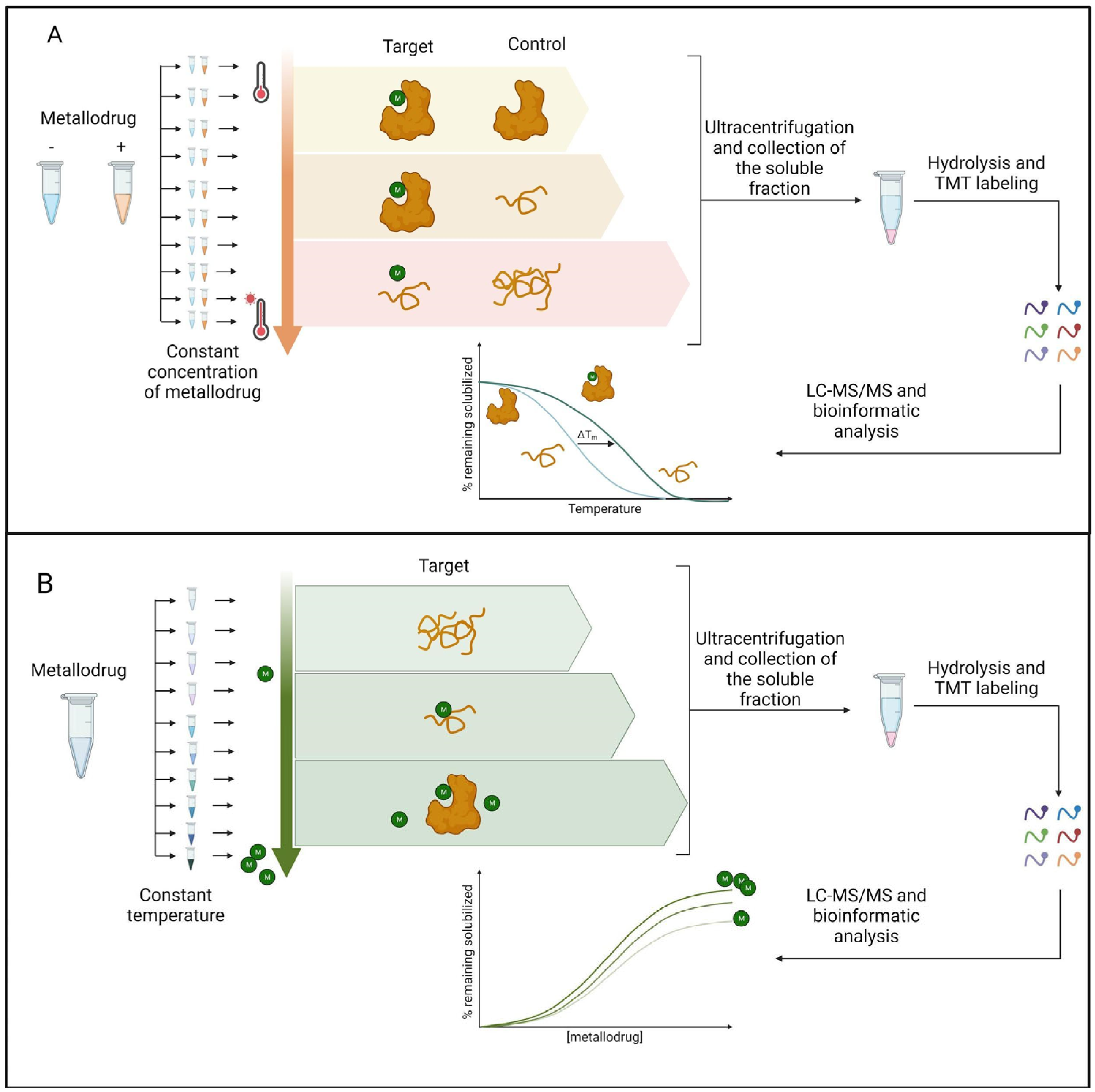
The extent of protein stabilization by the small molecule is proportional to the strength of the interaction and can be evaluated by multiplexed mass spectrometry analysis (see [59] for a comprehensive description of all the TPP applications). As shown in Figure 3, TPP can be performed by varying the temperature range (TR-TPP) (Figure 3A) or the drug concentration (CR-TPP) (Figure 3B). After the protein digestion and mass spectrometry analysis by LC-MS/MS, proteins are relatively quantified comparing the drug-treated and untreated conditions. The soluble fraction for each protein is reported as a temperature/ drug concentration function.
Figure 3.
A)TR-TPP experiment. Protein extract is incubated both with the vehicle and the metallodrug. For each condition, ten aliquots are made. Each aliquot is heated at one specific temperature and then the soluble fractions are digested with trypsin and labeled with a TMT isotope tag. Samples are analyzed by LC-MS/MS and the protein is identified. Melting curves are fitted and the melting temperatures are calculated for all the proteins. B) CR-TPP experiment. Protein extract untreated, as control, and treated with nine different concentrations of metallodrug, are heated at the same temperature Then the soluble fractions are digested with trypsin and labeled with a TMT isotope tag. Samples are analyzed by LC-MS/MS and the protein identification is made by Mascot and the isobarQuant software package. Dose-response curves are fitted and thermal stability parameters are calculated. Legend: M:metallodrug. Created with BioRender.com
TR-TPP has been recently applied to discover the antitumor Bis(N-Heterocyclic Carbene)Pt(II) complex targets in intact cells leading to the asparagine synthetase (ASNS) identification [60]. Another label-free based method used in the metallodrug-targets investigation is the Functional Identification of Target by Expression Proteomics (FITExP) approach [61]. The founding principle is that protein targets and main mechanistic-related proteins are modulated upon long drug exposure, in particular, they are overexpressed when the lethal concentration (LC50) is administered to the cells. Lee et al applied the FITExP methodology to evaluate the mechanism of action of Ru(II) complexes, RAPTA-T and RAPTA-EA. Although the approach suggested many protein targets, the best results were achieved in terms of disclosing the mechanism of action of the molecules. In fact, they found the overexpression of several oxidative stress-related and tumor-suppressing proteins for RAPTA-EA and RAPTA-T, respectively [62]. The combination of TPP and FITExP has been performed in the work carried out by Saei and colleagues, to have more chances to reduce the number of false positives and negatives [58]. In principle, other label-free strategies used in the study of protein-small molecule interactions could be applied also to the metallodrug field. These methods encompass the stability of proteins from rates of oxidation (SPROX) [63], pulse proteolysis (PP) [64], drug affinity responsive target stability (DARTS) [65] and limited proteolysis-coupled mass spectrometry (LiP-MS) [66]. However, no paper has been published yet, to the best of our knowledge.
3. Methods to enhance metallodrugs efficiency administration
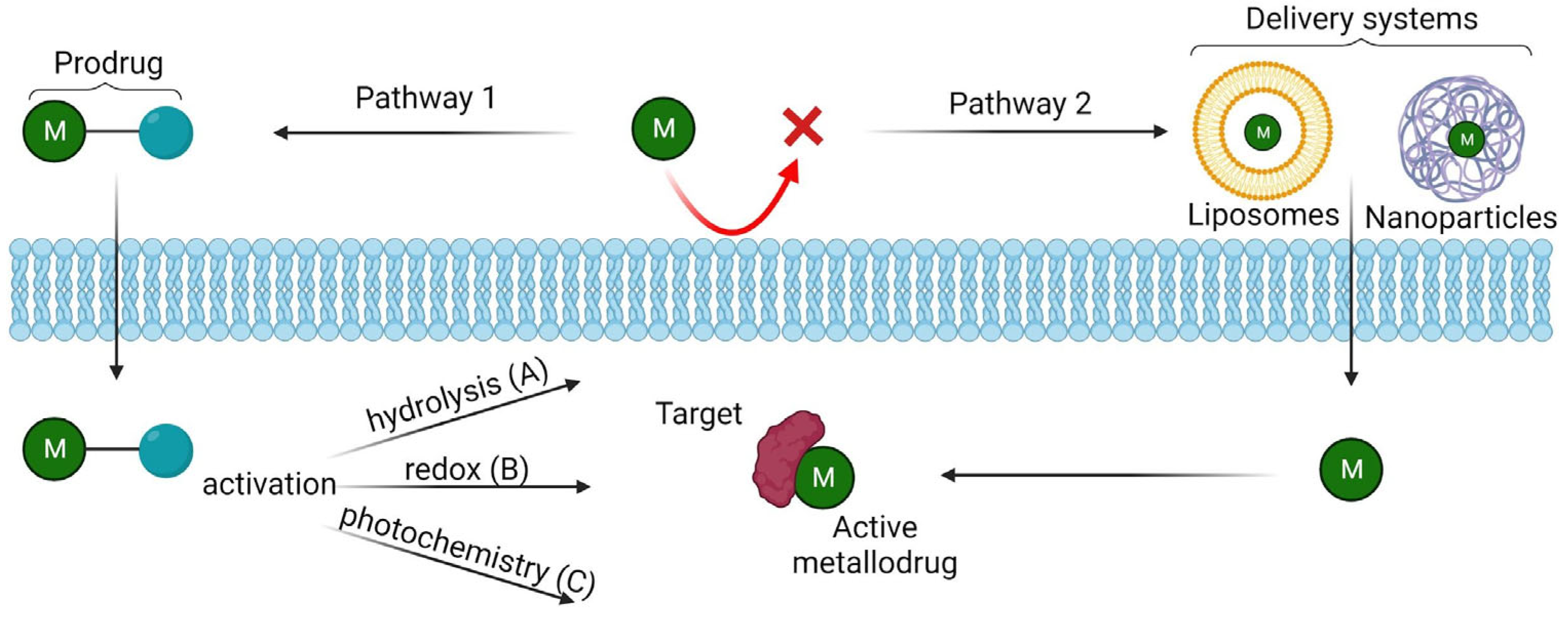
Another crucial aspect in medicinal chemistry is the development of innovative delivery systems able to recognise the target site or strategies to induce their selective activation in the drug target. Usually, once introduced in a biological environment (serum, a cell, or a biomimetic environment), metal complexes could interact with biological ligands such as lipids, amino acid residues of proteins, nucleic acids (DNA), or small molecules (i.e., vitamins, ions, neurotransmitters). This plethora of interactions can often lead to severe, deleterious side effects in most patients that limit their use [69]. New advances in the field, such as the design of pro-metallodrugs, or drug delivery systems (for example, liposomes or solid-lipid nanoparticles), offer opportunities to improve both the safety and efficacy of metallodrugs and to dispel the pervasive myth of inherent toxicity. Figure 4 shows an outline of the activation/delivery strategies discussed below.
Figure 4.
Most common strategies to enhance the bioavailability of the metallodrugs: Pathway 1) Pro-metallodrugs: the metallodrug is bound to a probe which allows the molecule to pass the phospholipid bilayer. After that, the metallodrug can be activated via hydrolysis (A), redox activation (B) or photochemistry activation (C); Pathway 2) the metallodrug is carried by delivery systems (i.e. liposomes, nanoparticles) transporting and protecting the drug properly. Created with BioRender.com
3.1. Pro-metallodrugs
Unlike most common small molecule drugs, metal-complex drugs are often “prodrugs” which undergo activation en route to or at the target site. There are many different activation strategies studied, and here we reported some recent examples:
- 1)
- Activation via hydrolysis
Regarding the activation via hydrolysis, this is a common activation mechanism for transitional metal drugs, involving the displacement of weakly bound σ-donor ligands by water (Figure 4a). Square-planar Pt(II) complexes are certainly the most used. The pioneering compound in this family is undoubtedly cisplatin, cis-[PtIICl2(NH3)2], a well-known anticancer drug widely used for the treatment of a wide variety of cancers. Cisplatin becomes activated once it enters the cell: its mode of action starts inside the cell with the hydrolysis of Pt–Cl bonds to form a Pt–H2O complex, resulting in more reactive mono-aquated [PtII(OH2)Cl(NH3)2]+ and/or di-aquated [PtII(NH3)2(OH2)2]2+ species (48). These intracellular products can react with DNA, where they cause cell cycle arrest and apoptosis. Unfortunately, the problem with cisplatin is that it may be inactivated into transplatin, trans-[PtIICl2(NH3)2], during the uptake into the cell. To avoid this problem several other derivatives of cisplatin, with a similar mechanism of action (MOA), have been synthesized, where two ligands are inter-connected and trans effect is decreased. One of the most common is Carboplatin, [Pt(NH3)2(CBDCA-O,O’)], where CBDCA is cyclobutane-1,1-dicarboxylate. Similarly to cisplatin, half-sandwich pseudo-octahedral Ru(II) and Os(II) η6-arene diamine anticancer complexes, [RuII/OsII(η6-arene)(N,N)Cl]+, also hydrolyze and bind to DNA, but monofunctionally as they have only one labile monodentate ligand [70].
- 2)
- Redox activation
The alteration of the redox balance is an effective anticancer strategy, owing to the distinct redox vulnerability of cancer cells, including hypoxia. The vast majority of examples reported consist of Pt(IV) complexes, less active than the Pt(II) analogues, that are reduced at the tumor site due to the higher levels of glutathione (GSH) and other antioxidants presented there (50). The general MOA is constituted by three main steps: octahedral Pt(IV) complexes reach the cancer cells intact, they are then activated by reductive elimination of the axial ligands (the two accepted electrons enter the dz2 orbital (LUMO), which destabilizes the ligands in axial positions) leading to the final release of the cytotoxic Pt(II) complex, thus acting as prodrugs (Figure 4b) (51). Four octahedral Pt(IV) prodrugs have entered clinical trials, namely, tetraplatin, iproplatin, satraplatin, and LA-12 but none of these have been currently approved for clinical use. As axial ligands they can contain bioactive carboxylate molecules: i) specific tumor-targeting molecules such as steroids, folates, amino acids and peptides; ii) enzyme inhibitors and iii) anticancer drugs having different targets than DNA able to synergize with the action of the cytotoxic Pt(II) metabolite. Recently, Ravera and co-workers studied several dual-action cisplatin-based Pt(IV) combos containing as axial ligands anticancer drugs such as ketoprofen (2-(3-benzoylphenyl)propanoic acid) or naproxen (2-(6-methoxynaphthalen-2-yl)propanoic acid. These complexes proved to act synergistically: the presence of Non-Steroidal Anti-Inflammatory Drugs (NSAIDs) in the structure increases the lipophilicity of the complex, facilitating its cellular accumulation. Once inside the tumor cells (HCT 116 and A-549), cisplatin is released after Pt(IV) → Pt(II) reduction and together with the NSAID induces the activation of NAG-1, a protein that has anti-tumorigenic and pro-apoptotic propensity (52). Similarly, in a recent study, a Pt(IV) combo contains as axial ligand an active antimetastatic metabolite of limonene, 4-isopropenylcyclohexene-1-carboxylic acid or perillic acid (PA), was designed. Also in this case, the increase in lipophilicity caused increased cellular accumulation and the consequent release of cisplatin moiety and PA ligand-induced cytotoxic and antimetastatic effects, respectively [71].
Interestingly, Pt(IV) complexes are not only being studied as anti-cancer agents but also as inhibitors of amyloid aggregation. It was largely reported that Pt(II) complexes can modulate Aβ peptide aggregation through the coordination of amino acids side chains (54); however, the main problem with these complexes is their poor uptake by the brain which limits their use in vivo. Kenche and co-workers studied a novel Pt(IV) complex [PtIV( N,N-dimethyl-2-[2-(quinolin-8-yl)-1H-- benzimidazol-1-yl] ethanamine) Cl4]. The Pt(IV) complex showed increased brain uptake in comparison to the Pt(II) complex, and upon reduction to Pt(II), it can limit peptide aggregation and toxicity in cortical neurons. The treatment of an APP/PS1 mouse model of Alzheimer’s Disease (AD) showed a statistically significant reduction in CSF Aβ1-42 levels and a reduction in plaque load [72].
- 3)
- Photoactivation (Light-activatable metallodrugs)
Metallodrugs can be selectively activated with high spatial resolution in cancer cells in photodynamic therapy (PDT), photothermal therapy (PTT) or photoactivated chemotherapy (PACT).
Ru(II) complexes are a class of molecules known for their rich photochemistry: they can undergo photoinduced ligand dissociation and the resulting Ru(II) aqua species can covalently bind to DNA similarly to cisplatin [73] (Figure 4c). This process is linked to 1MLCT (Metal-to-Ligand Charge Transfer) transitions: when irradiated by appropriate light, Ru(II) complexes first achieve the 1MLCT state and then reach the 3MLCT state through the ultra-fast intersystem crossing. The 3MLCT excited state of Ru(II) complexes can return to the ground state through non-radiative inactivation or luminescence pathways or can interact with other molecules such as O2 to generate singlet oxygen (through intersystem crossing to intra-ligand (IL, ILCT) states), showing potential as photodynamic agents or may populate the 3MC (metal-centered state or ligand-field) state by thermal activation that may lead to ligand dissociation and generate Ru(II) aqua species with DNA-binding ability, showing potential in photoactivated chemotherapy [69].
Ru(II)-TLD1433 is a novel water-soluble photosensitizer currently undergoing clinical trials. It has unique properties including bladder tumor selectivity: in the dark it has low cytotoxicity while, after activation with green laser light, it produces cytotoxic singlet oxygen (1O2) and radical oxygen species (ROS), causing cancer cell death [74].
As agents for PDT, another metal complex not containing Ru(II) has been already authorized in the EU since November 2017: TOOKAD® soluble (Pd II). It is a palladium bacteriopheophorbide monolysotaurine, also known as WST11 used for the treatment of adenocarcinoma of the prostate. It is a derivative of bacteriochlorophyll, the photosynthetic pigment of certain aquatic bacteria that draw their energy supply from sunlight, and becomes pharmaceutically active when illuminated by light. Tookad is retained within the vascular system and after being activated with 753 nm wavelength laser light, it generates oxygen radicals causing local hypoxia that induces the release of nitric oxide (•NO) radicals. This results in transient arterial vasodilation that triggers the release of the vasoconstrictor, endothelin-1. Rapid consumption of the •NO radicals, by oxygen radicals, leads to the formation of reactive nitrogen species (RNS) (e.g. peroxynitrite), in parallel to arterial constriction [75,76].
3.2. Delivery systems
Often, the administration of metallodrugs as such can be problematic due to their rapid metabolism, difficulties in reaching the drug site, and high systemic toxicity. To overcome these limitations, the research has moved to identify novel carriers capable of transporting and protecting the drug appropriately. The most studied delivery systems are generally polymeric or inorganic nanoparticles (NPs) and liposomes (Figure 4d).
The studies in this field were mainly focused on Pt chemotherapeutics aiming to overcome the disadvantages associated with the use of this class of drugs in clinical cancer chemotherapy. A liposomal formulation of cisplatin, named Lipoplatin, reached Phase III clinical trials. Lipoplatin, a nanoparticle of 110 nm average diameter composed of lipids and cisplatin, was revealed to be able to evade immune surveillance thus escaping clearance from macrophages, and to extravasate through the compromised endothelium of the vasculature in tumors [77,78].
Nanoparticle systems have also been developed for the two Ru(III)-drugs which reached clinical trials, KP1019 and NAMI-A. A nanosized drug conjugate of (NAMI-A)-block copolymer micelles showed improved inhibition of cell invasion and migration and simultaneous enhanced antimetastatic activity with respect to the metallodrug alone in pancreatic and ovarian cancer cells [79,80]. Concerning KP1019, its encapsulation into poly(lactic acid) (PLA) nanoparticles containing Tween-80 promoted higher cytotoxicity than the KP1019 alone in hepatoma cell lines and colon carcinoma [81].
A recent study reported on the design of diruthenium(II,III)-NSAID metallodrugs encapsulated into biocompatible terpolymer-lipid nanoparticles (TPLNs) to target glioblastoma cancer. The metal complex was formed by a Ru2(II,III) mixed-valence metal-metal multiply bonded core linked to four carboxylate ibuprofen (Ibp)drug ligands, [Ru2(Ibp)4Cl]. Its encapsulation into TPLNs was able to promote a significant enhancement of the antiproliferative effect in two human glioblastoma cancer cells, U87MG and T98G, which are chemoresistant to cisplatin [82].
Since a perfect drug delivery system should be characterized by high biocompatibility, stability and selectivity for a specific target site, research is moving towards the use of carrier systems consisting of molecules already present in our body, such as ferritins (Fts). They are natural proteins involved in the storage and release of iron that are able to self-assemble into hollow cage-like structures and are recognized by receptors overexpressed on cancer cells’ surfaces. These proteins were recently chosen to encapsulate the prototype of a novel class of metallodrugs containing a PtAs(OH)2 core, named Arsenoplatin-1(AP-1). Cellular experiments in human epidermoid carcinoma cell line and human keratinocyte cells (A431 and HaCaT) showed a significant increase in selectivity of AP-1-loadedFt against cancer cells with respect to normal cells [83].
4. Conclusions and Future Directions
To date, the use of drugs containing metal centers is prompted for the treatment of different classes of pathologies. This brief review aims to emphasize how these techniques can contribute to research in this important scientific area with the aim of advancing ambitious hypotheses on the introduction of new anticancer agents in clinical development and their use. There are numerous reviews illustrating the different chemical proteomics techniques that allow the investigation of metal–protein interactions in a biological system. Obviously, a lot depends on the type of investigation to be performed, the type of information to be obtained, and the type of interactions to be preserved. To date, increasingly advanced techniques of affinity purification, photolabeling, and quantification combined with mass spectrometry techniques allow us to easily identify putative interactors of metal-drug complexes while preserving labile interactions and providing useful information on drug mechanisms of action. Thus, it remains important to investigate the behavior of the different metal complexes to overcome problems of resistance and perhaps reduce toxicity. The information retrieved by chemical proteomics analysis might be useful also for the understanding of the mechanisms by which the metal-based drugs are conveyed to their targets.
In this field, significant advancements in the development of prodrugs and delivery systems have been obtained. The use of metallodrugs as prodrugs allow for targeted and controlled activation of the therapeutic effect, minimizing off-target effects and enhancing efficacy. Incorporating metal ions into delivery systems provides improved drug stability, controlled release, and targeted delivery to the desired site, enhancing therapeutic potential [69].
Looking ahead, the future of metallodrugs holds great promise. Continued research and technological advancements will further optimize prodrug design, delivery systems, and targeting strategies, leading to enhanced specificity, efficacy, and safety of metallodrug-based therapies. The ability to tailor metallodrugs for specific diseases and patient populations will pave the way for personalized medicine. Furthermore, the integration of metallodrugs with emerging fields such as nanotechnology, bioconjugation, and regenerative medicine opens up new avenues for innovative therapeutic approaches. Ultimately, the advancements in metallodrugs from prodrugs to delivery systems provide a solid foundation for the development of next-generation metal-based therapeutics with improved clinical outcomes.
Author Contributions
All authors contributed to the final version of the paper. All authors have read and agreed to the published version of the review.
Conflicts of Interest
The authors declare no conflict of interest.
References
- Kambe, T.; Tsuji, T.; Hashimoto, A.; Itsumura, N. The Physiological, Biochemical, and Molecular Roles of Zinc Transporters in Zinc Homeostasis and Metabolism. Physiological Reviews 2015, 95, 749–784. [Google Scholar] [CrossRef] [PubMed]
- Bagheri-Mohammadi, S.; Farjami, M.; Suha, A.J.; Zarch, S.M.A.; Najafi, S.; Esmaeili, A. The Mitochondrial Calcium Signaling, Regulation, and Cellular Functions: A Novel Target for Therapeutic Medicine in Neurological Disorders. J Cell Biochem 2023, 124, 635–655. [Google Scholar] [CrossRef] [PubMed]
- An, Y.; Li, S.; Huang, X.; Chen, X.; Shan, H.; Zhang, M. The Role of Copper Homeostasis in Brain Disease. Int J Mol Sci 2022, 23, 13850. [Google Scholar] [CrossRef] [PubMed]
- Ruiz, L.M.; Libedinsky, A.; Elorza, A.A. Role of Copper on Mitochondrial Function and Metabolism. Front. Mol. Biosci. 2021, 8, 711227. [Google Scholar] [CrossRef] [PubMed]
- Opazo, C.M.; Greenough, M.A.; Bush, A.I. Copper: From Neurotransmission to Neuroproteostasis. Front Aging Neurosci 2014, 6, 143. [Google Scholar] [CrossRef] [PubMed]
- Wang, L.; Yin, Y.-L.; Liu, X.-Z.; Shen, P.; Zheng, Y.-G.; Lan, X.-R.; Lu, C.-B.; Wang, J.-Z. Current Understanding of Metal Ions in the Pathogenesis of Alzheimer’s Disease. Transl Neurodegener 2020, 9, 10. [Google Scholar] [CrossRef]
- Haywood, S. Brain–Barrier Regulation, Metal (Cu, Fe) Dyshomeostasis, and Neurodegenerative Disorders in Man and Animals. Inorganics 2019, 7, 108. [Google Scholar] [CrossRef]
- Franz, K.J.; Metzler-Nolte, N. Introduction: Metals in Medicine. Chem. Rev. 2019, 119, 727–729. [Google Scholar] [CrossRef]
- Galib, null; Barve, M.; Mashru, M.; Jagtap, C.; Patgiri, B.J.; Prajapati, P.K. Therapeutic Potentials of Metals in Ancient India: A Review through Charaka Samhita. J Ayurveda Integr Med 2011, 2, 55–63. [CrossRef] [PubMed]
- Mukherjee, A.; Sadler, P.J. Metals in Medicine: Therapeutic Agents. In Wiley Encyclopedia of Chemical Biology; John Wiley & Sons, Inc.: Hoboken, NJ, USA, 2009; p. wecb333. ISBN 978-0-470-04867-2. [Google Scholar]
- Sodhi, R.K. Metal Complexes in Medicine: An Overview and Update from Drug Design Perspective. CTOIJ 2019, 14. [Google Scholar] [CrossRef]
- Rosenberg, B.; Van Camp, L.; Krigas, T. Inhibition of Cell Division in Escherichia Coli by Electrolysis Products from a Platinum Electrode. Nature 1965, 205, 698–699. [Google Scholar] [CrossRef] [PubMed]
- Sullivan, M.P.; Holtkamp, H.U.; Hartinger, C.G. 13. Antitumor Metallodrugs That Target Proteins. In Metallo-Drugs: Development and Action of Anticancer Agents; Sigel, A., Sigel, H., Freisinger, E., Sigel, R.K.O., Eds.; De Gruyter: Berlin, Boston, 2018; pp. 351–386. ISBN 978-3-11-047073-4. [Google Scholar]
- Khoury, A.; Deo, K.M.; Aldrich-Wright, J.R. Recent Advances in Platinum-Based Chemotherapeutics That Exhibit Inhibitory and Targeted Mechanisms of Action. J Inorg Biochem 2020, 207, 111070. [Google Scholar] [CrossRef] [PubMed]
- Zhong, T.; Yu, J.; Pan, Y.; Zhang, N.; Qi, Y.; Huang, Y. Recent Advances of Platinum-Based Anticancer Complexes in Combinational Multimodal Therapy. Adv Healthc Mater 2023, e2300253. [Google Scholar] [CrossRef] [PubMed]
- Wang, X.; Wang, X.; Guo, Z. Functionalization of Platinum Complexes for Biomedical Applications. Acc Chem Res 2015, 48, 2622–2631. [Google Scholar] [CrossRef]
- Barabas, K.; Milner, R.; Lurie, D.; Adin, C. Cisplatin: A Review of Toxicities and Therapeutic Applications. Vet Comparative Oncology 2008, 6, 1–18. [Google Scholar] [CrossRef]
- Rosenberg, B.; Vancamp, L.; Trosko, J.E.; Mansour, V.H. Platinum Compounds: A New Class of Potent Antitumour Agents. Nature 1969, 222, 385–386. [Google Scholar] [CrossRef]
- Williams, C.J.; Whitehouse, J.M. Cis-Platinum: A New Anticancer Agent. BMJ 1979, 1, 1689–1691. [Google Scholar] [CrossRef]
- Coffetti, G.; Moraschi, M.; Facchetti, G.; Rimoldi, I. The Challenging Treatment of Cisplatin-Resistant Tumors: State of the Art and Future Perspectives. Molecules 2023, 28, 3407. [Google Scholar] [CrossRef]
- Florio, D.; La Manna, S.; Annunziata, A.; Iacobucci, I.; Monaco, V.; Di Natale, C.; Mollo, V.; Ruffo, F.; Monti, M.; Marasco, D. Ruthenium Complexes Bearing Glucosyl Ligands Are Able to Inhibit the Amyloid Aggregation of Short Histidine-Peptides. Dalton Trans. 2023, 10.1039.D3DT01110K. [Google Scholar] [CrossRef]
- Paul, N.P.; Galván, A.E.; Yoshinaga-Sakurai, K.; Rosen, B.P.; Yoshinaga, M. Arsenic in Medicine: Past, Present and Future. Biometals 2023, 36, 283–301. [Google Scholar] [CrossRef]
- Florio, D.; Iacobucci, I.; Ferraro, G.; Mansour, A.M.; Morelli, G.; Monti, M.; Merlino, A.; Marasco, D. Role of the Metal Center in the Modulation of the Aggregation Process of Amyloid Model Systems by Square Planar Complexes Bearing 2-(2’-Pyridyl)Benzimidazole Ligands. Pharmaceuticals (Basel) 2019, 12, 154. [Google Scholar] [CrossRef] [PubMed]
- Pizarro, A.M.; Habtemariam, A.; Sadler, P.J. Activation Mechanisms for Organometallic Anticancer Complexes. In Medicinal Organometallic Chemistry; Jaouen, G., Metzler-Nolte, N., Eds.; Topics in Organometallic Chemistry; Springer Berlin Heidelberg: Berlin, Heidelberg, 2010; Vol. 32, pp. 21–56. ISBN 978-3-642-13184-4. [Google Scholar]
- Sullivan, M.P.; Holtkamp, H.U.; Hartinger, C.G. Antitumor Metallodrugs That Target Proteins. Met Ions Life Sci 2018, 18, /books/9783110470734/9783110470734-019/9783110470734-019.xml. [Google Scholar] [CrossRef]
- Zhou, Y.; Li, H.; Sun, H. Metalloproteomics for Biomedical Research: Methodology and Applications. Annu Rev Biochem 2022, 91, 449–473. [Google Scholar] [CrossRef]
- Kostova, I. Ruthenium Complexes as Anticancer Agents. CMC 2006, 13, 1085–1107. [Google Scholar] [CrossRef]
- Huang, H.; Cao, K.; Kong, Y.; Yuan, S.; Liu, H.; Wang, Y.; Liu, Y. A Dual Functional Ruthenium Arene Complex Induces Differentiation and Apoptosis of Acute Promyelocytic Leukemia Cells. Chem. Sci. 2019, 10, 9721–9728. [Google Scholar] [CrossRef]
- Moreno-Alcántar, G.; Picchetti, P.; Casini, A. Gold Complexes in Anticancer Therapy: From New Design Principles to Particle-Based Delivery Systems. Angew Chem Int Ed 2023, 62, e202218000. [Google Scholar] [CrossRef] [PubMed]
- Ahrweiler-Sawaryn, M.-C.; Biswas, A.; Frias, C.; Frias, J.; Wilke, N.L.; Wilke, N.; Berkessel, A.; Prokop, A. Novel Gold(I) Complexes Induce Apoptosis in Leukemia Cells via the ROS-Induced Mitochondrial Pathway with an Upregulation of Harakiri and Overcome Multi Drug Resistances in Leukemia and Lymphoma Cells and Sensitize Drug Resistant Tumor Cells to Apoptosis in Vitro. Biomed Pharmacother 2023, 161, 114507. [Google Scholar] [CrossRef]
- Ferraro, M.G.; Piccolo, M.; Misso, G.; Santamaria, R.; Irace, C. Bioactivity and Development of Small Non-Platinum Metal-Based Chemotherapeutics. Pharmaceutics 2022, 14, 954. [Google Scholar] [CrossRef]
- Barhamand, B.A. Difficulties Encountered in Implementing Guidelines for Handling Antineoplastics in the Physician’s Office. Cancer Nurs 1986, 9, 138–143. [Google Scholar] [CrossRef]
- Zanca, C.; Cozzolino, F.; Quintavalle, C.; Di Costanzo, S.; Ricci-Vitiani, L.; Santoriello, M.; Monti, M.; Pucci, P.; Condorelli, G. PED Interacts with Rac1 and Regulates Cell Migration/Invasion Processes in Human Non-Small Cell Lung Cancer Cells. J Cell Physiol 2010, 225, 63–72. [Google Scholar] [CrossRef]
- Fusco, S.; Aulitto, M.; Iacobucci, I.; Crocamo, G.; Pucci, P.; Bartolucci, S.; Monti, M.; Contursi, P. The Interaction between the F55 Virus-Encoded Transcription Regulator and the RadA Host Recombinase Reveals a Common Strategy in Archaea and Bacteria to Sense the UV-Induced Damage to the Host DNA. Biochimica et Biophysica Acta (BBA) - Gene Regulatory Mechanisms 2020, 1863, 194493. [Google Scholar] [CrossRef] [PubMed]
- Iacobucci, I.; Monaco, V.; Cozzolino, F.; Monti, M. From Classical to New Generation Approaches: An Excursus of -Omics Methods for Investigation of Protein-Protein Interaction Networks. J Proteomics 2021, 230, 103990. [Google Scholar] [CrossRef] [PubMed]
- Cozzolino, F.; Iacobucci, I.; Monaco, V.; Monti, M. Protein-DNA/RNA Interactions: An Overview of Investigation Methods in the -Omics Era. J Proteome Res 2021, 20, 3018–3030. [Google Scholar] [CrossRef] [PubMed]
- Kumara, B.N.; Kalimuthu, P.; Prasad, K.S. Synthesis, Properties and Potential Applications of Photoluminescent Carbon Nanoparticles: A Review. Anal Chim Acta 2023, 1268, 341430. [Google Scholar] [CrossRef]
- Xia, Y.; Fu, S.; Ma, Q.; Liu, Y.; Zhang, N. Application of Nano-Delivery Systems in Lymph Nodes for Tumor Immunotherapy. Nanomicro Lett 2023, 15, 145. [Google Scholar] [CrossRef] [PubMed]
- Kargozar, S.; Moghanian, A.; Rashvand, A.; Miri, A.K.; Hamzehlou, S.; Baino, F.; Mozafari, M.; Wang, A.Z. Nanostructured Bioactive Glasses: A Bird’s Eye View on Cancer Therapy. Wiley Interdiscip Rev Nanomed Nanobiotechnol 2023, e1905. [Google Scholar] [CrossRef] [PubMed]
- Peña, Q.; Wang, A.; Zaremba, O.; Shi, Y.; Scheeren, H.W.; Metselaar, J.M.; Kiessling, F.; Pallares, R.M.; Wuttke, S.; Lammers, T. Metallodrugs in Cancer Nanomedicine. Chem Soc Rev 2022, 51, 2544–2582. [Google Scholar] [CrossRef] [PubMed]
- Fedorov, I.I.; Lineva, V.I.; Tarasova, I.A.; Gorshkov, M.V. Mass Spectrometry-Based Chemical Proteomics for Drug Target Discoveries. Biochemistry (Mosc) 2022, 87, 983–994. [Google Scholar] [CrossRef]
- Skos, L.; Borutzki, Y.; Gerner, C.; Meier-Menches, S.M. Methods to Identify Protein Targets of Metal-Based Drugs. Current Opinion in Chemical Biology 2023, 73, 102257. [Google Scholar] [CrossRef]
- Steel, T.R.; Hartinger, C.G. Metalloproteomics for Molecular Target Identification of Protein-Binding Anticancer Metallodrugs. Metallomics 2020, 12, 1627–1636. [Google Scholar] [CrossRef]
- Ziegler, S.; Pries, V.; Hedberg, C.; Waldmann, H. Target Identification for Small Bioactive Molecules: Finding the Needle in the Haystack. Angew. Chem. Int. Ed. 2013, 52, 2744–2792. [Google Scholar] [CrossRef]
- Iacobucci, I.; Monaco, V.; Canè, L.; Bibbò, F.; Cioffi, V.; Cozzolino, F.; Guarino, A.; Zollo, M.; Monti, M. Spike S1 Domain Interactome in Non-Pulmonary Systems: A Role beyond the Receptor Recognition. Front Mol Biosci 2022, 9, 975570. [Google Scholar] [CrossRef] [PubMed]
- Federico, A.; Sepe, R.; Cozzolino, F.; Piccolo, C.; Iannone, C.; Iacobucci, I.; Pucci, P.; Monti, M.; Fusco, A. The Complex CBX7-PRMT1 Has a Critical Role in Regulating E-Cadherin Gene Expression and Cell Migration. Biochim Biophys Acta Gene Regul Mech 2019, 1862, 509–521. [Google Scholar] [CrossRef]
- Cozzolino, F.; Vezzoli, E.; Cheroni, C.; Besusso, D.; Conforti, P.; Valenza, M.; Iacobucci, I.; Monaco, V.; Birolini, G.; Bombaci, M.; et al. ADAM10 Hyperactivation Acts on Piccolo to Deplete Synaptic Vesicle Stores in Huntington’s Disease. Hum Mol Genet 2021, 30, 1175–1187. [Google Scholar] [CrossRef] [PubMed]
- Babak, M.V.; Meier, S.M.; Huber, K.V.M.; Reynisson, J.; Legin, A.A.; Jakupec, M.A.; Roller, A.; Stukalov, A.; Gridling, M.; Bennett, K.L.; et al. Target Profiling of an Antimetastatic RAPTA Agent by Chemical Proteomics: Relevance to the Mode of Action. Chem. Sci. 2015, 6, 2449–2456. [Google Scholar] [CrossRef] [PubMed]
- Wang, X.; Zhu, M.; Gao, F.; Wei, W.; Qian, Y.; Liu, H.-K.; Zhao, J. Imaging of a Clickable Anticancer Iridium Catalyst. Journal of Inorganic Biochemistry 2018, 180, 179–185. [Google Scholar] [CrossRef] [PubMed]
- Wang, X.; Zhang, J.; Zhao, X.; Wei, W.; Zhao, J. Imaging and Proteomic Study of a Clickable Iridium Complex. Metallomics 2019, 11, 1344–1352. [Google Scholar] [CrossRef]
- Neuditschko, B.; King, A.P.; Huang, Z.; Janker, L.; Bileck, A.; Borutzki, Y.; Marker, S.C.; Gerner, C.; Wilson, J.J.; Meier-Menches, S.M. An Anticancer Rhenium Tricarbonyl Targets Fe−S Cluster Biogenesis in Ovarian Cancer Cells. Angew Chem Int Ed 2022, 61. [Google Scholar] [CrossRef]
- Neuditschko, B.; Legin, A.A.; Baier, D.; Schintlmeister, A.; Reipert, S.; Wagner, M.; Keppler, B.K.; Berger, W.; Meier-Menches, S.M.; Gerner, C. Interaction with Ribosomal Proteins Accompanies Stress Induction of the Anticancer Metallodrug BOLD-100/KP1339 in the Endoplasmic Reticulum. Angew Chem Int Ed 2021, 60, 5063–5068. [Google Scholar] [CrossRef]
- Hu, D.; Liu, Y.; Lai, Y.-T.; Tong, K.-C.; Fung, Y.-M.; Lok, C.-N.; Che, C.-M. Anticancer Gold(III) Porphyrins Target Mitochondrial Chaperone Hsp60. Angew. Chem. Int. Ed. 2016, 55, 1387–1391. [Google Scholar] [CrossRef]
- Wang, S.; Tian, Y.; Wang, M.; Wang, M.; Sun, G.; Sun, X. Advanced Activity-Based Protein Profiling Application Strategies for Drug Development. Front. Pharmacol. 2018, 9, 353. [Google Scholar] [CrossRef] [PubMed]
- Lu, K. Chemoproteomics: Towards Global Drug Target Profiling. ChemBioChem 2020, 21, 3189–3191. [Google Scholar] [CrossRef] [PubMed]
- Jenmalm Jensen, A.; Cornella Taracido, I. Affinity-Based Chemoproteomics for Target Identification. In Methods and Principles in Medicinal Chemistry; Plowright, A.T., Ed.; Wiley, 2019; pp. 25–49. ISBN 978-3-527-34529-8. [Google Scholar]
- Savitski, M.M.; Reinhard, F.B.M.; Franken, H.; Werner, T.; Savitski, M.F.; Eberhard, D.; Molina, D.M.; Jafari, R.; Dovega, R.B.; Klaeger, S.; et al. Tracking Cancer Drugs in Living Cells by Thermal Profiling of the Proteome. Science 2014, 346, 1255784. [Google Scholar] [CrossRef] [PubMed]
- Saei, A.A.; Gullberg, H.; Sabatier, P.; Beusch, C.M.; Johansson, K.; Lundgren, B.; Arvidsson, P.I.; Arnér, E.S.J.; Zubarev, R.A. Comprehensive Chemical Proteomics for Target Deconvolution of the Redox Active Drug Auranofin. Redox Biology 2020, 32, 101491. [Google Scholar] [CrossRef]
- Mateus, A.; Kurzawa, N.; Becher, I.; Sridharan, S.; Helm, D.; Stein, F.; Typas, A.; Savitski, M.M. Thermal Proteome Profiling for Interrogating Protein Interactions. Molecular Systems Biology 2020, 16, e9232. [Google Scholar] [CrossRef]
- Hu, D.; Yang, C.; Lok, C.; Xing, F.; Lee, P.; Fung, Y.M.E.; Jiang, H.; Che, C. An Antitumor Bis(N-Heterocyclic Carbene)Platinum(II) Complex That Engages Asparagine Synthetase as an Anticancer Target. Angew. Chem. Int. Ed. 2019, 58, 10914–10918. [Google Scholar] [CrossRef]
- Chernobrovkin, A.; Marin-Vicente, C.; Visa, N.; Zubarev, R.A. Functional Identification of Target by Expression Proteomics (FITExP) Reveals Protein Targets and Highlights Mechanisms of Action of Small Molecule Drugs. Sci Rep 2015, 5, 11176. [Google Scholar] [CrossRef]
- Lee, R.F.S.; Chernobrovkin, A.; Rutishauser, D.; Allardyce, C.S.; Hacker, D.; Johnsson, K.; Zubarev, R.A.; Dyson, P.J. Expression Proteomics Study to Determine Metallodrug Targets and Optimal Drug Combinations. Sci Rep 2017, 7, 1590. [Google Scholar] [CrossRef]
- Strickland, E.C.; Geer, M.A.; Tran, D.T.; Adhikari, J.; West, G.M.; DeArmond, P.D.; Xu, Y.; Fitzgerald, M.C. Thermodynamic Analysis of Protein-Ligand Binding Interactions in Complex Biological Mixtures Using the Stability of Proteins from Rates of Oxidation. Nat Protoc 2013, 8, 148–161. [Google Scholar] [CrossRef]
- Park, C.; Marqusee, S. Pulse Proteolysis: A Simple Method for Quantitative Determination of Protein Stability and Ligand Binding. Nat Methods 2005, 2, 207–212. [Google Scholar] [CrossRef]
- Lomenick, B.; Jung, G.; Wohlschlegel, J.A.; Huang, J. Target Identification Using Drug Affinity Responsive Target Stability (DARTS). Curr Protoc Chem Biol 2011, 3, 163–180. [Google Scholar] [CrossRef]
- Feng, F.; Zhang, W.; Chai, Y.; Guo, D.; Chen, X. Label-Free Target Protein Characterization for Small Molecule Drugs: Recent Advances in Methods and Applications. Journal of Pharmaceutical and Biomedical Analysis 2023, 223, 115107. [Google Scholar] [CrossRef] [PubMed]
- Jia, S.; Wang, R.; Wu, K.; Jiang, H.; Du, Z. Elucidation of the Mechanism of Action for Metal Based Anticancer Drugs by Mass Spectrometry-Based Quantitative Proteomics. Molecules 2019, 24, 581. [Google Scholar] [CrossRef]
- Roberts, E.A.; Sarkar, B. Metalloproteomics: Focus on Metabolic Issues Relating to Metals. Current Opinion in Clinical Nutrition and Metabolic Care 2014, 17, 425–430. [Google Scholar] [CrossRef] [PubMed]
- Anthony, E.J.; Bolitho, E.M.; Bridgewater, H.E.; Carter, O.W.L.; Donnelly, J.M.; Imberti, C.; Lant, E.C.; Lermyte, F.; Needham, R.J.; Palau, M.; et al. Metallodrugs Are Unique: Opportunities and Challenges of Discovery and Development. Chem Sci 2020, 11, 12888–12917. [Google Scholar] [CrossRef] [PubMed]
- Coverdale, J.; Laroiya-McCarron, T.; Romero-Canelón, I. Designing Ruthenium Anticancer Drugs: What Have We Learnt from the Key Drug Candidates? Inorganics 2019, 7, 31. [Google Scholar] [CrossRef]
- Ravera, M.; Gabano, E.; Zanellato, I.; Rangone, B.; Perin, E.; Ferrari, B.; Bottone, M.G.; Osella, D. Cis,Cis,Trans -[Pt IV Cl 2 (NH 3 ) 2 (Perillato) 2 ], a Dual-Action Prodrug with Excellent Cytotoxic and Antimetastatic Activity. Dalton Trans. 2021, 50, 3161–3177. [Google Scholar] [CrossRef]
- Kenche, V.B.; Hung, L.W.; Perez, K.; Volitakes, I.; Ciccotosto, G.; Kwok, J.; Critch, N.; Sherratt, N.; Cortes, M.; Lal, V.; et al. Development of a Platinum Complex as an Anti-Amyloid Agent for the Therapy of Alzheimer’s Disease. Angew. Chem. Int. Ed. 2013, 52, 3374–3378. [Google Scholar] [CrossRef]
- Chen, Y.; Bai, L.; Zhang, P.; Zhao, H.; Zhou, Q. The Development of Ru(II)-Based Photoactivated Chemotherapy Agents. Molecules 2021, 26, 5679. [Google Scholar] [CrossRef]
- Kulkarni, G.S.; Lilge, L.; Nesbitt, M.; Dumoulin-White, R.J.; Mandel, A.; Jewett, M.A.S. A Phase 1b Clinical Study of Intravesical Photodynamic Therapy in Patients with Bacillus Calmette-Guérin–Unresponsive Non–Muscle-Invasive Bladder Cancer. European Urology Open Science 2022, 41, 105–111. [Google Scholar] [CrossRef]
- Hu, X.; Zhang, Y.-S.; Liu, Y.-C.; Wang, N.; Zeng, X.-T.; Zhang, L.-L. Emerging Photodynamic/Sonodynamic Therapies for Urological Cancers: Progress and Challenges. J Nanobiotechnology 2022, 20, 437. [Google Scholar] [CrossRef]
- Karges, J. Clinical Development of Metal Complexes as Photosensitizers for Photodynamic Therapy of Cancer. Angew Chem Int Ed Engl 2022, 61, e202112236. [Google Scholar] [CrossRef] [PubMed]
- Poursharifi, M.; Wlodarczyk, M.T.; Mieszawska, A.J. Nano-Based Systems and Biomacromolecules as Carriers for Metallodrugs in Anticancer Therapy. Inorganics 2018, 7, 2. [Google Scholar] [CrossRef]
- Boulikas, T. Clinical Overview on Lipoplatin: A Successful Liposomal Formulation of Cisplatin. Expert Opin Investig Drugs 2009, 18, 1197–1218. [Google Scholar] [CrossRef] [PubMed]
- Blunden, B.M.; Stenzel, M.H. Incorporating Ruthenium into Advanced Drug Delivery Carriers - an Innovative Generation of Chemotherapeutics. J. Chem. Technol. Biotechnol. 2015, 90, 1177–1195. [Google Scholar] [CrossRef]
- Fischer, B.; Heffeter, P.; Kryeziu, K.; Gille, L.; Meier, S.M.; Berger, W.; Kowol, C.R.; Keppler, B.K. Poly(Lactic Acid) Nanoparticles of the Lead Anticancer Ruthenium Compound KP1019 and Its Surfactant-Mediated Activation. Dalton Trans. 2014, 43, 1096–1104. [Google Scholar] [CrossRef] [PubMed]
- Alves, S.R.; Colquhoun, A.; Wu, X.Y.; de Oliveira Silva, D. Synthesis of Terpolymer-Lipid Encapsulated Diruthenium(II,III)-Anti-Inflammatory Metallodrug Nanoparticles to Enhance Activity against Glioblastoma Cancer Cells. J Inorg Biochem 2020, 205, 110984. [Google Scholar] [CrossRef]
- Monti, D.M.; Ferraro, G.; Merlino, A. Ferritin-Based Anticancer Metallodrug Delivery: Crystallographic, Analytical and Cytotoxicity Studies. Nanomedicine 2019, 20, 101997. [Google Scholar] [CrossRef]
- Kambe, T.; Tsuji, T.; Hashimoto, A.; Itsumura, N. The Physiological, Biochemical, and Molecular Roles of Zinc Transporters in Zinc Homeostasis and Metabolism. Physiological Reviews 2015, 95, 749–784. [Google Scholar] [CrossRef]
Disclaimer/Publisher’s Note: The statements, opinions and data contained in all publications are solely those of the individual author(s) and contributor(s) and not of MDPI and/or the editor(s). MDPI and/or the editor(s) disclaim responsibility for any injury to people or property resulting from any ideas, methods, instructions or products referred to in the content. |
© 2023 by the authors. Licensee MDPI, Basel, Switzerland. This article is an open access article distributed under the terms and conditions of the Creative Commons Attribution (CC BY) license (http://creativecommons.org/licenses/by/4.0/).
Copyright: This open access article is published under a Creative Commons CC BY 4.0 license, which permit the free download, distribution, and reuse, provided that the author and preprint are cited in any reuse.


