

Submitted:
04 June 2023
Posted:
06 June 2023
You are already at the latest version
Abstract
Psilocybin-containing mushrooms, commonly known as magic mushrooms have antidepressant effect, however, their safety in cardiovascular diseases such as heart failure is not fully known and needs to be investigated. Cardiac hypertrophy is an independent risk factor for heart failure morbidity and mortality. Angiotensin II (Ang-II) plays a major role in the pathogenesis of cardiac hypertrophy. We investigated the cardiovascular safety of extracts of Panaeolus cyanescens, Psilocybe natalensis, Psilocybe cubensis, and Psilocybe cubensis leucistic A+ strain mushrooms, well-known psilocybin-containing mushrooms in the Panaeolus and Psilocybe genus on Ang II-induced hypertrophy oxidative stress. The four mushrooms were grown, dried and extracted with 70% ethanol, cold and hot water. Extracts were tested for cytotoxicity on H9C2 cardiomyoblast cells. The cardiomyocytes were induced with (10 µM) AngII and treated with the three extracts of the four mushrooms over 48 hours. Control cells were serum starved but neither AngII induced nor treated while AngII cells were serum starved and stimulated with AngII but not treated. Losartan, an inhibitor of AngII type 1 receptor was used as positive control. Effects of the extracts on actin-filament labelling and cell surface area, mitochondrial activity, reactive oxygen species (ROS) and atrial natriuretic peptide levels were determined. Stimulation with AngII lowered cell viability, increased the cell width measurements and intracellular ROS levels significantly compared to control cells. The results indicated that the ethanol and water extracts of the four psilocybin mushrooms did not exacerbate the angiotensin II-induced hypertrophy conditions, but the extracts had cardio-protective activity against angiotensin II-induced oxidative stress. The phytochemical analysis of the extracts confirmed detections of known compounds with antioxidant and anti-inflammatory effects in the water and ethanol extracts of these four psilocybin mushrooms.
Keywords:
reactive oxygen species
; angiotensin II
; hypertrophy
; psilocybin mushrooms
; heart failure
1. Introduction
Depression is a burden to society and associated with chronic stress and aging [1]. Psilocybin-containing mushrooms have been used by different tribes to improve quality of life and for mind healing [2]. Many studies have also demonstrated the antidepressant effects of psilocybin (4-phospholoxy-N-N-dimethyltryptamine), the classic psychedelic agent occurring naturally in psilocybin-containing mushrooms [3,4]. Consequently, the use and awareness of psilocybin-containing mushrooms, commonly known as magic mushrooms is increasing. However, psilocybin and psilocybin mushrooms also lead to a temporary increase in heart rate and blood pressure which may pose as risk especially for users suffering from cardiovascular diseases [5]. Since depression is associated with aging people that are prone to cardiovascular disease such as hypertension and heart failure, investigating safety of the mushroom usage in these conditions is crucial.
Heart failure is an international public health problem of pandemic proportions and studies showed that about 64.3 million people globally are living with a heart failure condition [6,7]. Cardiomyocyte hypertrophy which is a major consequence of pressure and/or volume overload is considered a significant diagnostic component and plays a key role in the progression of heart failure [8]. Cell enlargement and apoptotic loss of cardiomyocytes are key pathological changes in cardiac hypertrophy [9,10]. Many factors are involved in the pathogenesis and regulation of cardiomyocyte hypertrophy including angiotensin II (AngII). Angiotensin II is a key factor of the renin-angiotensin system that induces cell hypertrophy, differentiation and apoptosis through activation of various intracellular signalling molecules including calcineurin, mitogen-activated protein kinase and many other factors [11].
Angiotensin II has two receptors, AngII type 1 (AT1R) which is known to mediate pro-hypertrophic effects of AngII and AngII type 2 (AT2R) that attenuates the AT1R activation-induced hypertrophy [12]. A fibroblast-derived factor is identified as a biochemical process used by AngII to stimulate direct cardiomyocyte hypertrophy which can be blocked by using bromodeoxyuridine, a fibroblast proliferation inhibitor [12]. Many studies also showed that AngII stimulated protein synthesis which could also be eliminated by losartan, the AT1R blocker, further indicating direct role of AngII in the production of some fibroblasts factor [12,13]. Furthermore, studies have also showed that the pro-hypertrophic effects of AngII are also mediated through mitochondrial and induced-cell death by activating NAD(P)H oxidase through ATR1 receptors leading to increased generation of reactive oxygen species (ROS) and oxidative stress [14,15]. Oxidative stress is the lack of balance state where production of ROS such as superoxide, hydrogen peroxide and hydroxyl radicals exceed the antioxidant defences [13].
Plasma levels of atrial natriuretic peptide (ANP) and brain natriuretic peptide (BNP), well-known as hall markers of heart failure, have been found to increase with the severity of heart failure [16]. Their main physiological effects are to suppress progression of heart failure by inducing several effects that includes inhibition of the renin-angiotensin-aldosterone and promoting vasodilation and natriuresis [16].
This study aimed at investigating for the first time the risks and/or safety of Panaeolus cyanescens, Psilocybe natalensis, Psilocybe cubensis and Psilocybe cubensis leucistic A+ strain mushrooms, well-known psilocybin-containing mushroom in the genus Panaeolus and Psilocybe, on AngII-induced hypertrophy using a rat H9C2 cardiomyoblast cells model which is well-known and a widely used in vitro cell model with accepted reliability in cardiovascular drug discovery [17].
2. Results
This section may be divided by subheadings. It should provide a concise and precise description of the experimental results, their interpretation, as well as the experimental conclusions that can be drawn.
2.1. Cytotoxicity of the Extractsn
The cytotoxicity results of the extracts on H9C2 cardiomyocytes showed that the three extracts of the four magic mushrooms were not toxic when comparing their LC50 values with the positive control doxorubicin, a well-known toxic drug, Table 6.1.

Table 1.
Cytotoxicity effects of the extracts over 48 hours on H9C2 cardiomyocytes.
| Sample | LC50 (µg/mL) |
|---|---|
| Pan cyanescens cold-water (PC) | 66.1 ± 1.8 |
| Pan cyanescens hot-water (PH) | >100 |
| Pan cyanescens 70% ethanol (PE) | > 100 |
| P. cubensis cold-water (GC) | >100 |
| P. cubensis hot-water (GH) | >100 |
| P. cubensis 70% ethanol (GE) | >100 |
| P. A+ strain cold-water (AC) | >100 |
| P. A+ strain hot-water (AH) | >100 |
| P. A+ strain 70% ethanol (AE) | >100 |
| P. natalensis cold-water (NC) | >100 |
| P. natalensis hot-water (NH) | >100 |
| P. natalensis 70% ethanol (NE) | >100 |
| Doxorubicin | 0.2169 ± 0.037 |
2.2. Effects of the Extracts on AngII-Induced Hypertrophy in H9C2 Cardiomyocytes
Morphological analysis of the actin filaments of the cardiomyocytes using rhodamine phalloidin reagent showed that AngII increased the cell size of the stimulated cardiomyocytes F-actin over 48 hours, Figure 1. Treatment with 50 µg/mL of the three extracts of Pan cyanescens (PC, PH and PE) and P. cubensis (GC, GH and GE) and the positive control losartan (100 µM) reduced the F-actin sizes of the cells, Figure 1.
Cell surface area measurements performed using CellSens Dimension 1,12 software on the cells showed in Figure 2 that AngII increased the cell width measurements of the induced cells 1.4-fold significantly (p< 0.001) compared to the non-induced negative control cells. The positive control losartan reversed these AngII effects significantly (p< 0.001). The cold-water (PC), hot-water (PH) and ethanol (PE) extracts of Pan cyanescens and P. cubensis’s cold-water (GC), hot-water (GH) and ethanol (GE) mushroom extracts also all reversed the AngII effects significantly, Figure 2.
Florescence morphological studies showed a decreased F-actin sizes in the AngII-induced cells treated with the cold-water, hot-water and ethanol extracts of P. A+ strain (AC, AH and AE) and P. natalensis (NC, NH and NE) mushrooms over 48 hours, in comparison to the AngII-stimulated cells, Figure 3.
The treatment with the cold-water, hot-water and ethanol extracts of P. A+ strain (AC, AN and AE) and P. natalensis (NC, NH and AE) mushrooms reduced the AngII-induced cell size measurements significantly compared to the AngII-stimulated cells, Figure 4.
2.3. Effects of the Extracts on ANP Levels in AngII-Induced Cardiomyocytes
The AngII non-significantly increased the ANP levels of stimulated cells compared to the control cells. All the extracts of the four mushrooms lowered AngII-induced ANP concentration, however non-significantly, results not showed. Only the positive control, losartan reversed the AngII-induced ANP concentration significantly (p= 0.0093).
2.4. Effects of the Extracts on Mitochondrial Activity of AngII-Induced Cells
Angiotensin II stimulation reduced the mitochondrial activity indicated by lowering viability of cells significantly (p< 0.001) below 80% in comparison to the control cells, Figure 5. Positive controls, losartan and L-NAME increased the viability of AngII-induced cells in a concentration-dose manner and the effects were more pronounced with L-NAME treatment that restored viability of cells in line with control cells and above 100% cell viability with the 100 µM treatment.
The cold-water extract of Pan cyanescens increased viability of AngII-induced cells above LNAME the positive control and the control cells with the concentration investigated in the study(50 µg/mL and 25 µg/mL concentration), Figure 5. The hot-water and ethanol extracts of Pan cyanescens increased the viability same as the P. cubensis above 80% also in a dose dependant manner, Figure 5. P. A+ strain treatment increased the % viability of the cells above 80% with lowest concentration (25 µg/mL) investigated in the study while the higher 50 µg/mL concentration slightly increased the viability around and below 80% with the three extracts. The P. natalensis increased viability of cells above 80% in a concentration-dependent manner and to the same level as losartan with the 25 µg/mL concentration.
2.5. Effects of the Extracts on Intracellular ROS Levels in AngII-Induced Cardiomyocytes
Angiotensin II stimulation increased ROS levels significantly (p< 0.001) when compared with the non-induced but serum starved control cells, Figure 6. The cold-water, hot-water and ethanol extracts of all the extracts reduced the AngII stimulated ROS levels significantly similar to losartan (p< 0.001) when compared to AngII-induced cells.
2.6. Phytochemistry Analysis of the Water and Ethanol Extracts of Pan cyanescens, P. cubensis and A+ Strain Mushrooms
Figure 7 show the GCMS-MS chromatograms of the cold-water, hot-water and ethanol extracts of Pan cyanescens mushrooms respectively. The compounds with known anti0xidant and anti-inflammatory biological activities from the chromatograms of the three extracts are tabulated in Table 2 with their different peak numbers, compound name, molecular weight, formulas, similarity, area%, diemention time and height per extracts. Four compounds (n-hexadecanoic acid; 3-octanone, nonadane and tetradecane) with known natural antioxidant and anti-inflammatory activities similar to the ones extracted from P. natalensis identified in [21] were also present in the Pan cyanescens mushroom extracts. The compounds 9-Octadecenamide, (Z)- and n-hexadecanoic acid were found in all the extracts. Tetradecane was found in the hot-water extract and the ethanol extract. Nonadecane and decane were detected in the ethanol extract together with 9,12-Octadecadienoic acid (Z,Z)- and heineicosane. Dodecane, 1,1-dimethoxy- compound was detected in different peaks only with cold water together with hexanoic acid, methyl ester while dotriacontane compound and olean-12-ene-3,28-diol, (3á)- was detected only in the hot water extract of Pan cyanescens, Table 2.
Figure 8 show the GCMS-MS chromatograms of the cold-water, hot-water and ethanol extracts of P. cubensis mushrooms respectively. The compounds with known biological activities from the chromatograms of the three extracts are tabulated in Table 3 with their different peak numbers, compound name, molecular weight, formulas, similarity, area%, diemention time and height per extracts. Four compounds (n-hexadecanoic acid; 3-octanone, nonadane and tetradecane) with natural antioxidant and anti-inflammatory activities similar to the ones extracted from P. ntalensis identified in [21] were also present in these mushroom extracts, Table 3. The compound n-hexadecanoic acid was present in all the extracts while tetradecane compound was detected only in the ethanol extract together with hexadecane, 9,12-Octadecadienoic acid (Z,Z)-, nonadecane, heneicosane, oleic acid and decane. Compound 9-Octadecenamide, (Z)- was detected in different peaks in both ethanol and hot-water extracts. In the cold and hot-water extracts, dodecane, 1,1-dimethoxy- was detected in many different peaks and hexadecanoic acid, methyl ester; 3-octanone and dodecanoic acid, methyl ester compounds were also detected in the extract.
Figure 9 show the GCMS-MS chromatograms of the cold-water, hot-water and ethanol extracts of P. A+ strain mushrooms respectively while the compounds with know biological activities from the chromatograms of the three extracts are tabulated in Table 4 with their molecular weight, formulas and area% per extracts. The four compounds (n-hexadecanoic acid; 3-octanone, nonadane and tetradecane) with natural antioxidant and anti-inflammatory activities similar to the ones extracted identified in [21] were also present in the P. A+ strain mushroom extracts. The compound n-hexadecanoic acid was detected in all the extracts of P. A+ strain. Decane, tetradecane, 9,12-octadecadienoic acid (Z,Z)-, and eicosane compounds were detected in the ethanol extract. The compounds hexadecanoic acid, methyl ester, 3-Octanone and 9-octadecenamide, (Z)-, were detected in both cold and hot water extracts with dodecane, 1,1-dimethoxy detected in many different peaks in both water extracts of P. A+ strain mushroom while glycine was detected only in the hot-water extract.
In Table 5 below, there are other compounds that were extracted and not included in Nkadimeng et al 2020 [21] and are tabulated here. Compound heneicosane, 9,12-octadecadienoic acid (Z,Z)-, 5-eicosene, (E)- and decane were detected only in the ethanol extract. Dodecane, 1,1-dimethoxy- was detected in many different peaks in both hot and cold-water extracts while hexadecanoic acid, methyl ester and dodecanoic acid, methyl ester was only detected in the cold-water extracts of P. natalensis mushroom.
3. Discussion
Previous studies have demonstrated that AngII plays a key role in progression of heart failure by affecting cell growth, differentiation and apoptosis, induction of pro-inflammatory cytokines and many other factors in cardiomyocytes [14]. In the study, morphological F-actin size results and cell width measurements showed that the cells that were induced with AngII increased the cell surface area of the cells significantly compared to non-induced serum starved control cells in agreement with previous studies [19]. Angiotensin II stimulation decreased mitochondrial activity by lowering cell viability < 80% signifying cell death and also increased levels of ROS production significantly while ANP level was non-significantly increased in cardiomyocytes. The results showed that the positive control losartan, which is an AngII inhibitor via blockage of ATR1 receptors, significantly reduced the AngII-induced cell surface area measurements, ROS and ANP levels of the stimulated cells. Losartan also improved cell viability of AngII-induced cells in a dose-dependent manner similar to L-NAME. This study showed that L-NAME, which is a non-selective NOS inhibitor, prevented the AngII-induced cell death in greater percentage indicating involvement of NOS uncoupling in the AngII-induced injury and cell death in the study.
Our study demonstrated that the cold-water, hot-water and ethanol extracts of Pan cyanescens, P. cubensis, P. A+ strain and P. natalensis mushroom extracts alleviated the cell enlargement induced by AngII stimulation same as the positive control, losartan. Cell enlargement is one of the key indices of hypertrophy and by reducing it, the extracts demonstrated not just safety but potential protective effects as well in AngII-induced hypertrophy conditions. The concentrations of ANP, which is a maker in heart failure, was also found to be lower however non-significant in the three extract-treatments. Since the increase levels of ANP are known to be associated with the severity of heart failure condition, their decrease agreed with the significant decrease in cell size enlargement supporting alleviation of hypertrophy observed with the mushroom extracts’ treatments.
Other well-investigated pro-hypertrophic mechanisms of AngII are also known to be mediated via activation of NAD(P)H oxidase through ATR1 receptors and inducing mitochondrial and induced-cell death leading to increased ROS generation and more oxidative stress [14,15]. In our study, we measured intracellular ROS especially superoxide and hydroxyl radicals both of which are known to increase significantly with AngII stimulation [13]. Our study showed that the cold-water, hot-water and ethanol extracts of all the four magic mushrooms alleviated these AngII-induced intracellular ROS generation of treated cells significantly similar to Losartan. By reducing accumulation of AngII-induced ROS, the extracts of Pan cyanescens, P. cubensis, P A+ strain and P. natalensis mushrooms demonstrated safety and protective potentials of the extracts in AngII-induced oxidative stress conditions in vitro in cardiomyocytes in the concentration investigated. These results agreed with our previous finding in endothelin-induced ROS activity following treatment with Pan cyanescens, P. cubensis water extracts [20]. Furthermore, we have also demonstrated invitro anti-inflammatory potential of the four extracts on LPS-induced human macrophage cells [21].
In addition, the four mushroom extracts also protected against AngII-induced mitochondrial and cell death signified by increasing in % viability of cells above 80% in safe margins with the concentrations investigated 50 µg/mL for Pan cyanescens and P. cubensis, and 25 µg/mL for P. A+ strain and P. natalensis mushrooms in the study. However, the study also showed that P. A+ strain mushroom extracts may be toxic if higher than 50 µg/mL concentrations are used in an AngII pathological condition.
The cytotoxicity assay results on H9C2 cardiomyocytes also showed that the three extracts of the four magic mushrooms were not toxic when compared to the positive control doxorubicin. The extracts were safe in the order ethanol> hot-water> cold-water for the three magic mushrooms and the order hot-water> ethanol> cold-water for P. cubensis. Moreover, in accordance to the American National Cancer Institution guidelines, it is indicated that extracts that exhibit an LC50 ≤ 20 µg/mL over 48 hours treatment are considered toxic. As a result, the three extracts of the four mushrooms will be considered to be safe with LC50 presented and also with the 50 µg/mL and 25 µg/mL concentration which were investigated in the study. However, further investigations in vivo to confirm this safety is recommended.
The phytochemistry analysis of the water and ethanol extracts showed presence of known natural compounds with antioxidant and anti-inflammatory activities in support of these ant-oxidative stress effects observed in the study. The two compounds, 9-Octadecenamide, (Z)- which is a potent antioxidant with antimicrobial activities, and n-hexadecanoic acid which is the most common saturated fatty acid known to have anti-inflammatory and antioxidant activities were detected in all the water and ethanol extracts of the four magic mushrooms [22,23,24].
Decane, an alkaline hydrocarbon compound, was found to possess activities such as phosphatase and membrane permeability inhibitor which means it prevents damage and preserve integrity of the cellular membrane [25]. The compound was also reported as platelet aggregation inhibitor and platelet activating factor beta antagonist which are known to inhibit thrombus production by decreasing platelet agglutination with potential therapeutic agents in different diseases including cardiovascular [25]. This compound was detected only in all the ethanol extracts of the four psilocybin mushrooms.
Tetradecane is an alkaline hydrocarbon compound which was found to have anti-inflammatory effects and also a potential membrane integrity agonist, cardiovascular analeptic (which are central nervous system stimulants agents that increases alertness, heart rate, blood pressure, breathing and blood glucose level, mood and euphoria among others) and nicotinic alpha6beta3beta4alpha5 receptor antagonist [25,26]. This compound was detected in all the ethanol extracts of the four psilocybin mushrooms extracts.
The three compounds, nonadecane, an alkaline hydrocarbon lipid molecule and vey hydrophobic with antioxidant, antibacterial and antimalarial activities [26,27,28]; 9,12-Octadecadienoic acid (Z,Z)- compound which has been reported to have antioxidant activity, and heneicosane, an aliphatic hydrocarbon compound that is reported to complements C5a chemotactic receptor antagonist which play important roles in inflammation and cell killing process [29], and also to possess anti-eczema atopic activities, phobic disorders treatment and betaadrenergic receptor kinase inhibitor which are known to ameliorate cardiac dysfunction and improve survival especially in heart failure [25] were all detected only in the ethanol extracts of the three psilocybin mushrooms, P. natalensis, Pan cyanescens and P. cubensis and it was not detected in any of the P. A+ strain mushroom extracts.
Hexadecanoic acid, methyl ester which is known to have antioxidant, anti-inflammatory (by inhibiting cyclooxygenase-2 enzymes) activities and also a blood cholesterol decreasing effect [30] was detected in the water extracts of P. A+ strain and P. cubensis, and also in the cold-water extracts of Pan cyanescens and P. natalensis mushrooms. Compound 3-Octanone reported to have antioxidant and anti-inflammation activities was detected in the water extracts of P. cubensis, P. A+ strain and P. natalensis mushrooms and in the cold water of Pan cyanescens mushroom [18,31]. Dotriacontane reported to have antioxidant activities were detected in the ethanol extracts of P. cubensis and P. natalensis mushrooms and also in the hot-water extracts of Pan cyanescens mushroom extracts [32].
Hexadecane has been found to have antibacterial, cognition disorder treatment, antianginal, nicotinic alpha6beta3beta4alpha5 and nicotinic alpha2beta2 receptor antagonist, kidney function stimulant and 5-hydroxytryptamine uptake stimulant. This compound was detected in ethanol extracts of P. cubensis [25,33]. Oleic acid is a mono-unsaturated omega9-fatty acid known to enhance antioxidant activity,inhibit adrenoleukodystrophy, boost memory, a key factor accounting for the hypotensive effects of olive oil [34] and generally known to improve and protect against cardiovascular disease [35], 2021) was detected in ethanol extracts of P. cubensis. Glycine, which is known to improves the body’s ability to use nitric oxide and relief blood pressure [36] was detected only in the hot-water extract of P. A+ strain mushroom. Olean-12-ene-3,28-diol, (3á)-, which is known to have anti-inflammatory and protecting activities against induced experimental autoimmune or allergic encephalomyelitis [37] was detected in the hot water extracts of Pan cyanescens mushroom extract.
In summary, the study showed that AngII induced cell enlargement and ANP levels signifying hypertrophy in the stimulated cells. Angiotensin II stimulation also induced cell injury and death by decreasing cell viability and increasing ROS generation and NOS activity in the induced cells. Losartan, the positive control reversed these AngII-induced hypertrophy effects and also cell injury effects similar to L-NAME in agreement with previous studies. The cold-water, hot-water and ethanol extracts of Pan cyanescens, P. cubensis, P. A+ strain and P. natalensis mushrooms reversed the cell size enlargement significantly and non-significantly lowered ANP levels (indices of AngII-induced hypertrophy effects) and protected the cardiomyocytes significantly against the AngII-induced oxidative stress and cell death in a manner similar to losartan at the 50 µg/mL (used for Pan cyanescens and P. cubensis) and 25 µg/mL (used for the P. A+ strain and P. natalensis) in the study. These findings suggested potential presence of compounds with antioxidant abilities known to neutralize the free radicals and alleviate intracellular ROS accumulation in the four mushrooms. The phytochemical analysis of the extracts confirmed these effects by showing detection of known compounds with antioxidant and anti-inflammatory effects in the water and ethanol extracts of these four psilocybin mushrooms.
4. Materials and Methods
4.1. Ethical Clearances
The protocol for this study was approved by the University of Pretoria research committee with the number REC045-18. The project was also approved by the Medical Control Council (MCC) of the South African Health Department with a permit license POS 223/2019/2020 since psilocybin mushrooms are schedule 7 substances in South Africa.
4.2. Growing Mushrooms and Making Extracts
The spores print syringe of Panaeolus (Copelandia) cyanescens (Pan cyanescens), Psilocybe nataleases (P. natalensis), Psilocybe cubensis (P. cubensis), and Psilocybe cubensis leucistic A+ strain (P. A+ strain) mushrooms were verified by the Sporespot Company and sterile substrate and they were grown and extracted with 70% ethanol, cold and hot-boiling water as described in [18].
4.3. Culturing of Cells
The rat H9C2 cardiomyoblast cells were obtained from American Type Culture Collection (ATCC ® CRL-1446™ ) and maintained using Dolbecco Modified Eargle media (DMEM) (Pan, Separations Scientific) supplemented with 10% fetal bovine serum (FBS) (Gibco, Sigma Aldrich) and 1% of 100 IUnits/mL penicillin and 100 µg/L streptomycin (Pan, Celtics diagnostic) in 75 cm2 tissue culture treated flasks (NEST, Whitehead Scientific). The cells were grown in an incubator (HERAcell 150, Thermo Electron Corporations, USA) at 370C in 5% CO2 balanced air.
4.3.1. Cytotoxicity Determination of the Mushroom
The cytotoxicity of the extracts was determined using the 3-(4,5-dimethylthiazol-2-yl)-2,5diphenyltetrazolium bromide (MTT) assay described by Mosmann (1983) with modifications by Nkadimeng et al. (2020a). When grown to confluence the H9C2 cardiomyocytes were washed with pre-warmed phosphate buffer (PBS) (Sigma-aldrich), passaged with trypsin-EDTA (Biochom biotch) and neutralised with DMEM. The cells were centrifuged for 7 minutes and pellet resuspended with 1 mL of fresh medium. Thereafter cells were counted and seeded 1 × 10⁴ cells (all the wells in column 2 to 12) in a 96 well tissue culture treated plates (NEST, Whitehead Scientific). After 24 hours cells had adhered fully, and medium was removed and replaced with fresh media 100 µl per well. Then the cells in column 2 to 10 were treated with the mushroom extracts (0.0075, 0.01, 0.025, 0.05, 0.075 and 0.1 µg/mL concentrations) and doxorubicin chloride (Pfizer Laboratories), a well-known toxic drug was used as a positive control (2, 4, 10 and 20 µM). Plates were incubated for 48 hours at 370C in 5% CO2 incubator. The wells of the 1st column without cells were used as blank while the wells of the last two columns 11 and 12 which were not treated were used as negative controls.
After 48 hours, medium was removed, and the cells were washed with 200 µl of pre-warmed PBS. Then 100 µl of fresh medium was added and 30 µl of MTT (Inqaba biotec, stock solution of 5 mg/ml in PBS) was added and the plates were incubated for 4 hours in dark at 370C in 5% CO2 incubator. After 4 hours, the media with MTT was removed and the formazon salts were dissolved with 50 µl of dimethyl sulfoxide (DMSO) (Sigma-aldrich) in dark. The plates were shaken for 1 minute and plates read using a microplate reader (Biotek, Synergy HT) at a wavelength of 570 nm and a reference wavelength of 630 nm. Untreated cells (negative control) were included. The treatment was performed in triplicates and the experiments were repeated three times. Viability of cells in percentages was calculated using the formula: % Viability= ((Sample Absorbance/control Absorbance) x 100). The results were expressed as lethal concentration (LC) LC50 value which is the concentration of the sample necessary to kill viability of cells by 50%.
4.3.2. Cell Culture for Treatment
The cells were cultured according to the method of [19] with modification. Briefly as soon as cells reached 70% confluence they were passaged, counted and 1 × 10⁶ cells seeded and grown on glass cover slips in 6 well plates (NEST, Whitehead scientific). After 24 hours medium was removed, the cells in the 6 well plates were washed with 1 mL serum free DMEM and deprived of serum for 18 hours by adding 2 mL of serum-free DMEM. After 18 hours sera-free media was removed, and the cells were treated AngII (10 µM) (Sigma-aldrich) and incubated for 45 min before treated with the three extracts (50 μg/mL) of Pan cyanescens and P. cubensis and the three extracts (25 µg/mL) of P. A+ strain and P. natalensis, and positive control 100 µM Nω-nitro-L-arginine methyl ester (L-NAME, Sigma-aldrich) over 48 hours in 1% FBS media. Losartan100 µM (Sigma-aldrich), another positive control which is the selective AT1R inhibitor was induced 45 minutes prior to stimulation with AngII and treated over 48 hours in media supplemented with 1% FBS and 1% penicillin-streptomycin. This medium was used to prepare and dilute all the treatment and drugs. Stock concentrations of chemicals were prepared in sterile pure water. AngII cells were induced with AngII but not treated, while the control cells were serum starved but neither induced with AngII nor treated.
Mitochondrial Activity
To test for mitochondrial activity, 1 × 10⁴ cells were seeded in 96 well plates (NEST, Whitehead scientific), deprived of serum the same way as above by removing old medium after 24 hours and adding 100 µl of serum-free DMEM over 18 hours prior to inducing the cells with 10 µM AngII for 45 minutes. Then cells were treated with the three extracts and positive controls over 48 hours in the presence of 1% FBS DMEM as above. AngII cells were cells that were induced with AngII but not treated and control cells were only serum starved but not stimulated with AngII nor treated. Mitochondrial activity was measured using the Resazurin assay kit AR002 (R & D, Whitehead scientific) according to the manufacture manual. The viability of cells in percentages was calculated using the formula: % Viability= ((Sample Absorbance/control Absorbance) x 100). The experiments were performed in triplicate and repeated in three different times.
Actin Filament Labelling and Surface Area Measurements
After 48 hours of treatment, the cells on coverslips in the 6 well plates were prepared for rhodamine phalloidin reagent fluorescence staining using ab235138 Rhodamine Phalloidin Reagent (Biocom Africa) for labeling, identifying and quantifying actin filaments (F-actin) in the cardiomyocyte cells according to the manual. Briefly the cells on coverslip were washed with PBS. Then cells were fixed with 3.5 % formaldehyde fixation in PBS at room temperature for 20 minutes. Formaldehyde solution was aspirated carefully, and cells washed with PBS. Then 0.1%Triton X-100 in PBS was added to the cells to increase permeability for 5 minutes and then washed with PBS. Then 1X phalloidin conjugate working solution was added into each well of fixed cells and incubated for 90 minutes. After 90 minutes, excess phalloidin conjugate was removed and mounting media added to preserve fluorescence and sealed. The cells were then observed using a fluorescents microscope (Olympus BX63) fitted with filter at Ex/Em=546/575 nm. Morphological images were taken using 50 µm lense and the cells were analysed for cell size width measurements using CellSens Dimension 1,12 software. The surface area of cells from each group (60-80 cells/group) were determined and compared with the AngII-induced cells. Negative control was neither treated nor induced with AngII. The results showed represented analysis from three independent experiments.
ANP Concentration Measurements
After 48 hours the effects of the extracts on levels of ANP were determined and quantified using the rat Atrial natriuretic peptide (ANP) ELISA kit (E-EL-R0017, Elabscience, Biocom Africa) following the same protocol as above using the instructor manual on the cell culture medium. The absorbance of samples and controls were inversely proportional to the concentrations of ANP in the media.
Intracellular ROS Measurements
To measure the reactive oxygen species (ROS) generated by the cells induced with AngII and treated with the extracts, the cells were seeded in 96 wells plates and deprived with serum for 18 hours. Thereafter the cells were induced with AngII over 2 hours before treated with 50 µg/mL for Pan cyanecsens and P. cubensis and 25 µg/mL for P. natalensis and P. A+ strain mushroom extracts and 100 µM losartan for 1 hour. Fluorometric Intracellular ROS assay kit (Green Fluorescence) MAK 143 (Sigma-Adrich) was used according to manual instructions to detect intracellular ROS (especially superoxide and hydroxyl radicals) in live cells with a green fluorescence intensity at λex= 485/20,520/25 nm on AngII-induced cardiomyocytes. The experiment was done in duplicate and repeated three times.
4.3.3. Phytochemical Determination of the Ethanol and Water Extracts
Phytochemical determination of extracts was performed using the gas chromatography-mass spectrometry (GCMS-MS) by the LC-MS (Synapt) facility at the Chemistry Department, University of Pretoria. The water and ethanol extracts of Pan cyanescens, P. cubensis and A+ strain mushrooms were dissolved in methanol (1mg/mL). Chromatograms and presence of compounds in the three extracts were produced. Phytochemistry analysis of P. natalensis water and ethanol mushroom extracts were done and the chromatograms and results are published in Nkadimeng et al 2020 [18].
4.3.4. Statistical Analysis
Results are expressed as mean ± standard deviations and statistically significant values were compared using one-way ANOVA analysis of variance using an interactive statistical program (Sigmastat, SPSS version 26, USA) and pairwise multiple comparison procedures using Holm-Sidak method according to [21]. Normality test was done using Shapiro-Wilk and equal variance test using Brown-Forsythe. The p-value of ≤ 0.050 was considered statistically significant. The cold-water, hot-water and ethanol extracts are symbolised in the chapter for Pan cyanescens as PC, PH and PE respectively; for P. cubensis as GC, GH and GE; for P. A+ strain as AC, AH and AE while P. natalensis is symbolised as NC, NH and NE respectively and losartan is symbolised with LOS.
5. Conclusions
In conclusion, 70% ethanol, hot-water and cold-water extracts of Panaeolus cyanescens, Psilocybe nataleases, Psilocybe cubensis, and Psilocybe cubensis leucistic A+ strain mushroom did not exacerbate the AngII-induced hypertrophy and the study revealed for the first time the potential cardio-protective effects of the four mushroom extracts against AngII-induces oxidative stress and hypertrophy in the concentration investigated in the study. Further investigations to support these findings in vivo and also to examine the underlying mechanisms in vivo and in vitro are recommended.
Author Contributions
Conceptualization, S.M.N.; Methodology, S.M.N.; writing—original draft preparation, S.M.N.; writing—review and editing S.M.N., C.M.L.S., J.N.E. All authors have read and agreed to the published version of the manuscript.
Funding
The study project (REC045-18) was funded by the Health and Welfare Sector Education and Training Authority (HWSETA) and MJ Medtech grants to S.M. Nkadimeng. The funders had no role in the design of the study; in the collection, analyses, or interpretation of data; in the writing of the manuscript; or in the decision to publish the results.
Data Availability Statement
Data for this research will be obtained from the corresponding author upon request.
Acknowledgments
We very much appreciate the support of Mr L Morland who assisted with the growing of mushrooms, and Ms LE Moagi for assistance with statistics. We higly appreciate and acknowledge the LC-MS Synapt Facility (Department of Chemistry, University of Pretoria) for chromatography and mass spectrometry services provided by Dr M Wooding and Dr Y Naude.
Conflicts of Interest
The authors declare no conflict of interest. The funders had no role in the design of the study; in the collection, analyses, or interpretation of data; in the writing of the manuscript; or in the decision to publish the results.
References
- World Health Organization. Depression and Other Common Mental Disorders: Global Health Estimates; World Health Organization: Geneva, Switzerland, 2017. [Google Scholar]
- Guzman, G.; Allen, J.W.; Gartz, J. A worldwide geographical distribution of the neurotropica fungi, an analysis and discussion. Ann. Mus. Civ. Stor. Nat. Giacomo Doria 1998, 14, 189–280. [Google Scholar]
- Kraehenmann, R.; Preller, K.H.; Scheidegger, M.; Pokomy, T.; Bosch, O.G.; Seifritz, E.; Vollenweider, F.X. Psilocybin-induced decrease in amygdala reactivity correlates with enhanced positive mood in healthy volunteers. Biol. Psychiatry 2015, 78, 572–581. [Google Scholar] [CrossRef] [PubMed]
- Hasler, F.; Grimberg, U.; Benz, M.A.; Huber, T.; Vollenweider, F.X. Acute psychological and physiological effects of psilocybin in healthy humans: A double-blind, placebo-controlled dose-effect study. Psychopharmacology 2004, 172, 145–156. [Google Scholar] [CrossRef]
- van Amsterdam, J.G.C.; Opperhuizen, A.; van den Brink, W. Harm potential of magic mushroom: A review. Regul. Toxicol. Pharmacol. 2011, 59, 423–429. [Google Scholar] [CrossRef] [PubMed]
- GBD 2017 Disease and Injury Incidence and Prevalence Collaborators. 2018 Global, regional, and national incidence, prevalence, and years lived with disability for 354 diseases and injuries for 195 countries and territories, 1990–2017: A systematic analysis for the Global Burden of Disease Study 2017. Lancet 392, 1789–1858.
- Groenewegen, A.; Rutten, F.H.; Mosterd, A.; Hoes, A.W. Epidemiology of heart failure. Eur. J. Heart Fail. 2020, 22, 1342–1356. [Google Scholar] [CrossRef]
- Curl, C.L.; Danes, V.R.; Bell, J.R.; Raaijmakers, A.J.A.; Ip, W.T.K.; Chandramouli, C.; Harding, T.W.; Porrello, E.R.; Erickson, J.R.E.; Charchar, F.J.; et al. Cardiomyocyte functional etiology in heart failure with preserved ejection fraction is distinctive; A new preclinical model. J. Am. Heart Assoc. 2018, 7, e007451. [Google Scholar] [CrossRef] [PubMed]
- Shah, A.K.; Bhullar, S.K.; Elimban, V.; Dhalla, N.S. Oxidative stress as a mechanism for functional alterations in cardiac hypertrophy and heart failure. Antioxidants 2021, 10, 931. [Google Scholar] [CrossRef]
- Rohini, A.; Agrawal, N.; Koyani, C.N.; Singh, R. Molecular targets and regulators of cardiac hypertrophy. Pharmacol. Res. 2010, 61, 269–280. [Google Scholar] [CrossRef]
- Molkentin, J.D.; Dorn, G.W. Cytoplasmic signaling pathways that regulate cardiac hypertrophy. Annual Rev. Physiol. 2001, 63, 391–426. [Google Scholar] [CrossRef]
- Sil, P.; Sen, S. Angiotensin II and myocyte growth role of fibroblasts. Hypertension 1997, 30, 209–216. [Google Scholar]
- Wen, H.; Gwathmey, J.K.; Xie, L.-H. Oxidative Stress-mediated effects of angiotensin II in the cardiovascular system. World J. Hypertens. 2012, 2, 34–44. [Google Scholar] [CrossRef]
- Liu, Q.; Wang, G.; Zhou, G.; Zhou, G.; Tan, Y.; Wang, X.; Wei, W.; Liu, L.; Xue, W.; Feng, W.; et al. Angiotensin II-induced p53-dependent cardiac apoptotic cell death: Its prevention by metallothionein. Toxicol. Lett. 2009, 191, 314–320. [Google Scholar] [CrossRef]
- Zhang, G.-X.; Lu, X.-M.; Kimura, S.; Nishiyama, A. Role of mitochondria in angiotensin II-induced reactive oxygen species and mitogenactivated protein kinase activation. Cardiovasc. Res. 2007, 79, 204–212. [Google Scholar] [CrossRef]
- Yoshimura, M.; Yasue, H.; Ogawa, H. Pathophysiological significance and clinical application of ANP and BNP in patients with heart failure. Can. J. Physiol. Pharmacol. 2001, 79, 730–735. [Google Scholar] [CrossRef]
- Singh, P.; Sharma, R.; McElhanon, K.; Allen, C.D.; Megyesi, J.K.; Beneš, H.; Singh, S.P. Sulforaphane protects the heart from doxorubicin-induced toxicity. Free. Radic. Biol. Med. 2015, 86, 90–101. [Google Scholar] [CrossRef]
- Nkadimeng, S.M.; Nabatanzi, A.; Steinmann, C.M.L.; Eloff, J.N. Phytochemical, cytotoxicity, antioxidant and anti-inflammatory effects of Psilocybe natalensis magic mushroom. Plants. 9, 127.Author 1, A.B. (University, City, State, Country); Author 2, C. (Institute, City, State, Country). Personal communication, 2020, 2012.
- Sholeb, M.S.; El-Kadi, A.O.S. Resveratrol protects against angiotensin II-induced cellular hypertrophy through inhibition of CYP1B1/Mid-Chain hydroxyeicosatetraenoic acid mechanism. FASEB J. 2019, 2019, 33. [Google Scholar]
- Nkadimeng, S.M.; Steinmann, C.M.L.; Eloff, J.N. Effects and safety of Psilocybe cubensis and Panaeolus cyanescens magic mushroom extracts on endothelin-1-induced hypertrophy and cell injury in cardiomyocytes. Sci. Rep. 2020, 10, 22314. [Google Scholar] [CrossRef] [PubMed]
- Nkadimeng, S.M.; Steinmann, C.M.L.; Eloff, J.N. Anti-inflammatory effects of four psilocybin-containing magic mushroom water extracts in vitro on 15-lipoxygenase activity and on lipopolysaccharide-induced cyclooxygenase-2 and inflammatory cytokines in human U937 macrophages. J. Inflamm. Res. 2021, 14, 3729–3738. [Google Scholar] [CrossRef] [PubMed]
- Aparna, V.; Dileep, K.V.; Mandal, P.K.; Karthe, P.; Sadasivan, C.; Haridas, M. Anti-inflammatory property of n-Hexadecanoic acid: Structural evidence and kinetic assessment. Chem. Biol. Drug Des. 2012, 80, 434–439. [Google Scholar] [CrossRef] [PubMed]
- Kumar, P.P.; Kumaravel, S.; Latlitha, C. Screening of antioxidant activity, total phenolics and GC-MS study of Vitex negundo. Afr. J. Biochem. Res. 2010, 4, 191–195. [Google Scholar]
- Korbecki, J.; Bajdak-Rusinek, K. The effect of palmitic acid on inflammatory response in macrophages: An overview of molecular mechanisms. Inflamm. Res. 2019, 68, 915–932. [Google Scholar] [CrossRef] [PubMed]
- Kumaresan, S.; Senthilkumar, V.; Stephen, A.; Balakumar, B.S. GC-MS analysis and pass-assisted predictions of biological activity spectra of extract of Phomopsis SP. Isolated from Andrographis paniculate. World J. Pharm. Res. 2015, 4, 1035–1053. [Google Scholar]
- Gurudeeban, S.; Sathyavani, K.; Ramanathan, T. Bitter apple (Citrulus colocynthis): An overview of chemical composition and biomedical potentials. Asian J. Plant Sci. 2010, 2010, 1–8. [Google Scholar]
- Mahmoodreza, M.; Forough, K.; Hossein, T.; Younes, G. Composition of the essential oil of Rosa damascene mill from South of Iran. Iran. J. Psychiatry Behav. Sci. 2010, 6, 59–62. [Google Scholar]
- Ombito, O.J.; Matasyoh, C.J.; Vulule, M.J. Chemical composition and larvicidal activity of Zantoxylem gilletii essential oil against Anopheles gambie. Afr. J. Biotechnol. 2014, 13, 2175–2180. [Google Scholar] [CrossRef]
- Horiuchi, T.; Tsukamoto, H. Complement-targeted therapy: Development of C5- and C5a-targeted inhibition. Inflamm. Regen. 2016, 36. [Google Scholar] [CrossRef] [PubMed]
- Hema, R.; Kumaravel, S.; Alagusundaram, K. GC/MS Determination of Bioactive Components of Murraya koenigii. J. Am. Sci. 2011, 2011, 7. [Google Scholar]
- Palariya, D.; Singh, A.; Dhami, A.; Pant, A.K.; Kumar, R.; Prakash, O. Phytochemical analysis and screening of antioxidant, antibacterial and anti-inflammatory activity of essential oil of Premna mucronata Roxb. leaves. Trends Phytochem. Res. 2019, 3, 275–286. [Google Scholar]
- Asong, J.A.; Amoo, S.O.; McGaw, L.J.; Nkadimeng, S.M.; Aremu, A.O.; Otang-Mbeng, W. Antimicrobial Activity, Antioxidant Potential, Cytotoxicity and Phytochemical Profiling of Four Plants Locally Used against Skin Diseases. Plants 2019, 8, 350. [Google Scholar] [CrossRef]
- Gideon, V.A. GC-MS analysis of phytochemical components of Pseudoglochidion anamalayanum Gamble: An endangered medicinal tree. Asian J. Plant Sci. 2015, 5, 36–4. [Google Scholar]
- Choulis, N.H. Chapter 49-Miscellaneous Drugs, Materials, Medical Devices, and Techniques. In Side Effects of Drugs Annual; Aronson, J.K., Ed.; Elsevier: Amsterdam, The Netherlands, 2011; Volume 33, pp. 1009–1029. [Google Scholar]
- Hernández, M.L.; Sicardo, M.; Dolores, B.A.; Martínez-Rivas, J.M. The oleic/linoleic Acid ratio in olive (Olea europaea L.) fruit Mesocarp is mainly controlled by OeFAD2–2 and OeFAD2–5 genes together with the different specificity of extraplastidial acyltransferase enzymes. Front. Plant Sci. 2021, 12, 345. [Google Scholar] [CrossRef] [PubMed]
- El Hafidi, M.; Pérez, I.; Zamora, J.; Soto, V.; Carvajal-Sandoval, G.; Baños, G. Glycine intake decreases plasma free fatty acids, adipose cell size, and blood pressure in sucrose-fed rats. Am. J. Physiol. Regul. Integr. Comp. Physiol. 2004, 287, R1387–R1393. [Google Scholar] [CrossRef] [PubMed]
- Senol, H.; Ozgun-Acar, O.; Dağ, A.; Eken, A.; Guner, H.; Aykut, Z.G.; Topcu, G.; Sen, A. Synthesis and Comprehensive in Vivo Activity Profiling of Olean-12-en-28-ol, 3β-Pentacosanoate in Experimental Autoimmune Encephalomyelitis: A Natural Remyelinating and Anti-Inflammatory Agent. J. Nat. Prod. 2023, 86, 103–118. [Google Scholar] [CrossRef] [PubMed]
Figure 1.
Morphological effects of Pan cyanescens (PC, PH and PE) and P. cubensis (GC, GH and GE) mushroom extracts (50 µg/mL) and positive control losartan (LOS) (100 µM), non-induced negative control (CTR) on the actin filaments of AngII-induced (ANG) H9C2 cardiomyocytes (50 µm) over 48 hours using a fluorescence filter at Ex/Em= 546/575 nm.
Figure 1.
Morphological effects of Pan cyanescens (PC, PH and PE) and P. cubensis (GC, GH and GE) mushroom extracts (50 µg/mL) and positive control losartan (LOS) (100 µM), non-induced negative control (CTR) on the actin filaments of AngII-induced (ANG) H9C2 cardiomyocytes (50 µm) over 48 hours using a fluorescence filter at Ex/Em= 546/575 nm.
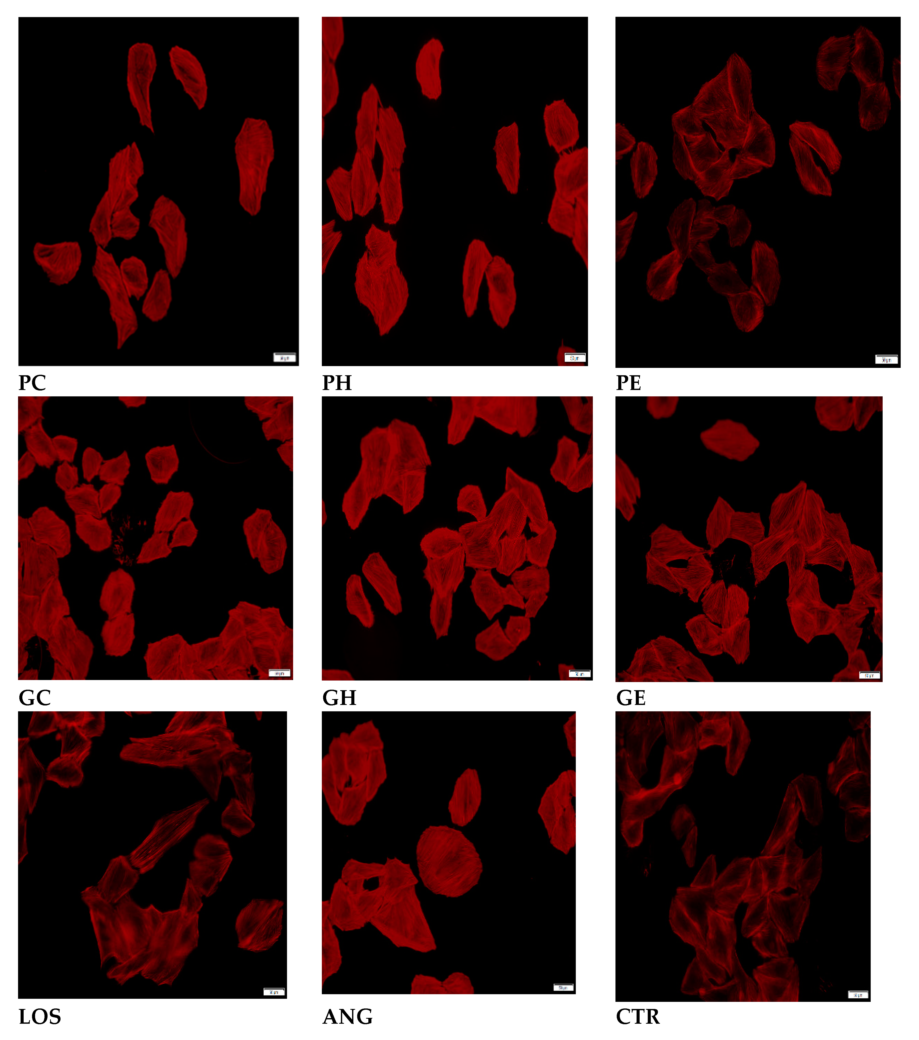
Figure 2.
The effects of Pan cyanescens (cold-water PC, hot-water PH and ethanol PE) and P. cubensis (cold-water GC, hot-water GH and ethanol GE) mushroom extracts (50 µg/mL) and positive control Losartan (100 µM) on the cell width measurements on AngII-induced hypertrophy on H9C2 cardiomyocytes over 48 hours. Control: non-induced negative control. (*: significant).
Figure 2.
The effects of Pan cyanescens (cold-water PC, hot-water PH and ethanol PE) and P. cubensis (cold-water GC, hot-water GH and ethanol GE) mushroom extracts (50 µg/mL) and positive control Losartan (100 µM) on the cell width measurements on AngII-induced hypertrophy on H9C2 cardiomyocytes over 48 hours. Control: non-induced negative control. (*: significant).
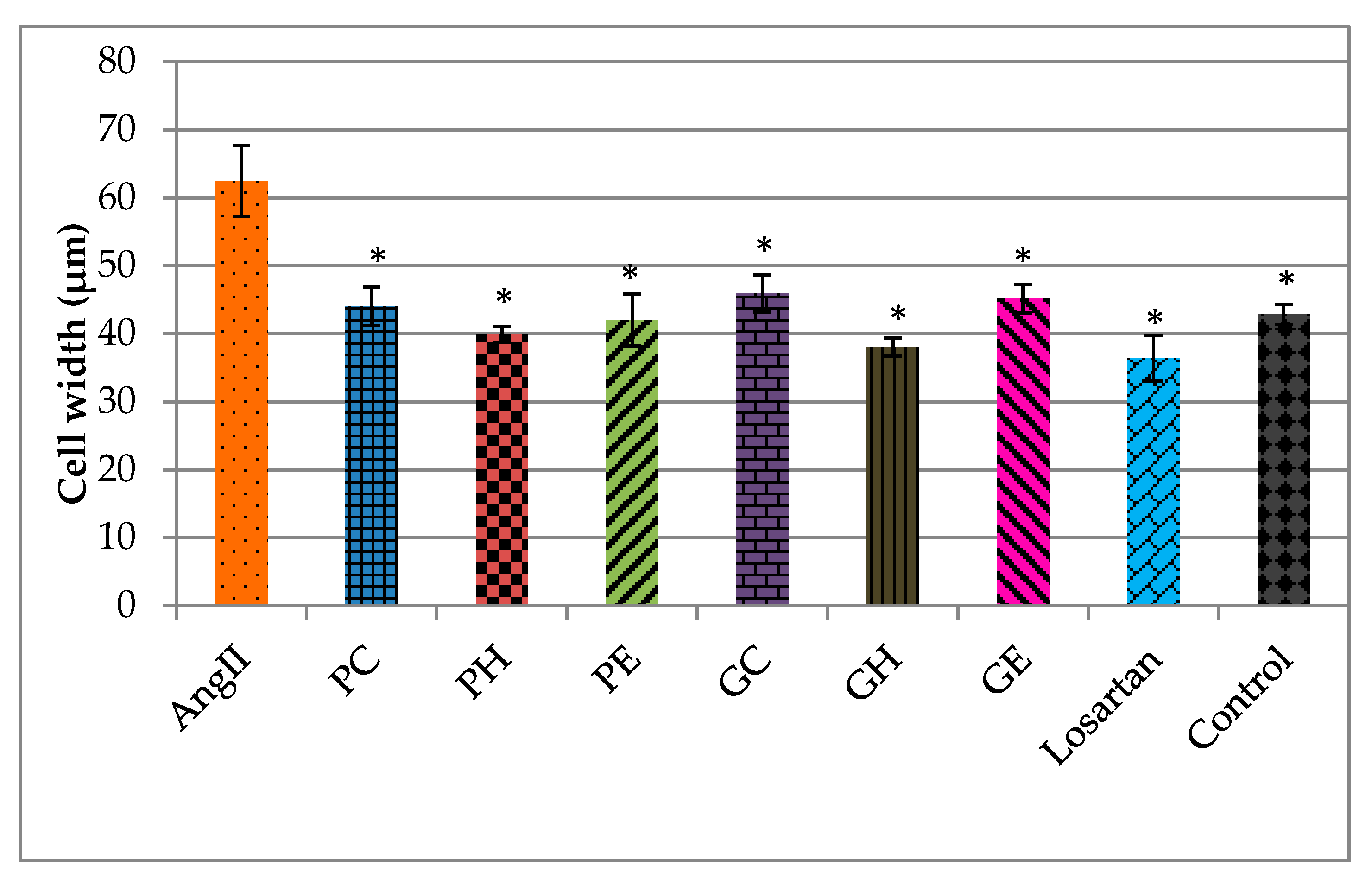
Figure 3.
Morphological effects of P. A+ strain (AC, AH and AE) and P. natalensis (NC, NH and NE) mushroom extracts (25 µg/mL) and positive control losartan (LOS) (100 µM) on the actin filaments of AngII-induced H9C2 cardiomyocytes (50 µm) over 48 hours using a fluorescence filter at Ex/Em= 546/575 nm. CTR: non-induced negative control cells.
Figure 3.
Morphological effects of P. A+ strain (AC, AH and AE) and P. natalensis (NC, NH and NE) mushroom extracts (25 µg/mL) and positive control losartan (LOS) (100 µM) on the actin filaments of AngII-induced H9C2 cardiomyocytes (50 µm) over 48 hours using a fluorescence filter at Ex/Em= 546/575 nm. CTR: non-induced negative control cells.
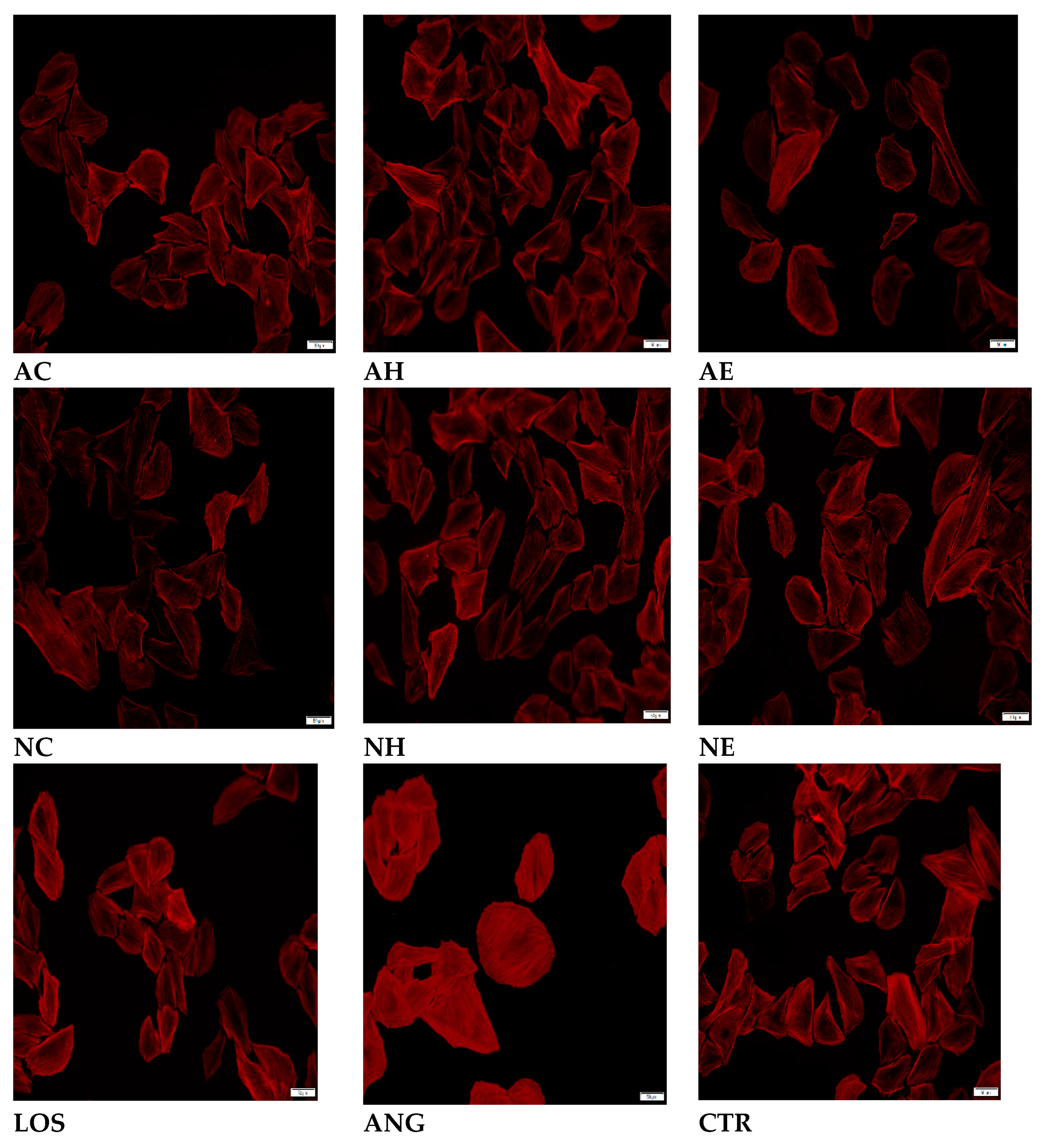
Figure 4.
The effects of P. A+ strain (cold-water AC, hot-water AH and ethanol AE) and P. natalensis (cold-water NC, hot-water NH and ethanol NE) mushroom extracts (25 µg/mL) and positive control Losartan (100 µM) on the cell width measurements on AngII-induced hypertrophy on H9C2 cardiomyocytes over 48 hours. Control: non-induced negative control. (*: significant).
Figure 4.
The effects of P. A+ strain (cold-water AC, hot-water AH and ethanol AE) and P. natalensis (cold-water NC, hot-water NH and ethanol NE) mushroom extracts (25 µg/mL) and positive control Losartan (100 µM) on the cell width measurements on AngII-induced hypertrophy on H9C2 cardiomyocytes over 48 hours. Control: non-induced negative control. (*: significant).
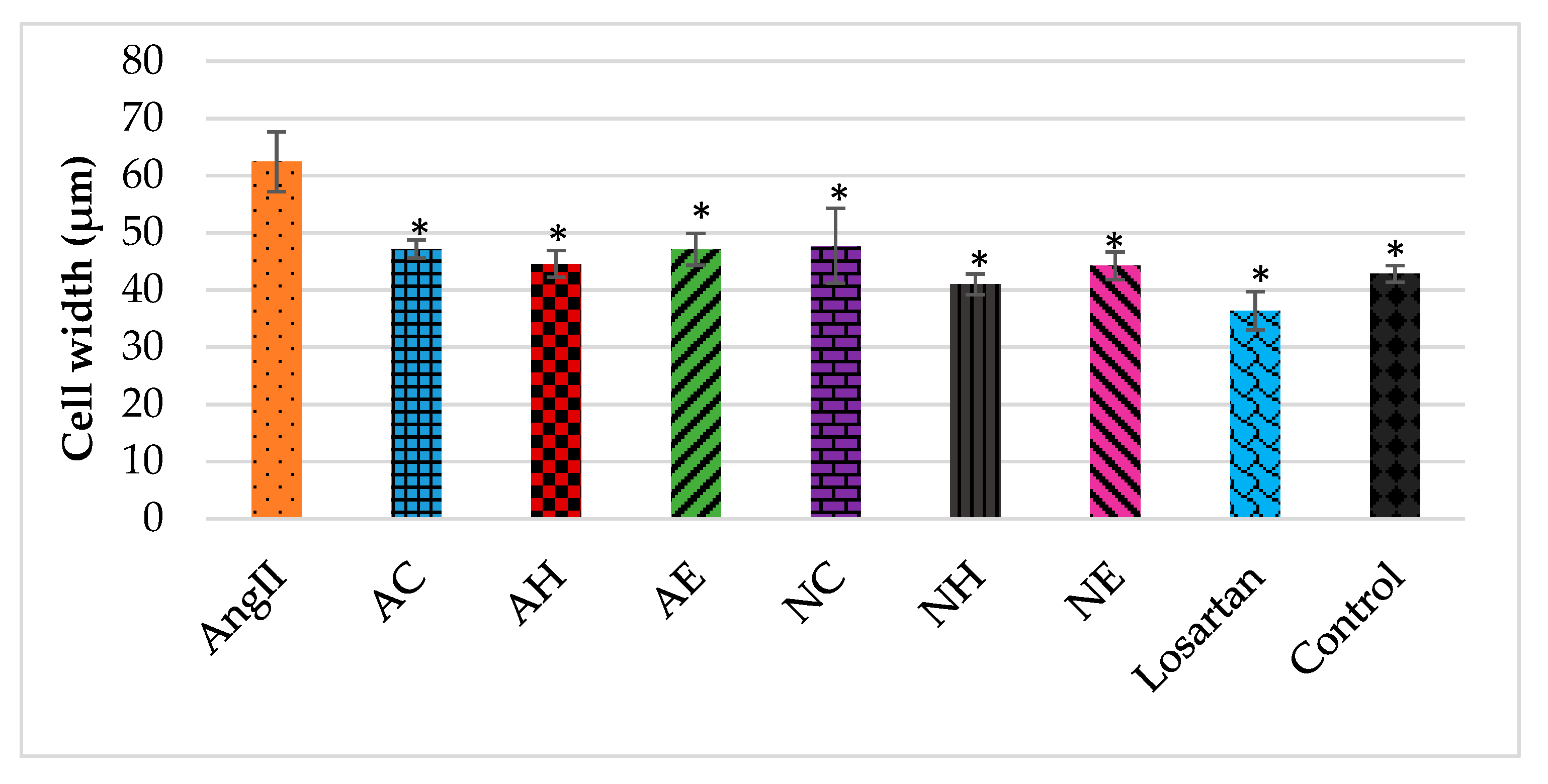
Figure 5.
Effects of the extracts (25 and 50 µg/mL) of Pan cyanescens (cold-water PC, hot-water PH and ethanol PE), P. cubensis (cold-water GC, hot-water GH and ethanol GE), P. A+ strain (cold-water AC, hot-water AH and ethanol AE) and P. natalensis (cold-water NC, hot-water NH and ethanol NE) mushroom and positive controls; losartan and LNAME (50, 100 µM) on the mitochondrial activity of AngII-induced hypertrophy over 48 hours. Control: non-induced negative control. (*: significant).
Figure 5.
Effects of the extracts (25 and 50 µg/mL) of Pan cyanescens (cold-water PC, hot-water PH and ethanol PE), P. cubensis (cold-water GC, hot-water GH and ethanol GE), P. A+ strain (cold-water AC, hot-water AH and ethanol AE) and P. natalensis (cold-water NC, hot-water NH and ethanol NE) mushroom and positive controls; losartan and LNAME (50, 100 µM) on the mitochondrial activity of AngII-induced hypertrophy over 48 hours. Control: non-induced negative control. (*: significant).
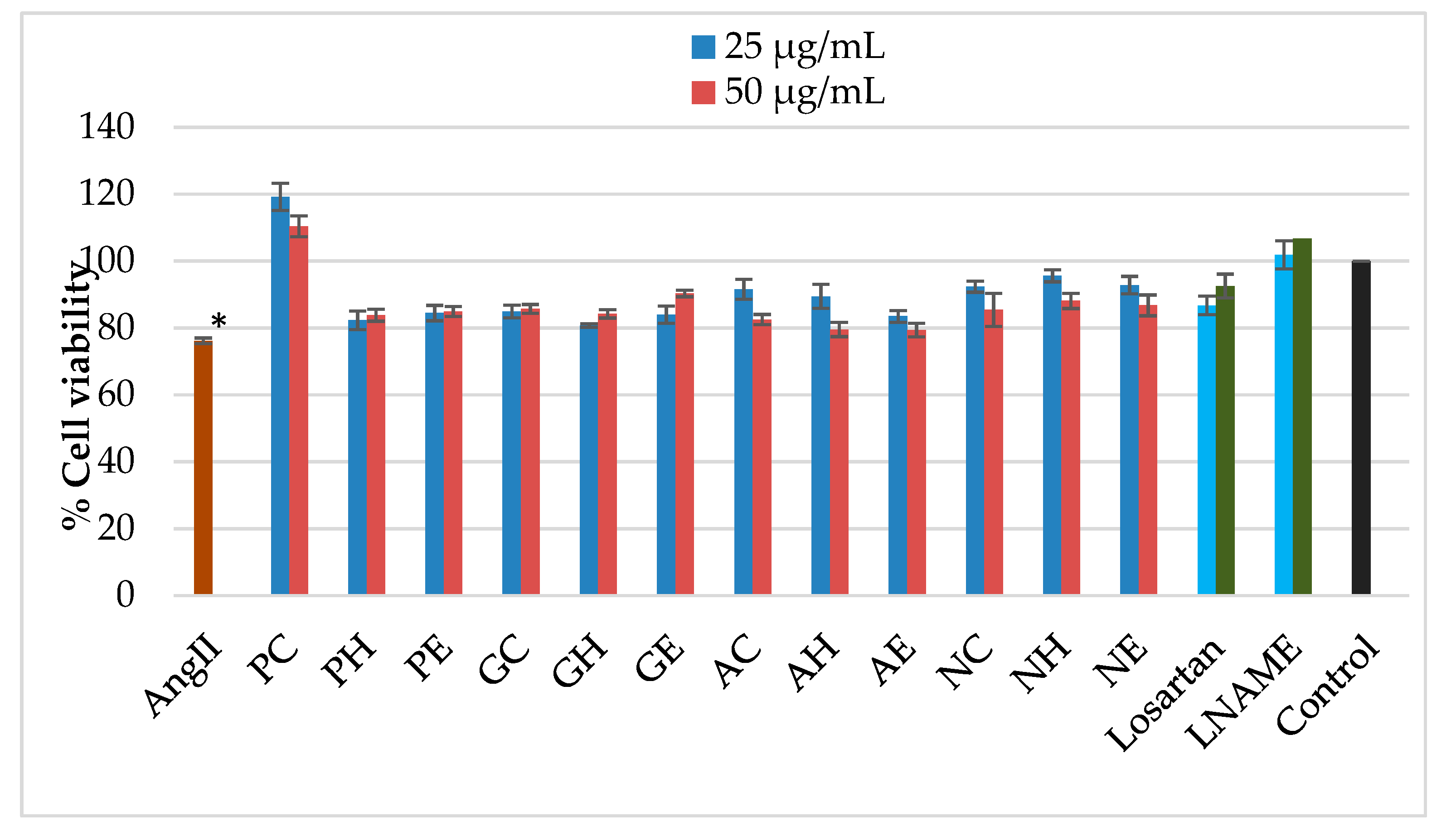
Figure 6.
Effects of 1-hour treatment with 50 µg/mL extracts of Pan cyanescens (cold-water PC, hot-water PH and ethanol PE), P. cubensis (cold-water GC, hot-water GH and ethanol GE), and 25 µg/mL of P. A+ strain (cold-water AC, hot-water AH and ethanol AE) and P. natalensis (cold-water NC, hot-water NH and ethanol NE) and the positive control Losartan (100 µM) treatments on fluorometric intracellular ROS (superoxide and hydroxyl radicals) production measured using a green fluorescence intensity at λex= 485/20,520/25 nm on AngII-induced cardiomyocytes. Control: non-induced negative control. (*: significant).
Figure 6.
Effects of 1-hour treatment with 50 µg/mL extracts of Pan cyanescens (cold-water PC, hot-water PH and ethanol PE), P. cubensis (cold-water GC, hot-water GH and ethanol GE), and 25 µg/mL of P. A+ strain (cold-water AC, hot-water AH and ethanol AE) and P. natalensis (cold-water NC, hot-water NH and ethanol NE) and the positive control Losartan (100 µM) treatments on fluorometric intracellular ROS (superoxide and hydroxyl radicals) production measured using a green fluorescence intensity at λex= 485/20,520/25 nm on AngII-induced cardiomyocytes. Control: non-induced negative control. (*: significant).
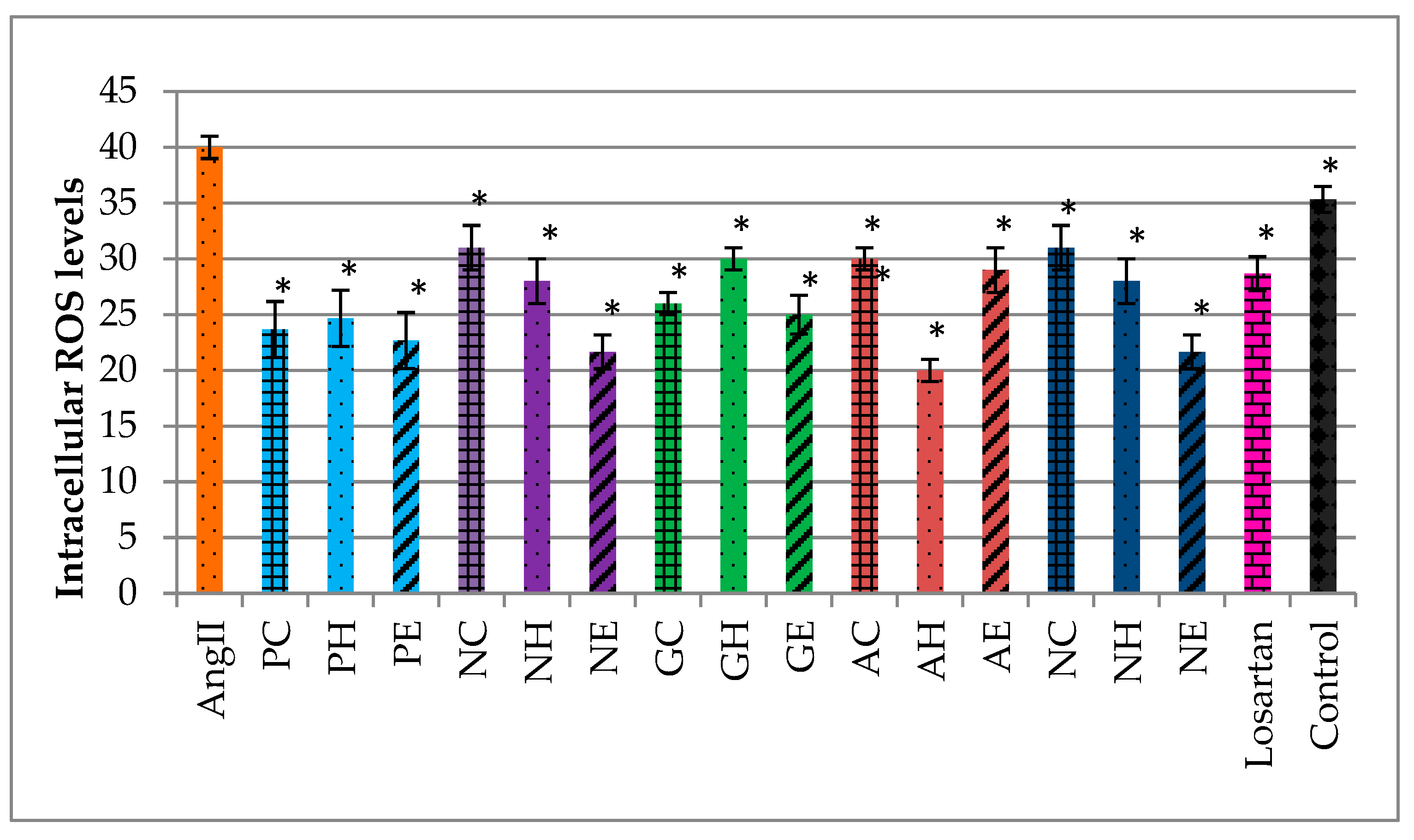
Figure 7.
The GCMS-MS chromatogram of cold-water (PC), hot-water (PH) and 70% ethanol (PE) extracts of Pan cyanescens mushroom.
Figure 7.
The GCMS-MS chromatogram of cold-water (PC), hot-water (PH) and 70% ethanol (PE) extracts of Pan cyanescens mushroom.
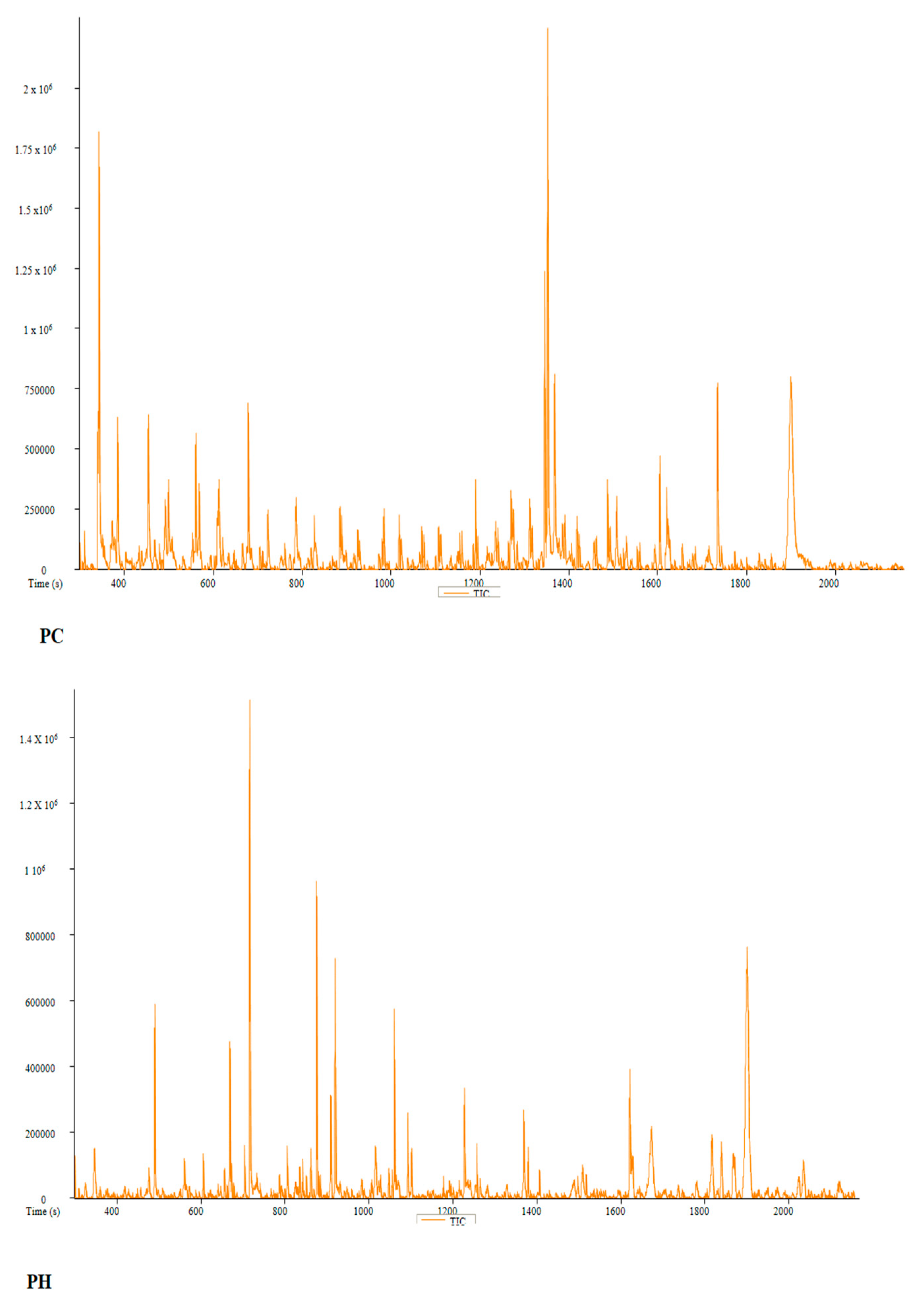
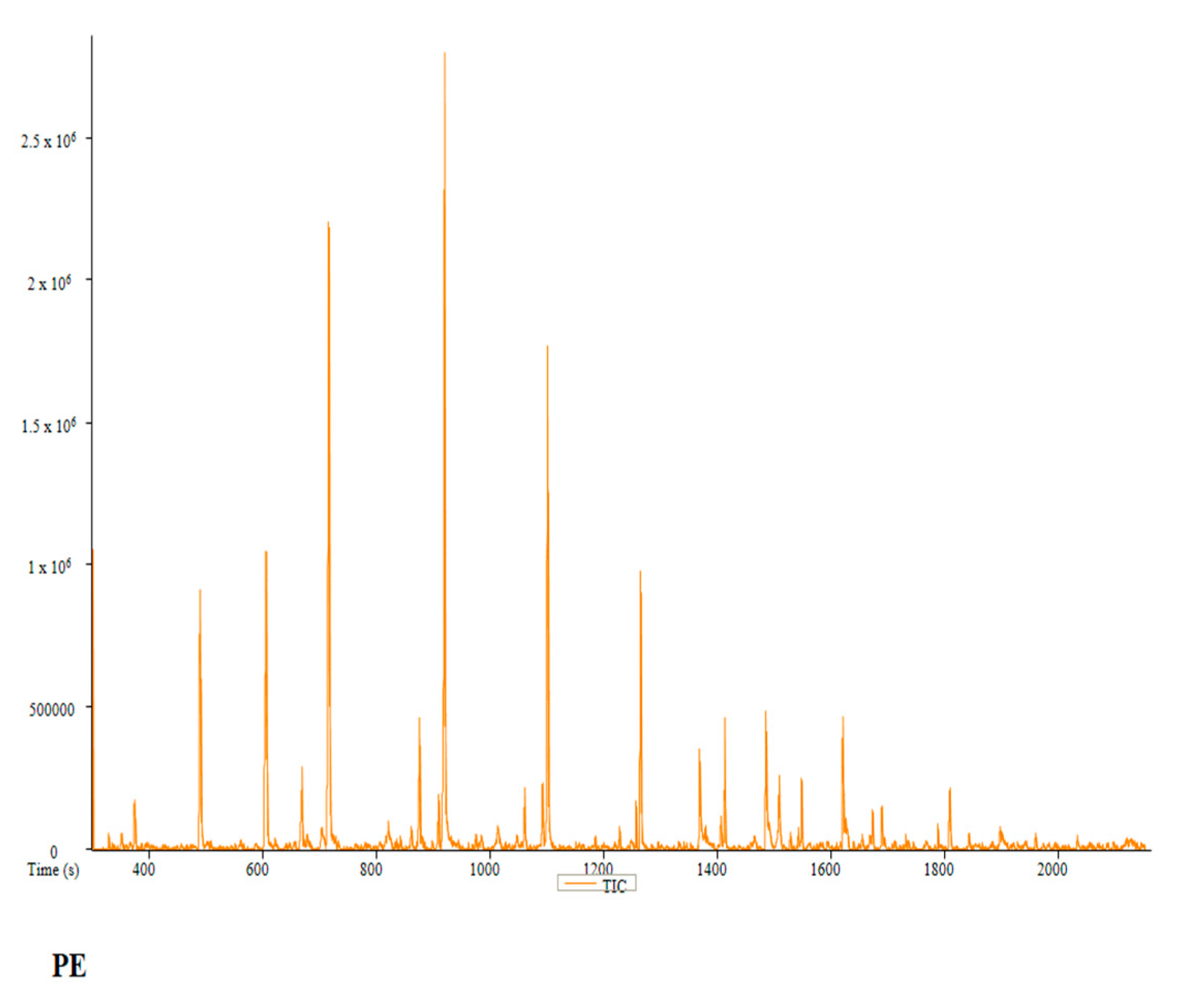
Figure 8.
The GCMS-MS chromatogram of cold-water (GC), hot-water (GH) and 70% ethanol (GE) extracts of P. cubensis mushroom.
Figure 8.
The GCMS-MS chromatogram of cold-water (GC), hot-water (GH) and 70% ethanol (GE) extracts of P. cubensis mushroom.
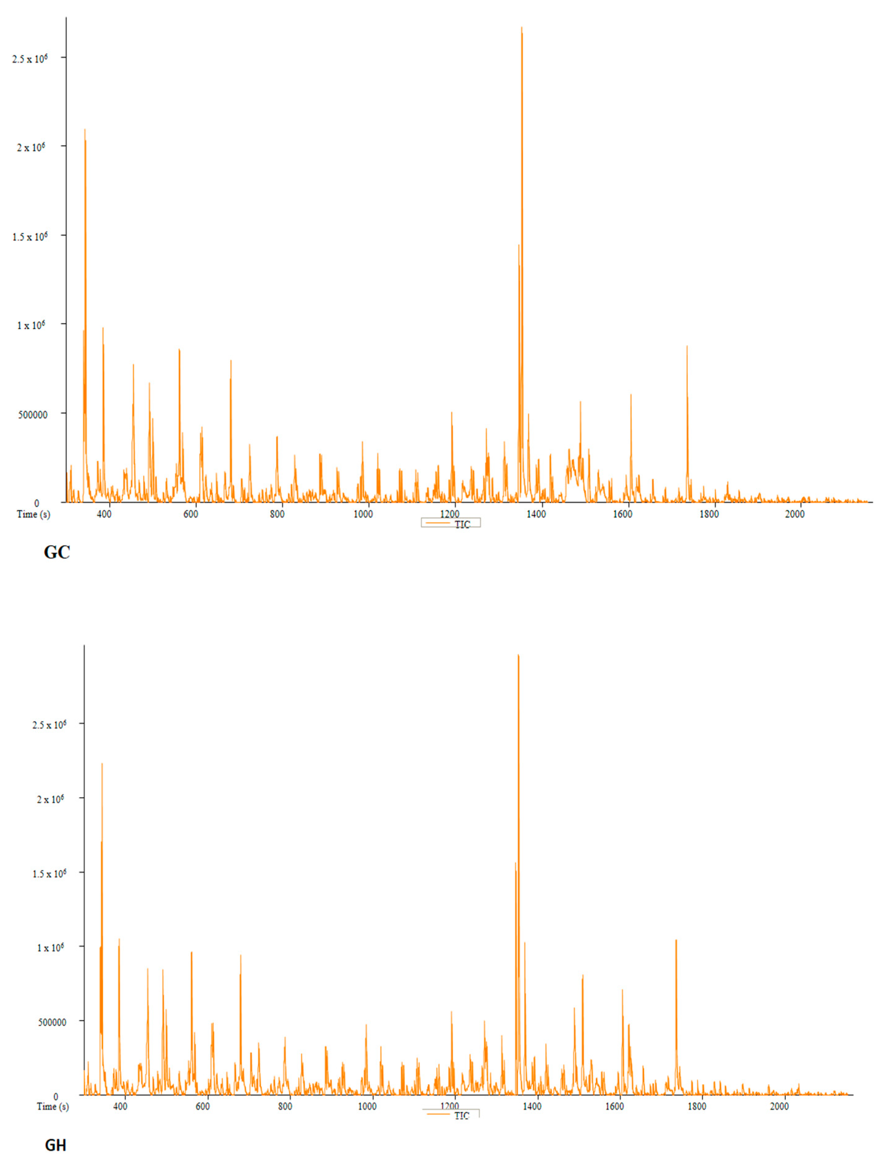
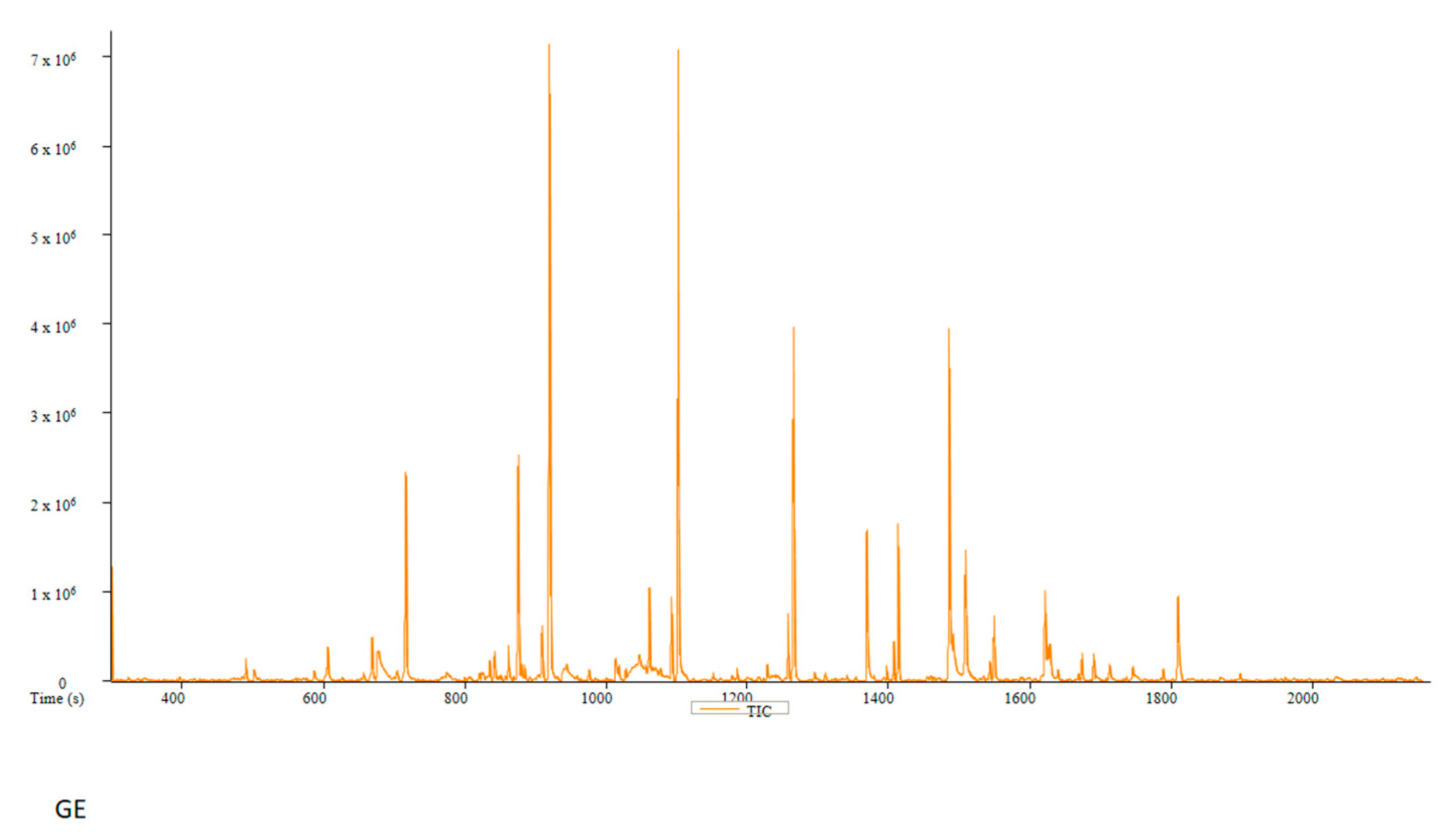
Figure 9.
The GCMS-MS chromatogram of cold-water (AC), hot-water (AH) and 70% ethanol (AE) extracts of P. A+ strain mushroom.
Figure 9.
The GCMS-MS chromatogram of cold-water (AC), hot-water (AH) and 70% ethanol (AE) extracts of P. A+ strain mushroom.
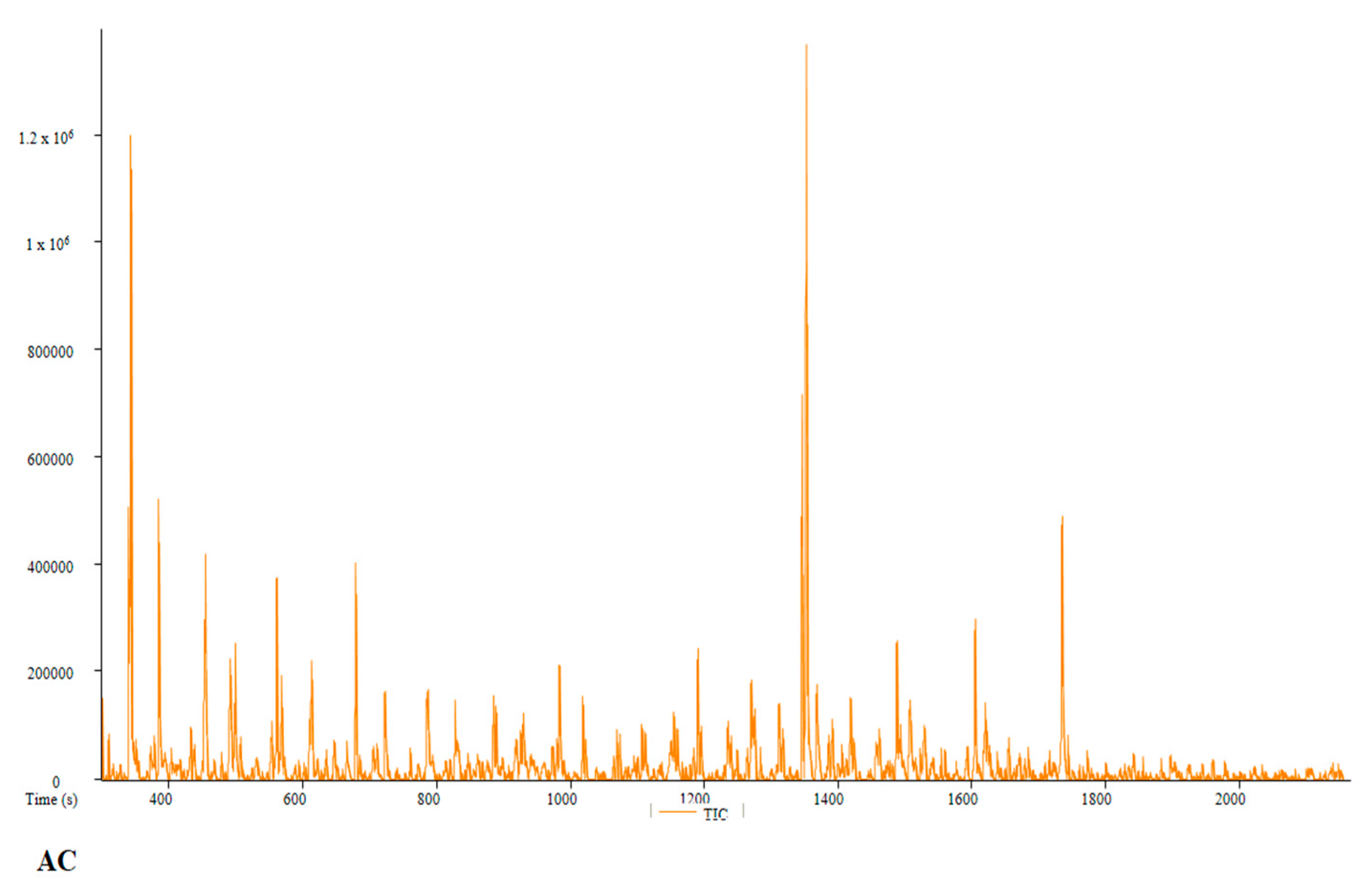
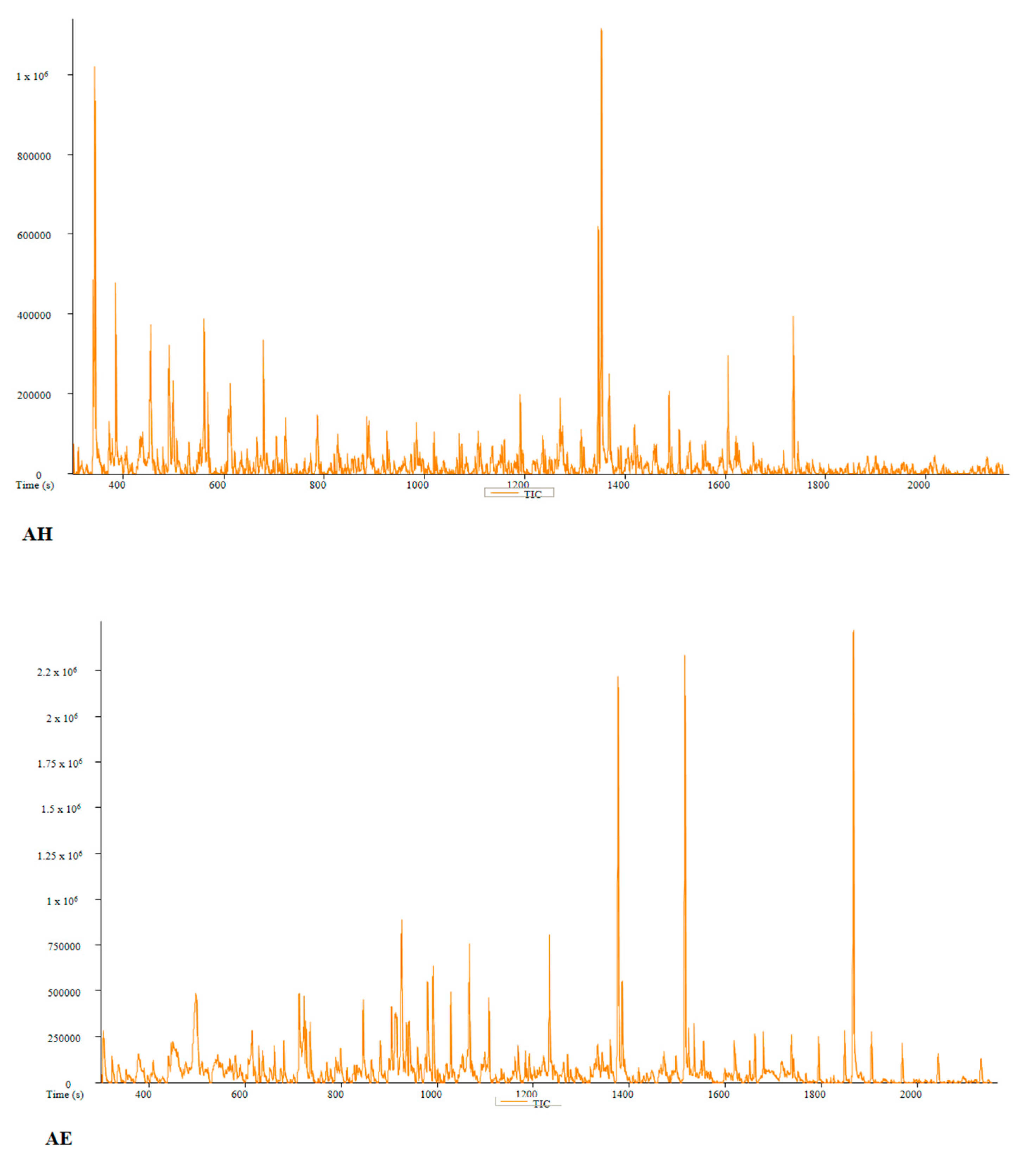
Table 2.
The compounds with anti-inflammatory and antioxidant effect identified in Pan cyanescens cold-water (PC), hot-water (PH) and 70% ethanol (PE) mushroom extracts.
Table 2.
The compounds with anti-inflammatory and antioxidant effect identified in Pan cyanescens cold-water (PC), hot-water (PH) and 70% ethanol (PE) mushroom extracts.
| PC | Peak # | Name | Weight | Formula | CAS | Similarity | Area % | 1st Dimension Time (s) | Height |
|---|---|---|---|---|---|---|---|---|---|
| 4 | Hexanoic acid, methyl ester | 130 | C7H14O2 | 106-70-7 | 927 | 2,0459 | 385,6 | 614562 | |
| 6 | 3-Octanone | 128 | C8H16O | 106-68-3 | 794 | 2,9534 | 453,6 | 647828 | |
| 7 | Dodecane, 1,1-dimethoxy- | 230 | C14H30O2 | 14620-52-1 | 705 | 2,8617 | 455 | 639963 | |
| 22 | Dodecane, 1,1-dimethoxy- | 230 | C14H30O2 | 14620-52-1 | 778 | 0,66502 | 785,8 | 265590 | |
| 26 | Dodecane, 1,1-dimethoxy- | 230 | C14H30O2 | 14620-52-1 | 816 | 0,79635 | 886 | 265712 | |
| 30 | Dodecane, 1,1-dimethoxy- | 230 | C14H30O2 | 14620-52-1 | 820 | 0,36008 | 980,6 | 132773 | |
| 35 | Dodecane, 1,1-dimethoxy- | 230 | C14H30O2 | 14620-52-1 | 843 | 0,4545 | 1070,3 | 185705 | |
| 38 | Dodecane, 1,1-dimethoxy- | 230 | C14H30O2 | 14620-52-1 | 815 | 0,46437 | 1155,4 | 155284 | |
| 42 | Dodecane, 1,1-dimethoxy- | 230 | C14H30O2 | 14620-52-1 | 794 | 0,93345 | 1236,3 | 196900 | |
| 43 | Dodecane, 1,1-dimethoxy- | 230 | C14H30O2 | 14620-52-1 | 800 | 0,56178 | 1241,5 | 176011 | |
| 47 | Dodecane, 1,1-dimethoxy- | 230 | C14H30O2 | 14620-52-1 | 781 | 1,0495 | 1313,4 | 297088 | |
| 51 | n-Hexadecanoic acid | 256 | C16H32O2 | 57-10-3 | 899 | 2,891 | 1369 | 754734 | |
| 52 | Dodecane, 1,1-dimethoxy- | 230 | C14H30O2 | 14620-52-1 | 840 | 0,65301 | 1386,9 | 155364 | |
| 53 | Dodecane, 1,1-dimethoxy- | 230 | C14H30O2 | 14620-52-1 | 774 | 0,66727 | 1392,4 | 200963 | |
| 58 | Dodecane, 1,1-dimethoxy- | 230 | C14H30O2 | 14620-52-1 | 798 | 0,22375 | 1524,4 | 90547 | |
| 62 | 9-Octadecenamide, (Z)- | 281 | C18H35NO | 301-02-0 | 908 | 1,1739 | 1621,3 | 345374 | |
| 63 | 9-Octadecenamide, (Z)- | 281 | C18H35NO | 301-02-0 | 816 | 0,81034 | 1624,9 | 215839 | |
| 64 | 9-Octadecenamide, (Z)- | 281 | C18H35NO | 301-02-0 | 733 | 0,51097 | 1628,1 | 131186 | |
| 66 | Dodecane, 1,1-dimethoxy- | 230 | C14H30O2 | 14620-52-1 | 770 | 0,35671 | 1716,6 | 102006 | |
| 68 | Dodecane, 1,1-dimethoxy- | 230 | C14H30O2 | 14620-52-1 | 791 | 0,20132 | 1774,1 | 81447 | |
| PH | Peak # | Name | Weight | Formula | CAS | Similarity | Area % | 1st Dimension Time (s) | Height |
| 13 | Tetradecane | 198 | C14H30 | 629-59-4 | 884 | 4,0398 | 920,1 | 731151 | |
| 19 | n-Hexadecanoic acid | 256 | C16H32O2 | 57-10-3 | 868 | 1,9932 | 1369,2 | 270343 | |
| 22 | 9-Octadecenamide, (Z)- | 281 | C18H35NO | 301-02-0 | 921 | 2,6151 | 1621,3 | 395816 | |
| 23 | 9-Octadecenamide, (Z)- | 281 | C18H35NO | 301-02-0 | 829 | 0,98533 | 1625 | 141739 | |
| 24 | 9-Octadecenamide, (Z)- | 281 | C18H35NO | 301-02-0 | 877 | 1,1489 | 1628,4 | 130670 | |
| 26 | Dotriacontane | 450 | C32H66 | 544-85-4 | 885 | 5,8278 | 1673,2 | 221028 | |
| 32 | Olean-12-ene-3,28-diol, (3á)- | 442 | C30H50O2 | 545-48-2 | 613 | 19,938 | 1900,8 | 766581 | |
| PE | Peak # | Name | Weight | Formula | CAS | Similarity | Area % | 1st Dimension Time (s) | Height |
| 5 | Decane | 142 | C10H22 | 124-18-5 | 846 | 8,8254 | 605,8 | 1051849 | |
| 10 | Tetradecane | 198 | C14H30 | 629-59-4 | 880 | 18,263 | 919,9 | 2801754 | |
| 14 | Nonadecane | 268 | C19H40 | 629-92-5 | 893 | 10,729 | 1101,5 | 1772907 | |
| 15 | Nonadecane | 268 | C19H40 | 629-92-5 | 904 | 5,3724 | 1265 | 980759 | |
| 17 | n-Hexadecanoic acid | 256 | C16H32O2 | 57-10-3 | 880 | 2,671 | 1369,1 | 353542 | |
| 19 | 9,12-Octadecadienoic acid (Z,Z)- | 280 | C18H32O2 | 60-33-3 | 910 | 3,4485 | 1485,3 | 489829 | |
| 22 | Heneicosane | 296 | C21H44 | 629-94-7 | 899 | 1,3394 | 1548,6 | 251573 | |
| 23 | 9-Octadecenamide, (Z)- | 281 | C18H35NO | 301-02-0 | 924 | 2,509 | 1621,2 | 466852 |
Table 3.
The compounds identified in P. cubensis cold-water (GC), hot-water (GH) and 70% ethanol (GE) mushroom extracts.
Table 3.
The compounds identified in P. cubensis cold-water (GC), hot-water (GH) and 70% ethanol (GE) mushroom extracts.
| GC | Peak # | Name | Weight | Formula | CAS | Similarity | Area % | 1st Dimension Time (s) | Height |
|---|---|---|---|---|---|---|---|---|---|
| 3 | Dodecane, 1,1-dimethoxy- | 230 | C14H30O2 | 14620-52-1 | 739 | 6,3304 | 343,2 | 2096696 | |
| 6 | Hexanoic acid, methyl ester | 130 | C7H14O2 | 106-70-7 | 928 | 2,8062 | 385,1 | 955717 | |
| 9 | 3-Octanone | 128 | C8H16O | 106-68-3 | 830 | 2,7283 | 452,8 | 762082 | |
| 27 | Dodecane, 1,1-dimethoxy- | 230 | C14H30O2 | 14620-52-1 | 779 | 0,71327 | 785,7 | 314589 | |
| 31 | Dodecane, 1,1-dimethoxy- | 230 | C14H30O2 | 14620-52-1 | 813 | 0,69149 | 885,8 | 276576 | |
| 35 | Dodecane, 1,1-dimethoxy- | 230 | C14H30O2 | 14620-52-1 | 855 | 0,35927 | 980,5 | 151860 | |
| 38 | Dodecanoic acid, methyl ester | 214 | C13H26O2 | 111-82-0 | 900 | 0,70445 | 1019,3 | 279675 | |
| 41 | Dodecane, 1,1-dimethoxy- | 230 | C14H30O2 | 14620-52-1 | 807 | 0,44597 | 1070,2 | 197083 | |
| 48 | Dodecane, 1,1-dimethoxy- | 230 | C14H30O2 | 14620-52-1 | 809 | 0,78095 | 1236,2 | 194599 | |
| 53 | Dodecane, 1,1-dimethoxy- | 230 | C14H30O2 | 14620-52-1 | 736 | 1,2754 | 1313,2 | 346174 | |
| 55 | Hexadecanoic acid, methyl ester | 270 | C17H34O2 | 112-39-0 | 910 | 3,7206 | 1346,9 | 1438966 | |
| 57 | n-Hexadecanoic acid | 256 | C16H32O2 | 57-10-3 | 876 | 1,7469 | 1368,7 | 479084 | |
| 58 | Dodecane, 1,1-dimethoxy- | 230 | C14H30O2 | 14620-52-1 | 837 | 0,81401 | 1386,7 | 214202 | |
| 59 | Dodecane, 1,1-dimethoxy- | 230 | C14H30O2 | 14620-52-1 | 761 | 0,89417 | 1392,1 | 245098 | |
| 65 | Dodecane, 1,1-dimethoxy- | 230 | C14H30O2 | 14620-52-1 | 851 | 0,21253 | 1524,3 | 93108 | |
| 71 | Dodecane, 1,1-dimethoxy- | 230 | C14H30O2 | 14620-52-1 | 729 | 0,20497 | 1716,4 | 85138 | |
| GH | Peak # | Name | Weight | Formula | CAS | Similarity | Area % | 1st Dimension Time (s) | Height |
| 3 | Dodecane, 1,1-dimethoxy- | 230 | C14H30O2 | 14620-52-1 | 718 | 5,7808 | 343,8 | 2236529 | |
| 11 | Dodecane, 1,1-dimethoxy- | 230 | C14H30O2 | 14620-52-1 | 739 | 3,5171 | 454,8 | 833100 | |
| 20 | Dodecane, 1,1-dimethoxy- | 230 | C14H30O2 | 14620-52-1 | 745 | 0,93596 | 569 | 403943 | |
| 28 | Dodecane, 1,1-dimethoxy- | 230 | C14H30O2 | 14620-52-1 | 759 | 0,65184 | 785,8 | 327037 | |
| 32 | Dodecane, 1,1-dimethoxy- | 230 | C14H30O2 | 14620-52-1 | 827 | 0,73365 | 885,8 | 331466 | |
| 37 | Dodecane, 1,1-dimethoxy- | 230 | C14H30O2 | 14620-52-1 | 833 | 0,44286 | 980,5 | 180459 | |
| 40 | Dodecanoic acid, methyl ester | 214 | C13H26O2 | 111-82-0 | 862 | 0,76451 | 1019,3 | 332902 | |
| 46 | Dodecane, 1,1-dimethoxy- | 230 | C14H30O2 | 14620-52-1 | 813 | 0,47338 | 1155,3 | 187669 | |
| 48 | Hexadecanoic acid, methyl ester | 270 | C17H34O2 | 112-39-0 | 829 | 1,4125 | 1191,3 | 568445 | |
| 50 | Dodecane, 1,1-dimethoxy- | 230 | C14H30O2 | 14620-52-1 | 802 | 0,88819 | 1236,2 | 252623 | |
| 55 | Dodecane, 1,1-dimethoxy- | 230 | C14H30O2 | 14620-52-1 | 784 | 1,2221 | 1313,2 | 405746 | |
| 56 | Dodecane, 1,1-dimethoxy- | 230 | C14H30O2 | 14620-52-1 | 789 | 0,80275 | 1318,5 | 238850 | |
| 59 | n-Hexadecanoic acid | 256 | C16H32O2 | 57-10-3 | 891 | 3,1125 | 1368,8 | 1020178 | |
| 60 | Dodecane, 1,1-dimethoxy- | 230 | C14H30O2 | 14620-52-1 | 827 | 0,72977 | 1386,7 | 208562 | |
| 61 | Dodecane, 1,1-dimethoxy- | 230 | C14H30O2 | 14620-52-1 | 772 | 0,67475 | 1392,2 | 260361 | |
| 69 | Dodecane, 1,1-dimethoxy- | 230 | C14H30O2 | 14620-52-1 | 845 | 0,23403 | 1524,2 | 119865 | |
| 74 | 9-Octadecenamide, (Z)- | 281 | C18H35NO | 301-02-0 | 901 | 1,5525 | 1620,9 | 476077 | |
| 75 | 9-Octadecenamide, (Z)- | 281 | C18H35NO | 301-02-0 | 828 | 0,94615 | 1624,4 | 327188 | |
| 76 | 9-Octadecenamide, (Z)- | 281 | C18H35NO | 301-02-0 | 768 | 0,71306 | 1627,7 | 212987 | |
| 78 | Dodecane, 1,1-dimethoxy- | 230 | C14H30O2 | 14620-52-1 | 839 | 0,41231 | 1716,3 | 134307 | |
| 80 | Dodecane, 1,1-dimethoxy- | 230 | C14H30O2 | 14620-52-1 | 772 | 0,22056 | 1773,7 | 103514 | |
| 81 | Dodecane, 1,1-dimethoxy- | 230 | C14H30O2 | 14620-52-1 | 735 | 0,27314 | 1829,2 | 96189 | |
| GE | Peak # | Name | Weight | Formula | CAS | Similarity | Area % | 1st Dimension Time (s) | Height |
| 1 | Decane | 142 | C10H22 | 124-18-5 | 897 | 0,44944 | 490,3 | 252016 | |
| 3 | Decane | 142 | C10H22 | 124-18-5 | 842 | 0,98073 | 605,7 | 379188 | |
| 8 | Tetradecane | 198 | C14H30 | 629-59-4 | 864 | 4,5504 | 716,3 | 2342975 | |
| 14 | Tetradecane | 198 | C14H30 | 629-59-4 | 873 | 12,505 | 919,9 | 7151813 | |
| 20 | Hexadecane | 226 | C16H34 | 544-76-3 | 914 | 11,926 | 1101,7 | 7087174 | |
| 24 | Nonadecane | 268 | C19H40 | 629-92-5 | 919 | 6,5048 | 1265 | 3973633 | |
| 26 | n-Hexadecanoic acid | 256 | C16H32O2 | 57-10-3 | 916 | 3,2652 | 1368,9 | 1698554 | |
| 29 | Heneicosane | 296 | C21H44 | 629-94-7 | 901 | 2,7681 | 1413,3 | 1765288 | |
| 30 | 9,12-Octadecadienoic acid (Z,Z)- | 280 | C18H32O2 | 60-33-3 | 920 | 7,7412 | 1485,4 | 3942406 | |
| 31 | Oleic Acid | 282 | C18H34O2 | 112-80-1 | 901 | 1,7768 | 1490,7 | 498823 | |
| 32 | Dodecanamide | 199 | C12H25NO | 1120-16-7 | 822 | 0,1794 | 1506,4 | 175567 | |
| 36 | 9-Octadecenamide, (Z)- | 281 | C18H35NO | 301-02-0 | 920 | 2,013 | 1620,9 | 1001169 | |
| 37 | 9-Octadecenamide, (Z)- | 281 | C18H35NO | 301-02-0 | 875 | 1,1399 | 1627,8 | 415638 | |
| 42 | 9-Octadecenamide, (Z)- | 281 | C18H35NO | 301-02-0 | 827 | 0,33757 | 1745 | 161336 |
Table 4.
The compounds identified in P. A+ strain cold-water (AC), hot-water (AH) and 70% ethanol (AE) mushroom extracts.
Table 4.
The compounds identified in P. A+ strain cold-water (AC), hot-water (AH) and 70% ethanol (AE) mushroom extracts.
| AC | Peak # | Name | Weight | Formula | CAS | Similarity | Area % | 1st Dimension Time (s) | Height |
|---|---|---|---|---|---|---|---|---|---|
| 3 | Dodecane, 1,1-dimethoxy- | 230 | C14H30O2 | 14620-52-1 | 732 | 7,2746 | 343,1 | 1201850 | |
| 5 | Hexanoic acid, methyl ester | 130 | C7H14O2 | 106-70-7 | 928 | 3,7608 | 385 | 524718 | |
| 7 | 3-Octanone | 128 | C8H16O | 106-68-3 | 852 | 3,7989 | 452,8 | 422676 | |
| 8 | Dodecane, 1,1-dimethoxy- | 230 | C14H30O2 | 14620-52-1 | 725 | 3,7989 | 454,5 | 422676 | |
| 24 | Dodecane, 1,1-dimethoxy- | 230 | C14H30O2 | 14620-52-1 | 742 | 0,77714 | 785,7 | 152661 | |
| 27 | Dodecane, 1,1-dimethoxy- | 230 | C14H30O2 | 14620-52-1 | 805 | 1,1092 | 885,8 | 160107 | |
| 30 | Dodecane, 1,1-dimethoxy- | 230 | C14H30O2 | 14620-52-1 | 841 | 0,47523 | 980,5 | 80179 | |
| 35 | Dodecane, 1,1-dimethoxy- | 230 | C14H30O2 | 14620-52-1 | 850 | 0,5429 | 1070,1 | 98825 | |
| 38 | Dodecane, 1,1-dimethoxy- | 230 | C14H30O2 | 14620-52-1 | 810 | 1,0146 | 1155,1 | 128129 | |
| 39 | Dodecane, 1,1-dimethoxy- | 230 | C14H30O2 | 14620-52-1 | 748 | 0,71579 | 1160,2 | 83648 | |
| 41 | Dodecane, 1,1-dimethoxy- | 230 | C14H30O2 | 14620-52-1 | 829 | 1,066 | 1236,1 | 112539 | |
| 45 | Dodecane, 1,1-dimethoxy- | 230 | C14H30O2 | 14620-52-1 | 754 | 1,2357 | 1313,2 | 144052 | |
| 46 | Dodecane, 1,1-dimethoxy- | 230 | C14H30O2 | 14620-52-1 | 732 | 0,83455 | 1318,6 | 97752 | |
| 47 | Hexadecanoic acid, methyl ester | 270 | C17H34O2 | 112-39-0 | 910 | 4,4828 | 1347 | 720709 | |
| 49 | n-Hexadecanoic acid | 256 | C16H32O2 | 57-10-3 | 884 | 2,0361 | 1369,1 | 181013 | |
| 50 | Dodecane, 1,1-dimethoxy- | 230 | C14H30O2 | 14620-52-1 | 802 | 0,68996 | 1386,7 | 85828 | |
| 60 | 9-Octadecenamide, (Z)- | 281 | C18H35NO | 301-02-0 | 895 | 1,3478 | 1621 | 146411 | |
| 61 | 9-Octadecenamide, (Z)- | 281 | C18H35NO | 301-02-0 | 764 | 0,62809 | 1624,5 | 93200 | |
| 63 | Dodecane, 1,1-dimethoxy- | 230 | C14H30O2 | 14620-52-1 | 859 | 0,36481 | 1716,4 | 56531 | |
| AH | Peak # | Name | Weight | Formula | CAS | Similarity | Area % | 1st Dimension Time (s) | Height |
| 3 | Dodecane, 1,1-dimethoxy- | 230 | C14H30O2 | 14620-52-1 | 729 | 7,5091 | 343,1 | 1023514 | |
| 6 | Hexanoic acid, methyl ester | 130 | C7H14O2 | 106-70-7 | 942 | 3,4369 | 384,8 | 481718 | |
| 8 | 3-Octanone | 128 | C8H16O | 106-68-3 | 822 | 0,94495 | 452,6 | ||
| 9 | Dodecane, 1,1-dimethoxy- | 230 | C14H30O2 | 14620-52-1 | 711 | 4,0912 | 454,3 | 366395 | |
| 26 | Dodecane, 1,1-dimethoxy- | 230 | C14H30O2 | 14620-52-1 | 783 | 0,82861 | 785,6 | 133376 | |
| 30 | Dodecane, 1,1-dimethoxy- | 230 | C14H30O2 | 14620-52-1 | 817 | 1,059 | 885,7 | 146601 | |
| 33 | Dodecane, 1,1-dimethoxy- | 230 | C14H30O2 | 14620-52-1 | 854 | 0,51922 | 980,3 | 80840 | |
| 38 | Dodecane, 1,1-dimethoxy- | 230 | C14H30O2 | 14620-52-1 | 827 | 0,58444 | 1070,1 | 106177 | |
| 41 | Dodecane, 1,1-dimethoxy- | 230 | C14H30O2 | 14620-52-1 | 817 | 0,60789 | 1155,2 | 76344 | |
| 43 | Hexadecanoic acid, methyl ester | 270 | C17H34O2 | 112-39-0 | 851 | 1,3131 | 1191,4 | 202951 | |
| 44 | Dodecane, 1,1-dimethoxy- | 230 | C14H30O2 | 14620-52-1 | 828 | 1,0656 | 1236,1 | 99142 | |
| 45 | Dodecane, 1,1-dimethoxy- | 230 | C14H30O2 | 14620-52-1 | 779 | 0,30158 | 1241,2 | 66062 | |
| 48 | Dodecane, 1,1-dimethoxy- | 230 | C14H30O2 | 14620-52-1 | 773 | 1,0275 | 1313,2 | 116536 | |
| 50 | Hexadecanoic acid, methyl ester | 270 | C17H34O2 | 112-39-0 | 910 | 3,9708 | 1347,1 | 619698 | |
| 52 | n-Hexadecanoic acid | 256 | C16H32O2 | 57-10-3 | 894 | 2,3541 | 1368,8 | 236893 | |
| 53 | Dodecane, 1,1-dimethoxy- | 230 | C14H30O2 | 14620-52-1 | 831 | 0,54835 | 1386,7 | 69077 | |
| 54 | Dodecane, 1,1-dimethoxy- | 230 | C14H30O2 | 14620-52-1 | 740 | 0,54021 | 1392,2 | 75964 | |
| 59 | Glycine | 75 | C2H5NO2 | 56-40-6 | 754 | 1,0692 | 1530,1 | 87122 | |
| 63 | 9-Octadecenamide, (Z)- | 281 | C18H35NO | 301-02-0 | 864 | 0,62408 | 1621,2 | 97425 | |
| 65 | Dodecane, 1,1-dimethoxy- | 230 | C14H30O2 | 14620-52-1 | 808 | 0,58739 | 1716,4 | 61884 | |
| AE | Peak # | Name | Weight | Formula | CAS | Similarity | Area % | 1st Dimension Time (s) | Height |
| 10 | Decane | 142 | C10H22 | 124-18-5 | 933 | 20,924 | 497,5 | 783673 | |
| 31 | Tetradecane | 198 | C14H30 | 629-59-4 | 888 | 3,381 | 925,5 | 289073 | |
| 39 | n-Hexadecanoic acid | 256 | C16H32O2 | 57-10-3 | 911 | 3,0487 | 1375,1 | 418335 | |
| 37 | Hexadecanoic acid, methyl ester | 270 | C17H34O2 | 112-39-0 | 851 | 0,54568 | 1353 | 74443 | |
| 42 | 9,12-Octadecadienoic acid (Z,Z)- | 280 | C18H32O2 | 60-33-3 | 914 | 2,1293 | 1491,4 | 204219 | |
| 35 | Eicosane | 282 | C20H42 | 112-95-8 | 896 | 1,4654 | 1107,3 | 168172 |
Table 5.
Other compounds identified in P. natalensis cold-water, hot-water and 70% ethanol mushroom extracts. (Chromatograms published in [18]).
Table 5.
Other compounds identified in P. natalensis cold-water, hot-water and 70% ethanol mushroom extracts. (Chromatograms published in [18]).
| NH | Peak # | Name | Weight | Formula | CAS | Similarity | Area % | 1st Dimension Time (s) | Height |
|---|---|---|---|---|---|---|---|---|---|
| 7 | Dodecane, 1,1-dimethoxy- | 230 | C14H30O2 | 14620-52-1 | 715 | 3,2977 | 454,6 | 567775 | |
| 23 | Dodecane, 1,1-dimethoxy- | 230 | C14H30O2 | 14620-52-1 | 817 | 1,1041 | 885,9 | 210887 | |
| 26 | Dodecane, 1,1-dimethoxy- | 230 | C14H30O2 | 14620-52-1 | 830 | 0,42934 | 980,6 | 105250 | |
| 28 | Dodecanoic acid, methyl ester | 214 | C13H26O2 | 111-82-0 | 907 | 1,1472 | 1019,6 | 224678 | |
| 30 | Dodecane, 1,1-dimethoxy- | 230 | C14H30O2 | 14620-52-1 | 843 | 0,63486 | 1070,3 | 134265 | |
| 33 | Dodecane, 1,1-dimethoxy- | 230 | C14H30O2 | 14620-52-1 | 810 | 0,86841 | 1155,4 | 139945 | |
| 36 | Dodecane, 1,1-dimethoxy- | 230 | C14H30O2 | 14620-52-1 | 806 | 0,92241 | 1236,2 | 152342 | |
| 37 | Dodecane, 1,1-dimethoxy- | 230 | C14H30O2 | 14620-52-1 | 821 | 0,4793 | 1241,4 | 120478 | |
| 40 | Dodecane, 1,1-dimethoxy- | 230 | C14H30O2 | 14620-52-1 | 750 | 1,677 | 1313,3 | 251622 | |
| 45 | Dodecane, 1,1-dimethoxy- | 230 | C14H30O2 | 14620-52-1 | 809 | 1,1774 | 1386,7 | 152379 | |
| 46 | Dodecane, 1,1-dimethoxy- | 230 | C14H30O2 | 14620-52-1 | 777 | 0,97402 | 1392,3 | 175193 | |
| 50 | Dodecane, 1,1-dimethoxy- | 230 | C14H30O2 | 14620-52-1 | 795 | 0,26298 | 1524,4 | 67498 | |
| 55 | 9-Octadecenamide, (Z)- | 281 | C18H35NO | 301-02-0 | 860 | 1,1976 | 1621,4 | 205738 | |
| 57 | Dodecane, 1,1-dimethoxy- | 230 | C14H30O2 | 14620-52-1 | 771 | 0,27229 | 1716,4 | 72493 | |
| 59 | Dodecane, 1,1-dimethoxy- | 230 | C14H30O2 | 14620-52-1 | 731 | 0,47328 | 1773,9 | 89699 | |
| NC | Peak # | Name | Weight | Formula | CAS | Similarity | Area % | 1st Dimension Time (s) | Height |
| 13 | Dodecane, 1,1-dimethoxy- | 230 | C14H30O2 | 14620-52-1 | 733 | 3,6742 | 454,9 | 751534 | |
| 31 | Dodecane, 1,1-dimethoxy- | 230 | C14H30O2 | 14620-52-1 | 779 | 0,68667 | 785,7 | 315435 | |
| 33 | Decanoic acid, methyl ester | 186 | C11H22O2 | 110-42-9 | 890 | 0,63556 | 827,8 | 263610 | |
| 35 | Dodecane, 1,1-dimethoxy- | 230 | C14H30O2 | 14620-52-1 | 831 | 0,74448 | 885,8 | 274267 | |
| 40 | Dodecane, 1,1-dimethoxy- | 230 | C14H30O2 | 14620-52-1 | 837 | 0,43806 | 980,5 | 161533 | |
| 43 | Dodecanoic acid, methyl ester | 214 | C13H26O2 | 111-82-0 | 913 | 0,67789 | 1019,4 | 277970 | |
| 46 | Dodecane, 1,1-dimethoxy- | 230 | C14H30O2 | 14620-52-1 | 797 | 0,44929 | 1070,1 | 184949 | |
| 49 | Dodecane, 1,1-dimethoxy- | 230 | C14H30O2 | 14620-52-1 | 789 | 0,58295 | 1155,3 | 183945 | |
| 53 | Dodecane, 1,1-dimethoxy- | 230 | C14H30O2 | 14620-52-1 | 783 | 0,87504 | 1236,2 | 203315 | |
| 58 | Dodecane, 1,1-dimethoxy- | 230 | C14H30O2 | 14620-52-1 | 751 | 0,94274 | 1313,2 | 320486 | |
| 59 | Dodecane, 1,1-dimethoxy- | 230 | C14H30O2 | 14620-52-1 | 809 | 0,79948 | 1318,7 | 210365 | |
| 63 | Dodecane, 1,1-dimethoxy- | 230 | C14H30O2 | 14620-52-1 | 820 | 0,75158 | 1386,7 | 170332 | |
| 64 | Dodecane, 1,1-dimethoxy- | 230 | C14H30O2 | 14620-52-1 | 759 | 0,71847 | 1392,2 | 224832 | |
| 65 | Hexadecanoic acid, methyl ester | 270 | C17H34O2 | 112-39-0 | 844 | 0,87915 | 1419,5 | 273632 | |
| 70 | Dodecane, 1,1-dimethoxy- | 230 | C14H30O2 | 14620-52-1 | 836 | 0,24407 | 1524,3 | 103354 | |
| 73 | Dodecane, 1,1-dimethoxy- | 230 | C14H30O2 | 14620-52-1 | 758 | 0,50675 | 1594,8 | 141017 | |
| 75 | 9-Octadecenamide, (Z)- | 281 | C18H35NO | 301-02-0 | 889 | 0,73559 | 1621,2 | 243511 | |
| 76 | 9-Octadecenamide, (Z)- | 281 | C18H35NO | 301-02-0 | 713 | 0,68859 | 1624,7 | 202379 | |
| 78 | Dodecane, 1,1-dimethoxy- | 230 | C14H30O2 | 14620-52-1 | 776 | 0,52186 | 1656,7 | 149748 | |
| NE | Peak # | Name | Weight | Formula | CAS | Similarity | Area % | 1st Dimension Time (s) | Height |
| 2 | Decane | 142 | C10H22 | 124-18-5 | 874 | 1,1285 | 490 | 365956 | |
| 17 | 5-Eicosene, (E)- | 280 | C20H40 | 74685-30-6 | 904 | 1,1568 | 1257,2 | 551903 | |
| 23 | 5-Eicosene, (E)- | 280 | C20H40 | 74685-30-6 | 904 | 0,76517 | 1406,5 | 329793 | |
| 24 | Heneicosane | 296 | C21H44 | 629-94-7 | 901 | 3,1921 | 1413,2 | 1359751 | |
| 25 | 9,12-Octadecadienoic acid (Z,Z)- | 280 | C18H32O2 | 60-33-3 | 884 | 1,6761 | 1485,1 | 574310 | |
| 27 | Oleic Acid | 282 | C18H34O2 | 112-80-1 | 828 | 1,6454 | 1508,6 | 534460 | |
| 28 | Dotriacontane | 450 | C32H66 | 544-85-4 | 921 | 1,3086 | 1548,6 | 624893 | |
| 29 | 9-Octadecenamide, (Z)- | 281 | C18H35NO | 301-02-0 | 927 | 2,1573 | 1621 | 787743 | |
| 30 | 9-Octadecenamide, (Z)- | 281 | C18H35NO | 301-02-0 | 857 | 0,99448 | 1624,7 | 316129 | |
| 31 | 9-Octadecenamide, (Z)- | 281 | C18H35NO | 301-02-0 | 847 | 1,0811 | 1627,8 | 304295 | |
| 35 | 9-Octadecenamide, (Z)- | 281 | C18H35NO | 301-02-0 | 827 | 0,26441 | 1745 | 100584 |
Disclaimer/Publisher’s Note: The statements, opinions and data contained in all publications are solely those of the individual author(s) and contributor(s) and not of MDPI and/or the editor(s). MDPI and/or the editor(s) disclaim responsibility for any injury to people or property resulting from any ideas, methods, instructions or products referred to in the content. |
© 2023 by the authors. Licensee MDPI, Basel, Switzerland. This article is an open access article distributed under the terms and conditions of the Creative Commons Attribution (CC BY) license (http://creativecommons.org/licenses/by/4.0/).
Copyright: This open access article is published under a Creative Commons CC BY 4.0 license, which permit the free download, distribution, and reuse, provided that the author and preprint are cited in any reuse.