Submitted:
05 May 2023
Posted:
09 May 2023
You are already at the latest version
Abstract
Purpose: This study aimed to evaluate the nanomechanical properties and chemical composition of restorative materials and dental surfaces using different toothpastes. Methods: Enamel (n=60) and dentin (n=60) bovine blocks were obtained and restored using resin-modified glass ionomer cement (RMGIC, n=30) or composite resin (CR, n=30) to form the dentin adjacent to RMGIC (DRMGIC), enamel adjacent to RMGIC (ERMGIC), dentin adjacent to CR (DCR), and enamel adjacent to CR (ECR). After restoration, one hemiface of each specimen was coated with an acid-resistant varnish to create the control (C) and eroded (E) sides (erosion: 5 days, 4 × 2 min/day; 1% citric acid / abrasion: 2 × 15 s followed by immersion on slurries 2 min). Three toothpastes were used: without fluoride (WF; n=10), sodium fluoride (NaF; n=10), and stannous fluoride (SnF2; n=10). The specimens were analyzed for nanohardness (H), elastic modulus (Er), and chemical composition using energy-dispersive X-ray spectroscopy (EDS) and Raman microscopy. Data were analyzed using ANOVA two-way repeated measures and Tukey’s test (α = 0.05). Results: The NaF presented lower values of H for DRMGIC-C, with a statistical difference for WF (p < 0.05). SnF2 resulted in lower Er values for ERMGIC-E and RMGIC-E than WF and NaF (p < 0.05). WF showed lower calcium and phosphorus concentrations for DCR-E than other types of toothpastes (p < 0.05). Only stannous-based toothpaste damaged the elasticity of eroded glass ionomer restorations performed in enamel. Toothpastes with fluoride was capable for maintaining main chemical elements of dentin adjacent to restorative materials under challenge conditions.
Keywords:
abrasion
; composite resin
; erosion
; glass ionomer cement
; stannous ion
1. Introduction
Erosive Tooth Wear (ETW) involves multiple factors and has increased in frequency over the past decade. The etiology is related to eating habits owing to the high consumption of acidic beverages and may be associated with bulimia, anorexia, and gastroesophageal disorders.1,2 Importantly, ETW results from a combination of constant contact with acids and the mechanical forces from tooth brushing, contributing to the removal of surface tissue that is softened by acids.3
Other factors may influence erosive progression, such as the dissolution rate of dental substrates, influenced by the presence of impurities in the substrate mineral content.4 Thus, numerous studies have investigated erosive dynamics considering different aspects such as the composition of eroded dental tissues; distinct in vitro protocols to simulate erosive processes,5,6,7 action of bioactive particles on eroded tissues;8 and the action of different toothpastes, rinses, or varnishes with various active ingredients in order to minimize tooth loss.9,10, In addition, caries-like and white spot lesions are also vulnerable to erosion once the substrate is damaged.11 A study comparing the abrasively level using different toothpastes and fluoride gel concluded that early carious lesions are more susceptible to erosive-abrasive processes than sound substrates, especially when brushing with high abrasively toothpaste.11
However, as the erosive process advances, pronounced dental substrate loss may occur, exposing the dentin and resulting in clinical restorative procedures.5 Scarce information about the impact of erosive-abrasive challenge on the surface of dental restorative materials interfaces have been described in the literature. The erosive process in restorative materials is different of the dental tissues.12,13 Repeated erosive cycles may affect mechanical properties of these materials reducing their longevity.14,15 Among the restoratives materials, the most frequently used for direct restorations are composite resin (CR) and resin-modified glass ionomer cements (RMGIC). Although RMGIC present chemical bond to enamel and dentin and fluoride release that may reduce the erosive effects on the adjacent dental tissues, studies have shown that this material can suffer higher degradation than CR.7,16 Therefore, a comparison of these restorative materials adjacent to dental substrates subjected to different toothpastes after erosive-abrasive cycles becomes relevant.
Thus, this study aimed to compare different types of toothpaste with different active compounds for nanomechanical properties and chemical composition of dental and restorative materials after erosion-abrasion cycles. Only a few studies in the literature have investigated the effect of different types of toothpaste in terms of these factors.
The null hypotheses were that (1) different types of toothpaste would not affect the nanomechanical properties of the dental substrates and restorative materials after erosion-abrasion cycles and (2) different types of toothpaste would not affect the chemical composition of the dental substrates after erosion-abrasion cycles.
2. Materials and Methods
2.1. Experimental design
Two experimental factors were investigated in this in vitro study, toothpastes and surfaces (dental and restorative materials) as follow: 1) Types of toothpastes - WF: without fluoride (negative control); NaF: sodium fluoride (positive control) and SnF2: stannous fluoride; 2) Types of surfaces following the sequence of dental surface, restorative material, and condition - ERMGIC-C: enamel adjacent to resin-modified glass ionomer cement on the control side; ECR-C: enamel adjacent to composite resin on the control side; RMGIC-C: resin-modified glass ionomer cement on the control side; CR-C: composite resin on the control side; DRMGIC-C: dentin adjacent to resin-modified glass ionomer cement on the control side; DCR-C: dentin adjacent to composite resin on the control side; ERMGIC-E: enamel adjacent to resin-modified glass ionomer cement on the eroded side; ECR-E: enamel adjacent to composite resin on the eroded side; RMGIC-E: resin-modified glass ionomer cement on the eroded side; CR-E: composite resin on the eroded side; DRMGIC-E: dentin adjacent to resin-modified glass ionomer cement on the eroded side; DCR-E: dentin adjacent to composite resin on the eroded side.
The characteristics of the toothpaste types and restorative materials are listed in Table 1. Figure 1 shows a flowchart of the study.
The response variables were the following: nanomechanical properties H and Er, of all surfaces; the chemical composition of dental surfaces and restorative materials using EDS and the chemical composition of only the dental surfaces using Raman spectroscopy.
2.2. Specimen preparation
This study was approved by the local animal ethics committee (process # 00243-2018). Bovine incisors were stored in a 0.1% aqueous solution of thymol for 30 days. A total of 60 enamel and 60 dentin blocks (4 × 4 × 2 mm2) were obtained using a precision saw and diamond disk (Isomet 1000; Buehler, Lake Bluff, IL, USA). The samples were then planed and flattened using silicon carbide papers (#320, #600, #1200, and #2000) under constant irrigation and polished using a felt disk with 1 μm diamond paste (Arotec, Cotia, SP, Brazil). The blocks were sonicated in distilled water for 15 min to remove debris. These procedures resulted in 1 mm thick enamel and dentin blocks. The blocks were analyzed by Knoop microhardness (Micromet 5114; OminiMet Software, Buehler, Lake Bluff, IL, USA) to standardize the samples with enamel hardness between 320 and 360 KHN and dentin hardness between 50 and 70 KHN. All blocks were stored at 100% humidity until use.
2.3. Restorative procedures
Two blocks (one dentin and one enamel) were embedded in acrylic resin using a metal matrix at 1 mm distance for future restoration with different materials.17 A cavity was prepared in the center of the samples using a diamond tip #1090 (KG Sorensen, Barueri, SP, Brazil) operated at a high rotational speed and replaced after every fifth preparation. When the preparation was complete, the box-shaped cavity was 2 × 2 mm2. The samples restored with CR were previously conditioned with 37% phosphoric acid for 20 s. Both cavities were filled with their respective restorative materials according to the manufacturer’s instructions, and then covered with a polyester strip. A glass slide was placed over the strip and a static load of 0.53 kg was applied using a heavy glass slab to allow excess material to extrude over the top of the cavity margins, which ensured that the material was flush with the surface of the enamel and dentin.14 Next, the glass slab was removed and the materials were photocured through a polyester strip and glass slide using a light-curing unit with an irradiance of 1000 mW/cm2 (Kavo, Joinville, SC, Brazil). Fifty samples were restored using CR (Filtek Z350 XT; 3M ESPE, St. Paul, MN, USA) and photocured for 20 s using a wave LED (Kavo, Joinville, SC, Brazil). Fifty other samples were restored using an RMGIC (Fuji II LC, GC, Tokyo, Japan), photocured for 40 s, protected with petroleum jelly.
All specimens were kept under humid conditions at 37°C for seven days. After storage, the samples were polished as previously described to remove excess material (#800, #1200, #2000, and felt disk). The hemiface of specimen was protected using an acid-resistant varnish (Colorama; São Paulo, SP, Brazil) to create the control and eroded sides.14
The specimens were randomly assigned to three experimental groups: (1) WF: without fluoride (Curaprox Enzycal Zero; Trybol, Neuhausen am Rheinfall, Switzerland), (2) NaF: sodium fluoride (Colgate total 12; Palmolive, São Bernardo do Campo, SP, Brazil), and (3) SnF2: stannous fluoride (Crest Pro-Health; Procter & Gamble, Cincinnati, OH, USA).
2.4. Erosion-abrasion cycling
The specimens were subjected to five-days of erosion-abrasion cycles. Erosion cycles were performed 4 ×/day, and abrasion cycles were applied after the first and last cycles daily. The samples were eroded by immersion in 250 mL of 1% citric acid (Merck; Darmstadt, Germany, pH=3.2) for 2 min under agitation in an orbital shaking table (Tecnal TE–420; Piracicaba, SP, Brazil) at 70 rpm. The toothpaste slurries (WF, NaF and SnF2) were prepared with distilled water (1:3), and 2 mL of this solution was pipetted onto the samples after the first and last erosion cycles, followed by an abrasion cycle with an electric toothbrush using a circular motion (Oral-B Plak Control Ultra; Braun, Frankfurt, Germany), which weighed 200 g for 15 s, and immersed in the slurry for 2 min.10 Each daily challenge was performed within a one-hour interval, and the samples were stored at 37 ºC in artificial saliva (1.5 mmol.L-1 Ca(NO3)2.4H20; 0.9 mmol.L-1 NaH2PO4.2H2O; 150 mmol.L-1 KCl, 0.1 mmol.L-1 buffer Tris; 0.03 ppm F; pH 7.0, Aphoticario, Araçatuba, SP, Brazil).18 At the end of the experimental period, the acid-resistant layer was removed and the samples were stored at 100% humidity.
2.5. Analyses of the nanohardness (H) and elastic modulus (Er)
Nanomechanical properties were measured using a nanohardness tester (UNAT; ASMEC, Zwick-Roell, Ulm, Germany). A Berkovich diamond tip was used at a load of 1000 µN and standard trapezoidal load function of 5-2-5 s.19 Three measurements were performed in each of the following regions for each specimen: control and eroded dental surfaces adjacent to the restorative interface, RMGIC, and CR at the center of the restoration. In total, there were 18 indentations for each specimen. H and Er were calculated from the load-displacement curves according to the following formulae:20
where Pmax is the maximum load, and A is the projected contact area between the indenter tip and specimen under maximum load.
where S is the initial unloading stiffness and A is the projected contact area between the indenter tip and the sample at maximum load.
2.6. Analyses of energy dispersive X-ray spectroscopy (EDS) and scanning electron microscopy (SEM)
The surface compositions of the dental substrates and restorative materials were obtained by EDS and SEM (EVO LS 15; Carl Zeiss, Oberkochen, Germany) and coated with gold using the Q150T coater (Quorum Technologies, Laughton, England). Three specimens from each group were selected for EDS analysis of the control and eroded surfaces of the dental substrates and restorative materials using INCAx-act (Oxford Instruments, Concorde, NH, USA) over a defined area of 200 × 200 μm2, using electronic mode (20 kV) with 2000× magnification. A representative image of all groups was also obtained by SEM at 2000× and 5000× magnifications.21
2.7. Analysis of micro-Raman spectroscopy
Raman measurements were performed using a micro-Raman spectrometer (Renishaw, in-Via model, London, UK) equipped with a CCD detector. The laser was applied at 785 nm with a diffraction grating of 1200 lines/mm. Spectra were recorded with an exposure time of 10 s and one accumulation. Optical images of the Raman spectrometer using 50× objective lens.22 The analyses were performed using the integrated areas of the Raman peaks attributed to phosphate and carbonate groups at 960 and 1070 cm−1, respectively.23
2.8. Statistical analysis
Statistical analyses were performed using the SigmaPlot version 12.5 software (Systat Software, San José, CA, USA). The data were analyzed for normality using the Shapiro-Wilk test. Nanomechanical property data (H and Er) and chemical composition (EDS) of dental surfaces and restorative materials were analyzed with a two-way repeated measures ANOVA and Tukey post hoc test. The enamel, dentin, and restorative materials were considered separately. Data Raman integrated area peaks were subjected to two-way repeated measures ANOVA and Tukey’s post hoc test only for the dental surfaces. The level of significance was set at α = 0.05.
3. Results
3.1. Nanomechanical properties (H and Er)
The nanomechanical properties are listed in Table 2 and Table 3. There were significant differences only for DRMGIC-C, with a lower H for NaF than for WF (p = 0.03). When comparing the control surfaces using the same toothpaste, there were differences in the WF toothpaste between ERMGIC-C and ECR-C (p = 0.04), and among RMGIC-C and CR-C for all toothpaste types (p < 0.05). Differences between the restorative materials were also observed for the eroded surfaces. Only surfaces that were altered after erosion-abrasion cycling had RMGIC-E for NaF toothpaste and eroded CR-E for all types of toothpaste (p > 0.05).
The Er values are shown in Table 3. NaF presented with lower values, with statistically significant differences for WF and SnF2 for ERMGIC-C (p < 0.001). However, WF showed lower values for ECR-C, with a statistically significant difference between NaF (p < 0.01) and SnF2 (p = 0.01). Furthermore, SnF2 resulted in lower Er values for ERMGIC-E and RMGIC-E, with statistically significant differences for the WF and NaF types of toothpaste (p < 0.05). The WF and SnF2 toothpastes resulted in lower Er values for the ECR-E, which was different from that of NaF (p < 0.05). When comparing the control and eroded surfaces, there was a statistically significant decrease in Er (p < 0.05), except for CR surfaces that did not undergo erosion-abrasion cycling (p > 0.05).
3.2. Energy dispersive spectroscopy (EDS)
The EDS analyses of the enamel and dentin surfaces are presented in Table 4 and Table 5, respectively. The calcium/phosphorus (Ca/P) ratio of the enamel surfaces showed no statistically significant differences for all toothpastes or eroded and sound surfaces (p > 0.05). However, no statistical analysis was performed for these elements because these ions were not observed in any of the specimens in the study. In contrast, the Ca/P ratio of the dentin surfaces showed statistically significant differences among the toothpastes, with lower WF values than NaF (p = 0.003) for the DCR-E surface. In the comparison among the dentin surfaces for a single toothpaste, there were statistically significant differences between WF and SnF2, with eroded surfaces presenting lower values than sound surfaces (p < 0.05).
3.3. Analysis of micro-Raman spectroscopy
The common peaks and areas detected in both enamel and dentin were phosphate (960 cm-1) and carbonate (1070 cm-1) in the Raman analysis (Figure 2 and Figure 3).
There were no statistical differences among the toothpaste types for enamel surfaces in relation to the phosphate and carbonate areas (p > 0.05). However, when using only one toothpaste, ECR-E presented a lower phosphate area than SnF2 (p < 0.05), and ERMGIC-C showed a lower carbonate area for NaF (p < 0.05) (Table 6). In relation to phosphate areas on dentin surfaces, SnF2 showed higher area values than NaF (p < 0.05). Regarding dentin surfaces using only one toothpaste, the control surfaces (DRMGIC and DCR) presented higher phosphate and carbonate area values than the eroded surfaces (DRMGIC and DCR) for all types of toothpaste (p < 0.05) (Table 7).
3.4. Scanning electron microscopy (SEM)
Representative SEM images are in Figure 4. As all eroded surfaces showed differences from the control, only images of the eroded surfaces are presented. Regarding the eroded enamel surfaces (Figure 4A, 4B and 4C), there were few differences among the various toothpaste types after the erosion-abrasion cycles. However, SnF2 (Figure 4C) showed hastened formation. Regarding the eroded dentin surfaces, in addition to the differences found between the control and eroded surfaces, larger dentinal tubules were observed in the WF group (Figure 4D), whereas partial obliteration of dentinal tubules with hastened formation was observed in the NaF and SnF2 toothpaste types (Figure 4E and 4F, respectively). Considerable alterations were found on the erosive surfaces for RMGIC (Figure 4G, 4H and 4I), irrespective of the toothpaste. The CR-E surfaces showed minimal morphological alterations in WF and NaF (Figure 4J and 4K). However, SnF2 (Figure 4L) exhibits a grooved surface.
4. Discussion
Hardness analysis is one of the most widely used quantitative methods for measuring the mechanical properties of substrates or materials.24 There are distinct types of hardness depending on the indenter type, load, and penetration depth.24 Depending on the substrate to be analyzed and the degree of tissue erosion, the surface microhardness becomes inadequate because the indentation limits are unclear. Thus, measurements are inaccurate or impossible.24 The measurement of H also produces small indentation regions, enabling the differentiation of intertubular, peritubular, or dentinal-tubular areas.25 In this study, dentin indentations were performed in the intertubular region. It is possible to analyze both the elastic deformation, which is transient, and plastic deformation, which is permanent.20,24 Nano-indentation also allows the calculation of the Er, offering another parameter to evaluate the impact of acids on substrates and restorative materials.24
The first null hypothesis was rejected because there were differences in nanomechanical properties among the toothpaste types, particularly for Er. One reason for the differences found in the control DRMGIC surfaces for H and control enamel surfaces for Er might have been associated with the diffusion of citric acid or toothpaste slurry through the control surface that was isolated by acid-resistant varnish. Blocks were previously selected based on surface Knoop microhardness. This effect was observed in a previous study.26
An interesting finding of H is that although there was no statistical difference among the enamel surfaces when using different toothpastes, enamel surfaces abraded with stannous-based toothpaste showed a hardness reduction of about 50% compared to other dentifrices. The reduced effect of stannous toothpaste may be associated with the binding between negative zeta potential abrasive silica particles and positive stannous ions (Sn2 +) which may reduce the anti-erosive action of the toothpaste.9 In the present study, regardless of the toothpaste employed, all eroded surfaces, except the CR surface, showed a decrease in H and Er values. Thus, no toothpaste was able to maintain the nanomechanical properties, possibly because a protective layer did not form. SEM images (Figure 4A–4C) show enamel surfaces with notable irregularities and without the presence of a significant protective layer. A study that evaluated the application of NaF and TiF4 varnishes concluded that NaF was not able to form a protective layer on enamel.27 Moreover, it is known that H and Er values may be affected by factors such as the region where indentation was performed.25 It is worth noting that no differences among these toothpaste types were found in another study by our group, where ultra-microhardness was used to evaluate dentin surfaces.26
Regarding restorative materials, hardness tests allow the indirect evaluation of the degree of monomer conversion to polymers (a material with higher hardness values has a better polymerization conversion rate.28 In contrast, the Er of an ideal restorative material should be slightly lower or similar to that of dentin, facilitating the transmission of adhesive interfacial forces.29 In general, RMGIC is vulnerable to erosion, with a decrease in H and Er values. Hence, it is important to highlight that the RMGIC indentations were performed on the polymeric matrix instead of inorganic particles. The ionomeric material naturally has a lower hardness than CR, as observed in a previous study.26 In addition, the association of the erosive process with abrasion using toothpaste types with different abrasive levels seems to have accentuated the modification of its structure and contributed to the decrease in its mechanical values,30 which was more notable after brushing with stannous-based toothpaste. Conversely, CR presented higher values than glass ionomer cement in terms of mechanical properties, as observed in other studies.30 In addition, no effects on the CR of erosion-abrasion cycles were noted, independent of the toothpaste used. This is likely associated with the composition of its organic matrix (Bis-GMA) and the arrangement or percentage of nanoparticles.31
Comparing the same type of surface when brushed with a single toothpaste, superior mechanical properties were found for ERMGIC compared to ECR in the WF groups, except for the H of the eroded enamel. The effects of fluoride released only from RMGIC may act on the enamel surface when brushed with fluoride-free toothpaste.32
The second null hypothesis was also rejected because there were differences in the chemical composition of the dental surfaces and restorative materials. EDS is widely used to investigate the chemical composition of surfaces and uses a semi-quantitative or quantitative method to analyze substrates and materials.4,21,24 Minerals from dental tissues are imperfect forms of hydroxyapatite, which result from the incorporation of ‘impure’ ion crystals from tissue fluids as well as from mineral crystals during hard tissue formation.4 When dental mineral tissues are calcium-deficient such as carbonated hydroxyapatite, they may contain ions such as Na, K, Mg, Cl, Zn, Pb, Cu, and Al.4,33 It is known that hydroxyapatite ion exchange can generate greater stress on enamel tissue, making it more susceptible to solubility.4 Thus, it is possible to notice the presence of chemical elements Na, Mg, Cl, and K, which corroborates the minerals detected through the EDS analysis in the present study.
Regarding eroded enamel surfaces, lower Ca and P concentrations were found for all types of toothpastes and were associated with the dissolution of hydroxyapatite. Thus, the loss of Ca and P ions after the erosion-abrasion cycle demonstrates that the toothpaste types did not prevent the dissolution of hydroxyapatite in relation to the control surface.34 Furthermore, Ca and P were more evident in enamel than in dentin, corroborating to another study that investigated the chemical composition of eroded dental tissues and concluded that enamel naturally contains a higher concentration of these compounds.4 NaF toothpaste seems to have a potential effect on ERMGIC because no differences were found between the control and eroded surfaces for Ca and P. Sn2 was detected in a few eroded dentin specimens. Although it is an anti-erosive toothpaste, in this case, it seemed to have acted more to obliterate the dentinal tubules (Figure 4F) and as a desensitizer. Some parts of the precipitates are loosely bound to the dentin surface and can be easily removed by brushing, which may reduce the protective effect. Lower tissue loss was observed in toothpaste types with lower pH values, higher fluoride concentrations, lower Ca and P concentrations, larger solid particles, and higher surface wettability.35 Dentinal tubule occlusion is also influenced by the presence of Sn+2.35 In the present study, the pH values of the toothpastes were as follows: WF=5.59, NaF=7.24, and SnF2=6.62, which are considered high. This may also have contributed to the lower protective effectiveness of the toothpaste. In another study using EDS analysis, the efficacy of solutions containing SnF2 is related to the incorporation of Sn2+ ions into the mineralized dentin when the organic portion was preserved on the subsurface.33 However, Sn2 precipitation occurs when the organic portion is removed from the surface.33 Furthermore, higher Sn2+ concentrations are associated with higher fluoride ppm concentrations.33 Another point to be highlighted is the presence of silica in the most eroded dentin surfaces for all toothpaste types. According to Ganss et al.9 silica concentrations of up to 10% could be more harmful to surfaces than concentrations above this value. However, specific compositional information on the toothpastes studied was not provided by the manufacturers, resulting in a limitation of the present study.
In relation to restorative materials, the ionomeric material was influenced by the action of the fluoride-based toothpaste types, as the eroded RMGIC surfaces showed increasing Ca and decreasing F for NaF and SnF2 toothpaste types after erosion-abrasion cycling. This was probably due to ion exchange with the environment. This might have been associated with the material's ability to stabilize the pH and simultaneously exhibit fluoride release to the environment.32 In addition, NaF and SnF2 promoted higher alterations on eroded surfaces, which may be compatible with the SEM images (Figure 4H and 4I), demonstrating greater changes suffered by the material after the erosion-abrasion cycles. In contrast, CR showed a similar composition to Si and Zr (Figure 4J–4L) after erosion-abrasion cycles, corroborating the nanomechanical properties that also remained constant. Guler et al.35 investigated the effect of beverages with different pH and citric acid levels on various resin-based restorative materials (CR and RMGIC), using Atomic Force Microscopy and SEM analysis. They observed that the ionomeric group presented deep cracks and spaces between the particles, while the CR group showed no significant changes, concurring with the images obtained in this study. Fluoride-based toothpaste affected the structural composition of the ionomeric material, as seen in the SEM images (Figure 4H and 4I), whereas CR showed no changes in the chemical composition or significant morphological surface alterations (Figure 4J, 4K and 4 L).
Raman spectroscopy is an analytical technique capable of measuring the molecular composition and vibration of a substrate or material and provides information about chemical changes in samples.23 In dentistry, it is useful to analyze calcium fluoride formation in the enamel, as well as a resin-to-dentin interface in restored teeth.36 Previous studies used phosphate (960 cm-1) which is indicative of the P-O stretch associated with hydroxyapatite.36 Therefore, analysis of the concentration of phosphate within the enamel is a good indicator of the degree of mineralization.36 In contrast, an in vitro study investigating caries lesions revealed that the carious tissue submitted to high abrasive toothpaste (without brushing) showed a characteristic mineral distribution.11 In the face of an erosive process, phosphate release can be expected once the hydroxyapatite is dissolved. In addition, biological apatite is calcium-deficient and contains substantial amounts of carbonate (1070 cm1).37 The bands represent the intensity of the signal according to the frequency, and the mathematical exploitation of this allows for comparative and quantitative analysis. It is expected that phosphate is released during erosive processes, resulting in a decrease in the intensity of this band.36 In the present study, there were no differences among the types of toothpastes used. However, the phosphate areas of the eroded enamel showed lower values than those of the control brushed with stannous toothpaste. Carbonate areas of eroded enamel presented lower values than those in the control brushed with NaF toothpaste, for other surfaces as well as teeth brushed with WF, no changes were found for phosphate and carbonate bands after erosion-abrasion cycles. One study revealed no differences between intact and eroded enamel in extracted primary teeth.37
Furthermore, because the volumes involved were small, there could be an overestimation of the amount of phosphate released from the apatite crystals.37 For dentin surfaces, there were differences between eroded and control surfaces, as decreased phosphate and carbonate were observed for all types of toothpaste. In other words, dentin surfaces are affected by the erosion-abrasion cycle and not only by the action of the toothpaste.
Further in situ and in vivo studies are required to thoroughly analyze the mechanical and chemical alterations of CR and glass ionomer cement restorations in eroded enamel and dentin, since the presence of saliva and salivary pellicle influences the dissolution and abrasive behavior of dental and restorative material surfaces, as well as the formation and stability of fluoride precipitates.
In conclusion, only stannous-based toothpaste damaged the elasticity of both surfaces involved in an eroded glass ionomer restorations performed in enamel. Toothpastes with fluoride was capable for maintaining the main chemical elements of dentin adjacent to restorative materials under erosive-abrasive conditions.
Conflict of Interest
The authors declare no potential conflicts of interest with respect to the authorship and/or publication of this article.
References
- Coupal, I. , Sołtysiak A. Dental erosion in archaeological human remains: A critical review of literature and proposal of a differential diagnosis protocol. Arch Oral Biol 2017, 84, 50–57. [Google Scholar] [CrossRef] [PubMed]
- Carvalho TS, Colon P, Ganss C, Huysmans MC, Lussi A, Schlueter N, Schmalz G, Shellis RP, Tveit AB, Wiegand A. Consensus report of the European federation of conservative dentistry: Erosive tooth wear-diagnosis and management. Clin Oral Investig 2015, 19, 1557–1561. [Google Scholar] [CrossRef] [PubMed]
- Schlueter N, Glatzki J, Klimek J, Ganss C. Erosive-abrasive tissue loss in dentine under simulated bulimic conditions. Arch Oral Biol 2012, 57, 1176–1182. [Google Scholar] [CrossRef] [PubMed]
- Ganss C, Lussi A, Schlueter N. The histological features and physical properties of eroded dental hard tissues. Monogr Oral Sci 2014, 25, 99–107. [Google Scholar]
- Lussi A, Buzalaf MAR, Duangthip D, Anttonen V, Ganss C, João-Souza SH, Baumann T, Carvalho TS. The use of fluoride for the prevention of dental erosion and erosive tooth wear in children and adolescents. Eur Arch Paediatr Dent 2019, 20, 517–527. [Google Scholar] [CrossRef] [PubMed]
- Schlueter N, Lussi A, Tolle A, Ganss C. Effects of erosion protocol design on erosion/abrasion study outcome and on active agent (NaF and SnF2) efficacy. Caries Res 2016, 50, 170–179. [Google Scholar] [CrossRef] [PubMed]
- Moda MD, Briso ALF, Oliveira RP, Pini NIP, Gonçalves DFM, Santos PHD, Fagundes TC. Effects of different toothpastes on the prevention of erosion in composite resin and glass ionomer cement enamel and dentin restorations. J Appl Oral Sci, 2020, 28, e20200493. [Google Scholar] [CrossRef]
- Dündar, A. , Şengün A., Başlak C, Kuş M. Effects of citric acid modified with fluoride, nano-hydroxyapatite and casein on eroded enamel. Arch Oral Biol, 2018, 93, 177–186. [Google Scholar] [CrossRef]
- Ganss C, Möllers M, & Schlueter N. Do abrasives play a role in toothpaste efficacy against erosion/abrasion? Caries Res 2017, 51, 52–57. [Google Scholar] [CrossRef]
- Pini NI, Lima DA, Lovadino JR, Ganss C, Schlueter, N. In vitro efficacy of experimental chitosan-containing solutions as anti-erosive agents in enamel. Caries Res 2016, 50, 337–345. [Google Scholar] [CrossRef]
- Kielbassa AM, Gillmann L, Zantner C, Meyer-Lueckel H, Hellwig E, Schulte-Mönting J. (). Profilometric and microradiographic studies on the effects of toothpaste and acidic gel abrasivity on sound and demineralized bovine dental enamel. Caries Res 2005, 39, 380–386. [Google Scholar] [CrossRef] [PubMed]
- Lussi A, Schlueter N, Rakhmatullina E, Ganss C. Dental erosion: an overview with emphasis on chemicaland histopathological aspects. Caries Res 2011, 45, 2–12. [Google Scholar] [CrossRef]
- Schlueter N, Jaeggi T, Lussi A. Is dental erosion really a problem? Adv Dent Res, 2012, 24, 68–71. [Google Scholar] [CrossRef] [PubMed]
- Alghilan MA, Cook NB, Platt JA, Eckert GJ, Hara AT. Susceptibility of restorations and adjacent enamel/dentine to erosion under different salivary flow conditions. J Dent 2015, 43, 1476–1482. [Google Scholar] [CrossRef] [PubMed]
- Viana Í, Alania Y, Feitosa S, Borges AB, Braga RR, Scaramucci, T. Bioactive materials subjected to erosion/abrasion and their influence on dental tissues. Oper Dent, 2020, 45, E114–E123. [Google Scholar] [CrossRef]
- Honório HM, Rios D, Francisconi LF, Magalhães AC, Machado MA, Buzalaf MA. Effect of prolonged erosive pH cycling on different restorative materials. J Oral Rehabil, 2008, 35, 947–53. [Google Scholar] [CrossRef]
- Souza BM, Comar LP, Vertuan M, Fernandes-Neto C, Buzalaf MA, Magalhães AC. Effect of an experimental paste with hydroxyapatite nanoparticles and fluoride on dental demineralisation and remineralisation in situ. Caries Res, 2015, 49, 499–507. [Google Scholar] [CrossRef]
- Cruz NV, Pessan JP, Manarelli MM, Souza MD, Delbem AC. In vitro effect of low-fluoride toothpastes containing sodium trimetaphosphate on enamel erosion. Arch Oral Biol, 2015, 60, 1231–1236. [Google Scholar] [CrossRef]
- Strazzi-Sahyon HB, Chimanski A, Yoshimura HN, Dos Santos PH. Effect of previous photoactivation of the adhesive system on the color stability and mechanical properties of resin components in ceramic laminate veneer luting. J Prosthet Dent 2018, 120, 631.e1–631.e6. [Google Scholar]
- Dos Santos PH, Karol S, Bedran-Russo AKB. Long-term nano-mechanical properties of biomodified dentin–resin interface components. J Biomech, 2011, 44, 1691–1694.
- Wiegand A, Schneider S, Sener B, Roos M. , Attin T. Stability against brushing abrasion and the erosion-protective effect of different fluoride compounds. Caries Res 2014, 48, 154–162. [Google Scholar] [CrossRef]
- Furini LN, Feitosa E, Alessio P, Shimabukuro MH, Riul Jr A, Constantino CJ. Tuning the nanostructure of DODAB/nickel tetrasulfonated phthalocyanine bilayers in LbL films. Mater Sci Eng C Mater Biol Appl, 2013, 33, 2937–2946. [Google Scholar] [CrossRef] [PubMed]
- Toledano M, Cabello I, Osorio E, Aguilera FS, Medina-Castillo AL, Toledano-Osorio M, Osorio R. Zn-containing polymer nanogels promote cervical dentin remineralization. Clin Oral Investig, 2019, 23, 1197–1208. [Google Scholar] [CrossRef] [PubMed]
- Schlueter N, Hara A, Shellis RP, Ganss C. Methods for the measurement and characterization of erosion in enamel and dentine. Caries Res, 2011, 45, 13–23. [Google Scholar] [CrossRef] [PubMed]
- Basting RT, Leme, AA, Bridi EC, Amaral FL, França FM, Turssi CP, Bedran-Russo, AK. Nanomechanical properties, SEM, and EDS microanalysis of dentin treated with 2.5% titanium tetrafluoride, before and after an erosive challenge. J Biomed Mater Res B Appl Biomater, 2015, 103, 783–789. [Google Scholar] [CrossRef] [PubMed]
- Gonçalves DFM, Briso ALF, Pini NIP, Moda MD, Parpinelli de Oliveira R, Santos PHD, Fagundes TC. Effects of dentifrices on mechanical resistance of dentin and restorative materials after erosion and abrasion. J Mech Behav Biomed Mater 2019, 97, 7–12. [Google Scholar] [CrossRef] [PubMed]
- Medeiros MI, Carlo HL, Lacerda-Santos R, Lima BA, Souza FB, Rodrigues JA, Carvalho FG. Thickness and nanomechanical properties of protective layer formed by TiF4 varnish on enamel after erosion. Braz Oral Res, 2016, 30, e75err. [Google Scholar]
- Cadenaro M, Antoniolli F, Sauro S, Tay FR, Di Lenarda R, Prati C, Biasotto M. , Contardo L, Breschi L. Degree of conversion and permeability of dental adhesives. Eur J Oral Sci, 2005, 113, 525–530. [Google Scholar] [CrossRef]
- Suzuki TY, Gomes-Filho JE, Gallego J, Pavan S, Dos Santos PH, Fraga-Briso AL. Mechanical properties of components of the bonding interface in different regions of radicular dentin surfaces. J Prosthet Dent 2015, 113, 54–61. [Google Scholar] [CrossRef]
- Kaur S, Makkar S, Kumar R, Pasricha S, Gupta, P. Comparative evaluation of surface properties of enamel and different esthetic restorative materials under erosive and abrasive challenges: An in vitro study. Indian J Dent 2015, 6, 172–180. [Google Scholar] [CrossRef]
- Gajewski VE, Pfeifer CS, Fróes-Salgado NR, Boaro LC, Braga RR. Monomers used in resin composites: Degree of conversion, mechanical properties and water sorption/solubility. Braz Dent J 2012, 23, 508–514. [Google Scholar] [CrossRef] [PubMed]
- Rolim FG, Sá AF, Silva-Filho GW, Brandim AS, Vale GC. Effect of high-fluoride dentifrice on enamel erosion adjacent to restorations in vitro. Oper Dent 2016, 41, 157–161. [Google Scholar] [CrossRef] [PubMed]
- Vieira-Junior, WF, Ferraz LN, Pini N, Ambrosano G, Aguiar F, Tabchoury C, Lima D. Effect of toothpaste use against mineral loss promoted by dental bleaching. Oper Dent, 2018, 43, 190–200. [Google Scholar] [CrossRef] [PubMed]
- Bezerra SJC, João-Souza SH, Aoki IV, Borges AB, Hara AT, Scaramucci T. Anti-Erosive effect of solutions containing sodium fluoride, stannous chloride, and selected film-forming polymers. Caries Res 2019, 53, 305–313. [CrossRef]
- Guler S, Unal M. The evaluation of color and surface roughness changes in resin based restorative materials with different contents after waiting in various liquids: An SEM and AFM study. Microsc Res Tech, 2018, 81, 1422–1433. [Google Scholar] [CrossRef]
- Osorio R, Toledano-Osorio M, Osorio E, Aguilera FS, Padilla-Mondéjar S, Toledano, M. Zinc and silica are active components to efficiently treat in vitro simulated eroded dentin. Clin Oral Investig, 2018, 22, 2859–2870. [Google Scholar] [CrossRef]
- Ganss C, Schlueter N, Klimek J. Retention of KOH-soluble fluoride on enamel and dentine under erosive conditions - A comparison of in vitro and in situ results. Arch Oral Biol 2007, 52, 9–14. [Google Scholar] [CrossRef]
Figure 1.
Flowchart of the study (a) Obtaining 120 bovine incisors, 60 enamel and 60 dentin blocks (4 × 4 mm2). (b) The blocks were then polished in an automatic polishing machine. (c) Blocks were selected using a surface microhardness analysis. (d) Enamel and dentin blocks were inserted in an acrylic base, 1 mm apart in each base. (e) A cavity was prepared on the mesial surface of the specimens, with a total surface area of 2 × 2 mm2. (f) The RMGIC or CR restorations were applied. (g) The restorations were polished to remove excess restorative material. (h) The hemiface of each specimen/restoration set was covered with an acid-resistant varnish. (i) The specimens were subjected to erosion (4 ×/day) (j) and abrasion (2 ×/day) challenges. (k) The specimens were stored in artificial saliva between erosion cycles. (l) The dental substrates and restorative materials were subjected to H and Er analysis. (m) SEM/EDS analyses of the dental surfaces and restorative materials. (n) Raman spectroscopy analysis of dental surfaces was performed.
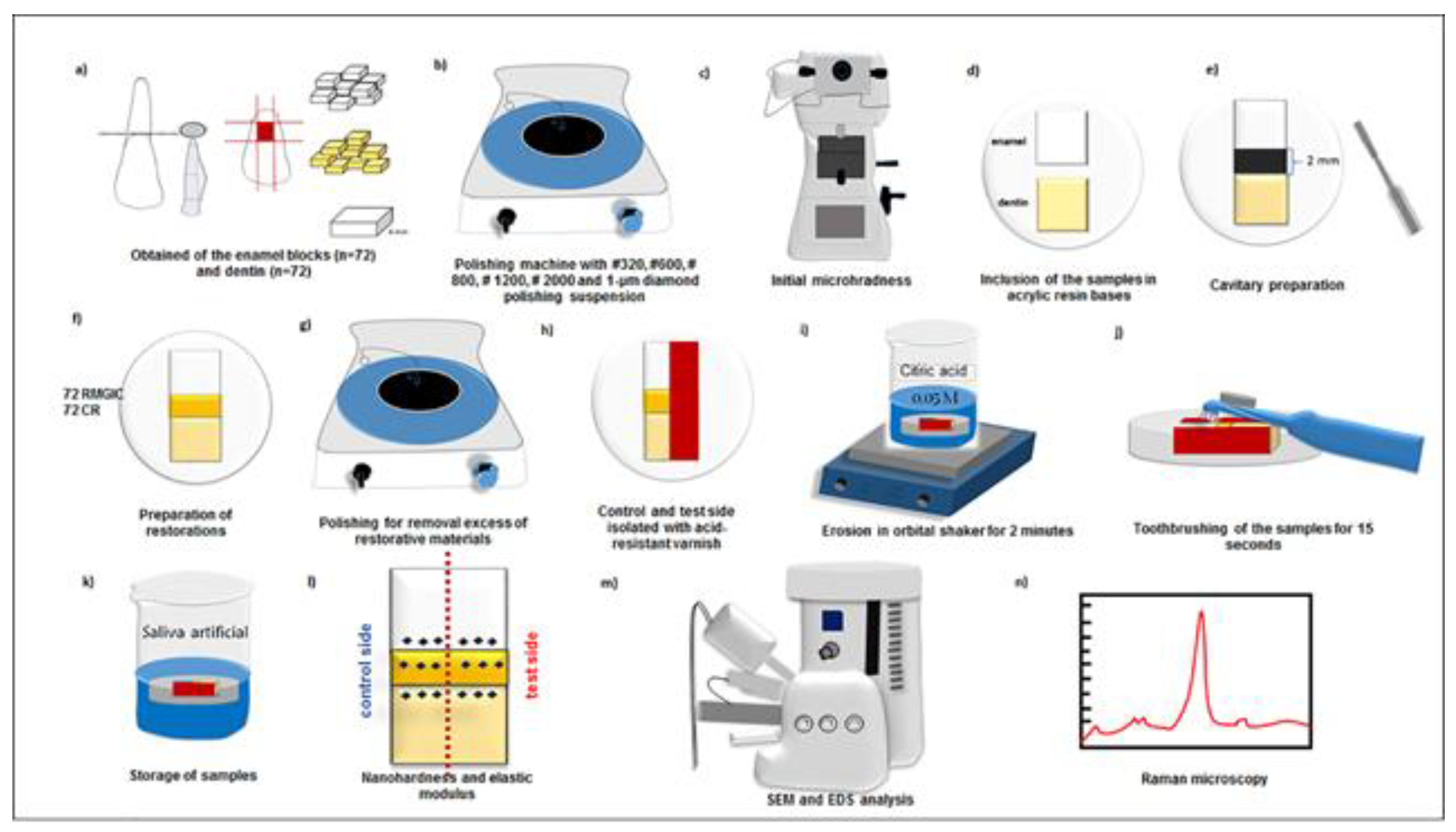
Figure 2.
Raman spectroscopy of control and eroded enamel with phosphate peak at 960 cm-1 and carbonate peak at 1070 cm-1. A) ERMGIC-C; B) ERMGIC-E; C) ECR-C; D) ECR-E.
Figure 2.
Raman spectroscopy of control and eroded enamel with phosphate peak at 960 cm-1 and carbonate peak at 1070 cm-1. A) ERMGIC-C; B) ERMGIC-E; C) ECR-C; D) ECR-E.
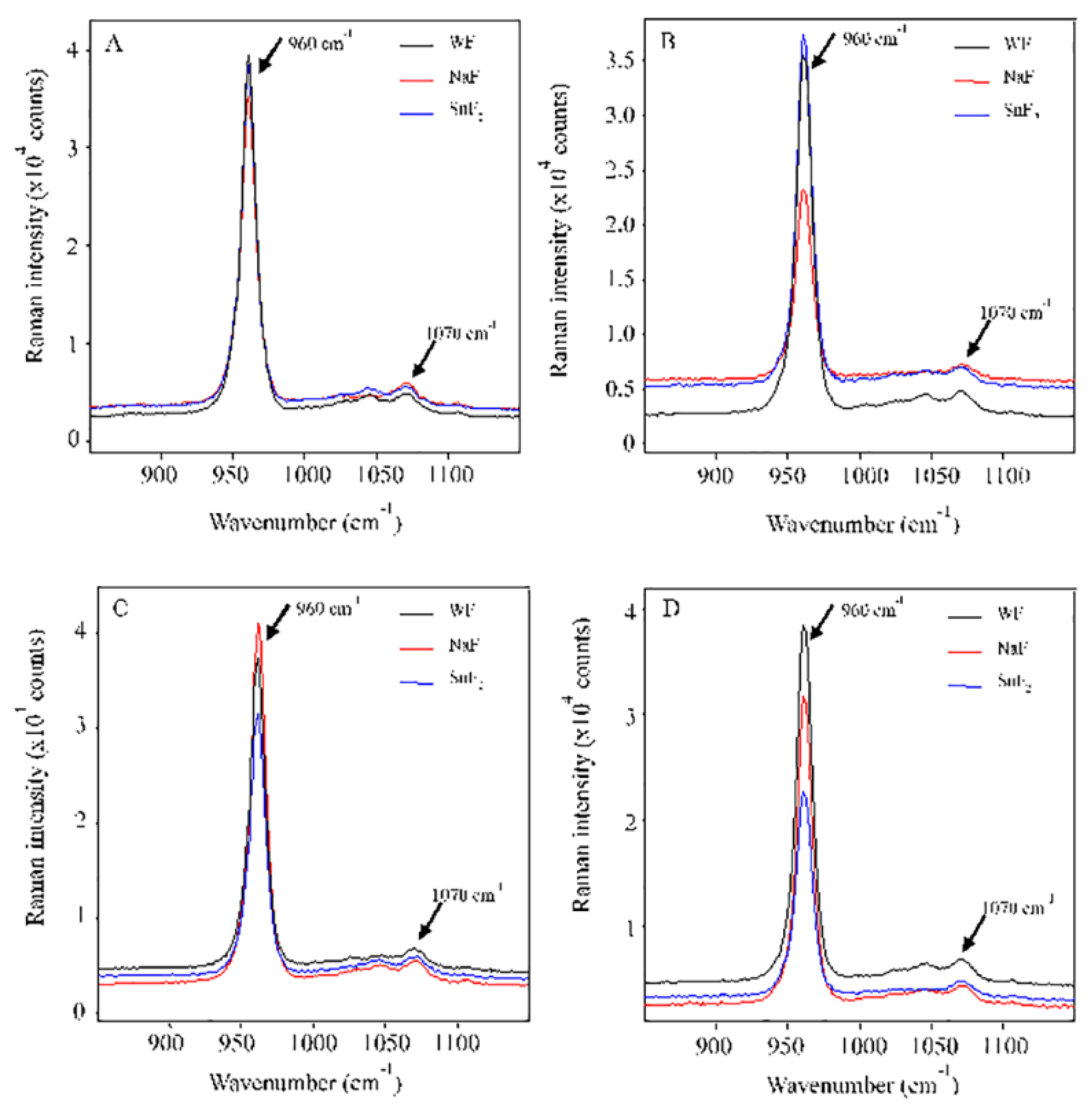
Figure 3.
Raman spectroscopy of control and eroded dentin with phosphate peak at 960 cm-1 and carbonate peak at 1070 cm-1. E) DRMGIC-C; F) DRMGIC-E; G) DCR-C; H) DCR-E.
Figure 3.
Raman spectroscopy of control and eroded dentin with phosphate peak at 960 cm-1 and carbonate peak at 1070 cm-1. E) DRMGIC-C; F) DRMGIC-E; G) DCR-C; H) DCR-E.
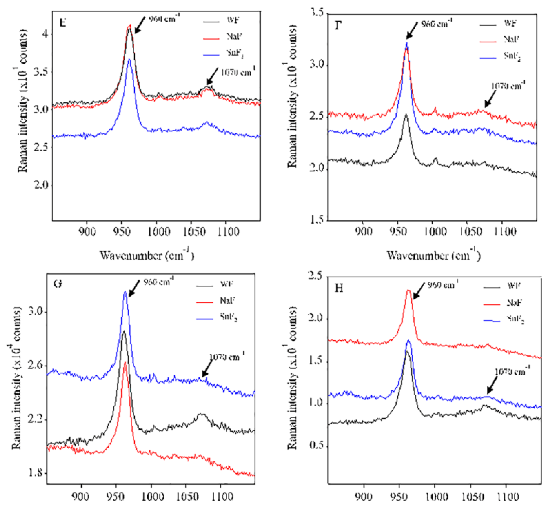
Figure 4.
Representative SEM images of eroded surfaces (5000×). A) Eroded enamel surface brushing with toothpaste WF toothpaste presented roughness. B) Eroded enamel surface brushing with NaF toothpaste presented roughness. C) Eroded enamel surface brushing with SnF2 toothpaste having mineral precipitation. D) Eroded dentin surface brushing with toothpaste WF, large dentinal tubules and presence of odontoblast processes. E) Eroded dentin surface brushing with NaF showed partial obliteration of the dentinal tubules. F) Eroded dentin surface brushing with SnF2 also presented partial obliteration of the dentinal tubules. G) RMGIC-E surface brushing with toothpaste WF showed some cracks. H) RMGIC-E surface brushing with NaF presented irregularities. I) RMGIC-E surface brushing with SnF2 showed cracks and concavities. J) CR-E surface brushing with toothpaste WF without alterations. K) CR-E surface brushing with NaF without alterations. (L) CR-E surface brushing with SnF2 showed grooves.


Table 1.
Materials used in this study.
| Material. | Application mode | Composition | Manufacturer |
|---|---|---|---|
|
Adper Single Bond 2 (Adhesive system) |
Apply one layer of adhesive, wait for 20 s, air stream for 5 s, and polymerize for 10 s | Bis-GMA, HEMA, dimethacrylates, ethanol, water, a novel photoinitiator system and a methacrylate functional copolymer of polyacrylic and polyitaconic acids | 3M ESPE, St. Paul, MN, USA. |
|
Filtek Z350 XT (color A2B) Batch: 672912 |
Apply increments of 2 mm and polymerize for 20 s each | Bis-GMA, UDMA, Bis-EMA, TEGDMA, PEGDMA, Zirconia and agglomerates of silica, camphorquinone | 3M ESPE, St. Paul, MN, USA. |
|
Fuji II LC (color A3) Batch: 17051316 |
GC conditioner was applied for 20 s, rinsed and dried for 10 s. 1 level scoop of powder to 2 drops of liquid was dispensed and mixed for 15-20 s. The mixture was transferred to the centrix syringe | Powder: fluor-amino-silicate glass. Liquid: aqueous solution of polycarboxylic acid, TEGDMA and HEMA | GC, Tokyo, Japan. |
|
Curaprox Enzycal Zero (RDA-60)* Batch: 442MHDEXP1121 |
Fluoride-free Toothpaste (WF) |
Water, Sorbitol, Hydrated Silica, Glycerin, Steareth-20, Titanium Dioxide (Cl 77891), Flavor, Sodium Phosphate, Carrageenan, Sodium Chloride, Citric Acid, Sodium Benzoate, Potassium Thiocyanate, Glucose Oxidase, Amyloglucosidase, Lactoperoxidase | Trybol, Neuhausen am Rheinfall, Swiss. |
|
Colgate Total 12 (RDA-70/80)* Batch: 6184BR121R |
Sodium Fluoride Toothpaste (NaF) |
Sodium Fluoride (1450 ppm as NaF) Water, Triclosan, Sorbitol, Silica, Sodium Lauryl Sulfate, PMV / MA Copolymer, Sodium Hydroxide, Saccharin Sodium, Titanium Dioxide |
Colgate-Palmolive, São Bernardo do Campo, SP, Brazil. |
|
Crest Pro-Health (RDA-155)* Batch: 6039GF |
Stannous Fluoride Toothpaste (SnF2) |
Stannous fluoride (1100 ppm F as SnF2) Glycerin, Hydrated Silica, Sodium Hexametaphosphate, Propylene Glycol, PEG 6, Water, Zinc Lactate, Trisodium Phosphate, Sodium Lauryl Sulfate, Sodium Lauryl Sulfate, Carrageenan, Sodium Saccharin, Xanthan Gum, Blue 1 |
Procter & Gamble, Cincinnati, OH, USA. |
Table 2.
Nanohardness values of surfaces and restorative materials using different toothpastes. Mean (SD) H values expressed in GPa.
Table 2.
Nanohardness values of surfaces and restorative materials using different toothpastes. Mean (SD) H values expressed in GPa.
| Factors | ERMGIC-C | ECR-C | RMGIC-C | CR-C | DRMGIC-C | DCR-C |
| WF | 2.97 (0.45) Aa | 2.66 (0.40) Ab | 0.47 (0.20) Ab | 0.69 (0.12) Aa | 0.68 (0.15) Aa | 0.63 (0.10) Aa |
| NaF | 2.89 (0.73) Aa | 2.96 (0.43) Aa | 0.41 (0.19) Ab | 0.67 (0.17) Aa | 0.59 (0.12) Ba | 0.61 (0.15) Aa |
| SnF2 | 3.09 (0.83) Aa | 2.98 (0.63) Aa | 0.49 (0.21) Ab | 0.70 (0.21) Aa | 0.65 (0.13) Aba | 0.67 (0.15) Aa |
| Factors | ERMGIC-E | ECR-E | RMGIC-E | CR-E | DRMGIC-E | DCR-E |
| WF | 0.51 (0.17) Aa* | 0.55 (0.22) Aa* | 0.29 (0.09) Ab* | 0.64 (0.08) Aa | 0.05 (0.02) Aa* | 0.10 (0.05) Aa* |
| NaF | 0.52 (0.24) Aa* | 0.50 (0.30) Aa* | 0.34 (0.16) Ab | 0.65 (0.18) Aa | 0.08 (0.04) Aa* | 0.06 (0.02) Aa* |
| SnF2 | 0.27 (0.07) Aa* | 0.23 (0.06) Aa* | 0.25 (0.14) Ab* | 0.63 (0.11) Aa | 0.08 (0.03) Aa* | 0.07 (0.02) Aa* |
| Upper case letters compare toothpastes in each control or eroded side. Lowercase letters compare surfaces separately (p < 0.05). *Statistical difference among the control and eroded surfaces. SD, standard deviation; H, nanohardness; GPa, gigapascal. | ||||||
Table 3.
Elastic modulus of surfaces and restorative materials using different toothpastes. Mean (SD) Er values expressed in GPa.
Table 3.
Elastic modulus of surfaces and restorative materials using different toothpastes. Mean (SD) Er values expressed in GPa.
| Factors | ERMGIC-C | ECR-C | RMGIC-C | CR-C | DRMGIC-C | DCR-C |
| WF | 79.92 (7.45) Aa | 64.74 (7.14) Bb | 12.99 (3.38) Aa | 13.60 (1.56) Aa | 21.34 (3.56) Aa | 18.70 (2.53) Aa |
| NaF | 76.25 (14.51) Ba | 82.37 (8.08) Aa | 13.50 (3.07) Aa | 13.31 (2.15) Aa | 18.62 (2.97) Aa | 18.26 (2.57) Aa |
| SnF2 | 87.73 (14.86) Aa | 75.22 (8.68) Aa | 14.71 (4.26) Aa | 14.26 (2.15) Aa | 19.08 (3.90) Aa | 19.70 (3.23) Aa |
| Factors | ERMGIC-E | ECR-E | RMGIC-E | CR-E | DRMGIC-E | DCR-E |
| WF | 30.23 (8.63) Aa* | 17.08 (8.22) Bb* | 10.18 (2.37) Ab* | 13.45 (1.51) Aa | 1.54 (0.40) Aa* | 2.65 (0.84) Aa* |
| NaF | 34.52 (12.91) Aa* | 34.59 (8.83) Aa* | 10.38 (3.55) Ab* | 13.52 (2.11) Aa | 1.97 (0.61) Aa* | 2.06 (0.79) Aa* |
| SnF2 | 20.72 (9.90) Ba* | 19.33 (3.26) Ba* | 6.47 (1.14) Bb* | 13.62 (1.50) Aa | 2.36 (0.66) Aa* | 1.99 (0.40) Aa* |
| Upper case letters compare toothpastes in each control or eroded side. Lowercase letters compare surfaces separately (p < 0.05). *Statistical difference among the control and eroded surfaces. SD, standard deviation; Er, elastic modulus; GPa, gigapascal | ||||||
Table 4.
Mean (SD) calcium/phosphorus ratios in enamel surfaces by EDS analysis.
| Factors | ERMGIC-C | ERMGIC –E | ECR-C | ECR-E |
| WF | 1.80 (0.10) Aa | 1.78 (0.12) Aa | 1.79 (0.10) Aa | 1.80 (0.12) Aa |
| NaF | 1.75 (0.14) Aa | 1.78 (0.16) Aa | 1.80 (0.10) Aa | 1.87 (0.08) Aa |
| SnF2 | 1.81 (0.02) Aa | 1.71 (0.09) Aa | 1.75 (0.12) Aa | 1.77 (0.09) Aa |
| Upper case letters compare toothpastes in each surface. Lowercase letters compare surfaces in each toothpaste (p < 0.05). SD, standard deviation. | ||||
Table 5.
Mean (SD) calcium/phosphorus ratios in dentin surfaces by EDS analysis.
| Factors | DRMGIC-C | DRMGIC –E | DCR-C | DCR-E |
| WF | 1.74 (0.08) Aa | 0.53 (0.83) Ab | 1.71 (0.09) Aa | 0.62 (0.96) Bb |
| NaF | 1.68 (0.08) Aa | 1.12 (0.87) Aa | 1.95 (0.35) Aa | 1.77 (0.12) Aa |
| SnF2 | 1.70 (0.08) Aa | 0.53 (0.81) Aa | 1.74 (0.10) Aa | 1.22 (0.95) ABab |
| Upper case letters compare toothpastes in each surface. Lowercase letters compare surfaces in each toothpaste (p < 0.05). SD, standard deviation. | ||||
Table 6.
Mean (SD) carbonate/phosphate ratios in enamel surfaces by Raman analysis (200 × 200 µm).
| Factors | ERMGIC-C | ERMGIC –E | ECR-C | ECR-E |
| WF | 0.06 (0.08) Aa | 0.04 (0.01) Aa | 0.04 (0.01) Aa | 0.04 (0.01) Aa |
| NaF | 0.05 (0.01) Aa | 0.05 (0.02) Aa | 0.03 (0.01) Aa | 0.04 (0.01) Aa |
| SnF2 | 0.04 (0.02) Aa | 0.08 (0.13) Aa | 0.04 (0.01) Aa | 0.04 (0.02) Aa |
| Upper case letters compare toothpastes in each surface. Lowercase letters compare surfaces in each toothpaste (p < 0.05). SD, standard deviation. | ||||
Table 7.
Mean (SD) carbonate/phosphate ratios in enamel surfaces by Raman analysis (200 × 200 µm).
| Factors | DRMGIC-C | DRMGIC –E | DCR-C | DCR-E |
| WF | 0.33 (0.20) Bb | 0.35 (0.05) Aab | 0.42 (0.07) Aa | 0.29 (0.04) Ab |
| NaF | 0.42 (0.06) Aa | 0.36 (0.08) Aab | 0.39 (0.04) Aa | 0.26 (0.08) Ab |
| SnF2 | 0.45 (0.07) Aa | 0.31 (0.08) Ab | 0.42 (0.06) Aa | 0.25 (0.09) Ab |
| Upper case letters compare toothpastes in each surface. Lowercase letters compare surfaces in each toothpaste (p < 0.05). SD, standard deviation. | ||||
Disclaimer/Publisher’s Note: The statements, opinions and data contained in all publications are solely those of the individual author(s) and contributor(s) and not of MDPI and/or the editor(s). MDPI and/or the editor(s) disclaim responsibility for any injury to people or property resulting from any ideas, methods, instructions or products referred to in the content. |
© 2023 by the authors. Licensee MDPI, Basel, Switzerland. This article is an open access article distributed under the terms and conditions of the Creative Commons Attribution (CC BY) license (http://creativecommons.org/licenses/by/4.0/).
Copyright: This open access article is published under a Creative Commons CC BY 4.0 license, which permit the free download, distribution, and reuse, provided that the author and preprint are cited in any reuse.


