
Submitted:
08 May 2023
Posted:
09 May 2023
You are already at the latest version
Abstract
Osmotic demyelination syndrome (ODS) is an acute demyelinating disorder characterized by the loss of myelin in the center of the basis pons which is defined as central pontine myelinolysis (CPM) and demyelination process in locations outside the pons which is defined as extrapontine myelinolysis (EPM). ODS including CPM and EPM is mainly caused by the rapid correction of hyponatremia. However, there are several reports of ODS in medical condition such as malnutrition, alcoholism, liver transplantation, malignancy, sepsis, electrolyte imbalance including hypernatremia, hypokalemia, hypophosphatemia, and chronic illness. Rarely, ODS was reported in patients with hyperammonemia or hyperbilirubinemia without sodium fluctuations. The symptoms usually correspond to the lesion site and EPM precedes CPM. Because ODS may be irreversible, prevention is more important. Here, we report a case of ODS secondary to the hyperammonemia and hyperbilirubinemia, as it can have unfavorable prognosis.
Keywords:
osmotic demyelination syndrome
; hyperammonemia
; hyperbilirubinemia
1. Introduction
Osmotic demyelination syndrome (ODS) is an acute demyelinating disorder in the central nerve system, which is classified into three different types according location of lesions. First, central pontine myelinolysis (CPM) occur due to an unique demyelination in the central pons. Second, neurological disorder that occur due to destruction of myelin in locations outside the pons is defined as extrapontine myelinolysis (EPM). Third type is when both CPM and EPM occur simutaneously [1].
Both CPM and EPM share common histology and pathogenesis. They commonly occur in patients with chronic hyponatremia. Some case reports showed that ODS is associated with liver transplantation, hypernatremia, hypokalemia, hypophosphatemia, hemodialysis, refeeding syndrome, malnutrition, diabetes mellitus, adrenal insufficiency, leukemia, lymphoma, systemic lupus erythematous, acquired immunodeficiency syndrome, and sepsis. ODS is commonly caused by rapid hyponatremia correction during only a few hours, but it can rarely occur when hyperbilirubinemia or hyperammonemia is corrected rapidly within normal serum sodium concentrations [1,2,3].
Here, we reports the case of ODS occurrence in patients with rapid correction of hyperammonemia and hyperbilirubinemia without changes in serum sodium level.
2. Case presentation
A 34-year-old man presented with loss of consciousness and admitted to our hospital. His initial blood pressure was 124/65 mmHg. He had no family history and past history, but drank 3 bottles of alcohol per day. On neurological examination, his consciousness was stupor, and he showed an avoidance response to the pain stimulus. Another neurologic deficit was not observed.
Initial blood test results were total billirubin 9.09 mg/dL, direct bilirubin 4.75 mg/dL, aspartate aminotransferase 2293 U/L, aspartate aminotransferase 304 U/L, alkaline phosphatase 64 U/L, gamma-glutamyl transpeptidase 276 U/L, ammonia 432 ug/dL, protein 4.8 g/dL, albumin 1.9 g/dL, amylase 730 U/L, lipase 73 U/L, blood urea nitrogen 15 mg/dL, creatinine 1.04 mg/dL, prothrombin time 62.7 sec and electrolyte levels were normal. International normalized ratio was 3.18. In addition, cerebrospinal fluid tests with immunoglobulin G index and aquaporin-4 antibody results were within the reference ranges. Moderate to severe ascities were observed in abdominal computed tomography. Initial brain magnetic resonance imaging (MRI) did not observe abnormal lesion. We diagnosed liver cirrhosis which is classfied as Child-Pugh class C. Because lateralizing and localizing sign were not observed, it was also considered that metabolic encephalopathy due to liver failure.
The ammonia level was initially controlled with lactulose enema, and the level was normal range on the third day of hospitalization. However, there was still no change in consciousness state. Also, progressive spastic quadriparesis and ophthalmoplegia was observed. Electroencephalography (EEG) results were continuous slow and generalized, and follow up brain MRI on the 14th day showed high signal intensity at bilateral caudate nucleus, putamen, thalami, midbrain, central pons and bilateral middle cerebellar peduncle on T2-weighted MRI (Figure 1). The lesion with no mass effect showed hypointense on T1-weighted MRI. Contrast enhancement is not seen in lesions. These were all new findings compared to prior brain MRI during the same admission. During these periods, electrolyte levels including sodium level maintained in normal range. It was diagnosed that the ODS was secondary to rapid correction of hyperammonemia and hyperbilirubinemia without changes in serum sodium level. Consciousness was still stupor and hyperbilirubinemia was shown during conservative manegement. The guardians did not agree to additional hemodyalysis and the transplantation of the liver. Eventually, he died on the 76th day of hospitalization due to severe metabolic acidosis.
3. Discussion and Review of Literature
The current case shows a patients with ODS that occurred at bilateral caudate nucleus, putamen, thalami, midbrain, central pons and bilateral middle cerebellar peduncle due to rapid correction of hyperammonemia and hyperbilirubinemia without changes in serum sodium level.
ODS is rare and reported within 1% of patients admitted to a neurology department and 0.06% of all admissions to general medical department. ODS in adult is more common than it is in children. ODS has a similar prevalance in both males and females. In general, ODS may present with a wide range of symptoms, including progressive spastic quadriparesis, pseudobulbar palsy, pseudobulbar affect, dysarthria, dysphagia, ophthalmoplegia, ataxia, nystagmus, and cranial nerve palsies. Before presenting the symptoms of ODS, patients may have neurological symptoms of encephalopathy. These symptoms usually correct following initial correction of electrolytes. Brain MRI is the recommended diagnostic modality to evaluate ODS including CPM and EPM. Furthermore, sequential brain MRI is recommended to perform over a period of weeks to months to reveal the progression or regression of the demylinating lesion depending on clinical symptoms. ODS is caused by rapid sodium correction of more than 12 mEq/L per day. Rapid correction of hyponatremia results in a rapid change in osmotic gradient, causing moisture in the cell to escape into the extracellular space, and damages to the vascular endothelium cells and glial cells that make up the blood brain barrier. Through the damaged blood-brain barrier, cytokines, lymphocytes, complement, and vasoactive amines, which are inflammatory mediators, flow into the central nervous system, resulting in inflammatory demyelination, which is defined as ODS including CPM and EPM. However, in this case, the blood sodium level was normal range and did not fluctuate during the treatment period [4,5].
To estimate the rarity of case, lethal speed for correction of hyperammonemia, threshold for a lethal level of hyperbilirubinemia, the cases reported in the scientific literature were reviewed. We performed a review of all reported ODS cases from 1960 to 2023 based upon a search of English language literature using Pubmed and Google Scholar databases. The literature search criteria were ODS, CPM, EPM, correction of ammonia, hyperammonemia, and hyperbilirubinemia. Very few cases of ODS, CPM and EPM were reported in patients with rapid correction of hyperammonemia or hyperbilirubinemia, which is summarized in Table 1. In children, most cases of hyperammonemia were caused by genetic abnormalities including OTCD, but most cases in adults were caused by chronic alcoholism. Most of ODS reports on MRI is seen as T2 hyperintensity in the central pons, with sparing of the ventrolateral pons, tegmentum, and corticospinal tracts. Contrast enhancement is usually not seen. Pontine lesions are usually symmetrical and extrapontine lesions are usually bilateral and most commonly observed in cerebellar peduncles, globus pallidus, thalamus, lateral geniculate body, putamen, external and extreme capsule, splenium of corpus callosum, and supratentorial white matter. Rapid correction of hyperammonemia or hyperbilirubinemia developed ODS that both CPM and EPM occur simutaneously. Only one CPM case report due to rapid correction of hyperammonemia or hyperbilirubinemia was identified. In general, EPM was even rarer CPM. Although, the presence of CPM rather than EPM is directly linked to poor prognosis including locked-in syndrome, coma or death, these extensive lesion including bilateral caudate nucleus, putamen, thalami, midbrain, central pons and bilateral middle cerebellar peduncle might be associated with poor prognosis of ODS due to rapid correction of hyperammonemia or hyperbilirubinemia.
The literature has been suggested that speed for correction of ammonia lower than 77.8 μmol /L/day showed non-lethal progenosis. Previous studies have compared the directly calculated changes in osmolarity due to changes in ammonia concentration over a 24-hour period with the safe range of osmolarity change rates due to changes in serum sodium concentration. The resulting osmolarity change rates directly caused by ammonia correction (previous cases: -0.459, -0.220, lower than -0.376 mOsm/L/day and our case: -0.077mOsm/L/day) were found to be very small in absolute value compared to the safe range of osmolarity change rates due to sodium. Therefore, previous studied suggested any other factor rather than osmolarity change rates directly caused by ammonia as the sole cause of ODS existed. The literature has been suggested that ammonia level higher than 204 μmol /L/day showed lethal progenosis. Cells have defense mechanisms to cope with changes in volume caused by alterations in osmolarity. In patients with hyperammonemia, glutamate plays a role in reducing intracellular ammonia by decreasing intracellular space and increasing extracellular space in the brain. Glutamate is an important osmolyte that is mainly released from cells when cell volume needs to be reduced, thereby contributing to the reduction of intracellular osmolarity. In patients with hyperammonemia, the mechanism described above can reduce the intracellular/extracellular concentration difference of glutamate, which may contribute to the disturbance of cell volume regulatory mechanisms [9,10].
Also, The literature review showed that only one case report with bilirubin level of 22.88 μmol /L showed non-lethal progenosis. Younger aged patients with ODS and CPM due to rapid correction of hyperammonemia or hyperbilirubinemia had better prognosis. A poor prognosis has been observed in most adult patients with ODS and CPM due to rapid correction of hyperammonemia or hyperbilirubinemia. Previous reports that severe hyponatremia (less than 115 mEq/L), associated hypokalemia, or low Glasgow Coma Scale score (less than 10) at presentation or ODS following liver transplant are reported to be poor prognostic factors in ODS. Also, the anatomic location and size of the demyelinating lesion are usually not associated with outcome [6]. However, for our case report and literature review, ODS and CPM due to rapid correction of hyperammonemia or hyperbilirubinemia observed to have poor prognosis. In present case and literature review, No effective treatment has been defined for ODS due to rapid correction of hyperammonemia or hyperbilirubinemia. Current management consists of general supportive care.
Although it is rare, ODS may occur in the presence of hyperammonemia or hyperbilirubinemia without rapid correction of sodium level. Hyperammonemia is most commonly associated with liver disease including chronic alcoholism and urea cycle disorders. Ammonia has a small molecular weight (17.03 g/mol) and potent of neurotoxin and does not directly affect osmotic pressure. However, it upregulates aquaporin-4 of astrocytes and penetrate the blood–brain barrier resulting in edema of astrocytes. Sudden correction of ammonia may lead to aquaporin-4 dysfunction similar to chronic neuro inflammatory diseases such as multiple sclerosis and neuromyelitis optica, leading to demyelinating lesions due to osmotic stress. Furthermore, hyperammonemia cause a multitude of neurotoxic effects such as cerebral edema, brain herniation and ultimately death. However, no significant correlation has yet been made between CPM and hyperammonemia [14,15]. In this case, rapid ammonia correction without fluctuation of sodium level was performed on the 3rd day of admission, and extensive lesion including bilateral caudate nucleus, putamen, thalami, midbrain, central pons, and both middle cerebellar peduncle were newly observed in follow up brain MRI on the 14th day. Although lesions affecting the basal ganglia, caudate nucleus, thalamus, cerebellum, and subcortical white matter are frequently observed in hyperammonemic encephalopathy, mainly insula, cigulate gyrus, and extensive cortical lesions are observed in hyperammonemic encephalopathy [16,17]. In regard to the treatment of hyperammonemia, lactulose, rifaximin and hemodialysis are common modalities used to lower ammonia levels, as were used in our patient. Also, alternate forms of treatment are available. For patients specifically with urea cycle disorders, nitrogen scavengers such as sodium benzoate, sodium phenylacetate, and arginine are administered for excretion of ammonia. Preventing toxicity to the brain by means of rapid removal of ammonia is the standard of care for patients presenting with urea cycle cycle disorders. However, for our case and literature review, physicians should be aware that ODS can occur when rapid correction of hyperammonemia is present in patients with liver failure.
Hyperbilirubinemia can also cause ODS. In the early stage of hyperbilirubinemia, secretion of interleukin or vascular endothelial growth factor from microvascular endothelial cells is suppressed. However, when hyperbilirubinemia persists for a long time, the production and secretion of cytokines and nitric oxide are promoted in vascular endothelial cells. As a result, hyperbilirubinemia weakens the blood-brain barrier by causing damage to vascular endothelial cells, then leading to ODS including CPM and EPM [7,18]. Previous report showed that high-dose methylprednisolone was initiated in an attempt to improve the integrity of the BBB by blocking inflammatory mediators. However, there was no clinical improvement. Furthermore, previous report have demonstrated that plasma exchange improves clinical outcomes in patients with ODS due to hyperbilirubinemia. However, the family of our patient did not want to treat plasma exchange.
In this case, continuous hyperbilirubinemia was observed during the treatment period, a new lesion suggesting ODS that was not observed in previous MRI. Although serum sodium level was continuously normal range and hyperammonemia was only found in the early stages of treatment, ODS caused by continuous hyperbilirubinemia could be suspected after identifying progressive spastic quadriparesis and ophthalmoplegia.
Although the change of sodium level is not significant, ODS can occur in patients with liver failure due to rapid correction of hyperammoniaemia or severe hyperbilirubinemia. Therefore, physicians pay attension to prevent occurrence of ODS during the treatment of patients with liver failure. As this was a single case report and literature review, cross-sectional retrospective observational studies are needed to accumulate further knowledge for ODS due to rapid correction of hyperammonemia or hyperbilirubinemia.
4. Conclusions
ODS due to rapid correction of hyperammonemia or hyperbilirubinemia is uncommon disease in adult and children, is multifactorial, and has poor prognosis. Physicians should be aware that ODS can occur when rapid correction of hyperammonemia or hyperbilirubinemia is present in patients with liver failure. Further studies are needed to accumulate further knowledge for ODS due to rapid correction of hyperammonemia or hyperbilirubinemia and to improve outcome of ODS.
Author Contributions
Conceptualization: Kunwoo Park, Sang-Beom Kim, Sung Sang Yoon, Ho Geol Woo; Data curation: Kunwoo Park, Sang-Beom Kim, Sung Sang Yoon, Ho Geol Woo; Formal analysis: Kunwoo Park, Ho Geol Woo; Investigation: Kunwoo Park, Ho Geol Woo; Methodology: Kunwoo Park, Ho Geol Woo; Project administration: Ho Geol Woo; Resources: Kunwoo Park, Ho Geol Woo; Software: Kunwoo Park, Ho Geol Woo; Supervision: Sang-Beom Kim, Sung Sang Yoon; Visualization: Kunwoo Park, Ho Geol Woo; Writing—original draft: Kunwoo Park, Ho Geol Woo; Writing—review & editing: Sang-Beom Kim, Sung Sang Yoon, Ho Geol Woo
Funding
This research received no external funding
Institutional Review Board Statement
Not applicable. The study was conducted according to the tenets of the Declaration of Helsinki. This was a CASE REPORT without any type of experimental intervention, and ethical approval was therefore waived.
Informed Consent Statement
Written informed consent has been obtained from the patient.
Data Availability Statement
Not applicable.
Acknowledgments
Not applicable.
Conflicts of Interest
The authors declare no conflict of interest.
References
- Bansal, L.R.; Zinkus, T. Osmotic Demyelination Syndrome in Children. Pediatr Neurol 2019, 97, 12–17. [Google Scholar] [CrossRef] [PubMed]
- Shah, M.K.; Mandayam, S.; Adrogué, H.J. Osmotic Demyelination Unrelated to Hyponatremia. American Journal of Kidney Diseases 2018, 71, 436–440. [Google Scholar] [CrossRef] [PubMed]
- Kumar, S.; Fowler, M.; Gonzalez-Toledo, E.; Jaffe, S.L. Central pontine myelinolysis, an update. Neurol Res 2006, 28, 360–366. [Google Scholar] [CrossRef] [PubMed]
- Burcar, P.J.; Norenberg, M.D.; Yarnell, P.R. Hyponatremia and central pontine myelinolysis. Neurology 1977, 27, 223–226. [Google Scholar] [CrossRef] [PubMed]
- Kleinschmidt-DeMasters, B.K.; Norenberg, M.D. Rapid correction of hyponatremia causes demyelination: relation to central pontine myelinolysis. Science 1981, 211, 1068–1070. [Google Scholar] [CrossRef] [PubMed]
- Singh, T.D.; Fugate, J.E.; Rabinstein, A.A. Central pontine and extrapontine myelinolysis: a systematic review. Eur J Neurol 2014, 21, 1443–1450. [Google Scholar] [CrossRef] [PubMed]
- Purdy, K.; Anderson, D.; Camicioli, R.; Khadaroo, R.G. Can osmotic demyelination syndrome be a complication of liver failure? eNeurologicalSci 2020, 18, 100223. [Google Scholar] [CrossRef] [PubMed]
- Rosenberg, C.; Rhodes, M. Hyperammonemia in the setting of Roux-en-Y gastric bypass presenting with osmotic demyelination syndrome. J Community Hosp Intern Med Perspect 2021, 11, 708–712. [Google Scholar] [CrossRef] [PubMed]
- Cardenas, J.F.; Bodensteiner, J.B. Osmotic demyelination syndrome as a consequence of treating hyperammonemia in a patient with ornithine transcarbamylase deficiency. J Child Neurol 2009, 24, 884–886. [Google Scholar] [CrossRef] [PubMed]
- Tanaka, Y.; Matsuki, M.; Furukawa, R.; Nakata, W.; Sakurai, Y.; Ajihara, S.; Kawano, A.; Mori, H. Imaging of extrapontine myelinolysis preceding central pontine myelinolysis in a case of ornithine transcarbamylase deficiency with hyperammonaemia and hypokalaemia. BMJ Neurol Open 2022, 4, e000354. [Google Scholar] [CrossRef] [PubMed]
- Kim, Y.; Oh, J.H.; Bae, D.-W.; Kim, S. A Case of Central Pontine Myelinolysis Caused by Hyperbilirubinemia in Hepatic Encephalopathy without Significant Osmotic Stress. J Korean Neurol Assoc 2022, 40, 192–194. [Google Scholar] [CrossRef]
- Mattson, L.R.; Lindor, N.M.; Goldman, D.H.; Goodwin, J.T.; Groover, R.V.; Vockley, J. Central pontine myelinolysis as a complication of partial ornithine carbamoyl transferase deficiency. American journal of medical genetics 1995, 60, 210–213. [Google Scholar] [CrossRef] [PubMed]
- Ho, K.C.; Hodach, R.; Varma, R.; Thorsteinson, V.; Hess, T.; Dale, D. Kernicterus and central pontine myelinolysis in a 14-year-old boy with fulminating viral hepatitis. Annals of neurology 1980, 8, 633–636. [Google Scholar] [CrossRef] [PubMed]
- Rama Rao, K.V.; Jayakumar, A.R.; Norenberg, M.D. Induction of the mitochondrial permeability transition in cultured astrocytes by glutamine. Neurochem Int 2003, 43, 517–523. [Google Scholar] [CrossRef] [PubMed]
- Popescu, B.F.; Bunyan, R.F.; Guo, Y.; Parisi, J.E.; Lennon, V.A.; Lucchinetti, C.F. Evidence of aquaporin involvement in human central pontine myelinolysis. Acta Neuropathol Commun 2013, 1, 40. [Google Scholar] [CrossRef] [PubMed]
- Miller, G.M.; Baker, H.L., Jr.; Okazaki, H.; Whisnant, J.P. Central pontine myelinolysis and its imitators: MR findings. Radiology 1988, 168, 795–802. [Google Scholar] [CrossRef] [PubMed]
- Jeon, S.J.; Choi, S.S.; Kim, H.Y.; Yu, I.K. Acute Acquired Metabolic Encephalopathy Based on Diffusion MRI. Korean J Radiol 2021, 22, 2034–2051. [Google Scholar] [CrossRef] [PubMed]
- Palmela, I.; Cardoso, F.L.; Bernas, M.; Correia, L.; Vaz, A.R.; Silva, R.F.; Fernandes, A.; Kim, K.S.; Brites, D.; Brito, M.A. Elevated levels of bilirubin and long-term exposure impair human brain microvascular endothelial cell integrity. Current neurovascular research 2011, 8, 153–169. [Google Scholar] [CrossRef] [PubMed]
Figure 1.
Osmotic demyelination syndrome at (A) bilateral caudate nucleus, putamen, thalami, (B) midbrain, (C) central pons and (D) bilateral middle cerebellar peduncle on T2-weighted magnetic resonance imaging in brain on the 14th day after rapid correction of hyperammonemia and hyperbilirubinemia.
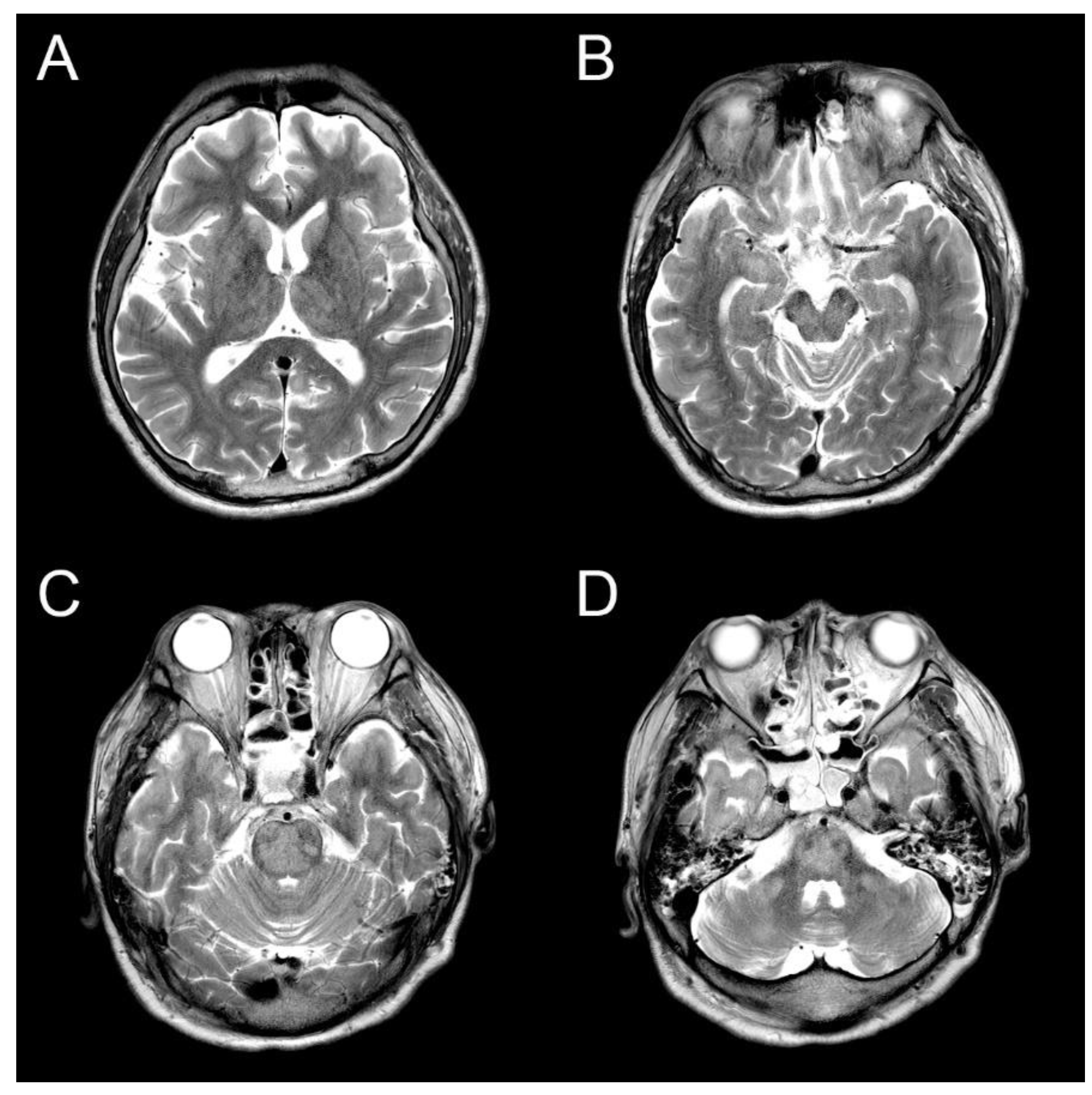

Table 1.
Other cases of central pontine myelinolysis due to hyperammonemia or hyperbilirubinemia reported.
Table 1.
Other cases of central pontine myelinolysis due to hyperammonemia or hyperbilirubinemia reported.
| Ref | Age/ Sex |
Clinical feature |
Ammonia (μmol /L/) / Correction rate (μmol /L/day) |
TB (μmol /L) |
MRI | Treatment | Associated disease | Survival |
|---|---|---|---|---|---|---|---|---|
| 1[7] | 30/M | Coma | Normal | 955 | EPM/CPM | Hemodialysis, steroid | Primary sclerosing cholangitis | No |
| 2[8] | 58/F | Status epilepticus | 290/87.5 | NA | EPM/CPM | Lactulose, rifaximin, hemodialysis | Abuse alcohol, opioid History of RYGB |
No |
| 3[9] | 7/F | Confusion | 508/475 | NA | EPM/CPM | sodium benzoate, sodium phenylacetate | OTCD, ADHD | Yes, but sequelae |
| 4[10] | 2-3/F | Somnolence | 555/430 | NA | EPM/CPM | Liver transplantation, Lactulose, hemodialysis, sodium benzoate, sodium phenylacetate | OTCD, Liver failure | Yes, but sequelae |
| 5[11] | 56/M | Coma | 204/77.8 | 22.88 | EPM/CPM | Lactulose | Chronic alcoholism | Yes |
| 6[12] | 5/F | Confusion | 376/NA | NA | EPM/CPM | sodium benzoate, sodium phenylacetate | OTCD | Yes, but sequelae |
| 7[13] | 14/M | semicoma | Normal | 1046 | CPM | NA | NA | No |
| Our case |
34/M | Stupor | 253.7/80 | 9.09 | EPM/CPM | Lactulose enema | Chronic alcoholism | No |
Ref: reference, EPM: Extra pontine myelinolysis, CPM: Central pontine myelinolysis, NA: Not applicable, OTCD: Ornithine Transcarboamylase deficiency, RYGB Roux-en-Y gastric bypass TB: Total bilirubin.
Disclaimer/Publisher’s Note: The statements, opinions and data contained in all publications are solely those of the individual author(s) and contributor(s) and not of MDPI and/or the editor(s). MDPI and/or the editor(s) disclaim responsibility for any injury to people or property resulting from any ideas, methods, instructions or products referred to in the content. |
© 2023 by the authors. Licensee MDPI, Basel, Switzerland. This article is an open access article distributed under the terms and conditions of the Creative Commons Attribution (CC BY) license (http://creativecommons.org/licenses/by/4.0/).
Copyright: This open access article is published under a Creative Commons CC BY 4.0 license, which permit the free download, distribution, and reuse, provided that the author and preprint are cited in any reuse.


