
Submitted:
13 April 2023
Posted:
14 April 2023
You are already at the latest version
Abstract
A variational analysis of dispersion medium (electrolytes and surfactants) was carried out for the best nucleotide interactions with the presence of nanoparticles and the ability to formation of homogeneous coatings upon drying / lyophilization. Using UV-spectroscopy and transmission electron microscopy, the processes of immobilization of nucleotides on the surface of nanoparti-cles of the composition ZrO2-3mol% Y2O3 (YSZ) in the presence of various dispersing agents were investigated. Promising dispersing agents were established, the optimal composition of suspen-sions based on DNA nucleotides and YSZ nanoparticles, which is potentially suitable for adaptive technologies of bionanoelectronics, was selected. The aim of the work is to obtain and study functional media for molecular electronics based on heterojunctions biological molecule – wide-gap dielectric.
Keywords:
immobilization of DNA molecules on the surface of dielectrics
; interaction of nanopowders with nucleotides
; molecular electronics
; nanotechnology
; molecular dynamics modeling
; DNA conformation in an electric field
; genotoxic effects
1. Introduction
Interdisciplinary research in the field of nanotechnology has a breakthrough potential and the main hopes of nanoscale technologies are associated with new effects at interface of chemistry, physics and biology. At the present time nanobioelectronics is rapidly developing [1]. The combination of biomolecules with solid-state nanoparticles gives rise to a new class of materials, primarily for new electronic sensor and optical systems; prospects for the development of molecular electronics, the creation of biochips, memory arrays and computer architectures of the future becomes real.
The DNA molecules are have good electrical conductivity [2,3], capable of storing and transmitting by copying of terabytes of information, reproducing themselves and moving in an electric field, therefore, extremely interesting as a functional element for bioelectronic devices.
Interests to bioelectronic computing are the questions of controlled conformation of DNA molecules. In particular, a controlled change in the secondary structure of DNA molecules, like the unwinding of double helixes (B-Z transition in DNA [4,5]) can be used to replicate and transfer large arrays of data. There is a reason to believe that such conformational effects can be achieved by changing of the electrodynamic parameters of the ionic environment of a DNA by an external electric field. As a polyanion, DNA can be in a close contact with various positively charged ions [6]. At the same time, some of ions are able to interact directly with DNA with formation of stable bonds, both with negatively charged groups of the sugar-phosphate core and with nitrogenous bases [4]. Obviously, by changing the potential or local concentration of such ions, the DNA structure can destabilize and trigger mechanism of the B-Z transition can be controlled launched. Conformational effects are already used in the development of new molecular electronic devices [7,8,9]. The simplest bioelectronic device that can be made based on DNA - molecules or their nitrogenous bases – nucleotides [10] is a biomolecular gate or diode. The principles of constructing such devices are actively discussed in the literature [11,12]. At the present time, “soft” electronic components for biological applications have already been demonstrated: quasi-liquid diodes and memristors based on liquid metals and hydrogels [13]. In case of DNA molecules and nucleotides even nucleotide polarization, is sufficient for functioning of molecular diode or valve (Figure 1, a – b), since the total elongation of molecules will lead to a reduction in interelectrode gap and a sharp increasing in conductivity of biomolecular functional layer in interelectrode space due to the high electrical conductivity of DNA molecules. The closest functional analogue of the molecular valve is a TRIAC (Figure 1, c – d).
As a rule, nanoelectronics at present stage of development presuppose not a scale, but a new principle of operation, often based on a quantum - size effects. In real devices, the architecture of the device itself and the spatial configuration of the structural elements of the functional medium are of decisive importance. For example, in case of nano-ionic capacitors [14,15], this basis is a porous carbon electrode. In the mentioned above bionanoelectronic devices, DNA molecules or their nitrogen bases nucleotides must have localized on some spatially distributed basis and have a certain spatial ordering. In this aspect it is of interest to use as an adsorber material a DNA molecules or nucleotides of spatially ordered ZrO2-based nanoparticle ensembles due to good biocompatibility and high dielectric constant (ε = 25) of this material. A nanodispersed system uniformly distributed in space will be the concentrated basis of the biomolecular functional element associated with the molecule through a capacitive bond [2]. The high dielectric constant , as follows from the formula for the capacitance of the dielectric ball C [16]:
where ε0 = 8.854∙10−12 F/m is the electric constant, R, ϕ = ϕDEL are the radius and potential of the surface of nanoparticles, respectively, which guarantees a high density of the surface charge qS and, as a result, a high efficiency of external influence on DNA molecule.
Indeed, within the framework of model representations of DEL in the form of a planar capacitor [17], it is obvious that the total charge of sorbed DNA molecules, as a specific type of DEL ions, will vary in proportion to the charge of surface qS of particles, only with sign inversion: qS = –qDEL. In the presence of an electrically continuous dispersion medium, for example, an electrolyte, a changing of a charge state of the ions in the nearest environment of the DNA molecule can lead to the conformational effect on it form and can be realized by direct changing of the electric potential of this medium ϕ. According to the mentioned above ideas, the actual scientific and technological problem is production and studying of liquid dispersed systems based on biomolecular functional elements as DNA molecule, biocompatible nanoparticle that can be useful for creating of objects of scientific research and devices of bionanoelectronics element base [18] by using of modern adaptive inkjet and aerosol printing technologies [19]. Obtaining of this solution using ensembled in electrically conductive electrolytes YSZ - nanoparticles, ionic surfactants, and commensurate DNA molecules fragments (nucleotides) was the aim of this work. The main task of the study was to optimize the composition of a dispersed system contained functional elements according to the efficiency of interaction of nucleotides with the surface of nanoparticles and the stability of the entire system to agglomeration both before and after polymerization (lyophilization). The studying of the influence of the amount of Y2O3 impurity (as an activator chemical activity of surface) on the physicochemical processes in the dispersed system under consideration was also included to the work tasks.
2. Experimental
The commercial drug of high polymer sodium salt of deoxyribonucleic acid (Reanol) was used. Acid hydrolysis of DNA was carried out using Schmidt-Tangauser method [20] in a one molar (1M) solution of HClO4. As a electrolytes and surfactants acetic acid (C2H5OH:CH3COOH), nitric acid (HNO3), ethyl alcohol (C2H5OH), potassium hydroxide KOH, sodium lauryl sulfate (C12H25SO4Na) and Cetylpyridinium-N-bromide (C21H38BrN) were used (Table A1 (in Appendix 1)).
The adsorption properties of nucleotides on YSZ powders were investigated under the influence of the next substances: 1) mixture of 96% ethyl alcohol and glacial acetic acid; 2) mixture of ethyl alcohol and acetic acid, diluted in 10 times; 3) aqueous solutions of KOH (10, 25 and 50%); 4) aqueous solutions of HNO3 (2, 5, 10%); 5) sodium lauryl sulfate (SDS) and 6) Cetylpyridinium-N-bromide (CPB). SDS and CPB are surface-active materials that form micelles at a constant of miceate formation Km = 3.84∙10–2 M and 1.3∙10–2 M, respectively. The concentration of DNA nucleotides in the solution was C = 500 mg/ml. Suspensions were homogenized in individual plastic containers immersed in water using an exponential emitter of the USDN-2 unit at a power of 50 W/cm2. In a chromatographic column filled with adsorbent 1 – 5, a V = 1 ml of DNA nucleotides was placed; fractions were mixed with an interval of 5 minutes. The content of nucleotides was determined by the spectrophotometric method (SF-2000) by the absorption of intensity at a wavelength of λ = 269 nm.
Used in the work the 3YSZ hydroxide nanopowders (3YSZ hydroxide, nanoparticle size 4 – 5 nm) were obtained using a co-precipitation of zirconium and yttrium salts (3%mol) with ammonia with followed dehydration of the precipitate in a specialized microwave oven at 120°C. To obtain powders of oxides with a nanoparticle size of 7.5 and 14 nm, the subsequent heat treatment of the hydroxide was used, respectively, at temperatures of 400°C and 700°C for a time t = 2 h yttrium oxide Y2O3 in an amount of 0, 3, 8 mol% was used as an alloying additive in a solid solution based on ZrO2. The distribution of the suspension over the substrate, as an indirect indicator of the dispersion stability of the dispersion system under study, was studied by scanning electron microscopy (SEM) using JSM640LV instrument; the spatial distribution of nanoparticles and nucleotides were investigated by transmission electron microscopy (TEM) using a JEM 200A instrument. A plate made of glass fiber laminate with gold electrodes 1 cm long was used as a substrate. Thickness of electrodes is 2 mm, electrode separation is 2 cm. Drops with a volume of 0.1 ml were formed on a substrate using a pipette dispenser.
Objects for TEM were prepared from suspensions containing DNA fragments using the ultrasonic mist method [21]. The specific surface area was determined using the BET (Brenauer – Emmetat – Taylor) method using a Sorbi 4.1 instrument. The pH of the air-dry state of the nanopowder surface was determined using an Eutech pH-700 pH-meter in distilled water. The ratio of water to powder: 50 ml of water / 20 mg of powder.
Used in the work the set of electrolytes and polar surfactants contains substances with pronounced acidic and basic properties, due to which optimization work to intensifying of the adsorption interaction of DNA nucleotides with the surface of the YSZ nanoparticle system and its spatial homogenization is possible.
3. Results
3.1. The effect of electrolytes and surfactants on the interaction of nanoparticles based on Zirconia with nucleotides.
The hydrolysis of polymeric DNA according to Schmidt-Tangauser passes mainly to nitrogenous bases: adenine, guanine, cytosine and thymine. The extinction coefficient at the maximum of the absorption length of the hydrolyzate at λ = 269 nm is ε = 7200. A typical UV-spectrum of a mixture of DNA nucleotides is presented on Figure 2, a. The results of the analysis are consistent with the literature data [22]. To identify the best conditions for immobilizing of DNA nucleotides on ZrO2 modified by Y2O3, the following groups of experiments were selected with variation of particle size, amount of Y2O3 dopant and degree of structural ordering of the nanoparticle material (Table 1).
As can be seen from the Table 1, the particles used in the work differ significantly in the amount of water in hydrate shell, number and nature of sorption centers, which is caused by the heat treatment mode. The surface of amorphous hydroxide YSZ is formed by both the main Lewis and Brønsted acidic active sites. Heat treatment of hydroxide at 400°C leads to loss of up to 20% by weight hydroxyl cover (ionic atmosphere around the particles), reduction of the specific area of the sorbing surface and change in the type of active centers and transformation of the polymer structure to the crystalline state. Temperature treatment at 700°C leads to annealing of the part of oxygen vacancies and the formation of terminal oxygen atoms, accompanied by the replacement of acid Brønsted centers to Lewis base centers. Thus, the presented set of powders allow to conduct in a number of parameters a comprehensive optimization experiment for the immobilization of DNA nucleotides from the surface of YSZ nanoparticles in the presence of electrolytes and surfactants. The results of investigation of the affect of electrolytes and surfactants on the interaction of nanopowder particles with DNA nucleotides.
3.2. The effect of electrolytes and surfactants to the nanopowder particles with DNA nucleotides interaction.
The typical spectra of spectrophotometric studies of the adsorption interaction of DNA nucleotides with YSZ powders are shown on Figure 2. The results of compared analysis of all combination of interacted components (Table 1) are shown on Figure 3. According to the results of spectrophotometric studies, the adsorption interaction of DNA nucleotides with powders is carried out in the best way in a mixture of ethyl alcohol 96% and glacial acetic acid, diluted 10 times (samples 1 – 3, 14 – 16, 27 – 29, 40 – 41, 44 – 46). Typical spectrum for this group on the example of adsorption of the polymer DNA hydrolyzate and nitrogenous bases with ZrO2 powder 700°C (sample 3) is shown on Figure 2, b.
As can be seen from Figure 2, b, after adding this electrolyte composition the content of nucleotides in the solution decreases in 2.5 times. It can be assumed that polar solvents (water, acetic acid, ethyl alcohol) have an orienting effect on negatively charged nitrogenous bases relative to the ionic atmosphere, the so-called "hydrated shell" of zirconium dioxide nanoparticles. The result of such weak interactions is the appearance of electrostatic forces. The second possible mechanism is the formation of a hydrogen bond between the negatively charged surface of the ZrO2 nanoparticle and the vacant orbital nucleotide (lonely electron pair in nitrogenous bases) in aqueous solutions of acetic acid and alcohol. It is noteworthy that in concentrated acetic acid with alcohol the adsorption of nucleotides is worse than at the presence of water. Those, less polar solvents reduce the nucleophilicity of the reacting particles.
3.3. Interaction with HNO3
In case of varying the concentration of nitric acid, nitro derivatives of purine bases were obtained (samples 7 – 9, 20 – 22, 33 – 35, 48). The spectrum of nucleotides for a group of samples 7 – 9 in the initial state and after interaction with HNO3 with a concentration of 2, 5, 10% is shown of Figure 2, c.
Obviously, if concentration of nitric acid higher, than the nitration reaction completed faster. The diagram of the intensities of spectrophotometric peaks which is characterized the adsorption property of the studied nano-sized Zirconia powders is shown on the Figure 3. As can be seen from Figure 2, c the content of nitro derivatives is increases with an increasing of the concentration of HNO3. It is impossible to determine the degree of sorption interaction of nucleotides with the surface of Zirconia nanoparticles according to spectrophotometry data, since the more intense peak of nitro derivatives at a wavelength of λ = 269 nm covers the peak of a lower intensity of nucleotides at a wavelength of λ = 269 nm.
Figure 3 indicates a positive effect of the adsorption interaction of nucleotides with the acidic medium. It can be assumed that present of acids increased the degree of polarization of nucleotides.
3.4. Interaction with KOH
The result of the interaction of nitrogenous bases with KOH (samples 4 – 6, 17 – 19, 30 – 32, 47) is shown on Figure 2, d and Figure 3. It can be assumed that the nature of the interaction of nanoparticles with DNA is largely determined by the pH of surface of the nanoparticles. In all cases the adsorption interaction of nucleotide bases with nanoparticles was worse than interaction of nanoparticles with dilute solutions of acetic acid and ethyl alcohol. At increasing of KOH concentration from ω = 10, 25, and 50% intensity of adsorption interaction decreases. Adsorption interaction is better in an aqueous diluted solution than in concentrated KOH. Based on the above facts, it can be assumed that such reaction of the system to a reducing agent is caused as by neutralizing the negative charge of DNA nucleotides, which are an anions in nature, or by passivating of the chemically active acid sites on the surface of YSZ nanoparticles.
According to spectrophotometry data (Figure 3, i – l) used in the work ionic surfactants which contain a sulfate group SDS, (samples 10 – 11, 23 – 24, 36 – 37, 49, or CPB (samples 12 – 13, 25 – 26, 38 – 39, 50) do not affect the adsorption properties of the investigated powders.
3.5. Objects morphology
On the example of samples № 1 – 13 (Table 1), the influence of dispersing agents used in the work on the distribution of the resulting suspensions over the substrate after drying is demonstrated on Figure 3. It can be seen that the sample that does not contain fillers – the original nucleotide solution after acid hydrolysis of DNA (Figure 3, a) is fairly uniformly distributed over the substrate except for visible elements, up to 250 nm in size, probably formed as a result of coagulation / spontaneous clumping of the initial solution. Acetic acid based suspensions (Figure 3, b, c) are relatively uniformly distributed throughout the substrate.
According to the pictures on Figure 3, k, l (including insert images), corresponding to the compositions № 12 Molar solution of CPB 2% (k), № 13 Molar solution of CPB 1.3∙10–2 M (l), surfactant CPB works relatively well as a dispersant agent. As can be seen from insert to images Figure 3, i, j, k, l sodium lauryl sulfate in the concentrations used in the work forms a foamy mass during drying and does not provide a sufficient degree of dispersion of the suspension being studied, whereas the CPB works relatively well in a small range of concentrations and forms a dense uniform coating at concentrations of more than 1.3∙10–2 Moles per liter.
A relatively small electrolyte concentration (Figure 3, d 10% KOH) leads to the formation of aggregated particles in a dense homogeneous mass covering them without pronounced morphological features. Increasing of the concentration of salts (more then 10% KOH and all used concentration HNO3) leads to the formation of inhomogeneous film (Figure 3, e, f, g, h) with high concentration of salts and not the higher uniformity of the distribution of the dispersed phase. This conclusion is confirmed by the insert images on Figure 3, e, f, h corresponding the compositions: № 5: aqueous solution of 25% KOH, №7: aqueous solution of 2% HNO3, and № 9: an aqueous solution of 10% HNO3 (h) respectively where as can be seen from insert on Figure 3, h, the bulk of the material has a pronounced crystal cut. These aqueous solutions do not provide the necessary degree of distribution of the suspension over the substrate. It can be concluded that the salt content is dispersing additives of more than 2%, regardless of their effect on the molecular adsorption of nucleotides on the surface of YSZ nanoparticles, are too large to form a functional layer of the desired property.
3.6. The influence of the chemical composition of the material of nanoparticles on the adsorption of nucleotides
The data of spectrophotometric studies with varying percentages of Y2O3 are shown on Figure 4. It can be seen that all of the samples of YSZ nanoparticles, including zirconium hydroxide regardless of the content of Y2O3 (1 – 5, Figure 4) are show a good sorption properties to DNA nucleotides in electrolyte based on 90% solution of acetic acid in alcohol.
Consequently, not only the chemical composition of the dopant, but also the size and structural ordering of the material of the particles do not have a significant effect to the adsorption of nucleotides. This indicates the absence of direct chemical interaction of nucleotides with the surface of nanoparticles. Therefore, the adsorption of nucleotides occurs on the already filled adsorption layer and has the character of physical adsorption. There is a slight (within 15.5%) difference in the adsorption capacity of the particles with an increase in the concentration of alcohol in the acetic acid solution to 50%. According to UV-spectroscopic data shown on Figure 4, in this case the adsorption interaction of the DNA bases with nanoscale powders increases in the line: ZrO2+3%molY2O3 400°C (Figure 4., axis e) > ZrO2+8%molY2O3 700°C (Figure 4., axis b) > ZrO2+0%molY2O3 700°C (Figure 4., axis d) > Zr(OH)2+3%molY2O3 (Figure 4., axis c) > ZrO2+3%molY2O3, 700°C (Figure 4., axis a). Worst of all the adsorption interaction of nucleotides with the surface of nanoparticles took place at the presence of surfactants. In this sense, YSZ hydroxide nanoparticles (3%molY2O3) and solid solution ZrO2+8%molY2O3 700°C are the limiting case. With hydroxide particles, the interaction proceeded worst of all in an alkaline medium (at the presence of KOH in various concentrations). Given the rather large molar mass of the used surfactants (288,38 g / mol in sodium lauryl sulfate and 384,45 g / mol in Cetylpyridinium-N-bromide, respectively), it can be assumed that they block the surface of the nanoparticles, not allowing the nucleotides to react, or replace them with adsorption layer.
The TEM images of typical structural objects of the dispersed systems under study are presented on Figure 5. The aggregate of the initial nanoparticles of the composition ZrO2+3%molY2O3 (700°C) (a), the initial DNA (b), the aggregate of the initial nanoparticles, surrounded by nucleotides formed at the presence of a solution of C2H5OH: CH3COOH 1/10 (c). It can be seen (Figure 5, c) that lamella-like fragments (1) of DNA surround aggregates of nanoparticles (2) with a relatively loose layer. In general, such object can be considered as a non-isometric two-level system with a dielectric core surrounded by a molecular conducting shell.
Based on Figure 5, we can conclude that at a certain density of structural elements of the nanodispersed system under consideration, one can experimentally try to catch the moment at which the percolation point appears on the dependences of electrical properties, near which the polarization of DNA fragments by the electric field will lead to significant changes in the conductivity of the system just like on Figure 1, d. Thus, the microscopic picture indirectly confirms the above-stated assumption about the possibility of using this material as a functional medium for molecular or bionanoelectronics.
4. Conclusions
According to the TEM data, the objects in the volume of the functional layer are a two-level system in the form of rounded aggregates of YSZ nanoparticles surrounded by a loose shell of nucleotides. According to spectroscopic data, DNA fragments in the best way reacted with nanoparticles in a medium of acetic acid (0.5 ml 1/10 solution C2H5OH: CH3COOH + 0.5 ml DNA). Complete blocking of the interaction was observed using ionic surfactants: lauryl sulfate and CPB. According to the SEM data, a surfactant CPB most clearly manifested itself as a dispersing agent in a range of concentration about 1.3∙10–2 Moles per liter. It gives a uniform, dense distribution of the suspension material over the substrate, but at the same time completely blocks the adsorption of nucleotides to the surface of the nanoparticles. A compromise option is to use an alcoholic solution of acetic acid in the range of ratios: C2H5OH: CH3COOH 1/1 – 1/10. This is the only compound that at the same time provides good adsorption of nucleotides on the surface of nanoparticles and a good distribution of the suspension on the substrate after it has dried. Thus, suspensions based on nucleotides localized on the surface of YSZ nanoparticles, obtained using acetic acid in an alcohol solution as a dispersing agent, are potentially suitable for producing functional media for molecular and bionanoelectronic devices when the percolation threshold of the dry layer is reached. Optimization work is required using the technique of measuring the electrical parameters of materials to ascertain the physical conditions of functioning of the resulting functional systems and to develop on their basis experimental samples of a volume-distributed biomolecular diode or controlled valve.
Acknowledgments
The study was performed in the scope of the Project H2020/MSCA/RISE/SSHARE number 871284 project and RO-JINR Projects №.267/2020 item 25 and № 268/2020 item 57, 59. № 366 / 2021 item 84, №.366 / 2021 item 81, No. 367 / 2021 item 27, Poland-JINR cooperation Projects PPPB/168-26/1128/2021, PPPB/120-25/1128/2022, PPPB/120-26/1128/2022, Serbia - JINR cooperation Projects № 178 2021 items 7 and 8.
Conflicts of Interest
The authors declare that they have no competing interests.
Appendix 1

Table A1.
List of tested DNA samples with nanoparticles of zirconium dioxide and hydroxide.
| № | Sample content |
| 1 | ZrO2 – 3%mol Y2O3 (700°С, 2h) + 1ml DNA |
| 2 | ZrO2 – 3%mol Y2O3 (700°С, 2h) + 1ml 1/1 sol. C2H5OH:CH3COOH + 0,5 ml DNA |
| 3 | ZrO2 – 3%mol Y2O3 (700°С, 2h) + 0,5 ml 1/10 sol. C2H5OH:CH3COOH + 0,5 ml DNA |
| 4 | ZrO2 – 3%mol Y2O3 (700°С, 2h) + 0,5 ml 10% КOH + 0,5 ml DNA |
| 5 | ZrO2 – 3%mol Y2O3 (700°С, 2h) + 0,5 ml 25% КOH + 0,5 ml DNA |
| 6 | ZrO2 – 3%mol Y2O3 (700°С, 2h) + 0,5 ml 50% КOH + 0,5 ml DNA |
| 7 | ZrO2 – 3%mol Y2O3 (700°С, 2h) + 0,5 ml 2% HNO3 + 0,5 ml DNA |
| 8 | ZrO2 – 3%mol Y2O3 (700°С, 2h) + 0,5 ml 5% HNO3 + 0,5 ml DNA |
| 9 | ZrO2 – 3%mol Y2O3 (700°С, 2h) + 0,5 ml 10% HNO3 + 0,5 ml DNA |
| 10 | ZrO2 – 3%mol Y2O3 (700°С, 2h) + 2% SDS + 1 ml DNA |
| 11 | ZrO2 – 3%mol Y2O3 (700°С, 2h) + 3,84∙10-2 M sol. lauryl sulfate + 1 ml DNA |
| 12 | ZrO2 – 3%mol Y2O3 (700°С, 2h) + 2% CPB + 1 ml DNA |
| 13 | ZrO2 – 3%mol Y2O3 (700°С, 2h) + 1,3∙10-2 М sol. CPB + 1 ml DNA |
| 14 | ZrO2 – 8%mol Y2O3 (700°С, 2h) + 1 ml DNA |
| 15 | ZrO2 – 8%mol Y2O3 (700°С, 2h) + 1 ml 1/1 sol. C2H5OH:CH3COOH + 0,5 ml DNA |
| 16 | ZrO2 – 8%mol Y2O3 (700°С, 2h) + 0,5 ml 1/10 sol. C2H5OH:CH3COOH + 0,5 ml DNA |
| 17 | ZrO2 – 8%mol Y2O3 (700°С, 2h) + 0,5 ml 10% КOH + 0,5 ml DNA |
| 18 | ZrO2 – 8%mol Y2O3 (700°С, 2h) + 0,5 ml 25% КOH + 0,5 ml DNA |
| 19 | ZrO2 – 8%mol Y2O3 (700°С, 2h) + 0,5 ml 50% КOH + 0,5 ml DNA |
| 20 | ZrO2 – 8%mol Y2O3 (700°С, 2h) + 0,5 ml 2% HNO3 + 0,5 ml DNA |
| 21 | ZrO2 – 8%mol Y2O3 (700°С, 2h) + 0,5 ml 5% HNO3 + 0,5 ml DNA |
| 22 | ZrO2 – 8%mol Y2O3 (700°С, 2h) + 0,5 ml 10% HNO3 + 0,5 ml DNA |
| 23 | ZrO2 – 8%mol Y2O3 (700°С, 2h) + 2% lauryl sulfate + 1 ml DNA |
| 24 | ZrO2 – 8%mol Y2O3 (700°С, 2h) + 3,84∙10-2 М sol. lauryl sulfate + 1 ml DNA |
| 25 | ZrO2 – 8%mol Y2O3 (700°С, 2h) + 2% CPB + 1 ml DNA |
| 26 | ZrO2 – 8%mol Y2O3 (700°С, 2h) + 1,3∙10-2 М sol. CPB + 1 ml DNA |
| 27 | Zirconium hydroxide with the addition of 3% mol of Yttrium hydroxide (120°С, 2h) + 1 ml DNA |
| 28 | 3YSZ hydroxide (120°С, 2h) + ml 1/1 sol. C2H5OH:CH3COOH + 0,5 ml DNA |
| 29 | 3YSZ hydroxide (120°С, 2h) + 0,5 ml 1/10 sol. C2H5OH:CH3COOH + 0,5 ml DNA |
| 30 | 3YSZ hydroxide (120°С, 2h) + 0,5 ml 10% КOH + 0,5 ml DNA |
| 31 | 3YSZ hydroxide (120°С, 2h) + 0,5 ml 25% КOH + 0,5 ml DNA |
| 32 | 3YSZ hydroxide (120°С, 2h) + 0,5 ml 50% КOH + 0,5 ml DNA |
| 33 | 3YSZ hydroxide (120°С, 2h) + 0,5 ml 2% HNO3 + 0,5 ml DNA |
| 34 | 3YSZ hydroxide (120°С, 2h) + 0,5 ml 5% HNO3 + 0,5 ml DNA |
| 35 | 3YSZ hydroxide (120°С, 2h) + 0,5 ml 10% HNO3 + 0,5 ml DNA |
| 36 | 3YSZ hydroxide (120°С, 2h) + 2% SDS + 1 ml DNA |
| 37 | 3YSZ hydroxide (120°С, 2h) + 3,84∙10-2 М р- SDS + 1 ml DNA |
| 38 | 3YSZ hydroxide (120°С, 2h) + 2% CPB + 1 ml DNA |
| 39 | 3YSZ hydroxide (120°С, 2h) + 1,3∙10-2 М sol. CPB + 1 ml DNA |
| 40 | ZrO2 (700°С, 2h) + 1 ml DNA |
| 41 | ZrO2 (700°С, 2h) + 1 ml 1/1 sol. C2H5OH:CH3COOH + 0,5 ml DNA |
| 42 | ZrO2 (700°С, 2h) + 3,84∙10-2 М s SDS + 1 ml DNA |
| 43 | ZrO2 (700°С, 2h) + 1,3∙10-2 М sol. CPB + 1 ml DNA |
| 44 | ZrO2 – 3%mol Y2O3 (400°С, 2h) + 1 ml DNA |
| 45 | ZrO2 – 3%mol Y2O3 (400°С, 2h) + 1 ml 1/1 sol. C2H5OH:CH3COOH + 0,5 ml DNA |
| 46 | ZrO2 – 3%mol Y2O3 (400°С, 2h) + 0,5 ml 1/10 sol. C2H5OH:CH3COOH + 0,5 ml DNA |
| 47 | ZrO2 – 3%mol Y2O3 (400°С, 2h) + 0,5 ml 10% КOH + 0,5 ml DNA |
| 48 | ZrO2 – 3%mol Y2O3 (400°С, 2h) + 0,5 ml 5% HNO3 + 0,5 ml DNA |
| 49 | ZrO2 – 3%mol Y2O3 (400°С, 2h) + 3,84∙10-2 М sol. SDS + 1 ml DNA |
| 50 | ZrO2 – 3%mol Y2O3 (400°С, 2h) + 1,3∙10-2 М sol. CPB + 1 ml DNA |
References
- Tarantina, O. A New Age Is Coming – the Age of Nanobioelectronics Available online:. Available online: http://www.dubnapress.ru/knowledge/286-2010-11-19-08-20-13 (accessed on 10 April 2023).
- Gray, H.B.; Winkler, J.R. Electron Transfer in Proteins. Annu Rev Biochem 1996, 65, 537–561. [Google Scholar] [CrossRef]
- Liu, C.; Xiang, L.; Zhang, Y.; Zhang, P.; Beratan, D.N.; Li, Y.; Tao, N. Engineering Nanometre-Scale Coherence in Soft Matter. Nat Chem 2016, 8, 941–945. [Google Scholar] [CrossRef] [PubMed]
- Pechlaner, M.; Sigel, R.K.O. Characterization of Metal Ion-Nucleic Acid Interactions in Solution. Met Ions Life Sci 2012, 10, 1–42. [Google Scholar] [CrossRef] [PubMed]
- Sigel, R.K.O.; Sigel, H. A Stability Concept for Metal Ion Coordination to Single-Stranded Nucleic Acids and Affinities of Individual Sites. Acc Chem Res 2010, 43, 974–984. [Google Scholar] [CrossRef] [PubMed]
- Misra, V.K.; Shiman, R.; Draper, D.E. A Thermodynamic Framework for the Magnesium-Dependent Folding of RNA. Biopolymers 2003, 69, 118–136. [Google Scholar] [CrossRef] [PubMed]
- Masillamani, A.M.; Osella, S.; Liscio, A.; Fenwick, O.; Reinders, F.; Mayor, M.; Palermo, V.; Cornil, J.; Samorì, P. Light-Induced Reversible Modification of the Work Function of a New Perfluorinated Biphenyl Azobenzene Chemisorbed on Au (111). Nanoscale 2014, 6, 8969–8977. [Google Scholar] [CrossRef] [PubMed]
- Müri, M.; Gotsmann, B.; Leroux, Y.; Trouwborst, M.; Lörtscher, E.; Riel, H.; Mayor, M. Modular Functionalization of Electrodes by Cross-Coupling Reactions at Their Surfaces. Adv Funct Mater 2011, 21, 3706–3714. [Google Scholar] [CrossRef]
- Crivillers, N.; Osella, S.; Van Dyck, C.; Lazzerini, G.M.; Cornil, D.; Liscio, A.; Di Stasio, F.; Mian, S.; Fenwick, O.; Reinders, F.; et al. Large Work Function Shift of Gold Induced by a Novel Perfluorinated Azobenzene-Based Self-Assembled Monolayer. Adv Mater 2013, 25, 432–436. [Google Scholar] [CrossRef] [PubMed]
- Knorre, D.G.; Myzina, S.D. Biological Chemistry; High School: Moscow, 2000. [Google Scholar]
- Sebechlebská, T.; Šebera, J.; Kolivoška, V.; Lindner, M.; Gasior, J.; Mészáros, G.; Valášek, M.; Mayor, M.; Hromadová, M. Investigation of the Geometrical Arrangement and Single Molecule Charge Transport in Self-Assembled Monolayers of Molecular Towers Based on Tetraphenylmethane Tripod. Electrochim Acta 2017, 258, 1191–1200. [Google Scholar] [CrossRef]
- Mishchenko, A.; Zotti, L.A.; Vonlanthen, D.; Bürkle, M.; Pauly, F.; Cuevas, J.C.; Mayor, M.; Wandlowski, T. Single-Molecule Junctions Based on Nitrile-Terminated Biphenyls: A Promising New Anchoring Group. J Am Chem Soc 2011, 133, 184–187. [Google Scholar] [CrossRef] [PubMed]
- Strukov, D.B.; Snider, G.S.; Stewart, D.R.; Williams, R.S. The Missing Memristor Found. Nature 2008, 453, 80–83. [Google Scholar] [CrossRef] [PubMed]
- Becker, H.J. Low Voltage Electrolytic Capacitor 1954.
- Rightmire, R.A. Electrical Energy Storage Apparatus U. S. Patent 3288641 1962.
- Maxwell, J.C. 1873.
- Damaskin, B.B.; Petri, O.A. Introduction to Electrochemical Kinetics; Higher School: Moscow, 1983. [Google Scholar]
- Majumder, S.; Mishra, I.; Subudhi, U.; Varma, S. Enhanced Biocompatibility for Plasmid DNA on Patterned TiO2 Surfaces. Appl Phys Lett 2013, 103, 063103. [Google Scholar] [CrossRef]
- Zlenko, M.A.; Nagaytsev, M.V.; Dovbysh, V.M. Additive Technologies in Mechanical Engineering; GNTs RF Federal State Unitary Enterprise: Moscow, 2015. [Google Scholar]
- Schmidt, G.; Thannhauser, S.J. A Method for the Determination of Desoxyribonucleic Acid, Ribonucleic Acid, and Phosphoproteins in Animal Tissues. J Biol Chem 1945, 161, 83–89. [Google Scholar] [CrossRef] [PubMed]
- Doroshkevich, A.S.; Danilenko, I.A.; Konstantinova, T.E.; Glazunova, V.A.; Sinyakina, S.A. Diagnostics of Nanopowder Systems Based on Zirconium Dioxide by Methods of Transmission Electron Microscopy. Electron microscopy and strength of materials 2006, 13, 151. [Google Scholar]
- Maniatis, T.I.; Fritsch, E.; Sambrook, J. Molecular Cloning; Mir: Moscow, 1984. [Google Scholar]
Figure 1.
Schematic illustration of the operation principle of the molecular polarization / conformational valve in the rest mode: ϕ = 0 (a) and in the active state ϕ = U (conformation / polarization, b). Schematic representation of a microelectronic device closest in functionality (TRIAC, c) and its electrical modes (d), where 1 is nucleotide; 2 – electrodes, 3 – power supply, 4 – electrical load, U – switching potential (conformation / polarization); T1, T2 – cathode and anode, respectively, G – control electrode.
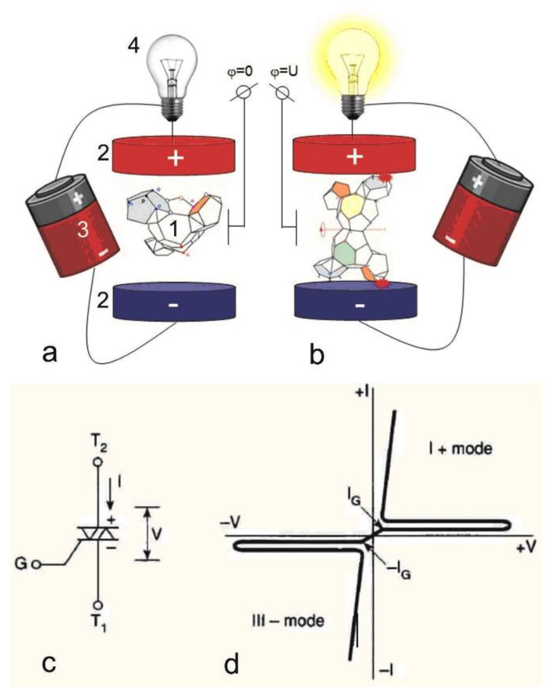
Figure 2.
Spectra: a – typical UV spectrum of a mixture of DNA nucleotides obtained by hydrolysis, b – spectrum of the polymer DNA hydrolyzed (curve 1) and nitrogenous bases after interaction with ZrO2 700°C in a solution of acetic acid and ethyl alcohol without water (curve 2) and diluted with water in 10 times (curve 3), c – spectra of nucleotides at a wavelength of 269 nm (1) and nitro (or nitroso) derivatives of nitrogenous bases at a wavelength of 300 nm as a result of the interaction of 2, 5, 10% HNO3: curves (2), (3), (4) respectively, d ¬ nucleotide spectra in the initial state (1) and after interaction with KOH at a concentration of 10, 25, 50%: curves (2), (3) and (4), respectively.
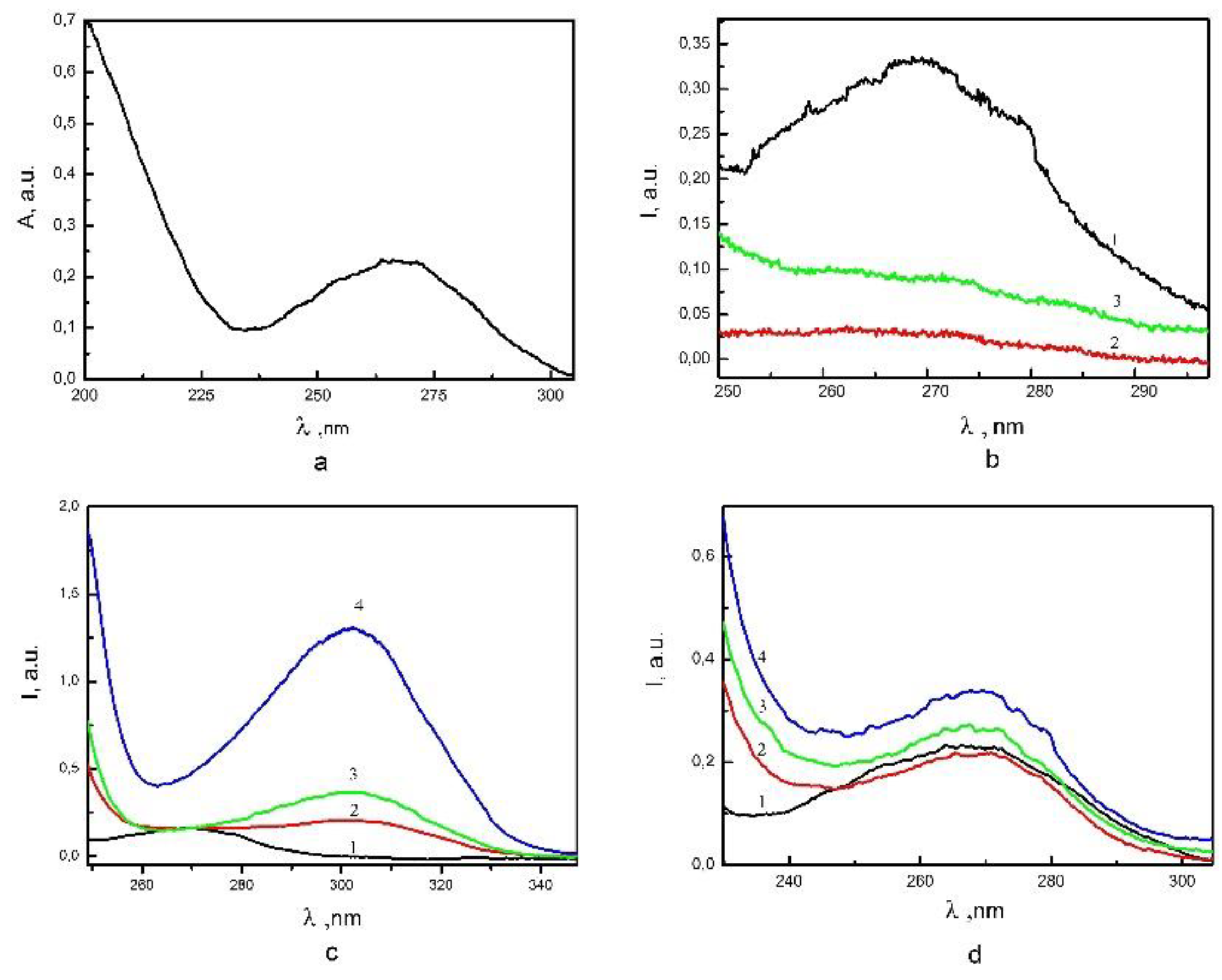
Figure 3.
Diagram of the intensities of spectrophotometric peaks characterizing the adsorption properties of the studied nano-sized powders of Zirconia with images of SEM (magnification x30 and x1000), reflecting the nature of the distribution on the substrate – suspensions that numbers are shown according to Table 1.
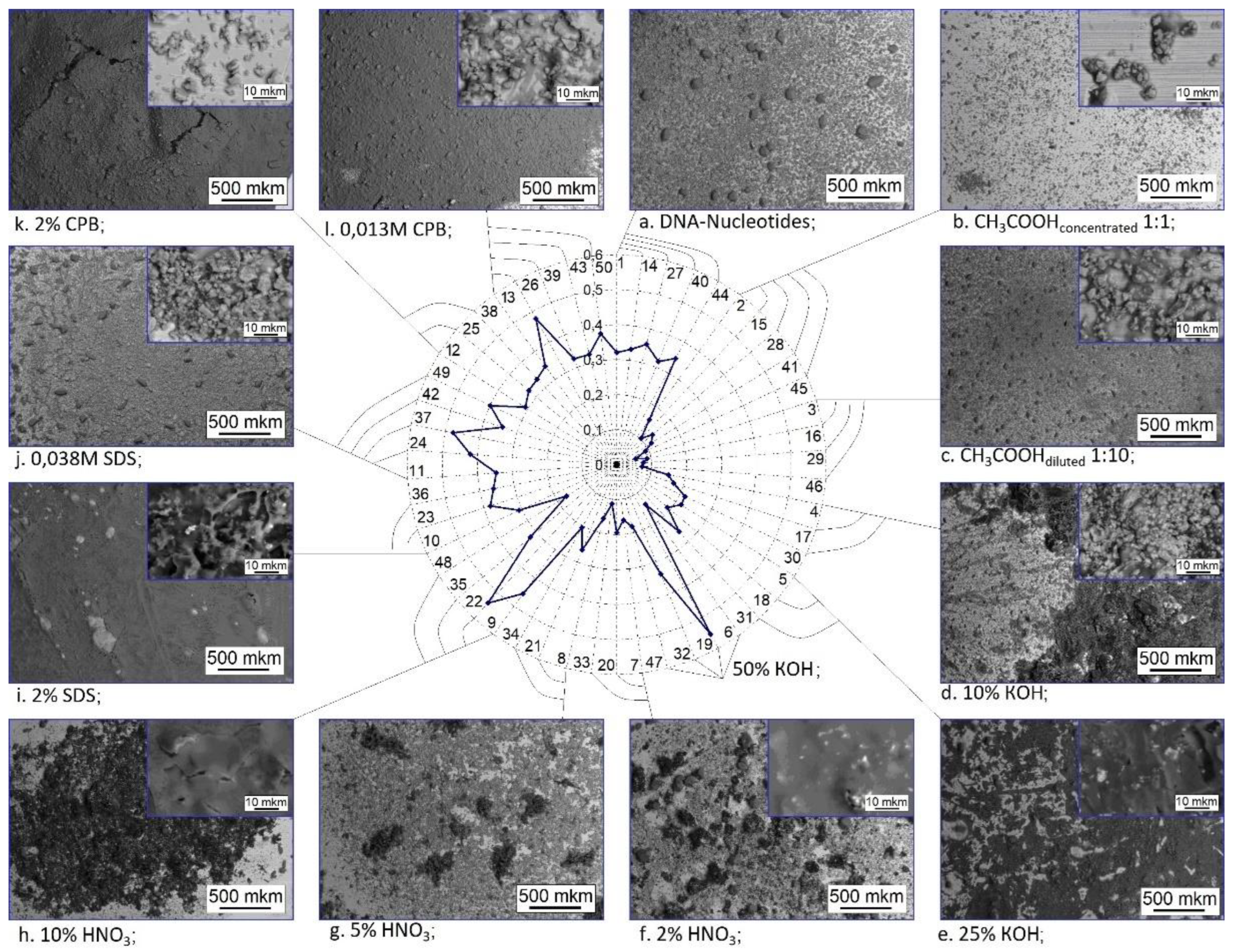
Figure 4.
Diagram of the intensities of spectrophotometric peaks characterizing the adsorption properties of the studied nano-sized powders of zirconia with SEM-images of these powders. Axes shows following powders: a – ZrO2–3%mol Y2O3 (700oС, 2h), b – ZrO2–8%mol Y2O3 (700oС, 2h), c – Zirconium hydroxide with the addition of 3% mol of Yttrium hydroxide (120℃, 2h), d – ZrO2 (700℃, 2h), e – ZrO2–3%mol Y2O3 (400oС, 2h). Curves show the surfactants, the adsorption interaction of that increases nearing to the center of the diagram.
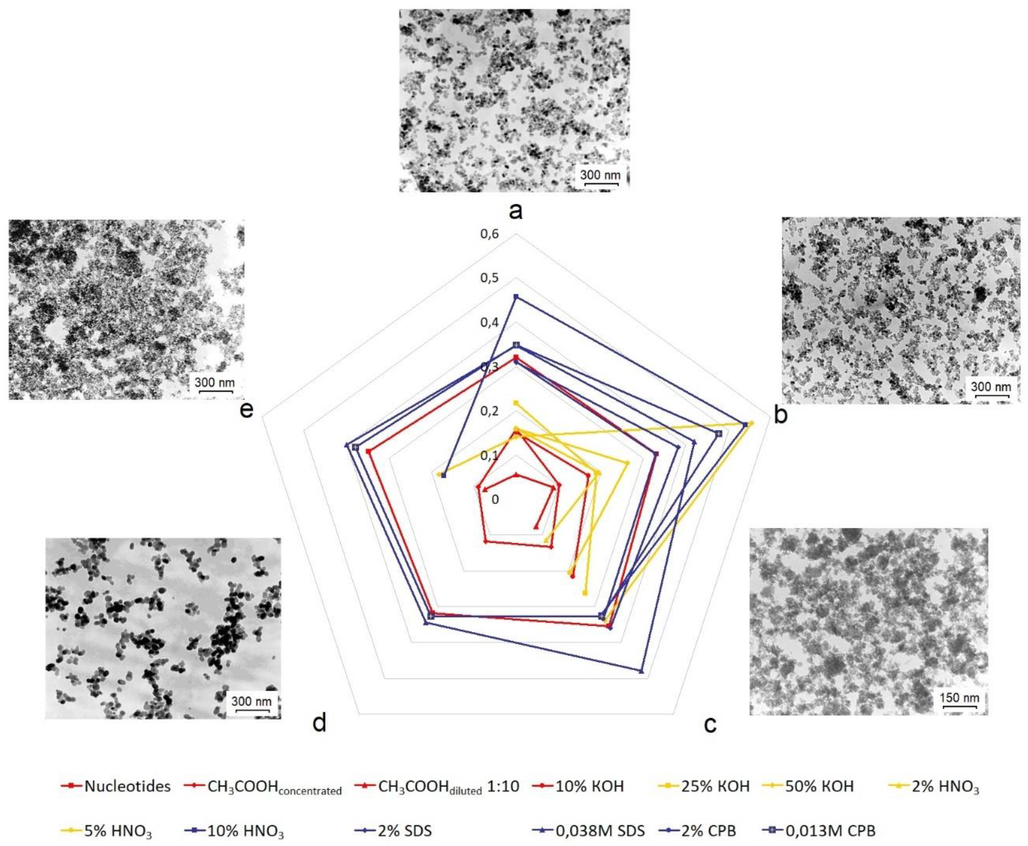
Figure 5.
TEM images of typical structural objects of functional structures under study: a – the aggregate of the initial nanoparticles of the composition ZrO2+3%molY2O3 (700°C), b – the initial DNA, c – the aggregate of the initial nanoparticles surrounded by nucleotides, formed in the solution C2H5OH: CH3COOH 1/10.
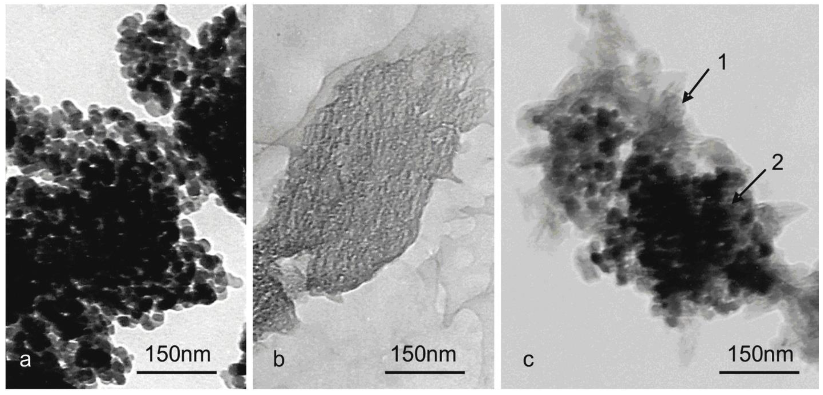
Table 1.
The main functional characteristics of nanopowders used in the work.
| Group Number |
Chemical structure | Sintering temperature / Particle size |
Structural ordering |
pHair-dry | SBET, m2/g |
| 1 | ZrO2+3%molY2O3 | 700°C / 17 nm | Crystalline, P42 / nmc | 5,95 | 42,4 |
| 2 | ZrO2+8%molY2O3 | 700°C / 15 nm | Crystalline, Fm3m | 4,71 | 59,39 |
| 3 | (Zr+3%molY2O2)O(OH)2 | 120°C / 3-4 nm | Middle order | 5,26 | 310 |
| 4 | ZrO2 | 700°C / 18 nm | Crystalline, P21C | 5,79 | 34,7 |
| 5 | ZrO2+3%molY2O3 | 400°C / 7,5-9 nm | Crystalline, P42 / nmc | 4,89 | 113,8 |
Disclaimer/Publisher’s Note: The statements, opinions and data contained in all publications are solely those of the individual author(s) and contributor(s) and not of MDPI and/or the editor(s). MDPI and/or the editor(s) disclaim responsibility for any injury to people or property resulting from any ideas, methods, instructions or products referred to in the content. |
© 2023 by the authors. Licensee MDPI, Basel, Switzerland. This article is an open access article distributed under the terms and conditions of the Creative Commons Attribution (CC BY) license (https://creativecommons.org/licenses/by/4.0/).
Copyright: This open access article is published under a Creative Commons CC BY 4.0 license, which permit the free download, distribution, and reuse, provided that the author and preprint are cited in any reuse.


