Submitted:
02 February 2023
Posted:
03 February 2023
Read the latest preprint version here
Abstract
The lumpy skin disease virus (LSDV) is an animal virus and a member of the Poxviridae family, which causes lumpy skin disease (LSD) in livestock animals like cows and buffaloes. LSD is an important transboundary disease of economic importance that was first discovered in 1929 in Zambia. LSDV has been prevalent in African countries, where several outbreaks have been reported previously. However, the virus has spread rapidly across the Middle East in the past two decades, reaching Russia and, recently, the Asian subcontinent. With the unprecedented cluster outbreaks reported across Asian countries, LSDV is certainly undergoing an epidemiological shift and expanding its geographical footprint globally. The recent LSD outbreaks have gained attention from global regulatory authorities and raised serious concerns among epidemiologists and veterinary researchers. Although there is no dearth of knowledge about LSDV, the disease lacks networked global surveillance and management, consequently making the current statistics deficient, fragmented, and unreliable. Hence, recurrent LSD outbreaks seriously threaten the global livestock industry. This review provides recent insights into LSDV by augmenting latest literature associated with its epidemiology, pathogenesis, transmission, currently-available intervention strategies, and economic implications on the dairy industries. The review also critically examines the changing epidemiological footprint of LSD and speculates on the possible reasons contributing to the ongoing multi-country LSD outbreak.
Keywords:
Lumpy skin disease virus
; lumpy skin disease
; epidemiological footprint and multi-country outbreak
; transboundary spread and disease resurgence
; diagnosis and vaccines
1. Introduction
In the past two decades, the world has experienced the wrath of several disease outbreaks, particularly those resulting from re-emerging viruses like the severe acute respiratory syndrome virus 2 (SARS-CoV-2), monkeypox virus (MPXV), Zika virus, and Nipah virus [1]. These have been attributed to the spillovers from various animal species due to increased contact (with humans), genomic mutations eventually leading to the emergence of variants, and crossing of species barrier [2]. With the prolonged coronavirus disease 2019 (COVID-19) pandemic and global monkeypox outbreak, several Asian countries are experiencing an unprecedented outbreak of the lumpy skin disease (LSD). It is a highly contagious viral infection of cattle that is caused by the lumpy skin disease virus (LSDV). This bovine virus is a member of the Capripovirus genus and subfamily Chordopoxviriniae, one of the most prominant animal poxviruses because of its serious complications in cattle, buffaloes, and other large ruminants, which directly hits the economics of the livestock sector [3]. The disease is known as 'Neethling virus disease,' 'knopvelsiekte,' 'pseudo-urticaria,' and 'exanthema nodularis bovis' [3,4]. Moreover, LSDV is closely related to the goat poxvirus (GTPV) and sheep poxvirus (SPPV), as it shares high sequence similarity and antigenic relationships with them [5]. LSD is a vector-borne, non-zoonotic, and transboundary disease that has emerged over the decades as a global catastrophic threat to livestock. Although LSD is a bovine ailment and does not affect or pose any threat to humans directly, it has indirect repercussions on human life. These include economic and financial losses and disruption of livestock, dairy, and meat industries, which ultimately lowers the gross domestic product (GDP) in countries that fundamentally depend on agricultural and dairy-related sectors [6]. The virus can infect all cattle, regardless of age and breed, but young calves and lactating mothers are reported to be more vulnerable to high infection rates [7]. The LSDV finds easy access to susceptible animals through contaminated food (feed), water, and milk. Common arthropod vectors like biting flies, lice, ticks, mosquitoes, and wasps also play a critical role in transmitting LSD [6]. Although there are no references to natural LSD infection in goats and sheep despite close interactions with infected cattle, experimental infections characterized by the development of skin lesions and nodules have been reported in giant gazelles, giraffes, goats, impalas, sheep, and wildebeest [8]. Considering its potentially high transmissibility across territorial borders and its negative economic impact, the World Organization for Animal Health (WOAH) has listed LSD as an important notifiable cross-border disease [9]. The LSDV has also been considered a potent agro-terrorism agent due to its recent global emergence, transboundary spread, and notably high transmission rates [4]. Moreover, high morbidity and low mortality rates are attributed to LSD. The viral infection extends both short- and long-term symptoms, depending on the immunological state of the infected animal [8]. Short-term symptoms include lymphadenitis, anorexia, rhinorrhea, and bilateral epiphora [8], while prolonged illness results in ailments like mastitis, pneumonia, and deep holes in the body [10]. Infected animals may also experience temporary or permanent infertility. Eventually, LSD reduces the economic value of the livestock in terms of milk yield and meat production, quality of animal hide, reproductive fitness, and long-term health. Earlier, LSDV was known to be restricted only to African nations; but it was subsequently reported across distinct geographical locations (non-endemic) worldwide. Recently, LSDV has been reported across regions of Asian and Middle-Eastern countries like India, Pakistan, Israel, Kuwait, Oman, and Yemen [11,12]. Moreover, multiple cluster outbreaks of LSD have erupted in the past five years in the Asian subcontinent itself, creating havoc and resulting in the death of cattle on a large scale. As per recent estimates from India, a country that relies heavily on its agricultural and livestock sectors, over 155,000 cattle deaths were reported in 2022 alone, making this animal disease a serious concern among epidemiologists, researchers, and veterinary scientists. In light of the recent events and considering the expanding geographical footprint of this viral disease, it becomes increasingly important for the scientific community to bridge the existing gaps in the biology of LSDV, its etiology, changing epidemiology, transmissibility, pathogenesis, diagnosis, and disease control and management. Hence, this review aims to augment latest knowledge on the complex biology of LSDV and its economic repercussions. In an attempt to gain more insights, this review also examines the recent LSD outbreaks and speculates the probable reasons behind the epidemiological shift and sudden resurgence of LSDV.
2. Etiology of LSDV: a brief overview of virus structure
LSDV is an enveloped virus with a brick-shaped structure of roughly 320 x 260 nm in size, which belongs to the Poxviridae [13]. Under an electron microscope, the structure of LSDV closely resembles that of the vaccinia virus, displaying a characteristic dumbbell-shaped core with lateral bodies (Figure 1). The virus belongs to the genus Capripoxvirus of the Chordopoxvirinae subfamily along with two other species, i.e., the GTPV and SPPV [14]. The virus harbors a double-stranded DNA genome, roughly about 151 kb in size, and comprises a chief coding region at the center, surrounded by identical terminals of 2.4 kb inverted repeats. The covalently-linked DNA strands contain palindromic hairpins at their terminals. The genome codes for nearly 156 putative genes, of which 30 structural and non-structural genes share about 97% sequence similarity with GTPV and SPPV [15]. The viral DNA contains roughly 146 conserved genes that are critical in driving molecular processes like DNA replication, transcription, virion production, and assembly. However, LSDV completely depends on the host cellular machinery to translate viral mRNA. Also, the LSDV genome contains homologous genes such as G protein-coupled CC chemokine receptor (GPCR), interleukin-10 (IL-10), IL-1 binding proteins, and epidermal growth factor-like protein, which are commonly observed in related poxviruses [16]. The viral DNA also contains an exclusive gene, LSDV132, which differs from other members of Capripoxvirus.
LSDV exists in two forms: the enveloped virion (EV) and the mature virion (MV) (Figure 1). These infectious forms have been categorized based on the existence of different surface glycoproteins and membrane layers [2]. MVs harbor a single lipid bilayer that is acquired from the endoplasmic reticulum of the infected cells. At the same time, EVs are characterized by the presence of an additional outer membrane (host-derived) that bears several entry-fusion complexes and even surface microtubules [8]. The virus is known to be resistant to both physical and chemical treatments, and shows high stability under ambient conditions for prolonged periods [13]. It has an exclusive survival potential in desiccated skin crusts (35 days), necrotic nodules (35 days), and air-dried hides (18 days). LSDV is unable to withstand incubation under high temperatures of 55˚C (2 h) and 65˚C (30 min). It remains persistent between pH 6.6 and 8.6 at 37˚C for five days but is highly susceptible to extreme alkaline or acidic conditions [13]. Lipidophilic detergents and sunlight can immediately predispose and inactivate the virus. LSDV is labile to chemical disinfectants such as chloroform, ether (20%), phenol (2% for 15 min), iodine compounds (1:33 dilution), formalin (1%), sodium hypochlorite (2–3%), and quaternary ammonium compounds (0.5%) [13].
4. Transmission, reservoirs, and hosts of LSDV: a panoramic yet distal view
The transmission of poxviruses is multifactorial, involving direct contact through aerosols, bodily fluids like semen, and indirect dissemination through animal or insect vectors, reservoirs, and even fomites [27]. Like any other poxvirus, LSD is a host-specific disease that is transmitted mechanically through arthropod vectors, with its infective host(s) being bovine animals like cows and buffaloes (Figure 3). Interestingly, LSD is a non-zoonotic disease with no traceable history or evidence of human infection to date.
Recent studies have demonstrated that both direct and indirect contact can spread LSDV. The virus can also be acquired vertically through the intrauterine route in infected cattle [28]. It is known to be transmitted from an infected dam to its calf through skin lesions on the udder or via contaminated milk [3]. Experimental evidence documents the transmission of LSD by tainted bovine sperm [29]. Sharing of water troughs and fodder with cattle that have been exposed to nasal discharge or saliva from suspected/infected animals may also result in indirect transmission of the LSDV. Another critical route for the transmission of LSD infection within a herd is the possible use of infected needles between cattle during mass vaccination drives [3]. The transmission routes of LSDV, both direct and indirect, have been shown in Figure 3.
LSD is primarily transmitted through insect vectors that act as natural reservoirs of the virus and infest healthy livestock. The mechanical transmission of LSD occurs via various species of mosquitoes, biting flies, and ticks [8]. Biting flies like Stomoxy calictrans and Biomyia fasciata are known to be responsible for the vector-borne pathophysiology of LSD. Nonetheless, the potential role of non-biting flies in transmitting LSD has also been elucidated [30]. Ticks like Amblyomma hebraeum, Rhipicephalus appendiculatus, and Rhipicephalus decoloratus also serve as reservoirs of the LSDV [30]. Additionally, mosquitoes like Culex mirificens and Aedes natrionus are also known to transmit LSD. On the other hand, Anopheles stephensi Liston and Culex quinquefasciatus Say have been identified as potent carriers of LSDV. Still, their role in disease transmission has not been documented [31]. Amidst the LSD outbreaks in Russia in 2015, ixodid ticks were speculated to play a critical role in the transmitting LSD. This has been recently confirmed using molecular studies where ixodid ticks recovered from LSD-infected cattle tested positive for viral DNA [32]. Apart from this, surveillance studies in Bulgaria have also indicated the presence of LSDV DNA in other ticks like Hyalomma marginatum and Rhipicephalus bursa [33].
The Asian water buffalos (Bubalus bubalis) and cattle (Bos Taurus and Bos indicus) are both severely affected by LSD [17]. Compared to cattle, buffalos have a significantly lower risk of morbidity associated with LSDV [3]. This fact has been hypothesized and correlated based on the thick skin texture of buffaloes, which is difficult to be pricked by the frail mouthparts of blood-sucking vectors such as flies, ticks, and mosquitoes thereby lowering the possibility of viral transmission and susceptibility to LSD [31,34]. Since buffaloes tend to evade hot summer seasons by taking refuge in ponds, it has been postulated that this physiological behavior lowers the tendency to be attacked by insect vectors [35]. Hence, it becomes challenging for the insect vectors to establish direct contact with the animal skin, consequently lowering the risk of LSD infection. Regardless of age, cattle of both sexes are vulnerable to this virus. The immunological status and physiological well-being of the animal also plays a pivotal role in determining the disease severity [3]. Bos indicus shows low vulnerability to clinical illness than Bos taurus [24]. Furthermore, younger animals show greater vulnerability and severity toward LSD infection than adult cattle [36]. Wild animals are naturally immune to LSD infection, but in experimental settings, oryx (Oryx gazelle), springbok (Antidorcas marsupialis), Thomson's gazelle, giraffe (Giraffe camelopardalis), and impala (Aepyceros melampus), and have all been shown to develop clinical lesions and disease symptoms [37,38]. Typically, it has been shown that natural wildlife plays a minor role in the spread and persistence of LSDV.
The exact mechanism behind the mechanical transmission of LSDV remains unclear. It is difficult to claim whether the transmittance is attained by contaminated mouthpart or if other intricate interactions are involved. In severe infections, high viral titers are present in the skin lesions, which serve as a potential source of contamination for arthropod vectors [39]. For biting and blood-feeding insects like mosquitoes, a lower level of viremia has been detected that usually lasts for 12 days or less [40]. Interestingly, the caseload of LSD attains a peak during the summer and rainy seasons, which usually coincides with the high prevalence of arthropod-based vectors, especially the blood-feeding insects [30,41]. This raises further speculations that such insects may be critical to transmitting LSDV. However, outbreaks beyond the vector prevalence period support the existence of an additional yet undiscovered mode of transmission of LSDV. Moreover, a few reports also propose that LSDV transmission is not confined to any specific season [27]. Frequently, migration of domestic or wild cattle has also been correlated to the widespread transmission of LSDV [27]. Hence, other naturally-existing reservoirs of LSDV must be identified, and their vectoring potentials be scrutinized, especially for the insects pertaining to livestock and farm animals. Despite numerous reports indicating vector-borne transmission, LSD outbreaks have been observed to occur, even in the complete absence of insect vectors. This suggests that LSDV may possibly employ other means for viral transmission, in addition to vector-assisted routes.
5. Pathogenesis and clinical representations of LSDV: from signs to symptoms
The clinical representation of LSDV infections shows a remarkable variation, including short- and long-term subclinical infections, and even death [42]. Once the virus has been successfully transmitted to its natural host, the incubation period varies from 7 to 28 days [22]. LSD is characterized by the presence of numerous skin lesions which are well-circumscribed and range between 2 to 7 cm in diameter, appear solid with flat-topped papules and nodules, and multiple coalescing centers [8]. The virus persists in skin lesions, blood, scabs, oral, nasal, and ocular fluids, semen, and occasionally in animal skin without any noticeable symptoms [43]. Following LSDV infection, the virus replicates in the epidermal tissue, resulting in viremia and sudden onset of fever in the animal. LSDV localizes in the cutaneous tissue and then causes the nodules to develop [9]. The nodules involve both the dermis as well as epidermis, but sometimes extend to the hypodermis, and rarely to the adjacent striated muscle. LSDV exhibits a wide tissue tropism, but the preferred sites are the skin on the neck, head, limbs, perineum, udder, and genitalia [44,45]. During the initial days of viral infection, the nodules appear grayish-white (internally) and may also exude serum. However, following disease progression (~ 14 days), the nodules may develop a cone-shaped central core or sequestrum of necrotic material called the "sit-fast" [46]. As soon as the infected nodules on the mouth, nose, eyes, udder, genitalia, and rectum; begin to ulcerate, LSDV finds passage into all bodily fluids like saliva, nasal and ocular secretions, and even the genital discharge. Consequently, many cattle suffer from significant emaciation and weakness, resulting in the loss of animal productivity for several months, which may further inflict permanent damage to the hides [43].
According to recent studies, most tissues and organs of the infected animals exhibit pathological alterations such as mastitis, orchitis, necrotic hepatitis, lymphadenitis, and disseminated vasculitis [43]. Tracheitis, cardiac damage, and other pathological alterations are also seen in a few cattle. These pathological abnormalities might induce varying degrees of injury to the animal, making LSDV infection more detrimental [47]. A clinical study on LSDV surfaced previously, indicating the oxidation-anti-oxidation state imbalance in infected cattle, thereby invoking a significant rise in the levels of pro-inflammatory cytokines, extending negative consequences on animal health [48]. This was positively correlated with histopathological outcomes in infected animals, which showed signs of profuse necrosis, mononuclear cell infiltration, intracytoplasmic inclusion bodies, and severe vasculitis. The dysregulation of organ functions is known to be triggered by the metabolite buildup in the heart, liver, and kidney, causing hypophosphatemia, which exacerbates the symptoms of hemolytic anemia. Experimental findings from hematological and biochemical studies in LSD-infected animals have also revealed that infected animals suffer from pancytopenia, hyperproteinemia, hyperkalemia, hyperchloremia, and decreased creatinine content [8,49]. Hence, these indicators/markers may be used as an index for assessing the disease prognosis, severity, and timely management or control of LSD. It has been speculated that young cows, lactating mothers, and underweight livestock are more vulnerable to infection by LSDV, possibly due to poor or impaired immunity [39,50]. Interestingly, disease-recovered animals have been shown to harbor lifelong immunity against the virus [9]. Calves from the infected mothers exhibit resistance towards LSDV for nearly six months because of the acquired maternal antibodies [40]. Nonetheless, animals that withstand the wrath of LSD infection show complete clearance of the viral load and do not act as carriers for LSDV [3].
6. Diagnosis, preventive measures, and treatment of LSD
LSDV infection is diagnosed based on classical clinical symptoms, such as lymphadenopathy and typical nodular skin lesions, in conjunction with confirming the presence of the virus or viral antigen in immunodiagnostic tests. Conventional PCR [51] and real-time PCR [39] are molecular techniques that are often employed to validate LSDV infections. Real-time PCR is employed to diagnose and clinically differentiate LSDV from other animal-associated poxviruses like GTPV and SPPV [52]. Furthermore, restriction fragment length polymorphism (RFLP) is another technique that is being exploited to distinguish vaccine strains from virulent LSDV [53]. Besides RFLP, LSDV is also identified using electron microscopy, virus isolation, and virus neutralization tests (VNT) [9]. Virus neutralization is known to be the gold-standard for detecting antibodies raised against Capripoxviruses. Nonetheless, the disease can also be diagnosed by serological tests, including VNT, indirect fluorescent antibody test (IFAT), serum neutralization test (SNT), and indirect immunofluorescence test [54]. However, ELISA is more sensitive and selective than IFTA or VNT [55]. Another approach for LSD diagnosis is the immuno-peroxidase monolayer assay (IPMA), a relatively cheap and convenient technique. It has greater sensitivity and specificity than VNT and commercially available ELISA kits [56]. Moreover, owing to higher costs and tedious operations, western blot, an extremely sensitive and specific technique, is seldom used to detect LSDV [9].
In recent years, the spread and recurrent outbreaks of the Capripoxviruses point towards major issues like inconsistencies and inefficiencies in vaccination programs, poor economic conditions, and unawareness among farmers in endemic and non-endemic areas, legal as well as illegal trade of livestock, and global climatic changes. To date, the line of action taken to cure LSD is solely symptomatic which mainly targets on prevention against secondary microbial infections. This includes various combinations of anti-inflammatory, antimicrobials, supportive therapy, and anti-septics [9]. Currently, no effective antiviral drugs are available to treat LSD. Nonetheless, FDA-approved drugs and phytocompounds that are effective against other poxviruses may be repurposed against the LSDV [8]. The disease can only be controlled in endemic areas via mass vaccinations, imposing movement restrictions (quarantine), and removing suspected or infected animals [45]. Culling of infected/suspected animals, transportation/movement restrictions, and mandatory and uniform immunization have all been suggested as control measures to minimize the possible transboundary spread of this disease [3,44]. Moreover, due to the cardinal role of arthropod vectors in transmitting LSD, their eradication becomes more challenging. In addition, the delayed disposal of diseased animals or carcasses makes the situation even worse [3]. However, to stop the spread of disease through vectors, certain control measures, such as the use of insecticides, pesticides, and vector traps, has been recommended in regions with a high vector population [6]. Besides, there are risk factors associated with such control activities. Further, creating awareness among veterinarians and farm/livestock workers regarding the disease will also enable rapid diagnosis of clinical cases, permitting timely management of the disease, thereby breaking the transmission chain and preventing cluster outbreaks [44].
The widespread administration of appropriate vaccines is quintessential for preventing and eradicating the virus. Currently, live-attenuated vaccines based on the LSDV, SPPV or GTPV strains make up most of the commercially available LSD vaccines. The presently-administered LSDV vaccines and efficacies have been summarized in Table 1. Live-attenuated LSD vaccine are usually formulated by the conventional South African Neethling strain or the Kenyan sheep and goat pox strains, KSGP O-180 and O-240, respectively [57]. For preparing attenuated vaccines, the Neethling strain (virulent) has been subjected to serial passaging (61 times) in lamb kidney cells (LK), followed by 20 passages in the chorioallantoic membrane of embryonated chicken eggs, and subsequently back in LK cells (3 times) [58]. Another virulent strain, i.e., the Madagascan LSDV strain, requires 101 passages in rabbit kidney cells, followed by five passages in fetal calf kidney cells for its potential use as a vaccine [58]. After immunization with homologous booster LSD vaccines, animals may experience adverse effects such as allergic reactions at vaccination site or typical skin nodules accompanied by reduction in lactation [57]. This reaction is often called the "Neethling response/disease."
In 2021, the homologous live-attenuated LSD vaccines, including Herbivac LS, Lumpy Skin Disease Vaccine, Kenyavac (South Africa), and Lumpyvax (South Africa), and Vaccin LSD Neethling O vivant (Morocco) were clinically tested by researchers [59]. Interestingly, none of the aforementioned vaccines adversely affected general health or animal behavior in any of the experimental groups, including feed intake, albeit these were known to induce fever in some animals [59]. Nevertheless, in animals immunized with Herbivac LS vaccine, swollen lymph nodes were detected, while the other three South African vaccines showed clinical manifestations of Neethling disease upon vaccination. Small nodules, not as large as those reported in sick animals, appeared in the Moroccan Neethling vaccine group [59]. Since LSDV also shares more than 97% of its nucleic acid sequences with that of GTPV and SPPV, immunization with live-attenuated goat or sheep pox vaccines also confers cross-immunity in susceptible animals against the LSDV. This has been typically employed in clinical settings to circumvent LSD.
In the recent past, various immunization studies have reported that a live-attenuated ‘Neethling’ strain can be chemically inactivated using ethylenimine and coupled with various adjuvants like the Montanide adjuvant [60] and a low molecular weight copolymer (Polygen, MVP Adjuvants®, named as Adjuvant A) [61], to provide adequate protection against the LSDV. Interestingly, inactivated vaccines have been shown to elicit a heightened immune response which was 37% higher than that of live-attenuated jabs [60]. Another bivalent inactivated vaccine conjugated with oil adjuvants against the LSDV and bluetongue virus was reported recently, which could stimulate the production of neutralizing antibodies at high titers [62]. Moreover, recombinant LSDV vaccines, namely, LSDV-WB005KO and LSDV-WB008KO, have also been developed using a homologous recombination technique by deleting the LSDV open reading frames 005 and 008 [8]. Further, clinical investigations have discovered that combining these two vaccines can significantly enhance the titers of neutralizing antibodies in immunized cattle, which can eventually fend off any infection or invasion by LSDV [63].
7. Current scenario & economic repercussions of LSD outbreaks
Over the past few years, the world has witnessed an unprecedented wave of LSD outbreaks across diverse geographical boundaries, pioneering from Africa and spreading beyond the Middle East to southeast Europe and the Asian subcontinent [64]. Several factors have influenced the transboundary spread of LSD to non-endemic countries. These constitute both legal/illegal transportation and trade of livestock, cross-border passage of insect vectors/reservoirs, deceleration of vaccination drives, and reduced global surveillance [8]. In recent times, animal disease databases like EMPRES-i (FAO) and WAHIS (WOAH) have allowed real-time monitoring of the global disease situation, which helps in collating data for continuous risk assessments and associated trade recommendations for animals and related products [8,17]. The LSDV has been spreading like wildfire across the Asian subcontinent, inflicting high mortality and incurring huge economic losses in some countries. Statistics from the past two years suggest a total of 3,562 outbreaks worldwide, with most cases reported in Asian countries like India, China, Nepal, Vietnam, Thailand, and Sri Lanka [26]. Countries that share territorial borders with regions having active LSDV infections are at a significantly higher risk of incursion by the virus. With the first outbreaks of this disease in non-endemic countries like China, India, Pakistan, Afghanistan, and Bangladesh, LSDV has been expanding its geographical range at a global level [8]. In 2022 alone, India witnessed around 3 million LSD infections of cattle, with a mortality rate of nearly 6%, resulting in 155,000 deaths [26]. Figure 4 represents a comprehensive geographical overview of LSD outbreaks reported in the past five years at the global level. The statistics have been quantified based on the categorization made by WOAH with respect to LSDV infections (Cases reported, deaths, susceptible, and vaccinated animals). Considering these grim statistics in the wake of the current multi-country LSD outbreak, this viral disease has raised serious concerns for the livestock industry. Although a few authorities actively undertake global surveillance of LSD, their consistent efforts towards disease tracking have not been successful enough to curb the transboundary spread of this virus. Hence, it is the need of the hour that animal healthcare agencies, state governments, academia, and relevant stakeholders collaborate to establish synchrony for ensuring active surveillance programs for the timely identification of LSD outbreaks/clusters.
To curb the spread of LSD, the Food and Agricultural Organization of the United Nations (FAO) has drafted necessary guidelines and protocols, laid down templates for contingency plans, and awareness measures [65]. To provide the best protection, FAO recommends annual vaccination of livestock and dairy animals in LSD-affected nations and coordinated vaccination drives across countries. Newly-born calves from uninfected mothers must be immunized at any early age, while calves from naturally-infected or immunized dams should be vaccinated between three to six months post-partum. Regionally-harmonized vaccinations have also been proposed before massive herd movements, for instance, before the commencement of seasonal grazing [65]. According to the FAO guidelines, the principal foundation for LSD surveillance programs should be passive disease reporting and, secondarily, risk-based surveillance based on detecting clinical signs in both wild and domestic farm animals. Along with vector surveillance, emphasis must be placed on monitoring susceptible hosts, wildlife animals, and even small ruminants. Serological surveillance can also be used for retrospective analyses in affected areas or to predict possible LSD outbreaks [65]. However, insights into the recent genomic studies highlight the need for large-scale genome surveillance and close monitoring to track the LSDV for building better algorithms, disease prediction models, logistics, and diagnostics in the coming future. For example, genome sequences of six viral isolates retrieved from infected animals during the recent LSDV outbreak in India have indicated the presence of several genomic mutations, strongly suggesting that the presently-circulating LSDV strains have evolved from a distinct lineage, giving rise to genetic variants of this animal virus [66]. Hence, immediate attention must be given to the global surveillance of LSD, which may add new dimensions to understanding the viral disease better and controlling future outbreaks.
Since LSDV primarily targets livestock animals like cattle and buffaloes, any outbreak in countries that heavily depend on the productivity of dairy industries takes a huge financial hit. With more than 650 million head of cattle and buffaloes, Asia is a major contributor towards the global livestock industry, accounting for a mammoth share of 39% [67]. Most of these animals are concentrated in South and Southeast Asia, with India being on the top with a whopping 300 million head, followed by China and Pakistan with approximately 90 and 85 million, respectively [68]. India is also a lead exporter in the beef market, with nearly 527 tons of carcass weight equivalent exported in 2018 alone [68]. With the death of nearly 155,000 cows in India during the 2022 LSD outbreak, it has been estimated that the country faced a direct economic loss of nearly 3 billion Indian Rupees [26]. Therefore, the transboundary spread of LSD is bound to substantially impact the economies of agro- and dairy-based Asian countries. The recurrent outbreaks of LSDV directly affect the dairy, meat, and tannery industries because of decreased meat and milk production, damaged cattle skins, fertility problems, abortions, and, ultimately, the death of severely affected animals [69]. Restrictions on intra- and inter-country trade and movement of cattle also incur indirect losses. The high costs of disease diagnosis, management, treatment, and vaccination also add to the economic burden [70]. Considering the strategic positions held by China and India in the global meat and dairy markets, any significant impact on their livestock industries will certainly be felt across the global markets. Despite the gruesome figures coming out of India, the United States Department of Agriculture (USDA) suggests that the disease outbreak in India had a marginal impact on the gross milk production and net revenues [71]. This points towards Western nations' ignorance and double standards in undermining infectious diseases like LSD that are not prevalent or endemic within their territorial boundaries. The present LSD outbreak must be viewed very critically by the global economies as a wake-up call to adopt integrated approaches toward the surveillance and management of LSD. In addition, the unprecedented spread of LSDV has a direct implication towards escalating antimicrobial and anthropogenic resistance in the environment due to the extensive use of broad-spectrum antibiotics in livestock for treating secondary bacterial infections and large-scale application of insecticides to kill insect vectors [57].
8. Causes of LSDV resurgence: from ground reality to speculations, and beyond
Several reasons can be speculated for the unprecedented outbreaks of LSDV in recent years. The scenario draws an analogy with many other re-emerging viruses like the MPXV, SARS-CoV-2, Nipah, and influenza virus [2]. Of lately, the recent outbreaks of LSDV have majorly affected the developing countries of the Asian subcontinent and the Middle East. In most South Asian territories, the disease erupted unexpectedly and began to spread rapidly during monsoons. High rainfall creates ambient environments for the propagation and multiplication of insect vectors like mosquitoes, flies, and ticks, which directly increases the risk of disease transmission [72]. Parallels can be drawn with chikungunya and dengue, which are vector-borne viral diseases and are also known to peak in Asian countries during rainy seasons when mosquito populations are at their maximum [73]. Notably, the sudden rise in LSDV infections coincided with weather conditions that resonate with high amounts of precipitation. The increased rainfall and humidity make it difficult to control insect vectors and treat the infected animals as open skin wounds (lesions) take a longer time to heal. This increases the susceptibility of animals to secondary bacterial infections. Interestingly, climate change is also conjectured to play a pivotal role in expanding the geographical niche of this disease (Figure 5). Reports have shed light on this matter, stating that the LSDV may be transported internationally across territorial borders due to altered wind direction and velocity [28]. Therefore, the climate is believed to be critical in shaping LSD outbreaks.
Since LSDV is a DNA virus, its genome was believed to be stable for many years. Field isolates of LSDV recovered over decades in Africa exhibited minimal genetic alterations from the parental strain, which was first identified in Zambia in 1929 [7]. Moreover, LSDV strains retrieved from the successive outbreaks that occurred in the Middle East and Europe, post-2012 and -2015, respectively, did not show any signs of divergence or mutations in their DNA genomes [3,74,75]. As a result, the genomic stability of the virus was exploited to develop live-attenuated vaccines against LSDV, where the virus could be easily differentiated from the contemporary field isolates [53,76]. But surprisingly, LSDV strains recovered from infected animals in Russia between 2017 and 2019 exhibited vaccine-like characteristics, prompting an immediate shift towards this dynamics [77,78]. Some of these variants contained a 12-nucleotide insertion in the GPCR gene, similar to the vaccine strains, while others displayed a 27-nucleotide deletion in the ORF LSDV 126, like the LSDV Neethling vaccine strain. Recombination between the field virus strains and the Neethling vaccine strain was believed to be the primary cause for the emergence of these LSDV variants [78]. On similar lines, the virus strains recovered from the LSD outbreaks in China also demonstrated GPCR profiles similar to that of LSDV vaccines with the 27-nucleotide deletion [30,78]. Moreover, recent studies have demonstrated that these recombinant LSDV strains can induce more severe disease than their parental field strains [79]. Other than contributing towards increased virulence, these genomic mutations may also play a critical role in altering the virus’ mode of transmission (Figure 5). These new variants can possibly lead to a direct, cattle-to-cattle transfer of viral infection through semen or other body fluids like saliva and nasal discharge. Similar concerns have been raised in the case of MPXV, which was previously known to be transmitted only through direct contact and aerosols. But recent studies hint towards a possible role of sexual transmission since most of the monkeypox infections were reported in homosexual men [2]. Hence, the recently identified mutations in the LSDV genome may be attributed to the ongoing multi-country outbreak of LSD.
In most developing South-Asian countries, domestic livestock are allowed to roam outdoors freely and consume wild vegetation. From a broader perspective, this communal grazing system can also be deemed responsible for the escalating LSDV infections. The impact of open grazing is two-fold for disease epidemiology. The nutritional composition of wild vegetation in the meadows is not defined as compared to regular farm fodder, which upon consumption, may lead to the deficiency of essential nutrients in the livestock [80]. This scarcity of nutrients can impair the immune system of farm animals, making them more susceptible to diseases [81], such as LSD. Secondly, infected animals grazing in open fields are more likely to spread infection via insect vectors (Figure 5). Such animals can transmit the disease to arthropod vectors, which can infest healthy livestock, making them more vulnerable to viral infection. This can ultimately result in an unprecedented outbreak at livestock farms, making disease containment even more challenging.
LSDV is not a novel virus; the disease is almost a century old. Despite the availability of LSD vaccines for several decades, the virus is currently spreading like wildfire across countries. It must be noted that vaccines formulated from the erstwhile primitive LSDV strains (parental) are still being used [82]. Hence, the lack of vaccine upgradation is speculated to be another possible reason behind the resurgence of LSD. These vaccines have not been upgraded according to the currently-circulating LSDV strains and, therefore, are now failing to protect animals worldwide (Figure 5). This implies serious doubts over vaccine efficacy. Similar issues have been brought up with the COVID-19 vaccines, as the current vaccine formulations are based on the SARS-CoV-2 strain that first emerged in the Wuhan province of China more than 3 years ago [83]. Since then, more than a hundred variants of SARS-CoV-2 with distinct PANGO lineages have emerged, including the alpha, beta, gamma, delta, and presently-spreading omicron variants [84]. These variants are known to evade the vaccine-induced neutralizing antibodies and even the host immune system [85]. The limited protection against the new sub-variants is believed to be a key factor in recurrent cluster outbreaks causing multiple unprecedented infection waves across international borders [85]. Hence, there is a pressing need to modernize and re-formulate LSD vaccines with the presently existing or circulating virus strains. Also, most existing research and clinical trials on LSD have been conducted with mildly virulent strains of LSDV [8]. Therefore, the actual efficacy of existing vaccines is not completely dependable regarding their application in field animals. In addition, the recombination events between live-attenuated (vaccine) and field strains of LSD have recently been shown to result in the emergence of new variants [78]. Hence, the presently used and commercially available live-attenuated LSD vaccines incur serious risks of generating new LSDV variants. Such vaccines also put livestock animals at risk of vaccine-derived LSD (Figure 5), a scenario that has been previously reported with the live-attenuated oral polio vaccine (OPV). The OPV is infamously associated with the incidence of vaccine-derived polio, wherein the OPV strains undergo partial reversion to become virulent again [86]. Consequently, the developed nations have ceased administering OPV and replaced it with the inactivated polio vaccine (IPV) [86]. Similar instances may be possible with LSDV, where partial reversion to a virulent type may be responsible for inflicting vaccine-derived LSD. Nonetheless, such possibilities warrant strong scientific upholding and further scrutiny to determine the fate of existing LSDV vaccines.
WOAH has categorized LSDV as a notifiable transboundary disease [12]. The movement of the arthropod vectors or other reservoirs over long distances due to high wind currents or natural migration may also be attributed to virus transmission across borders. Parallel to the meat and dairy industries, illegal wildlife trade and unlawful selling of livestock across territories is another potential reason behind the transboundary spread of LSDV [87]. When introduced into the unaffected areas through illegal activities, the infected animals from endemic regions can also transmit the virus to healthy livestock, eventually causing a widespread outbreak of the disease (Figure 5). Importantly, the ongoing COVID-19 pandemic also played a critical role in shaping today’s scenario concerning the LSD outbreak. The deadly SARS-CoV-2 occupied the center stage and drew the attention of researchers/clinicians globally, sidelining other important human diseases, including cancer(s), leave alone animal diseases [88]. The unprecedented COVID-19 pandemic compelled the global economies to enforce nationwide lockdowns and restrict human movement, which exacerbated the existing challenges for veterinary personnel and scientific laboratories working on disease diagnosis and prediction of possible outbreaks. This ultimately impeded disease monitoring, surveillance, and the subsequent enactment of containment strategies (Figure 5). In summary, the present-day multi-country outbreak of LSD has multidimensional aspects that have been illustrated in Figure 5. Considering the speculations raised in this review, it can be believed that the current scenario was already in the making until the LSDV infections exploded beyond comprehension. Hence, necessary control measures are needed to end the wrath of LSDV. Ensuring coordinated attempts from the international regulatory bodies, governmental authorities, and veterinary scientists is critical to managing disease treatment, prevention, and relevant control measures against LSDV. Devising alternate intervention strategies, creating awareness among livestock farm owners, promoting regular vaccination campaigns for susceptible animals, and implementing strict laws to prevent wildlife trafficking across borders are a few necessary steps that can be taken to curb recurrent LSD outbreaks. Apart from this, regular screening of farm animals and maintaining high-level surveillance programs lay a strong foundation for preventing cluster outbreaks of LSD.
Conclusion
Livestock animals are a critical pillar of the dairy industries that contribute greatly to the world economy. The global livestock sector has been severely affected by recurrent LSDV outbreaks, resulting in the large-scale death of cows and buffaloes. Apart from being fatal, the virus reduces overall productivity in livestock animals which incurs tremendous revenue losses to the agro-dependent nations. LSDV was previously thought to be endemic in Africa, but the recent trends and unprecedented resurgence indicate the virus’ expanding geographical foothold in non-endemic countries. This becomes critically important since LSD is an economically important transboundary disease that lacks proper global surveillance and data acquisition, making the existing statistics deficient, fragmented, and unreliable. The present situation has worsened with the identification of recombinant virus strains and several mutations in the LSDV genome. With the ongoing COVID-19 pandemic and multi-country monkeypox outbreak, the gravity of the present situation has increased significantly, demanding immediate attention towards stringent global surveillance and healthcare systems. There is a dire need to upgrade the vaccine formulations with the presently-circulating LSDV strains and devise alternative intervention strategies to combat LSD outbreaks. Extensive research from academia, strong inter-organization engagement, and collaborations between the relevant stakeholders are paramount to controlling and managing the recurrent outbreaks of LSD. It is high time that all nations join hands to work collaboratively on a common platform to ensure that such viral outbreaks are not transformed into widespread epidemics or pandemics.
Funding
The authors did not receive support from any organization for the submitted work.
Data Availability Statement
The datasets generated and analyzed during the current study are available at the World Organization for Animal Health (https://wahis.woah.org/#/dashboards/qd-dashboard).
Acknowledgments
SC would like to thank the Indian Council of Medical Research (ICMR) for their financial support. Financial assistance from ICMR, New Delhi for providing fellowship (SRF) to JC and LK is also appreciated.
Conflicts of Interest
The authors report there are no competing interests to declare.
Abbreviations
COVID-19: Coronavirus disease 2019, EV: Enveloped virion, FAO: Food and agricultural organization, GTPV: Goat poxvirus, IMPA: Indirect fluorescent antibody test, IPV: Inactivated polio vaccine, LSD: Lumpy skin disease, LSDV: Lumpy skin disease virus, MPXV: Monkeypox virus, MV: Mature virion, OPV: Oral polio vaccine, RFLP: Restriction fragment length polymorphism, SARS-CoV-2: Severe acute respiratory syndrome virus 2, SNT: Serum neutralization test, SPPV: Sheep poxvirus, VNT: Virus neutralization test, WHOA: World organization for animal health.
References
- Mourya, D.; Yadav, P.; Ullas, P.T.; et al. Emerging/re-emerging viral diseases & new viruses on the Indian horizon. Indian Journal of Medical Research 2019, 149, 447–467. [Google Scholar] [PubMed]
- Chadha, J.; Khullar, L.; Gulati, P.; et al. Insights into the monkeypox virus: Making of another pandemic within the pandemic? Environmental Microbiology 2022, 24, 4547–4560. [Google Scholar] [CrossRef] [PubMed]
- Tuppurainen, E.S.M.; Venter, E.H.; Shisler, J.L.; et al. Review: Capripoxvirus Diseases: Current Status and Opportunities for Control. Transboundary and Emerging Diseases 2017, 64, 729–745. [Google Scholar] [CrossRef] [PubMed]
- Khan, Y.R.; Ali, A.; Hussain, K.; et al. A review: Surveillance of lumpy skin disease (LSD) a growing problem in Asia. Microbial Pathogenesis 2021, 158, 105050. [Google Scholar] [CrossRef] [PubMed]
- Diallo, A.; Viljoen, G.J. Genus Capripoxvirus. In: Mercer, A.A., Schmidt, A., Weber, O. (eds) Poxviruses. Birkhäuser Advances in Infectious Diseases. Birkhäuser Basel 2007; 167-181.
- Gupta, T.; Patial, V.; Bali, D.; et al. A review: Lumpy skin disease and its emergence in India. Veterinary Research Communications 2020, 44, 111–118. [Google Scholar] [CrossRef] [PubMed]
- Tuppurainen, E.S.M.; Stoltsz, W.H.; Troskie, M.; et al. Potential Role for Ixodid (Hard) Tick Vectors in the Transmission of Lumpy Skin Disease Virus in Cattle. Transboundary and Emerging Diseases 2011, 58, 93–104. [Google Scholar] [CrossRef] [PubMed]
- Liang, Z.; Yao, K.; Wang, S.; et al. Understanding the research advances on lumpy skin disease: A comprehensive literature review of experimental evidence. Frontiers in Microbiology 2022, 13, 1065894. [Google Scholar] [CrossRef] [PubMed]
- Namazi, F.; Khodakaram-Tafti, A. Lumpy skin disease, an emerging transboundary viral disease: A review. Veterinary Medicine and Science 2021, 7, 888–896. [Google Scholar] [CrossRef]
- Selim, A.; Manaa, E.; Khater, H. Molecular characterization and phylogenetic analysis of lumpy skin disease in Egypt. Comparative Immunology, Microbiology and Infectious Diseases 2021, 79, 101699. [Google Scholar] [CrossRef] [PubMed]
- Ayelet, G.; Haftu, R.; Jemberie, S.; et al. Lumpy skin disease in cattle in central Ethiopia: outbreak investigation and isolation and molecular detection of the virus. Revue Scientifique et Technique de l'OIE 2014, 33, 877–887. [Google Scholar] [CrossRef]
- Anwar, A.; Na-Lampang, K.; Preyavichyapugdee, N.; et al. Lumpy Skin Disease Outbreaks in Africa, Europe, and Asia (2005–2022): Multiple Change Point Analysis and Time Series Forecast. Viruses 2022, 14, 2203. [Google Scholar] [CrossRef] [PubMed]
- Scientific Opinion on lumpy skin disease: EFSA Panel on Animal Health and Welfare (AHAW). EFSA Journal 2015, 13, 3986.
- Bhanuprakash, V.; Hosamani, M.; Singh, R.K. Prospects of control and eradication of capripox from the Indian subcontinent: A perspective. Antiviral Research 2011, 91, 225–232. [Google Scholar] [CrossRef] [PubMed]
- Tulman, E.R.; Afonso, C.L.; Lu, Z.; et al. The Genomes of Sheeppox and Goatpox Viruses. Journal of Virology 2002, 76, 6054–6061. [Google Scholar] [CrossRef] [PubMed]
- Tulman, E.R.; Afonso, C.L.; Lu, Z.; et al. Genome of Lumpy Skin Disease Virus. Journal of Virology 2001, 75, 7122–7130. [Google Scholar] [CrossRef] [PubMed]
- Azeem, S.; Sharma, B.; Shabir, S.; et al. Lumpy skin disease is expanding its geographic range: A challenge for Asian livestock management and food security. The Veterinary Journal 2022, 279, 105785. [Google Scholar] [CrossRef] [PubMed]
- Ali, A.A.; Esmat, M.; Attia, H.; et al. Clinical and pathological studies on lumpy skin disease in Egypt. Vet Rec 1990, 127, 549–550. [Google Scholar] [PubMed]
- Yeruham, I.; Nir, O.; Braverman, Y.; et al. Spread of lumpy skin disease in Israeli dairy herds. Veterinary Record 1995, 137, 91–93. [Google Scholar] [CrossRef] [PubMed]
- Lojkić, I.; Šimić, I.; Krešić, N. Complete Genome Sequence of a Lumpy Skin Disease Virus Strain Isolated from the Skin of a Vaccinated Animal. Genome Announcements 2018, 6, e00482-18. [Google Scholar] [CrossRef] [PubMed]
- Byadovskaya, O.; Prutnikov, P.; Shalina, K.; et al. The changing epidemiology of lumpy skin disease in Russia since the first introduction from 2015 to 2020. Transboundary and Emerging Diseases 2022, 69, e2551–e2562. [Google Scholar] [CrossRef] [PubMed]
- Das, M.; Chowdhury, M.; Akter, S.; et al. An updated review on lumpy skin disease: a perspective of Southeast Asian countries. Journal of Advanced Biotechnology and Experimental Therapeutics 2021, 4, 322–333. [Google Scholar] [CrossRef]
- Lu, G.; Xie, J.; Luo, J.; et al. Lumpy skin disease outbreaks in China, since 3 August 2019. Transboundary and Emerging Diseases 2020, 68, 216–219. [Google Scholar] [CrossRef] [PubMed]
- Sudhakar, S.B.; Mishra, N.; Kalaiyarasu, S.; et al. Lumpy skin disease (LSD) outbreaks in cattle in Odisha state, India in August 2019: Epidemiological features and molecular studies. Transboundary and Emerging Diseases 2020, 67, 2408–2422. [Google Scholar] [CrossRef] [PubMed]
- Koirala, P.; Meki, I.K.; Maharjan, M.; et al. Molecular Characterization of the 2020 Outbreak of Lumpy Skin Disease in Nepal. Microorganisms 2022, 10, 539. [Google Scholar] [CrossRef] [PubMed]
- Kumar, N.; Tripathi, B.N. A serious skin virus epidemic sweeping through the Indian subcontinent is a threat to the livelihood of farmers. Virulence 2022, 13, 1943–1944. [Google Scholar] [CrossRef] [PubMed]
- Sprygin, A.; Pestova, Y.; Wallace, D.B.; et al. Transmission of lumpy skin disease virus: A short review. Virus Research 2019, 269, 197637. [Google Scholar] [CrossRef] [PubMed]
- Rouby, S.; Aboulsoud, E. Evidence of intrauterine transmission of lumpy skin disease virus. The Veterinary Journal 2016, 209, 193–195. [Google Scholar] [CrossRef] [PubMed]
- Annandale, C.H.; Holm, D.E.; Ebersohn, K.; et al. Seminal Transmission of Lumpy Skin Disease Virus in Heifers. Transboundary and Emerging Diseases 2014, 61, 443–448. [Google Scholar] [CrossRef] [PubMed]
- Melcher, U.; Sprygin, A.; Babin, Y.; et al. Analysis and insights into recombination signals in lumpy skin disease virus recovered in the field. Plos One 2018, 13, e0207480. [Google Scholar]
- Chihota, C.M.; Rennie, L.F.; Kitching, R.P.; et al. Attempted mechanical transmission of lumpy skin disease virus by biting insects. Medical and Veterinary Entomology 2003, 17, 294–300. [Google Scholar] [CrossRef]
- El-Ansary, R.E.; El-Dabae, W.H.; Bream, A.S.; et al. Isolation and molecular characterization of lumpy skin disease virus from hard ticks, Rhipicephalus (Boophilus) annulatus in Egypt. BMC Veterinary Research 2022, 18, 1–10. [Google Scholar] [CrossRef] [PubMed]
- Sprygin, A.; Pestova, Y.; Prutnikov, P.; et al. Detection of vaccine-like lumpy skin disease virus in cattle and Musca domestica L. flies in an outbreak of lumpy skin disease in Russia in 2017. Transboundary and Emerging Diseases 2018, 65, 1137–1144. [Google Scholar] [CrossRef] [PubMed]
- Neamat-Allah, A.N.F.; Mahmoud, E.A. Assessing the possible causes of hemolytic anemia associated with lumpy skin disease naturally infected buffaloes. Comparative Clinical Pathology 2019, 28, 747–753. [Google Scholar] [CrossRef]
- Jainudeen, M.R. Buffalo husbandry. Asia. Encyclopedia of Dairy Sciences 2002; 186-193.
- Elhaig, M.M.; Selim, A.; Mahmoud, M. Lumpy skin disease in cattle: Frequency of occurrence in a dairy farm and a preliminary assessment of its possible impact on Egyptian buffaloes. Onderstepoort Journal of Veterinary Research 2017, 84, e1–e6. [Google Scholar] [CrossRef] [PubMed]
- Dao, T.D.; Tran, L.H.; Nguyen, H.D.; et al. Characterization of Lumpy skin disease virus isolated from a giraffe in Vietnam. Transboundary and Emerging Diseases 2022, 69, e3268–e3272. [Google Scholar] [CrossRef] [PubMed]
- Fagbo, S.; Coetzer, J.A.W.; Venter, E.H. Seroprevalence of Rift Valley fever and lumpy skin disease in African buffalo Syncerus caffer in the Kruger National Park and Hluhluwe-iMfolozi Park, South Africa. Journal of the South African Veterinary Association 2014, 85, e1–e7. [Google Scholar] [CrossRef] [PubMed]
- Babiuk, S.; Bowden, T.R.; Parkyn, G.; et al. Quantification of Lumpy Skin Disease Virus Following Experimental Infection in Cattle. Transboundary and Emerging Diseases 2008, 55, 299–307. [Google Scholar] [CrossRef] [PubMed]
- Tuppurainen, E.S.M.; Venter, E.H.; Coetzer, J.A.W. The detection of lumpy skin disease virus in samples of experimentally infected cattle using different diagnostic techniques. Onderstepoort J Vet Res 2005, 72, 153–164. [Google Scholar] [CrossRef] [PubMed]
- Kahana-Sutin, E.; Klement, E.; Lensky, I.; et al. High relative abundance of the stable fly Stomoxys calcitrans is associated with lumpy skin disease outbreaks in Israeli dairy farms. Medical and Veterinary Entomology 2017, 31, 150–160. [Google Scholar] [CrossRef]
- Badhy, S.C.; Chowdhury, M.G.A.; Settypalli, T.B.K.; et al. Molecular characterization of lumpy skin disease virus (LSDV) emerged in Bangladesh reveals unique genetic features compared to contemporary field strains. BMC Veterinary Research 2021, 17, 61. [Google Scholar] [CrossRef] [PubMed]
- Khalafalla, A. Lumpy Skin Disease: An Economically Significant Emerging Disease. In: Cattle Diseases - Molecular and Biochemical Approach 2022; 1-14.
- Beard, P.M. Lumpy skin disease: a direct threat to Europe. Veterinary Record 2016, 178, 557–558. [Google Scholar] [CrossRef] [PubMed]
- Şevik, M.; Doğan, M. Epidemiological and Molecular Studies on Lumpy Skin Disease Outbreaks in Turkey during 2014-2015. Transboundary and Emerging Diseases 2017, 64, 1268–1279. [Google Scholar] [CrossRef] [PubMed]
- Nielsen, S.S.; Alvarez, J.; Bicout, D.J.; et al. Assessment of the control measures for category A diseases of Animal Health Law: Lumpy Skin Disease. EFSA Journal 2022, 20, e07069. [Google Scholar] [PubMed]
- Ali, A.A.; Neamat-Allah, A.N.F.; Sheire, H.A.; et al. Prevalence, intensity, and impacts of non-cutaneous lesions of lumpy skin disease among some infected cattle flocks in Nile Delta governorates, Egypt. Comparative Clinical Pathology 2021, 30, 693–700. [Google Scholar] [CrossRef] [PubMed]
- Kamr, A.; Hassan, H.; Toribio, R. Oxidative stress, biochemical, and histopathological changes associated with acute lumpy skin disease in cattle. Veterinary World 2022; 1916-1923.
- Abutarbush, S.M.; Ababneh, M.M.; Al Zoubi, I.G.; et al. Lumpy Skin Disease in Jordan: Disease Emergence, Clinical Signs, Complications and Preliminary-associated Economic Losses. Transboundary and Emerging Diseases 2015, 62, 549–554. [Google Scholar] [CrossRef] [PubMed]
- Gaber, A.; Rouby, S.; Elsaied, A.; et al. Assessment of heterologous lumpy skin disease vaccine-induced immunity in pregnant cattle vaccinated at different times of gestation period and their influence on maternally derived antibodies. Veterinary Immunology and Immunopathology 2022, 244, 110380. [Google Scholar] [CrossRef] [PubMed]
- Zheng, M.; Liu, Q.; Jin, N.; et al. A duplex PCR assay for simultaneous detection and differentiation of Capripoxvirus and Orf virus. Molecular and Cellular Probes 2007, 21, 276–281. [Google Scholar] [CrossRef] [PubMed]
- Lamien, C.E.; Lelenta, M.; Goger, W.; et al. Real time PCR method for simultaneous detection, quantitation and differentiation of capripoxviruses. Journal of Virological Methods 2011, 171, 134–140. [Google Scholar] [CrossRef] [PubMed]
- Menasherow, S.; Rubinstein-Giuni, M.; Kovtunenko, A.; et al. Development of an assay to differentiate between virulent and vaccine strains of lumpy skin disease virus (LSDV). Journal of Virological Methods 2014, 199, 95–101. [Google Scholar] [CrossRef]
- Molla, W.; de Jong, M.C.M.; Frankena, K. Temporal and spatial distribution of lumpy skin disease outbreaks in Ethiopia in the period 2000 to 2015. BMC Veterinary Research 2017, 13, 310. [Google Scholar] [CrossRef] [PubMed]
- Aleksandr, K.; Olga, B.; David, W.B.; et al. Non-vector-borne transmission of lumpy skin disease virus. Scientific Reports 2020, 10, 7436. [Google Scholar] [CrossRef]
- Bedeković, T.; Šimić, I.; Krešić, N.; et al. Detection of lumpy skin disease virus in skin lesions, blood, nasal swabs and milk following preventive vaccination. Transboundary and Emerging Diseases 2018, 65, 491–496. [Google Scholar] [CrossRef]
- Tuppurainen, E.; Dietze, K.; Wolff, J.; et al. Review: Vaccines and Vaccination against Lumpy Skin Disease. Vaccines 2021, 9, 1136. [Google Scholar] [CrossRef] [PubMed]
- Kitching, R.P. Vaccines for lumpy skin disease, sheep pox and goat pox. Dev Biol (Basel) 2003, 114, 161–167. [Google Scholar]
- Haegeman, A.; De Leeuw, I.; Mostin, L.; et al. Comparative Evaluation of Lumpy Skin Disease Virus-Based Live Attenuated Vaccines. Vaccines 2021, 9, 473. [Google Scholar] [CrossRef] [PubMed]
- Hamdi, J.; Boumart, Z.; Daouam, S.; et al. Development and Evaluation of an Inactivated Lumpy Skin Disease Vaccine for Cattle. Veterinary Microbiology 2020, 245, 108689. [Google Scholar] [CrossRef] [PubMed]
- Wolff, J.; Tuppurainen, E.; Adedeji, A.; et al. Characterization of a Nigerian Lumpy Skin Disease Virus Isolate after Experimental Infection of Cattle. Pathogens 2021, 11, 16. [Google Scholar] [CrossRef] [PubMed]
- Es-sadeqy, Y.; Bamouh, Z.; Ennahli, A.; et al. Development of an inactivated combined vaccine for protection of cattle against lumpy skin disease and bluetongue viruses. Veterinary Microbiology 2021, 256, 109046. [Google Scholar] [CrossRef]
- Kara, P.D.; Mather, A.S.; Pretorius, A.; et al. Characterisation of putative immunomodulatory gene knockouts of lumpy skin disease virus in cattle towards an improved vaccine. Vaccine 2018, 36, 4708–4715. [Google Scholar] [CrossRef]
- Tuppurainen, E.S.M.; Oura, C.A.L. Review: Lumpy Skin Disease: An Emerging Threat to Europe, the Middle East and Asia. Transboundary and Emerging Diseases 2012, 59, 40–48. [Google Scholar] [CrossRef] [PubMed]
- LUMPY SKIN DISEASE A field manual for veterinarians. In Food and Agriculture Organization of the United Nations (FAO). Available online: https://www.fao.org/3/i7330e/i7330e.pdf (accessed on 31 January 2023).
- Bhatt, L.; Bhoyar, R.C.; Jolly, B.; et al. The genome sequence of the Lumpy Skin Disease virus from the outbreak in India suggests a distinct lineage of the virus. bioRxiv 2022. [Google Scholar] [CrossRef] [PubMed]
- Pineda, P.S.; Flores, E.B.; Herrera, J.R.V.; et al. Opportunities and Challenges for Improving the Productivity of Swamp Buffaloes in Southeastern Asia. Frontiers in Genetics 2021, 12, 629861. [Google Scholar] [CrossRef] [PubMed]
- Xavier-Roche, A.R.; TagoPacheco, D.; Kamata, A. Introduction and spread of lumpy skin disease in South, East and Southeast Asia - Qualitative risk assessment and management. FAO animal production and health 2020; 1-50.
- Kiplagat, S.K.; Kitala, P.M.; Onono, J.O.; et al. Risk Factors for Outbreaks of Lumpy Skin Disease and the Economic Impact in Cattle Farms of Nakuru County, Kenya. Frontiers in Veterinary Science 2020, 7, 259. [Google Scholar] [CrossRef] [PubMed]
- Casal, J.; Allepuz, A.; Miteva, A.; et al. Economic cost of lumpy skin disease outbreaks in three Balkan countries: Albania, Bulgaria and the Former Yugoslav Republic of Macedonia (2016-2017). Transboundary and Emerging Diseases 2018, 65, 1680–1688. [Google Scholar] [CrossRef]
- The Hindu (Businessline) Slight impact Lumpy skin disease had marginal impact on India’s milk output this year:, U.S.D.A. Available online: https://www.thehindubusinessline.com/economy/agri-business/lumpy-skin-disease-outbreak-marginally-impacted-indias-milk-output-this-year-usda/article66065825.ece (accessed on 31 January 2023).
- Campbell-Lendrum, D.; Manga, L.; Bagayoko, M.; et al. Climate change and vector-borne diseases: what are the implications for public health research and policy? Philosophical Transactions of the Royal Society B: Biological Sciences 2015, 370, 20130552. [Google Scholar] [CrossRef]
- Tuladhar, R.; Singh, A.; Banjara, M.R.; et al. Effect of meteorological factors on the seasonal prevalence of dengue vectors in upland hilly and lowland Terai regions of Nepal. Parasites & Vectors 2019, 12, 42. [Google Scholar]
- Alkhamis, M.A.; VanderWaal, K. Spatial and Temporal Epidemiology of Lumpy Skin Disease in the Middle East, 2012–2015. Frontiers in Veterinary Science 2016, 3, 19. [Google Scholar] [CrossRef] [PubMed]
- Agianniotaki, E.I.; Mathijs, E.; Vandenbussche, F. Complete Genome Sequence of the Lumpy Skin Disease Virus Isolated from the First Reported Case in Greece in 2015. Genome Announcements 2017, 5, e00550-17. [Google Scholar] [CrossRef] [PubMed]
- Gelaye, E.; Belay, A.; Ayelet, G.; et al. Capripox disease in Ethiopia: Genetic differences between field isolates and vaccine strain, and implications for vaccination failure. Antiviral Research 2015, 119, 28–35. [Google Scholar] [CrossRef]
- Kononov, A.; Prutnikov, P.; Shumilova, I.; et al. Determination of lumpy skin disease virus in bovine meat and offal products following experimental infection. Transboundary and Emerging Diseases 2019, 66, 1332–1340. [Google Scholar] [CrossRef]
- Uddin, J.M.; Sprygin, A.; Pestova, Y.; et al. Evidence of recombination of vaccine strains of lumpy skin disease virus with field strains, causing disease. Plos One 2020, 15, e0232584. [Google Scholar]
- Kononova, S.; Kononov, A.; Shumilova, I.; et al. A lumpy skin disease virus which underwent a recombination event demonstrates more aggressive growth in primary cells and cattle than the classical field isolate. Transboundary and Emerging Diseases 2020, 68, 1377–1383. [Google Scholar] [CrossRef] [PubMed]
- Zeballos, E.; Chelius, C. The effects of grazing on daily caloric intake and dietary quality. International Journal of Behavioral Nutrition and Physical Activity 2021, 18, 163. [Google Scholar] [CrossRef] [PubMed]
- Kegley, E.B.; Ball, J.J.; Beck, P.A.; et al. Impact of mineral and vitamin status on beef cattle immune function and health. Journal of Animal Science 2016, 94, 5401–5413. [Google Scholar] [CrossRef] [PubMed]
- Morgenstern, M.; Klement, E. The Effect of Vaccination with Live Attenuated Neethling Lumpy Skin Disease Vaccine on Milk Production and Mortality—An Analysis of 77 Dairy Farms in Israel. Vaccines 2020, 8, 324. [Google Scholar] [CrossRef] [PubMed]
- Nohynek, H.; Wilder-Smith, A. Does the World Still Need New Covid-19 Vaccines? New England Journal of Medicine 2022, 386, 2140–2142. [Google Scholar] [CrossRef] [PubMed]
- Young, M.; Crook, H.; Scott, J.; et al. Covid-19: virology, variants, and vaccines. BMJ Medicine 2022, 1, e000040. [Google Scholar] [CrossRef] [PubMed]
- Chadha, J.; Khullar LMittal, N. Facing the wrath of enigmatic mutations: a review on the emergence of severe acute respiratory syndrome coronavirus 2 variants amid coronavirus disease-19 pandemic. Environmental Microbiology 2021, 24, 2615–2629. [Google Scholar] [CrossRef] [PubMed]
- Lai, Y.A.; Chen, X.; Kunasekaran, M.; et al. Global epidemiology of vaccine-derived poliovirus 2016–2021: A descriptive analysis and retrospective case-control study. eClinicalMedicine 2022, 50, 101508. [Google Scholar] [CrossRef] [PubMed]
- Rush, E.R.; Dale, E.; Aguirre, A.A. Illegal Wildlife Trade and Emerging Infectious Diseases: Pervasive Impacts to Species, Ecosystems and Human Health. Animals 2021, 11, 1821. [Google Scholar] [CrossRef] [PubMed]
- Boniface, D.; Tapia-Rico, G. Oncology During the COVID-19 Pandemic: a Lockdown Perspective. Current Oncology Reports 2022, 24, 1219–1235. [Google Scholar] [CrossRef] [PubMed]
Figure 1.
The structure of LSDV. The mature virus (MV) exhibits host ER-derived lipid membrane surrounded by an envelope of surface glycoproteins. The enveloped virus (EV) possesses an additional host CM-derived external membrane.
Figure 1.
The structure of LSDV. The mature virus (MV) exhibits host ER-derived lipid membrane surrounded by an envelope of surface glycoproteins. The enveloped virus (EV) possesses an additional host CM-derived external membrane.
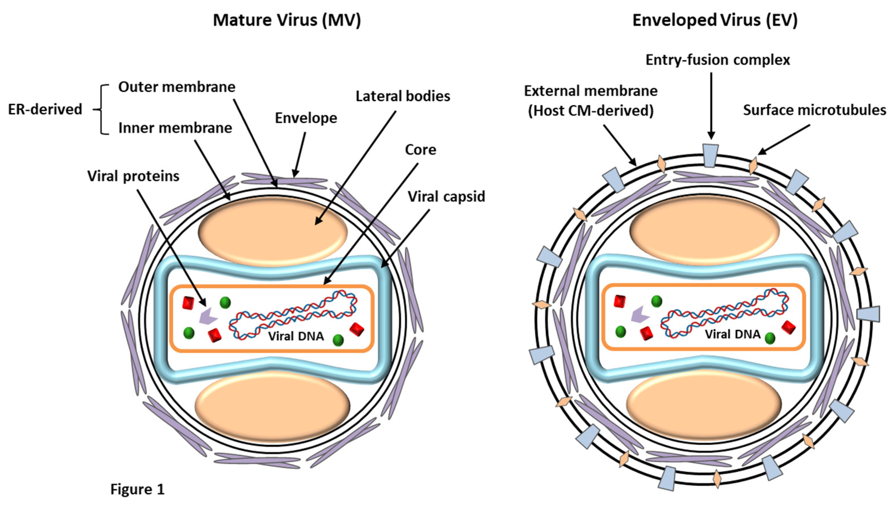
Figure 2.
The timeline of key events and major outbreaks associated with LSDV.
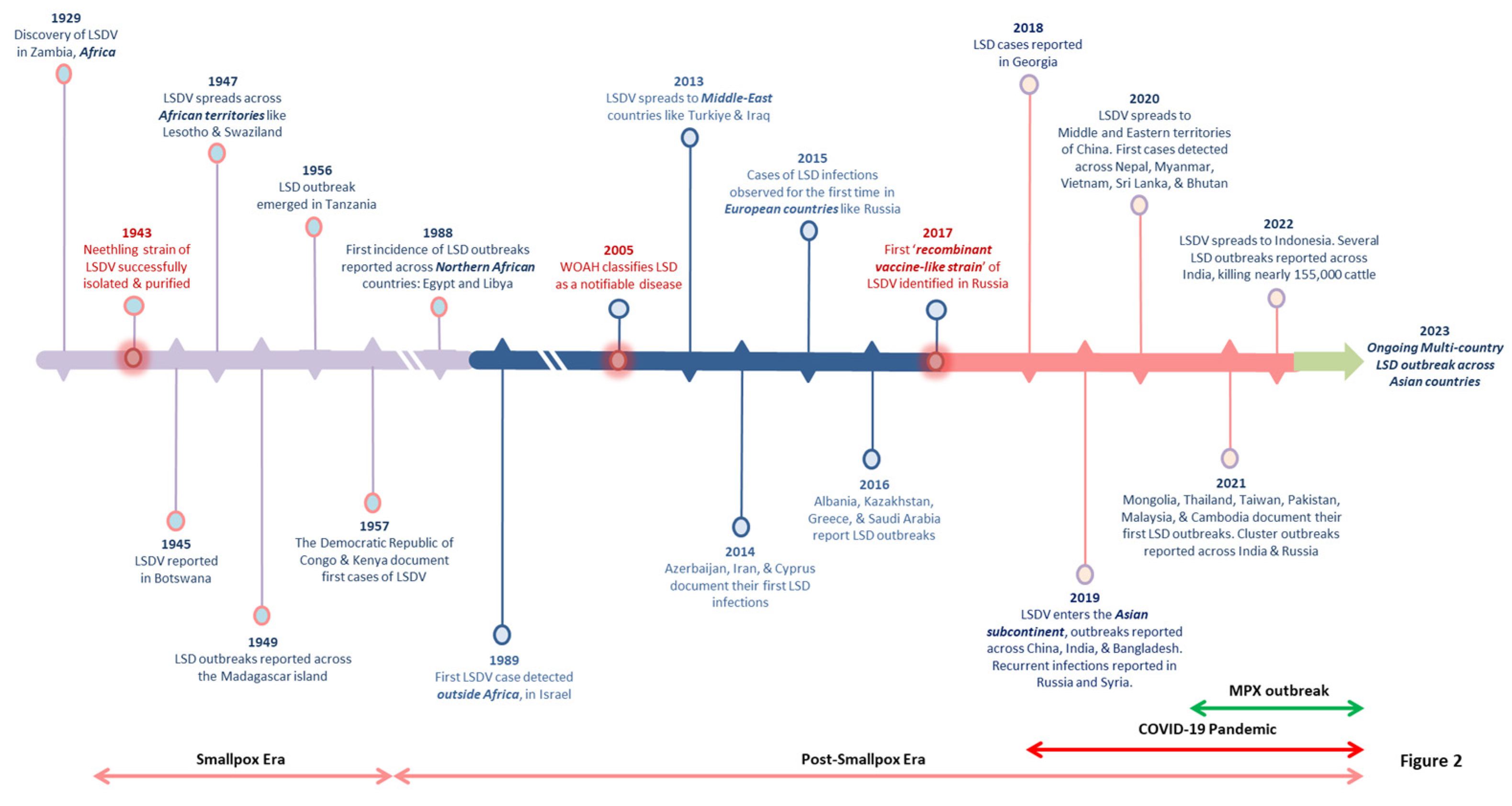
Figure 3.
The modes of LSDV transmission. The virus can be transmitted via (1) direct routes. Direct physical contact includes transmission through skin lesions, bodily fluids, etc. LSDV can also be acquired vertically from the mothers’ milk and through the intrauterine route. (2) Indirect transmission can occur by arthropod vectors and cross-border transportation (legal/illegal) of infected livestock.
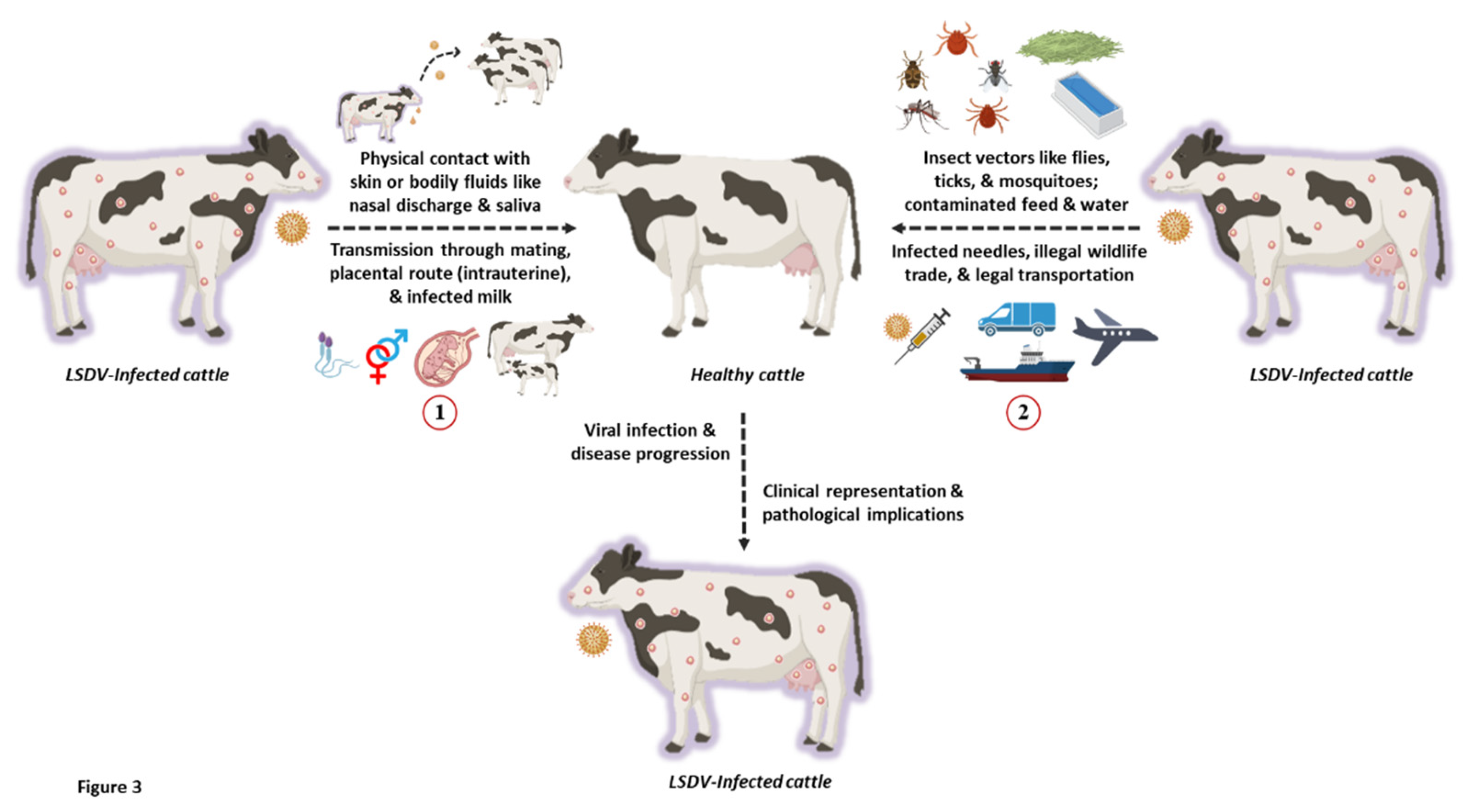
Figure 4.
The geographical distribution of LSDV infections across various countries over the past five years. The pie charts depict the proportion of LSDV associated with susceptible animals, reported cases, deaths, and animals vaccinated.
Figure 4.
The geographical distribution of LSDV infections across various countries over the past five years. The pie charts depict the proportion of LSDV associated with susceptible animals, reported cases, deaths, and animals vaccinated.
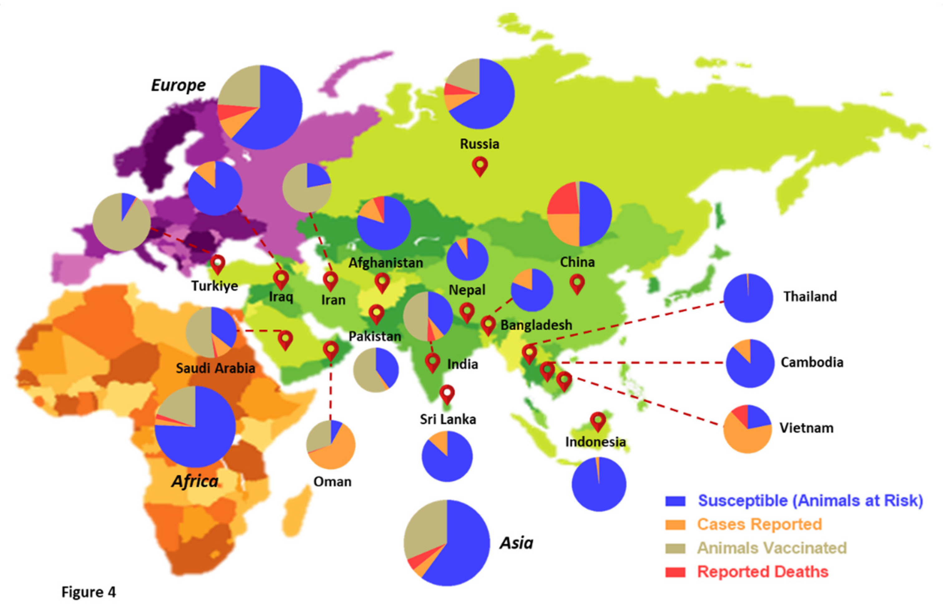
Figure 5.
The possible causes for the global resurgence and multi-country LSD outbreaks. Various factors have been speculated for the re-emergence and recurrent LSD outbreaks, including: (1) Legal/illegal trade and cross-border transportation of infected livestock. (2) Genetic mutations and recombination events influencing the LSDV virulence and transmissibility. (3) Climate change and high rainfall owing to elevated vector population further increasing the risk of viral infection. (4) Open grazing system also incurs a high risk of exposure to LSDV. (5) Reduced vaccine efficacy against the presently-circulating LSDV strains and a probable reversion of live-attenuated vaccine to virulent strains. (6) Lack of active surveillance, fragmented and inaccurate statistics, as well as monitoring of LSDV infections.
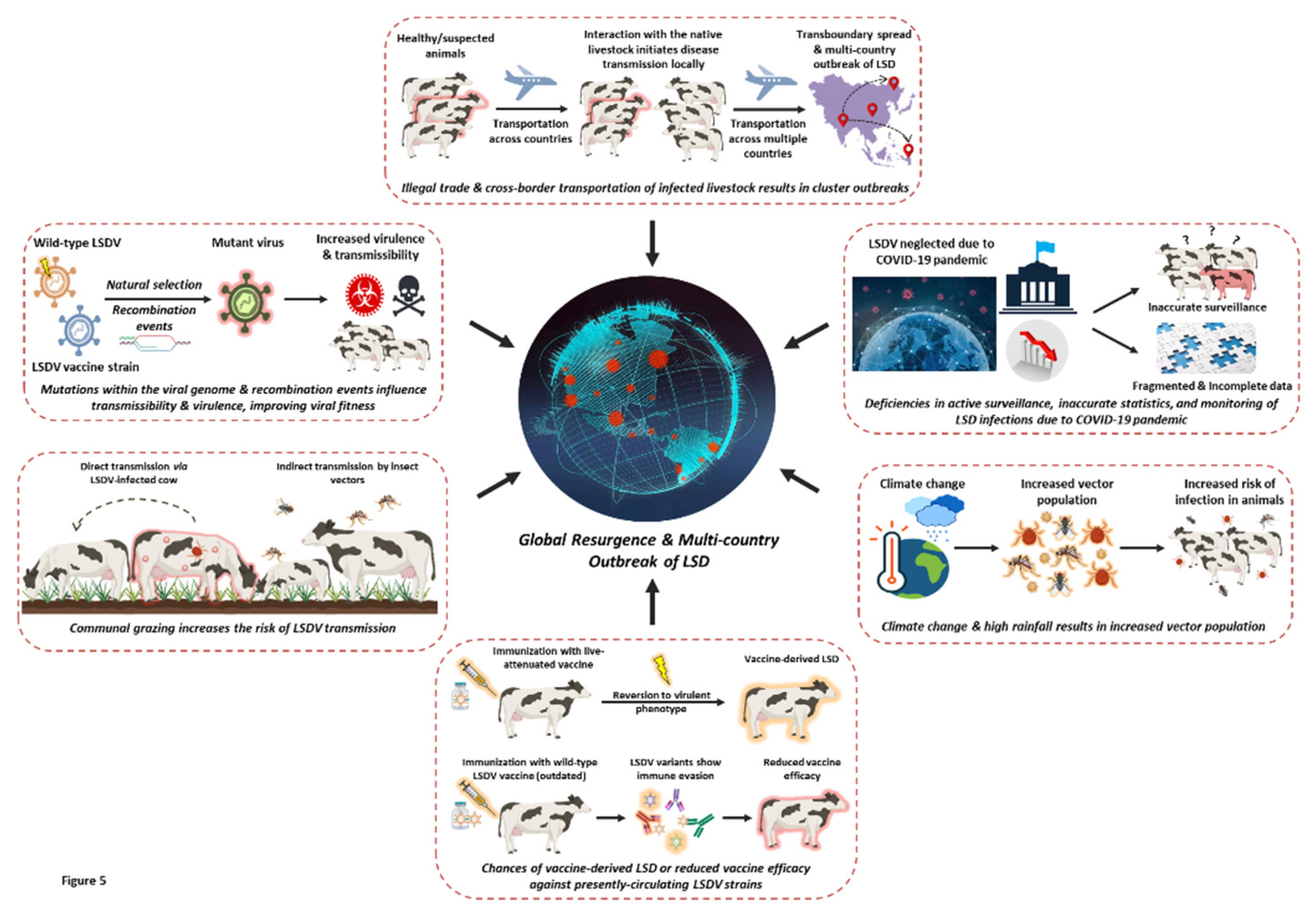

Table 1.
Commercially available live-attenuated LSDV vaccines and information about their formulations and protective efficacies.
Table 1.
Commercially available live-attenuated LSDV vaccines and information about their formulations and protective efficacies.
| S. No. | Commercial name | Viral strain | Target animal | Viral titer (per dose) | Vaccine efficacy | Reference |
|---|---|---|---|---|---|---|
| 1. | Lumpyvax™ (Intervet (Pty) South Africa/MSD Animal Health) |
LSD SIS Neethling type strain | Cattle | 104.0 TCID50 | ~ 80 % | http://www.msd-animal-health.co.za |
| 2. | Bovivax-LSD™ | LSD Neethling strain | Cattle | 103.5 TCID50 | 100 % | http://www.mci-santeanimale.com/en/ |
| 3. | Lumpy Skin Disease Vaccine for Cattle ( Onderstepoort Biological Products (OBP) South Africa) |
LSD Neethling strain | Cattle | Not known | ~ 70 % | http://www.obpvaccines.co.za |
| 4. | LumpyShield-N™ | LSD Neethling strain | Cattle | 104.0 TCID50 | Not available | http://www.jovaccenter.com |
| 5. | MEVAC LSD | LSD Neethling strain | Cattle | 103.5 TCID50 | ~ 41 % | https://www.me-vac.com/about |
| 6. | Lumpy Skin Disease Vaccine ( National Veterinary Institute (NVI) Ethiopia) |
LSD Neethling strain | Cattle | 103.0TCID50 | 100% |
https://www.nvi.com.et |
| 7. | Lumpivax™ [Kenya Veterinary Vaccines Production Institute (KEVEVAPI)] |
Live attenuated LSDV | Cattle | Not known | 100 % | http://www.kevevapi.org/ |
| 8. | Penpox-M™ Live SPPV |
Bakirköy SPPV strain | Cattle | 102.5 TCID50 | Not available | https://vetkontrol.tarimorman.gov.tr/pendik/Sayfalar/EN/AnaSayfa.aspx |
| 9. | Poxvac™ | Bakirköy SPPV strain | Sheep, Cattle |
102.5 TCID50 | Not available | http://www.vetal.com.tr |
| 10. | Lumpyvac™ | LSD Neethling strain | Cattle | 103.5 TCID50 | Not available | http://www.vetal.com.tr |
| 11. | Poxdoll™ |
Bakirköy SPPV strain | Cattle Sheep Goat |
102.5 TCID50 | Not available | http://www.dollvet.com.tr |
| 12. | LSD-NDOLLTM | LSD Neethling strain | Cattle | 103.5 TCID50 | Not available | http://www.dollvet.com.tr |
| 13. | Sheep Pox Cultural Dry™ |
Arriah SPPV Strain |
Sheep Cattle |
Not known | Not available | http://www.arriah.ru |
| 14. | Herbivac-LS | LSD Neethling strain | Cattle | Not known | 100 % |
https://deltamune.co.za/ |
| 15. | Kenyavac | KSGP 0240 | Sheep, goat, cattle | Not known | 100 % | https://jovaccenter.com |
| 16. | Jovivac | Yugoslavian SPPV RM-65 strain | Sheep, cattle | 103.9 TCID50 | Not available | https://jovaccenter.com |
Disclaimer/Publisher’s Note: The statements, opinions and data contained in all publications are solely those of the individual author(s) and contributor(s) and not of MDPI and/or the editor(s). MDPI and/or the editor(s) disclaim responsibility for any injury to people or property resulting from any ideas, methods, instructions or products referred to in the content. |
© 2023 by the authors. Licensee MDPI, Basel, Switzerland. This article is an open access article distributed under the terms and conditions of the Creative Commons Attribution (CC BY) license (http://creativecommons.org/licenses/by/4.0/).
Copyright: This open access article is published under a Creative Commons CC BY 4.0 license, which permit the free download, distribution, and reuse, provided that the author and preprint are cited in any reuse.


