PreprintArticleVersion 1Preserved in Portico This version is not peer-reviewed
Comparison of In vivo [18F]Fluoro-desoxyglucose and [18F]Fluoro-thymidine Positron Emission Tomography for Disease Monitoring in a Mouse Model of Higher-Risk Myelodysplastic Syndrome
Version 1
: Received: 2 April 2021 / Approved: 5 April 2021 / Online: 5 April 2021 (10:14:33 CEST)
How to cite:
Sarda-Mantel, L.; Gou, P.; Hontonnou, F.; Hosten, B.; Vignal, N.; San, C.; Brumpt, C.; Fenaux, P.; Chomienne, C.; Giraudier, S.; Padua, R. Comparison of In vivo [18F]Fluoro-desoxyglucose and [18F]Fluoro-thymidine Positron Emission Tomography for Disease Monitoring in a Mouse Model of Higher-Risk Myelodysplastic Syndrome. Preprints.org2021, 2021040087. https://doi.org/10.20944/preprints202104.0087.v1
Sarda-Mantel, L.; Gou, P.; Hontonnou, F.; Hosten, B.; Vignal, N.; San, C.; Brumpt, C.; Fenaux, P.; Chomienne, C.; Giraudier, S.; Padua, R. Comparison of In vivo [18F]Fluoro-desoxyglucose and [18F]Fluoro-thymidine Positron Emission Tomography for Disease Monitoring in a Mouse Model of Higher-Risk Myelodysplastic Syndrome. Preprints.org 2021, 2021040087. https://doi.org/10.20944/preprints202104.0087.v1
Cite as:
Sarda-Mantel, L.; Gou, P.; Hontonnou, F.; Hosten, B.; Vignal, N.; San, C.; Brumpt, C.; Fenaux, P.; Chomienne, C.; Giraudier, S.; Padua, R. Comparison of In vivo [18F]Fluoro-desoxyglucose and [18F]Fluoro-thymidine Positron Emission Tomography for Disease Monitoring in a Mouse Model of Higher-Risk Myelodysplastic Syndrome. Preprints.org2021, 2021040087. https://doi.org/10.20944/preprints202104.0087.v1
Sarda-Mantel, L.; Gou, P.; Hontonnou, F.; Hosten, B.; Vignal, N.; San, C.; Brumpt, C.; Fenaux, P.; Chomienne, C.; Giraudier, S.; Padua, R. Comparison of In vivo [18F]Fluoro-desoxyglucose and [18F]Fluoro-thymidine Positron Emission Tomography for Disease Monitoring in a Mouse Model of Higher-Risk Myelodysplastic Syndrome. Preprints.org 2021, 2021040087. https://doi.org/10.20944/preprints202104.0087.v1
Abstract
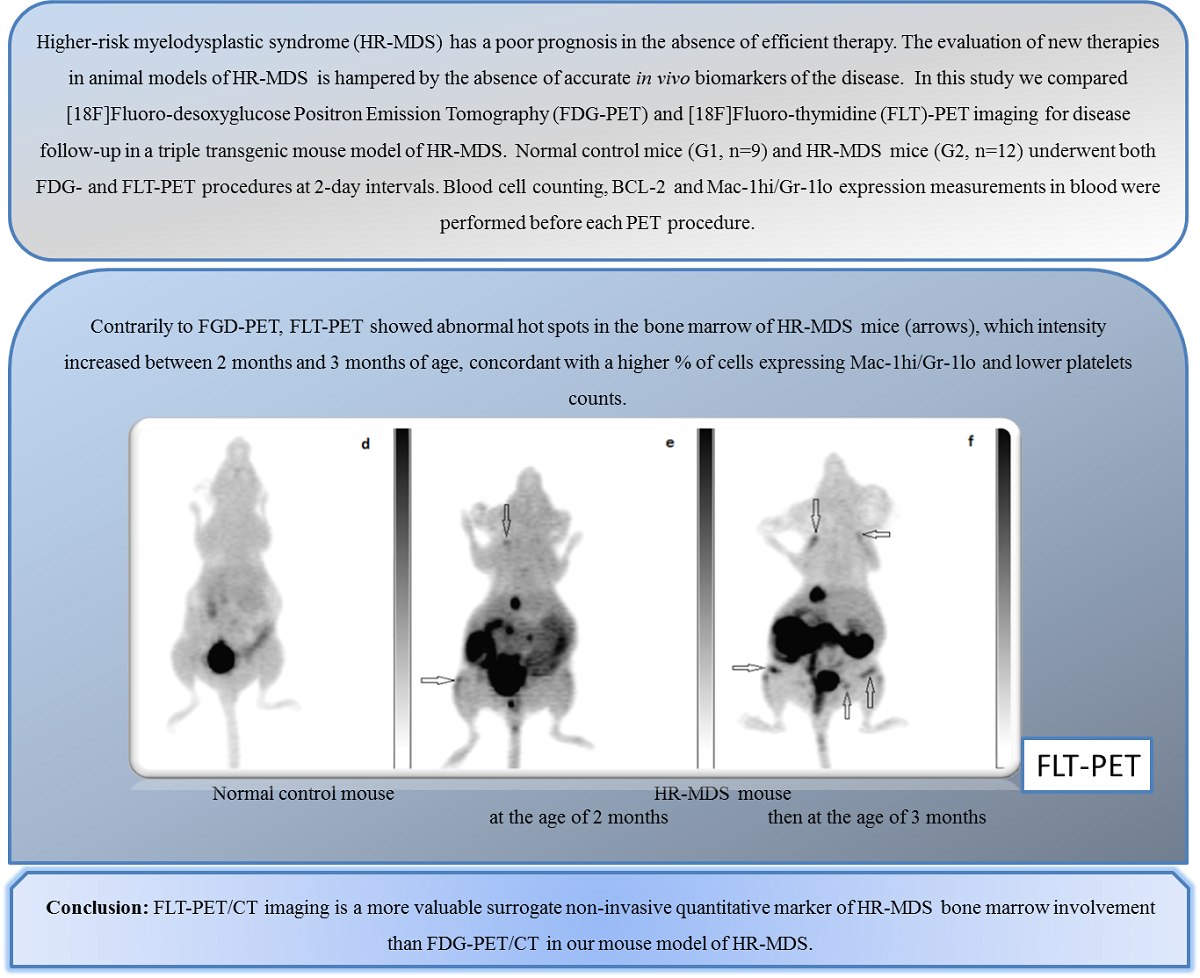
Higher-risk myelodysplastic syndrome (HR-MDS) has a poor prognosis in the absence of efficient therapy. The evaluation of new therapies in animal models of HR-MDS is hampered by the absence of accurate in vivo biomarkers of the disease. In this study we compared [18F]Fluoro-desoxyglucose Positron Emission Tomography (FDG-PET) and [18F]Fluoro-thymidine (FLT)-PET imaging for disease follow-up in a triple transgenic MMTVtTA/TetoBCL-2/MRP8NRASD12 mouse model of HR-MDS. Normal control FVB/N mice (G1,n=9) and HR-MDS mice (G2,n=12) underwent both FDG- and FLT-PET procedures at 2-day intervals, on a dedicated small animal device. Blood cell counting, BCL-2 and Mac-1hi/Gr-1lo expression measurements in blood were performed before each PET procedure. Visually, PET images of G2 mice demonstrated homogeneous FDG uptake in the whole skeleton similar to that observed in G1 mice, and abnormal FLT hot spots in bone marrow not observed in G1 mice. The intensity of FLT hot spots in bone marrow was higher in 3-months old G2 mice than in 2-months old G2 mice, concordant with a higher percentage of cells expressing Mac-1hi/Gr-1lo and lower platelets counts. We conclude that FLT-PET/CT imaging is a more valuable surrogate non-invasive quantitative marker of HR-MDS bone marrow involvement than FDG-PET/CT in our mouse model of HR-MDS.
Keywords
Myelodysplastic syndrome; Positron Emission Tomography; [18F]fluoro-thymidine; small animal imaging
Subject
Biology and Life Sciences, Biochemistry and Molecular Biology
Copyright:
This is an open access article distributed under the Creative Commons Attribution License which permits unrestricted use, distribution, and reproduction in any medium, provided the original work is properly cited.