Submitted:
20 January 2021
Posted:
21 January 2021
You are already at the latest version
Abstract
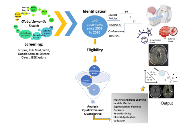
Medical brain image analysis is a necessary step in the Computers Assisted /Aided Diagnosis (CAD) systems. Advancements in both hardware and software in the past few years have led to improved segmentation and classification of various diseases. In the present work, we review the published literature on systems and algorithms that allow for classification, identification, and detection of White Matter Hyperintensities (WMHs) of brain MRI images specifically in cases of ischemic stroke and demyelinating diseases. For the selection criteria, we used the bibliometric networks. Out of a total of 140 documents we selected 38 articles that deal with the main objectives of this study. Based on the analysis and discussion of the revised documents, there is constant growth in the research and proposal of new models of deep learning to achieve the highest accuracy and reliability of the segmentation of ischemic and demyelinating lesions. Models with indicators (Dice Score, DSC: 0.99) were found, however with little practical application due to the uses of small datasets and lack of reproducibility. Therefore, the main conclusion is to establish multidisciplinary research groups to overcome the gap between CAD developments and their complete utilization in the clinical environment.
Keywords:
deep learning
; machine learning
; ischemic stroke
; demyelinating disease
; image processing
; computer aided diagnostics
; brain MRI
; CNN
; White Matter Hyperintensities
; VOSViewer
Copyright: This open access article is published under a Creative Commons CC BY 4.0 license, which permit the free download, distribution, and reuse, provided that the author and preprint are cited in any reuse.


