Submitted:
16 June 2018
Posted:
19 June 2018
You are already at the latest version
Abstract
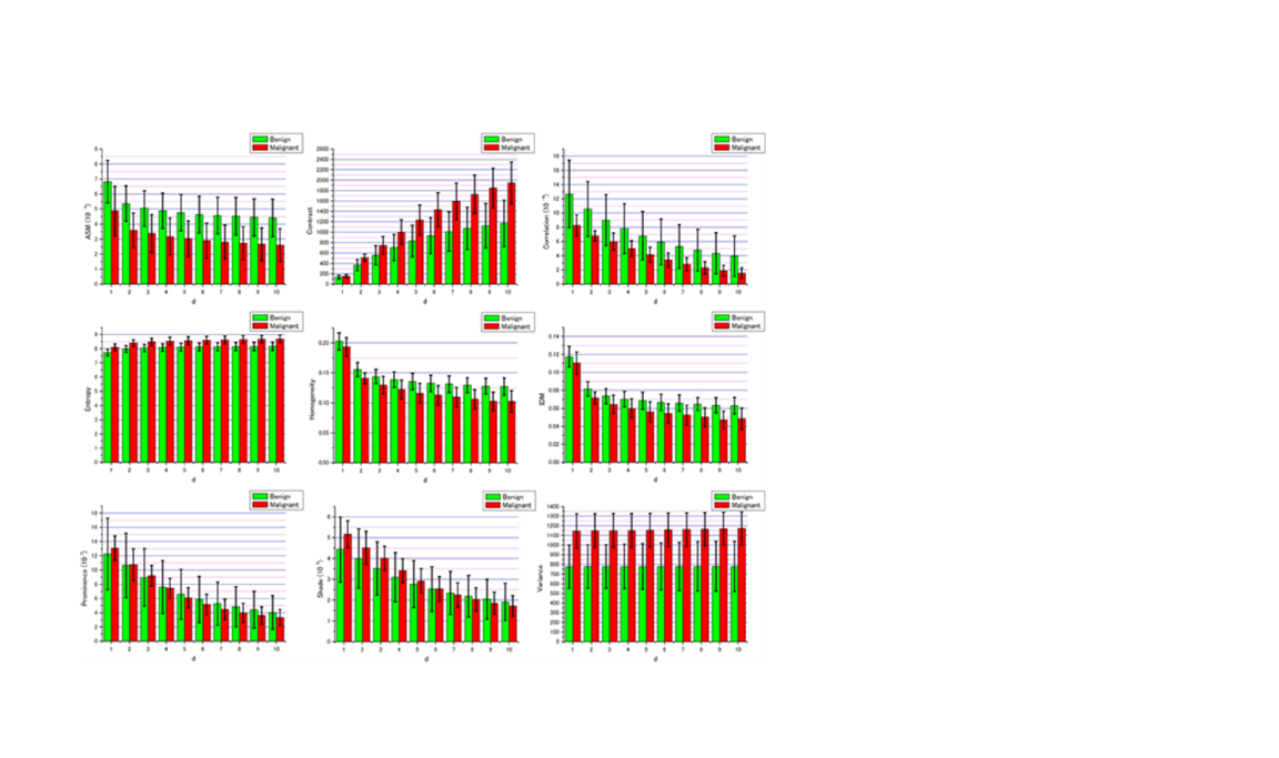
Label-free confocal photothermal (CPT) microscopy was utilized for the first time to investigate malignancy in mouse skin cells. A laser diode (LD) with 405nm or 488nm was used as a pump and 638nm LD as a probe for the CPT microscope. The Grey Level Cooccurrence Matrix (GLCM) for texture analysis was applied to the CPT images. Nine parameters of GLCM were calculated for the intracellular super-resolved CPT images, and the parameters Entropy and Prominence were found to be most suited among the nine parameters to discriminate between healthy cells and MM cells in case pump wavelength of 488nm is used.
Keywords:
photothermal microscopy
; label-free imaging
; melanoma
; texture analysis
Copyright: This open access article is published under a Creative Commons CC BY 4.0 license, which permit the free download, distribution, and reuse, provided that the author and preprint are cited in any reuse.


