
Submitted:
16 November 2026
Posted:
18 November 2026
You are already at the latest version
Abstract
Background: Obesity-related glomerulopathy (ORG) is characterised by glomerulomegaly, with or without an adaptive focal segmental glomerulosclerosis lesion, and its incidence is rising in parallel with the obesity epidemic. Case presentation: We report the case of a 43-year-old man with class III obesity (body mass index 40.2 kg/m²) and a 20-year history of arterial hypertension who was referred for newly detected renal impairment. He was admitted with elevated serum creatinine (261 µmol/L, eGFR 26 mL/min/1.73 m²), moderately increased proteinuria (UPCR 172.6 mg/mmol) with normal serum albumin, urinary sediment without erythrocyturia, and features of metabolic syndrome. An oral glucose tolerance test and HbA1c confirmed previously undiagnosed type 2 diabetes mellitus. Kidney biopsy showed glomerulomegaly with the perihilar variant of FSGS, superimposed on moderate arteriosclerosis and 40–50% interstitial fibrosis and tubular atrophy, without nodular diabetic glomerulosclerosis or electrondense immune deposits. Although electron microscopy showed diffuse foot-process effacement, the integrated clinicopathological findings supported ORG. After adding empagliflozin to renin–angiotensin–aldosterone system blockade, along with dietary and lipid-lowering measures, proteinuria decreased (urine protein/creatinine ratio from 172.6 to 132.8 mg/mmol), and kidney function remained stable. Conclusion: This case highlights the role of kidney biopsy in patients with obesity and advanced CKD in identifying coexisting lesions, interpreting ultrastructural findings in clinical context, and screening for diabetes in patients with CKD.
Keywords:
obesity-related glomerulopathy
; glomerulomegaly
; SGLT2 inhibitors
1. Introduction and Clinical Significance
In 2024, approximately 940 million adults worldwide were living with obesity [1]. The global prevalence of obesity has risen dramatically every year. By 2050, an estimated 3.8 billion adults aged ≥25 years are projected to be living with overweight or obesity globally [2].
Obesity is an independent risk factor for the development and progression of CKD and end-stage kidney disease (ESKD), beyond its association with type 2 diabetes mellitus, hypertension, and dyslipidaemia [3]. In a historical series of 6,818 native kidney biopsies examined between 1986 and 2000, the proportion showing ORG increased from 0.2% in 1986–1990 to 2.0% in 1996–2000 [4].
ORG is defined pathologically as glomerulomegaly with or without perihilar focal segmental glomerulosclerosis (FSGS) in patients with a body mass index (BMI) ≥30 kg/m². When present, the associated FSGS lesion is typically interpreted as secondary and adaptive [5].
The pathophysiology of ORG is multifactorial, involving both hemodynamic and metabolic mechanisms [5]. Obesity-induced afferent arteriolar dilatation, increased proximal tubular sodium reabsorption, and activation of the renin–angiotensin–aldosterone system (RAAS) lead to glomerular hyperfiltration and intraglomerular hypertension [5,6]. Insulin resistance, adipokine imbalance, ectopic renal lipid accumulation with lipotoxicity, and oxidative stress further contribute to this maladaptive process [7]. These changes drive compensatory glomerulomegaly; however, adaptive podocyte hypertrophy ultimately fails to keep pace with glomerular tuft expansion, resulting in podocyte depletion and segmental sclerosis [5,7]. A predominance of the perihilar FSGS variant, together with relatively mild foot-process effacement, helps distinguish ORG from primary FSGS [5,8].
Clinically, ORG typically presents with slowly progressive, moderately increased proteinuria, while full nephrotic syndrome is distinctly unusual. Despite this generally indolent course, up to one-third of patients develop progressive renal failure and ESKD [5,9].
Diagnosis of ORG requires kidney biopsy and careful clinicopathological correlation, because patients with obesity frequently have other or coexisting kidney diseases. In a large biopsy series of patients with severe obesity, ORG was found in only 29% of cases, and in most of these it coexisted with another kidney disease [10].
This is particularly challenging when obesity coexists with long-standing hypertension and diabetes, which may produce overlapping histological lesions [11,12].
Management of ORG rests on two complementary pillars: long-term weight reduction to address the underlying cause, and renoprotective pharmacotherapy to reduce proteinuria and slow functional decline [13]. Weight reduction includes lifestyle interventions (dietary modification and physical activity), anti-obesity pharmacotherapy, and bariatric/metabolic surgery, applied stepwise according to BMI [13]. The cornerstones of renoprotective therapy are RAAS inhibitors for their antiproteinuric effect and sodium–glucose cotransporter-2 (SGLT2) inhibitors, which combine renoprotective and weight-loss benefits [13,14]. Mineralocorticoid receptor antagonists are a further component of renoprotective treatment [13].
Here, we present a 43-year-old man with class III obesity, long-standing hypertension, proteinuric CKD, and previously undiagnosed type 2 diabetes mellitus. The clinicopathological findings favoured ORG with coexisting chronic vascular injury, although extensive foot-process effacement in a limited electron microscopy sample complicated interpretation. The case highlights the value and limitations of integrated biopsy assessment and describes the short-term clinical course following combined therapeutic and dietary interventions.
2. Case Presentation
During a routine examination by his general practitioner, elevated serum creatinine (249 µmol/L) and proteinuria (+++ on urine dipstick) had been found, prompting referral.
He had a more than 20-year history of arterial hypertension. He also had hyperlipidaemia and class III obesity and was a non-smoker. On presentation, he reported foamy urine and intermittent lower-leg oedema, without dysuria. His family history included diabetes mellitus in his father. His long-term medications comprised a maximal dose of fixed combination of perindopril/indapamide/amlodipine, nebivolol, moxonidine, and atorvastatin.
On physical examination, he had abdominal obesity, with a body weight of 119 kg, a height of 172 cm, BMI of 40.2 kg/m², and a waist circumference of 123 cm. Blood pressure was 129/88 mmHg and heart rate was 64 beats/min. Bilateral pitting oedema of the lower legs was present. The remainder of the examination was unremarkable.
Seven months before admission, the patient’s serum creatinine was already elevated (201 µmol/L), indicating that kidney dysfunction had persisted for more than 3 months and met the KDIGO chronicity criterion for CKD. Laboratory findings on admission were consistent with CKD stage G4, category A3 (severe increased albuminuria), according to the Kidney Disease: Improving Global Outcomes (KDIGO) classification with serum creatinine having risen to 261 µmol/L, estimated glomerular filtration rate (eGFR) 26 mL/min/1.73m² (calculated using the 2021 CKD-EPI creatinine equation), urine albumin/creatinine ratio 135.8 mg/mmol, urine protein/creatinine ratio (172.6 mg/mmol), and serum albumin 40.1 g/L (shown in Table 1). The patient had dyslipidaemia with marked hypertriglyceridaemia (triglycerides 6.72 mmol/L) and low HDL cholesterol (0.72 mmol/L). Urinary sediment analysis was negative, with no erythrocyturia detected.
Renal ultrasound showed kidneys of normal size with irregular contours, reduced parenchymal thickness, and marked hyperechogenicity, consistent with advanced chronic renal parenchymal damage. The hepatic parenchyma showed features of diffuse steatosis. Corticomedullary differentiation was preserved, and no hydronephrosis, cysts, or calculi were seen; colour Doppler showed patent vessels.
In the differential diagnosis of moderately increased proteinuria, viral serology for hepatitis B, hepatitis C, and human immunodeficiency virus was negative, as well as immunological screening for glomerular disease, including antinuclear antibodies (ANA), myeloperoxidase-ANCA (MPO-ANCA), proteinase 3-ANCA (PR3-ANCA), anti-glomerular basement membrane (anti-GBM) antibodies, and anti-PLA2R. Serum complement levels (C3 and C4) were within the reference range, and no monoclonal gammopathy was detected.
An ultrasound-guided core biopsy of the left kidney was performed, yielding three cores. On light microscopy, the specimen was of borderline adequacy, containing ten glomeruli (none globally sclerosed). These showed mild-to-moderate glomerulomegaly, and three glomeruli had segmental sclerosis of the perihilar variant of focal segmental glomerulosclerosis (FSGS). Light microscopy showed no mesangial expansion or Kimmelstiel-Wilson nodules. Light microscopy identified no overt glomerular basement membrane thickening. Interstitial fibrosis and tubular atrophy (IFTA) involved approximately 40–50% of the tissue, and the vessels showed mild-to-moderate medial and fibrointimal hyperplasia with moderate luminal narrowing (cv2) (Figure 1). Immunofluorescence revealed only weak, nonspecific mesangial IgA and IgM with tubular casts and negative amyloid staining, insufficient to support IgA nephropathy. Electron microscopy (one glomerulus) showed segmental sclerosis, irregularly thickened glomerular basement membranes with subendothelial lucencies and focal double contours, and diffuse foot-process effacement (80–100%), without electron-dense immune deposits (Figure 2).
During hospitalisation, an oral glucose tolerance test (oGTT) revealed previously undiagnosed type 2 diabetes mellitus, with a fasting plasma glucose of 5.7 mmol/L and a 2-hour value of 12.2 mmol/L; HbA1c was 54.1 mmol/mol (IFCC). Fasting serum C-peptide was 4.09 µg/L and fasting insulin 7.8 mU/L (HOMA-IR 1.8), consistent with preserved endogenous insulin secretion.
Taken together, the clinical and pathological findings favoured obesity-related glomerulopathy with an adaptive perihilar FSGS pattern and coexisting chronic vascular injury. Interpretation of the extensive foot-process effacement was limited by the available electron-microscopy sample
Because of advanced CKD, the fixed-dose combination of perindopril/indapamide/amlodipine and moxonidine were discontinued. Antihypertensive therapy was continued with perindopril at [10 mg once daily] and nebivolol at [5 mg once daily], and empagliflozin 10 mg once daily was initiated. The lipid-lowering therapy was intensified by adding ezetimibe to atorvastatin. Ketoanalogues of amino acids were added at 12 tablets per day to support the newly initiated low-protein diet (0.8 g/kg/day), together with sodium restriction (< 2 g/day). The patient was advised to reduce body weight, sodium, and animal-fat intake, and early follow-up at a diabetology clinic was arranged.
At outpatient follow-up approximately four months later, the patient remained clinically stable. At the four-month follow-up visit, the patient reported no treatment-related adverse effects. Home blood pressure measurements ranged from 114–130/63–73 mmHg. Renal function remained stable, with serum creatinine 237 µmol/L and eGFR 29 mL/min/1.73m². Proteinuria decreased with continued empagliflozin treatment, with a UACR of 95.1 mg/mmol and a UPCR of 132.8 mg/mmol.
3. Discussion
Obesity is a chronic metabolic disease and an independent risk factor for the development and progression of CKD. ORG is its specific renal manifestation, and its incidence in native kidney biopsies increased tenfold within 15 years [4]. ORG typically affects middle-aged adults (mean age 37–46 years) and presents with non-nephrotic proteinuria in 52–90% of patients and normal serum albumin, whereas full nephrotic syndrome occurs in fewer than 5% [5]. Despite this indolent course, up to one-third progress to ESKD [5].
Our patient’s clinical picture was consistent with ORG. We present a 43-year-old man with class III obesity, long-standing hypertension, dyslipidaemia, newly diagnosed type 2 diabetes mellitus, moderately increased proteinuria (UPCR 172.6 mg/mmol), and an eGFR of 26 mL/min/1.73 m². Despite normoalbuminaemia, the lower-leg oedema was likely multifactorial, related to dihydropyridine calcium channel blocker therapy and obesity-associated venous hypertension, rather than nephrotic physiology [15,16].
However, the clinical phenotype alone is not sufficient to establish the diagnosis, because ORG frequently coexists with other kidney diseases. Choung et al. analysed 248 native kidney biopsies from patients with a BMI ≥40 kg/m²; ORG was found in 73 patients (29%), of whom only 29 had ORG as the sole diagnosis. Diabetic nephropathy and hypertensive nephrosclerosis were among the most frequent alternative diagnoses, and the biopsy yielded a management-changing diagnosis in 49% of patients [10]. Importantly, an eGFR <30 mL/min/1.73 m² was the strongest predictor of non-ORG lesions in that cohort [10]. With an eGFR of 26 mL/min/1.73 m², our patient therefore had a high pre-test probability of an alternative or coexisting pathology, which justified kidney biopsy.
Light microscopy revealed the histological hallmarks of ORG: glomerulomegaly and the perihilar variant of FSGS. The perihilar localisation is pathophysiologically consistent with hyperfiltration, as the vascular pole is exposed to the highest filtration pressure [5]. The principal histological differential diagnoses were early diabetic nephropathy and hypertensive nephrosclerosis. ORG may show mesangial matrix expansion and GBM thickening resembling mild diabetic glomerulosclerosis, reflecting shared molecular pathways [5]. In our patient, diabetes was newly diagnosed and mild [HbA1c (54.1 mmol/mol; 7.1%)], and no nodular mesangial sclerosis (Kimmelstiel–Wilson nodules) was present, making established diabetic nephropathy unlikely as the dominant lesion. Nevertheless, the irregular GBM thickening, subendothelial lucencies, and focal double contours suggest a component of chronic endothelial injury, most likely related to long-standing hypertension, and early hyperglycaemia-related changes cannot be fully excluded. Moderate arteriosclerosis (cv2) and IFTA involving 40–50% of the cortex indicate a substantial hypertensive and chronic component. This degree of tubulointerstitial scarring exceeds that typically described in ORG, where interstitial fibrosis is generally milder than in primary FSGS [5], and likely reflects the synergistic effect of obesity, hypertension, and dysglycaemia on glomerular and vascular lesions [11]. Immunofluorescence showed only weak, nonspecific mesangial IgA and IgM staining, without corresponding electron-dense deposits on electron microscopy; therefore, these findings were insufficient to support a diagnosis of IgA nephropathy [17].
A seemingly discordant finding was the diffuse foot-process effacement (80–100%). The clinical phenotype and light-microscopic findings favoured an adaptive FSGS pattern in the setting of severe obesity and long-standing hypertension. However, electron microscopy was limited to one glomerulus containing segmental sclerosis, which restricted reliable assessment of the distribution of foot-process effacement [5]. The reported extensive effacement therefore remains a diagnostic limitation.
Our patient’s metabolic profile—abdominal obesity, marked hypertriglyceridaemia, low HDL cholesterol, and hepatic steatosis—corresponded to metabolic syndrome, in which insulin resistance, adipokine imbalance, and renal lipotoxicity contribute to ORG development [5,7]. The low HOMA-IR value (1.8) should be interpreted with caution, as reduced renal insulin clearance in advanced CKD limits the reliability of fasting insulin-based indices. Type 2 diabetes was diagnosed based on a 2-hour plasma glucose of 12.2 mmol/L on oGTT and an HbA1c of 54.1 mmol/mol (7.1%), both above diagnostic thresholds, whereas fasting plasma glucose (5.3 mmol/L) remained within the normal range. Screening based on fasting glucose alone would therefore have missed the diagnosis. Moreover, HbA1c is less reliable in advanced CKD (eGFR <30 mL/min/1.73 m²), mainly due to altered erythrocyte turnover and haemoglobin carbamylation, although our patient had no anaemia [18]. HbA1c may thus misclassify glycaemic status in either direction. In this setting, oGTT gives a direct measure of glucose tolerance that does not depend on erythrocyte turnover. It can reveal postprandial hyperglycaemia even when fasting glucose is normal, which allows diabetes to be diagnosed and treated before it causes systemic, including renal, complications. Diabetes screening practices vary widely, as also shown in kidney transplant candidates and recipients, which underlines the need for a systematic approach to diabetes screening in patients with advanced CKD [19].
Managing ORG is complex and targets the underlying cause, combining long-term weight reduction with renoprotective pharmacotherapy. RAAS blockade reduces proteinuria, but its effect may be transient, and many patients progress despite treatment [13]. SGLT2 inhibitors reduce intraglomerular pressure via tubuloglomerular feedback, decrease proximal tubular workload, promote modest weight loss, and reduce albuminuria independently of glycaemic control [9]. In experimental ORG, empagliflozin reduced albuminuria, serum lipids, and glomerular diameter; however, clinical data specific to ORG remain limited [9]. Their use in our patient is supported by large outcome trials in CKD; in the EMPA-KIDNEY trial, empagliflozin reduced the risk of kidney disease progression or cardiovascular death across a wide range of causes of CKD, irrespective of diabetes status, and in patients with an eGFR as low as 20 mL/min/1.73 m² [21]. SGLT2 inhibitors also facilitate continued RAAS blockade in albuminuric CKD [21].
In our case, adding empagliflozin to perindopril reduced proteinuria by approximately 23% (UPCR from 172.6 to 132.8 mg/mmol) and albuminuria by approximately 30% (UACR from 135.8 to 95,1 mg/mmol) over four months, with stable kidney function (eGFR from 26 to 29 mL/min/1.73 m²) and controlled blood pressure. This reduction is modest, and because lipid-lowering therapy, dietary sodium restriction, a low-protein diet with ketoanalogues, and lifestyle advice were introduced simultaneously, it cannot be attributed to empagliflozin alone.
Given the patient’s risk of CKD progression, additional therapeutic options could be considered during subsequent follow-up, subject to individual eligibility and tolerability. KDIGO 2024 suggests a non-steroidal mineralocorticoid receptor antagonist with proven kidney or cardiovascular benefit for patients with type 2 diabetes, an eGFR >25 mL/min/1.73 m², normal serum potassium, and albuminuria (>30 mg/g) despite a maximum tolerated dose of a renin-angiotensin system inhibitor [23]. GLP-1 receptor agonists are particularly attractive in this phenotype; in the FLOW trial, semaglutide reduced major kidney disease events by 24% in patients with type 2 diabetes and CKD, in addition to inducing weight loss [22]. With a BMI of 40.2 kg/m², bariatric/metabolic surgery is also an option, as weight reduction has consistently reduced proteinuria in ORG, although large prospective trials are lacking [5,13].
Our report has some limitations. Follow-up was short (four months), and kidney function was estimated using a creatinine-based equation, which may be less accurate in severe obesity. Proteinuria and albuminuria were assessed at baseline and at a single four-month follow-up visit, without repeated measurements to confirm the persistence of the observed reductions. These findings should therefore be interpreted as short-term changes observed during combined management rather than evidence of a sustained antiproteinuric effect attributable to empagliflozin or of long-term renoprotection.
Clinically, this case represents a scenario nephrologists will encounter increasingly often. ORG should be considered in the differential diagnosis of proteinuric CKD in patients with severe obesity. In selected patients with an uncertain aetiology of kidney disease, kidney biopsy may help identify coexisting lesions and refine the clinicopathological diagnosis. This case also shows that clinicians can establish the diagnosis of ORG on integrated clinicopathological grounds even when individual ultrastructural features deviate from the classic description. Another important message for clinical practice is the need for systematic screening for diabetes, including oGTT, in patients with CKD and metabolic risk factors.
4. Conclusions
ORG should be considered in patients with severe obesity and proteinuric CKD, but coexisting causes of kidney injury require careful assessment. In selected patients with an uncertain aetiology, kidney biopsy may refine the clinicopathological diagnosis. In this case, the clinical and light-microscopic findings favoured ORG with chronic vascular injury, while extensive foot-process effacement in a limited electron-microscopy sample remained a diagnostic limitation. Management should address kidney, cardiovascular, and metabolic risk through an individualised multidisciplinary approach. The short-term follow-up does not establish a treatment-specific effect or long-term renoprotection.
Author Contributions
Writing — original draft preparation, P.K.; writing—review and editing, K.G.; visualisation, T.J.; project administration, M.V.; supervision, I.D. All authors have read and agreed to the published version of the manuscript.
Funding
This project was funded through the Slovak Research and Development Agency under the project no. VV-MVP-24-0252.
Institutional Review Board Statement
This study was conducted in accordance with the Declaration of Helsinki. The Ethical Committee of University Hospital Martin has waived the ethical approval for reporting individual cases or case series when the patient provides informed consent.
Informed Consent Statement
The patient provided written informed consent for publication of this paper.
Data Availability Statement
The original contributions presented in this study are included in this article; further inquiries can be directed to the corresponding author.
Acknowledgments
We would like to thank Peter Talarčík and Magdaléna Drobná at Cytopathos spol. s.r.o., in Bratislava, Slovakia, for renal biopsy analysis.
Conflicts of Interest
The authors declare no conflicts of interest.
Abbreviations
| ALP | alkaline phosphatase |
| ALT | alanine aminotransferase |
| ANA | antinuclear antibodies |
| anti-GBM | anti-glomerular basement membrane antibodies |
| anti-PLA2R | anti-phospholipase A2 receptor antibodies |
| AST | aspartate aminotransferase |
| BMI | body mass index |
| CKD | chronic kidney disease |
| eGFR | estimated glomerular filtration rate |
| ESKD | end-stage kidney disease |
| FSGS | focal segmental glomerulosclerosis |
| GBM | glomerular basement membrane |
| GGT | gamma-glutamyltransferase |
| GLP-1 | glucagon-like peptide-1 |
| HbA1c | glycated haemoglobin |
| HCO₃⁻ | bicarbonate |
| HDL | high-density lipoprotein |
| HOMA-IR | homeostasis model assessment of insulin resistance |
| IFCC | International Federation of Clinical Chemistry and Laboratory Medicine |
| IFTA | interstitial fibrosis and tubular atrophy |
| KDIGO | Kidney Disease: Improving Global Outcomes |
| LDL | low-density lipoprotein |
| MPO-ANCA | myeloperoxidase–antineutrophil cytoplasmic antibodies |
| oGTT | oral glucose tolerance test |
| ORG | obesity-related glomerulopathy |
| PR3-ANCA | proteinase 3–antineutrophil cytoplasmic antibodies |
| PTH | parathyroid hormone |
| RAAS | renin–angiotensin–aldosterone system |
| SGLT2 | sodium–glucose cotransporter-2 |
| UACR | urine albumin/creatinine ratio |
| UPCR | urine protein/creatinine ratio |
| WBCs | white blood cells |
References
- World Health Organization. Obesity and Overweight. Available online: https://www.who.int/news-room/fact-sheets/detail/obesity-and-overweight (accessed on 20 October 2026).
- GBD 2021 Adult BMI Collaborators. Global, regional, and national prevalence of adult overweight and obesity, 1990–2021, with forecasts to 2050: A forecasting study for the Global Burden of Disease Study 2021. Lancet 2025, 405, 813–838. [CrossRef]
- Hsu, H.C.; Shih, L.J.; Hou, Y.C.; Lu, K.C. Mechanisms of obesity-related kidney disease: From adipose depot biology to the chymase-aldosterone and ghrelin-leptin axes. Biomolecules 2026, 16, 1155. [CrossRef]
- Kambham, N.; Markowitz, G.S.; Valeri, A.M.; Lin, J.; D’Agati, V.D. Obesity-related glomerulopathy: An emerging epidemic. Kidney Int. 2001, 59, 1498–1509. https://doi.org/10.1046/j.1523-1755.2001.0590041498.x. 5. D’Agati, V.D.; Chagnac, A.; de Vries, A.P.; Levi, M.; Porrini, E.; Herman-Edelstein, M.; Praga, M. Obesityrelated glomerulopathy: Clinical and pathologic characteristics and pathogenesis. Nat. Rev. Nephrol. 2016, 12, 453–471. [CrossRef]
- Cortinovis, M.; Perico, N.; Ruggenenti, P.; Remuzzi, A.; Remuzzi, G. Glomerular hyperfiltration. Nat. Rev. Nephrol. 2022, 18, 435–451. [CrossRef]
- Ahmed, N.; Dalmasso, C.; Turner, M.B.; Arthur, G.; Cincinelli, C.; Loria, A.S. From fat to filter: The effect of adipose tissue-derived signals on kidney function. Nat. Rev. Nephrol. 2025, 21, 417–434. [CrossRef]
- Yau, K.; Kuah, R.; Cherney, D.Z.I.; Lam, T.K.T. Obesity and the kidney: Mechanistic links and therapeutic advances. Nat. Rev. Endocrinol. 2024, 20, 321–335. [CrossRef]
- Liu, Y.; Eirin, A.; Lerman, L.O. Novel therapeutic strategies for obesity-related glomerulopathy. Curr. Hypertens. Rep. 2026, 28, 3. [CrossRef]
- Choung, H.G.; Bomback, A.S.; Stokes, M.B.; Santoriello, D.; Campenot, E.S.; Batal, I.; Markowitz, G.S.; D’Agati, V.D. The spectrum of kidney biopsy findings in patients with morbid obesity. Kidney Int. 2019, 95, 647–654. [CrossRef]
- Sasaki, T.; Tsuboi, N.; Okabayashi, Y.; Haruhara, K.; Kanzaki, G.; Koike, K.; Takahashi, H.; Ikegami, M.; Shimizu, A.; Yokoo, T. Synergistic impact of diabetes and hypertension on the progression and distribution of glomerular histopathological lesions. Am. J. Hypertens. 2019, 32, 900–908. [CrossRef]
- Martinez, A.; Letourneau, P.; Rabeyrin, M.; Picard, C.; Guebre-Egziabher, F.; Fouque, D.; Caussy, C.; Soulage, C.; Koppe, L. Kidney biopsy findings in patients with obesity exhibit a wide spectrum of disease entities. Clin. Kidney J. 2026, 19, sfag092. [CrossRef]
- Herman-Edelstein, M.; Weinstein, T.; Chagnac, A. Obesity-related glomerulopathy: Clinical management. Semin. Nephrol. 2021, 41, 358–370. [CrossRef]
- Caravaca-Fontán, F.; Stevens, K.; Padrón, M.; Huerta, A.; Montomoli, M.; Villa, J.; González, F.; Vega, C.; Rivas, B.; Gutiérrez, E.; et al. Sodium-glucose cotransporter 2 inhibition in primary and secondary glomerulonephritis. Nephrol. Dial. Transplant. 2024, 39, 328–340. [CrossRef]
- Liang, L.; Kung, J.Y.; Mitchelmore, B.; Cave, A.; Banh, H.L. Comparative peripheral edema for dihydropyridines calcium channel blockers treatment: A systematic review and network meta-analysis. J. Clin. Hypertens. 2022, 24, 536–554. [CrossRef]
- Fukaya, E.; Kolluri, R. Nonsurgical management of chronic venous insufficiency. N. Engl. J. Med. 2024, 391, 2350–2359. [CrossRef]
- Stamellou, E.; Seikrit, C.; Tang, S.C.W.; Boor, P.; Tesař, V.; Floege, J.; Barratt, J.; Kramann, R. IgA nephropathy. Nat. Rev. Dis. Primers 2023, 9, 67. [CrossRef]
- Hassanein, M.; Shafi, T. Assessment of glycemia in chronic kidney disease. BMC Med. 2022, 20, 117. [CrossRef]
- Laghrib, Y.; Hilbrands, L.; Oniscu, G.C.; Crespo, M.; Gandolfini, I.; Mariat, C.; Mjøen, G.; Sever, M.S.; Watschinger, B.; Velioglu, A.; et al. Current practices in prevention, screening, and treatment of diabetes in kidney transplant recipients: European survey highlights from the ERA DESCARTES Working Group. Clin. Kidney J. 2024, 18, sfae367. [CrossRef]
- Martínez-Montoro, J.I.; Morales, E.; Cornejo-Pareja, I.; Tinahones, F.J.; Fernández-García, J.C. Obesity-related glomerulopathy: Current approaches and future perspectives. Obes. Rev. 2022, 23, e13450. [CrossRef]
- The EMPA-KIDNEY Collaborative Group; Herrington, W.G.; Staplin, N.; Wanner, C.; Green, J.B.; Hauske, S.J.; Emberson, J.R.; Preiss, D.; Judge, P.; Mayne, K.J.; et al. Empagliflozin in patients with chronic kidney disease. N. Engl. J. Med. 2023, 388, 117–127. [CrossRef]
- Limonte, C.P.; Pilla, S.J.; de Boer, I.H. Diabetes and chronic kidney disease. JAMA 2025, 333, 343–344. [CrossRef]
- Kidney Disease: Improving Global Outcomes (KDIGO) CKD Work Group. KDIGO 2024 Clinical Practice Guideline for the Evaluation and Management of Chronic Kidney Disease. Kidney Int. 2024 Apr;105(4S):S117-S314. PMID: 38490803. [CrossRef]
Figure 1.
Histological examination of renal biopsy. (a) Light microscopy with haematoxylin and eosin staining (magnification ×400): glomerulomegaly (width of glomerulus 311 µm) and segmental glomerulosclerosis (red circle); (b) electron microscopy (magnification ×5000): extensive foot process effacement (red arrows), no electron-dense deposits or segmental sclerosis; (c) electron microscopy (magnification ×5000): irregular basal glomerular membrane thickening (red arrows).
Figure 1.
Histological examination of renal biopsy. (a) Light microscopy with haematoxylin and eosin staining (magnification ×400): glomerulomegaly (width of glomerulus 311 µm) and segmental glomerulosclerosis (red circle); (b) electron microscopy (magnification ×5000): extensive foot process effacement (red arrows), no electron-dense deposits or segmental sclerosis; (c) electron microscopy (magnification ×5000): irregular basal glomerular membrane thickening (red arrows).
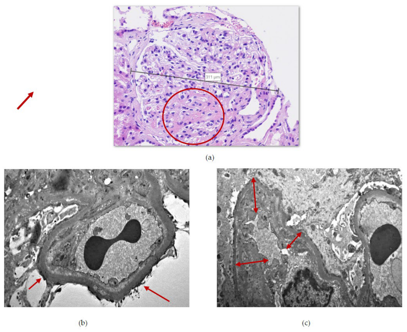

Table 1.
Admision and follow-up clinical and laboratory parameters: eGFR—estimated glomerular filtration rate, LDL-cholesterol—low-density lipoprotein cholesterol, HDL-cholesterol—high-density lipoprotein cholesterol, ALT—alanine aminotransferase, AST—aspartate aminotransferase, GMT—gamma-glutamyl aminotrasferase, ALP—alkaline phosphatase, UACR—urine albumin/creatine ratio, UPCR—urine protein/creatinine ratio, BMI—body mass index, HCO₃⁻—hydrogen carbonate, PTH—parathormone, WBCs—white blood cells, HbA1c—glycated hemoglobine.
Table 1.
Admision and follow-up clinical and laboratory parameters: eGFR—estimated glomerular filtration rate, LDL-cholesterol—low-density lipoprotein cholesterol, HDL-cholesterol—high-density lipoprotein cholesterol, ALT—alanine aminotransferase, AST—aspartate aminotransferase, GMT—gamma-glutamyl aminotrasferase, ALP—alkaline phosphatase, UACR—urine albumin/creatine ratio, UPCR—urine protein/creatinine ratio, BMI—body mass index, HCO₃⁻—hydrogen carbonate, PTH—parathormone, WBCs—white blood cells, HbA1c—glycated hemoglobine.
| Baseline | Fourth month of follow-up | |
|---|---|---|
| Creatinine (µmol/L) | 261 [59–104] | 237 |
| Urea (mmol/L) | 16.4 [2.8-7.2] | 12.5 |
| Uric acid (µmol/L) | 421 [208-428] | 374 |
| Albumin (g/L) | 40.1 [35.0-52.0] | 41.2 |
| eGFR (mL/min/1.73m2) | 26 | 29 |
| Total protein (g/L) | 75.3 [66.0-83.0] | 80.2 |
| Fasting glycaemia (mmol/L) | 5.7 [4.1-5.9] | 6.1 |
| Total cholesterol (mmol/L) | 4.88 [< 5.17] | 2.58 |
| Triglycerides(mmol/L) | 6.72 [0.40-1.70] | 4.71 |
| LDL-cholesterol (mmol/L) | 2.78 [1.00-3.30] | 1.54 |
| HDL-cholesterol (mmol/L) | 0.72 [1.03-2.00] | 0.66 |
| ALT (µkat/L) | 0.24 [0.10-0.85] | 0.31 |
| AST (µkat/L) | 0.32 [0.10-0.85] | 0.26 |
| GGT (µkat/L) | 0.55 [0.03-0.92] | 0.76 |
| ALP (µkat/L) | 1.26 [0.50-2.15] | 1.66 |
| UACR (mg/mmol) | 135.8 [< 3.0] | 95.1 |
| UPCR (mg/mmol) | 172.6 [<15.0] | 132.8 |
| Erythrocyteuria (erytrocytes/µL) | 0.0 [< 10.0] | 0.0 |
| BMI (kg/m2) | 40.2 [< 25] | 39.7 |
| Weight (kg) | 119 | 117,5 |
| Sodium (mmol/L) | 141 [136-146] | 136 |
| Potassium (mmol/L) | 3.9 [3.5-5.1] | 3.8 |
| HCO₃⁻ (mmol/L) | 20 [22.0-26.0] | 20.6 |
| Calcium (mmol/L) | 2.52 [2.2-2.65] | 2.52 |
| Phosphate (mmol/L) | 1.29 [0.81-1.45] | 1.12 |
| PTH (ng/L) | 108.8 [12-88] | 103.1 |
| 25-OH vitamin D (µg/L) | 26.5 [30-100] | 40.3 |
| Haemoglobin (g/L) | 143 [140-179] | 143 |
| WBCs (109/L) | 9.3 [3.90-10.0] | 9.6 |
| Thrombocytes (109/L) | 258 [140-400] | 300 |
| HbA1c (mmol/mol,IFCC) | 54.1 [< 38] | – |
Disclaimer/Publisher’s Note: The statements, opinions and data contained in all publications are solely those of the individual author(s) and contributor(s) and not of MDPI and/or the editor(s). MDPI and/or the editor(s) disclaim responsibility for any injury to people or property resulting from any ideas, methods, instructions or products referred to in the content. |
© 2026 by the authors. Licensee MDPI, Basel, Switzerland. This article is an open access article distributed under the terms and conditions of the Creative Commons Attribution (CC BY) license (http://creativecommons.org/licenses/by/4.0/).
Copyright: This open access article is published under a Creative Commons CC BY 4.0 license, which permit the free download, distribution, and reuse, provided that the author and preprint are cited in any reuse.