
Submitted:
16 November 2026
Posted:
18 November 2026
You are already at the latest version
Abstract
Therapeutic cancer vaccines have long promised durable, tumour-specific immunity but have repeatedly failed to deliver it in the clinic. The combination of synthetic messenger RNA (mRNA) with ionizable lipid nanoparticles (LNPs) has changed that trajectory. What began as a delivery solution for a fragile nucleic acid has become a programmable immunotherapy platform: an mRNA molecule encodes any antigen within days of sequence identification, and the LNP that carries it simultaneously serves as a potent, self-adjuvanting immunostimulant. This review integrates the mechanistic literature of the past five years with the maturing clinical dataset. We first dissect how LNP composition, intracellular trafficking and mRNA chemistry determine the amount, duration and anatomical location of antigen expression. We then examine the innate immune circuit that converts encapsulated RNA into a TH1-biased adjuvant signal, and explain why the same type I interferon (IFN-I)-PKR axis that licenses dendritic cell maturation also imposes a ceiling on antigen output. Building on this, we trace the path from cross-presentation by cDC1 to effector CD8+ T cell function inside the tumour, and we map the tumour-intrinsic and microenvironmental lesions that constrain efficacy. Clinically, individualised neoantigen therapy has produced the first randomised evidence of benefit in resected melanoma (KEYNOTE-942), while autogene cevumeran has shown that vaccine-expanded, long-lived neoantigen-specific CD8+ T cells can be generated in pancreatic cancer-a tumour previously regarded as vaccine-refractory. We synthesise these data into design rules that link antigen selection, LNP chemistry, dosing schedule and combination partner to the immunological objective, and we identify the mechanistic questions-extrahepatic targeting, repeat dosing, immunity to the carrier, and the definition of surrogate endpoints-that will determine whether mRNA-LNP vaccines become part of standard oncological care.
Keywords:
mRNA vaccine
; lipid nanoparticle
; neoantigen
; cancer immunotherapy
; cross-presentation
; type I interferon
; individualised neoantigen therapy
1. Introduction
The concept of vaccinating a patient against their own tumour is older than most of the technologies now used to execute it. What has changed is the resolution at which a tumour can be read and the speed at which a matching immunogen can be manufactured. Whole-exome and RNA sequencing, mass-spectrometry-based immunopeptidomics and machine-learning epitope prediction together allow the mutant peptide landscape of an individual tumour to be enumerated within weeks [1,2,3]. Synthetic mRNA can then be produced by in vitro transcription, encapsulated in LNPs and released for administration faster than any bespoke biologic in oncology [4,5].
The clinical translation of mRNA-LNP vaccines for cancer has been shaped by an unusual convergence. The platform was validated at pandemic scale as a prophylactic vaccine against SARS-CoV-2, which generated manufacturing capacity, regulatory familiarity and-critically-a large safety database in millions of individuals [6,7]. At the same time, the therapeutic programme advanced from first-in-human studies of peptide and RNA neoantigen vaccines to a randomised phase 2b trial in melanoma [8,9]. The result is a field that now has both mechanistic depth and clinical signal, and that is beginning to be constrained less by immunology than by chemistry, manufacturing and trial design [10,11].
What distinguishes mRNA-LNP vaccines from earlier therapeutic vaccine platforms is that the delivery vehicle is not immunologically inert. Ionizable lipids, the RNA itself, and process-related double-stranded RNA (dsRNA) impurities engage pattern-recognition receptors and drive a type I interferon response that matures antigen-presenting cells and shapes T cell differentiation [12,13]. This intrinsic adjuvanticity has been treated both as an asset and as a liability, because the same signalling axis suppresses translation of the delivered mRNA and can drive exhaustion of primed T cells [14]. Much of the recent engineering effort in the field can be understood as an attempt to separate these two consequences.
The field is not starting from zero. Three decades of therapeutic vaccination in oncology produced a consistent pattern: immunological activity was readily demonstrable, and clinical benefit was not. Peptide, protein, viral-vector and dendritic-cell vaccines each induced measurable T cell responses in a fraction of patients, and each failed to improve survival in randomised trials [15,16]. Retrospective analysis of these failures identified recurring design faults: antigens chosen for immunogenicity rather than for tumour specificity, vaccination in patients with bulky disease, absence of a rational combination partner, and endpoints that measured immunogenicity rather than clinical effect. The mRNA-LNP platform does not automatically solve any of these problems, but it addresses two of them structurally: antigen selection can be individualised at scale, and the delivery vehicle provides its own adjuvant, removing the need for a separately optimised immunostimulant [8].
This review focuses on mechanisms and their clinical consequences. We do not attempt an exhaustive catalogue of preclinical constructs; instead we ask how the physical and immunological properties of the platform determine what the immune system sees, and how the clinical data obtained so far inform the next generation of designs. We draw principally on literature published between 2020 and 2026, a period in which the field moved from proof of concept to randomised evidence.
2. The mRNA-LNP Platform: Design Principles
2.1. Encoding the Antigen
An mRNA vaccine is a minimalist genetic construct: a 5′ cap, a 5′ untranslated region (UTR), an open reading frame, a 3′ UTR and a poly(A) tail. Each element has been engineered for translational efficiency and stability. The identity and methylation status of the first transcribed nucleotide determines cap-proximal translation initiation and innate sensing, and optimised cap analogues are now standard in clinical-grade mRNA [17]. Poly(A) tail length governs both stability and translational efficiency, with tails in the 100-150 nucleotide range routinely used [18].
The most consequential chemical modification is the substitution of uridine with N1-methylpseudouridine (m1Ψ). By reducing recognition by endosomal and cytosolic RNA sensors, nucleoside modification increases protein output by an order of magnitude in vivo and reduces inflammatory toxicity [19,20]. The trade-off is not trivial: modification also removes much of the intrinsic adjuvant effect of RNA, and recent work shows that the balance between modification and ionizable lipid chemistry jointly sets both translation and immune activation [19]. Clinical constructs therefore now vary the degree of modification deliberately rather than by default.
Process-related impurities matter as much as design. In vitro transcription generates dsRNA and abortive products that activate TLR3, RIG-I and MDA5, and the purification strategy used during manufacture can change innate activation by more than an order of magnitude at constant dose [20]. For therapeutic cancer vaccines, where repeated dosing and high antigen load are required, this is not a minor issue: it determines whether the vaccine is limited by immunogenicity or by reactogenicity.
Beyond the vector backbone, the choice of antigen format is a design decision with immunological consequences. Full-length ORFs present the entire mutational landscape of a protein and permit presentation of both class I and class II epitopes, at the cost of diluting the response across many subdominant specificities [21]. String-of-beads poly-epitope constructs concentrate the response on selected epitopes and can be designed to avoid self-tolerance, but risk junctional neoepitopes and unequal epitope processing 1. Linking antigen to signals that direct it to the endosomal or lysosomal compartment, or to MHC class II, can bias presentation toward CD4+ help, which is increasingly recognised as necessary for durable CD8+ memory [22].
2.2. Lipid Nanoparticles: Assembly, Trafficking and the Escape Problem
A standard mRNA-LNP contains four components: an ionizable lipid, a helper phospholipid, cholesterol and a PEG-lipid, assembled at defined molar ratios during rapid microfluidic mixing [23,24]. The ionizable lipid is the functional core. Its apparent pKa-optimised between roughly 6.2 and 6.8-determines the charge transition that drives both nucleic acid encapsulation at low pH and membrane destabilisation within the acidifying endosome [25,26]. Combinatorial libraries and, more recently, machine-learning-guided design have expanded the accessible chemical space and produced lipids with improved potency and organ selectivity [25,27].
Endosomal escape remains the principal inefficiency of the platform. Quantitative studies indicate that only a small fraction of internalised mRNA reaches the cytosol, with the majority degraded in the endolysosomal pathway or recycled to the cell surface [28,29]. The mechanism is now understood in some detail: as the endosome acidifies, ionizable lipids become protonated, associate with anionic endosomal phospholipids, and destabilise the bilayer in a manner that permits mRNA release through transient defects rather than wholesale rupture [29]. Helper lipids modulate this process, and substituting the neutral helper lipid for a charged alternative shifts both potency and organ tropism [30].
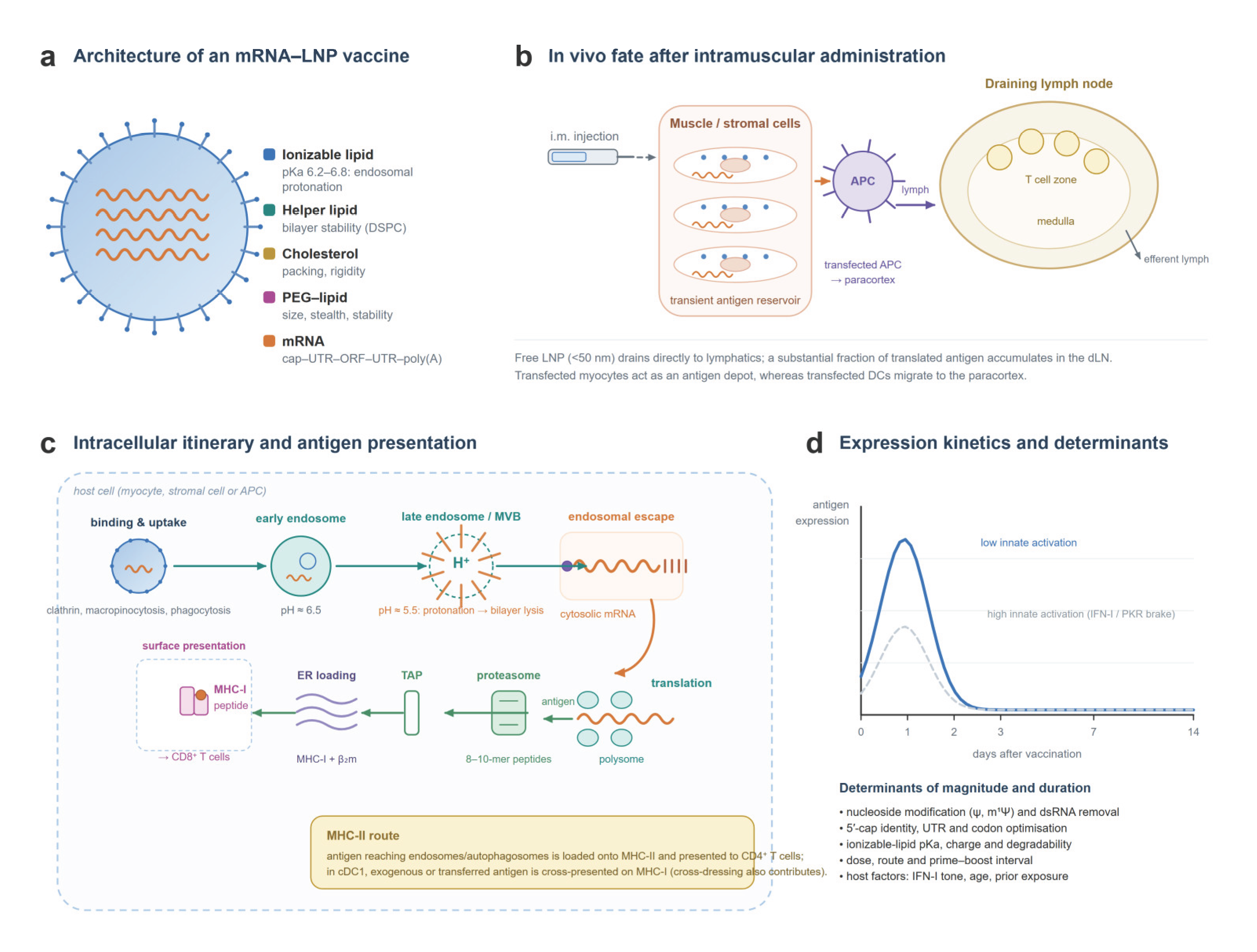
Once in the cytosol, the mRNA is translated by ribosomes and the nascent protein enters the antigen-processing pathway. For vaccines, the relevant consequence is that the density and persistence of peptide-MHC complexes on the surface of the transfected cell-not the amount of protein produced per se-is the proximate determinant of T cell priming [22] (Fig. 1c).
2.3. Where the Antigen Goes, and Why it Matters
After intramuscular injection, LNP distribution is governed by particle size, charge and the adsorbed protein corona. Particles smaller than roughly 50 nm drain through the lymphatic capillaries and reach the draining lymph node (dLN) directly, whereas larger particles are retained at the injection site and transfected into muscle and stromal cells, which then act as an antigen depot [31]. Apolipoprotein E adsorption to the particle surface promotes hepatocyte uptake via LDL receptor family members, and this is the principal reason that systemically administered LNPs are largely liver-tropic [32,33].
Both routes contribute to immunogenicity, and their relative weight depends on the vaccine construct. Transfected muscle cells are poor antigen-presenting cells but release antigen that is taken up and cross-presented by dendritic cells; transfected dendritic cells that migrate to the dLN present antigen directly [31,34]. In the dLN, subcapsular sinus macrophages capture LNP-associated material and contribute to innate activation, shaping the subsequent germinal centre and T cell response [34,35] (Fig. 1b).
Organ-selective delivery has been substantially advanced by the recognition that the composition of the LNP itself-not only its targeting ligand-determines tropism. Selective organ targeting (SORT) uses the addition of a fifth, permanently charged or anionic lipid to redirect particles to lung, spleen or liver [36,37]. These approaches have obvious relevance for cancer immunotherapy, where the objective may be to reach the spleen for systemic T cell priming, to reach a tumour-draining node, or to transfect tumour-infiltrating myeloid cells in situ [30,38].
2.4. Dose, Schedule and the Geometry of Boosting
Three variables other than the construct itself determine the immunological outcome: dose, interval and route. Dose escalation in mRNA vaccines is limited less by toxicity than by the shape of the dose-response curve. Because innate activation suppresses translation of the delivered mRNA, increasing the dose increases the adjuvant signal and the antigen signal simultaneously but not proportionally, so that the relationship between dose and T cell response plateaus earlier than the relationship between dose and reactogenicity [12,14]. This is a strong argument for dose-sparing formats-self-amplifying RNA, or constructs that reduce innate activation without reducing expression [39].
Interval matters because of the kinetics of the response. A single dose generates a wave of antigen expression lasting roughly a week, followed by contraction of the effector population and formation of memory [40]. Boosting too early, while antigen is still abundant and effector cells are still expanding, may drive terminal differentiation rather than memory; boosting too late may encounter a contracted and less responsive population. Clinical protocols for individualised neoantigen therapy have used intervals of several weeks, with priming at the start of adjuvant chemotherapy and boosts thereafter [41]. The optimal interval has not been systematically determined in humans and is likely to differ between prophylactic and therapeutic settings.
Route has already been discussed in terms of biodistribution, but it also determines the anatomical geometry of priming. Intramuscular delivery distributes antigen across many small draining nodes and generates a broad, disseminated response; intratumoral delivery concentrates antigen in the node draining the injected lesion and generates a response skewed toward the antigens expressed by that lesion [42]. Lymph-node-targeted delivery, in which the particle is engineered to reach the subcapsular sinus directly, bypasses the need for cell migration and amplifies the germinal centre and T cell response per unit dose [35,43].
Figure 1.
Architecture, in vivo fate and intracellular itinerary of mRNA-LNP vaccines. a, Composition of a four-component mRNA-LNP and the function of each lipid class. b, After intramuscular injection, small particles drain to the lymph node while larger ones transfect muscle and stromal cells, which act as an antigen depot; transfected antigen-presenting cells migrate to the paracortex. c, Intracellular route: endocytic uptake, endosomal acidification, protonation of the ionizable lipid and escape of mRNA into the cytosol, translation, proteasomal processing and TAP-dependent loading onto MHC class I; the MHC class II route and cross-presentation are indicated. d, Expression kinetics after a single dose, showing how the strength of innate activation sets both the peak and the duration of antigen expression. dLN, draining lymph node; ER, endoplasmic reticulum; MVB, multivesicular body; TAP, transporter associated with antigen processing.
Figure 1.
Architecture, in vivo fate and intracellular itinerary of mRNA-LNP vaccines. a, Composition of a four-component mRNA-LNP and the function of each lipid class. b, After intramuscular injection, small particles drain to the lymph node while larger ones transfect muscle and stromal cells, which act as an antigen depot; transfected antigen-presenting cells migrate to the paracortex. c, Intracellular route: endocytic uptake, endosomal acidification, protonation of the ionizable lipid and escape of mRNA into the cytosol, translation, proteasomal processing and TAP-dependent loading onto MHC class I; the MHC class II route and cross-presentation are indicated. d, Expression kinetics after a single dose, showing how the strength of innate activation sets both the peak and the duration of antigen expression. dLN, draining lymph node; ER, endoplasmic reticulum; MVB, multivesicular body; TAP, transporter associated with antigen processing.
3. Innate Immune Engagement: Adjuvant and Brake
3.1. The Sensors
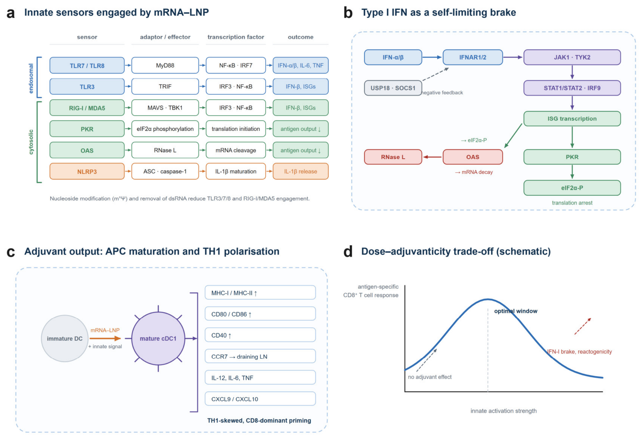
Encapsulated mRNA activates a defined set of pattern-recognition receptors. Endosomal TLR7 and TLR8 sense single-stranded RNA and signal through MyD88 to NF-κB and IRF7; TLR3 senses dsRNA and signals through TRIF to IRF3 [12,13]. In the cytosol, RIG-I and MDA5 recognise short dsRNA and long dsRNA respectively and signal through MAVS and TBK1; PKR phosphorylates eIF2α to arrest translation; 2′-5′-oligoadenylate synthetase activates RNase L to degrade RNA; and NLRP3 inflammasome activation generates IL-1β [12]. Recent work has shown that the ionizable lipid component itself, independent of cargo, can elicit NF-κB and IRF responses through TLR signalling, blurring the distinction between delivery vehicle and adjuvant 13.
The anatomical site of innate activation matters. IFN-β produced at the injection site acts on recruited monocytes and dendritic cells and is required for the cellular immune response to mRNA vaccines in mice, while innate activation within the dLN-particularly by lymph node macrophages-shapes the magnitude and quality of the anti-tumour T cell response [34,44]. This has a practical implication: the adjuvant effect of a given LNP is not a scalar property but a function of where the particle accumulates.
3.2. The IFN-I-PKR Axis as a Self-Limiting Circuit
The most important negative feedback loop in the platform operates as follows. Cytosolic RNA sensing induces IFN-β, which acts on the producing cell and on neighbours through IFNAR, JAK1/TYK2 and STAT1/STAT2/IRF9 to induce interferon-stimulated genes. Among these, PKR phosphorylates eIF2α and globally suppresses cap-dependent translation, while RNase L degrades the delivered mRNA [12,14]. The result is that a strongly immunostimulatory formulation can reduce the amount of antigen expressed from the same dose, and negative regulators including USP18 and SOCS1 attenuate the response over time.
This circuit has a clear experimental corollary: unmodified, highly immunogenic mRNA can generate stronger anti-tumour immunity than modified mRNA in some settings, because the IFN-I response itself contributes to dendritic cell activation and TH1 polarisation [14]. Conversely, excessive IFN-I signalling drives PD-L1 expression, recruits suppressive myeloid cells and can promote T cell dysfunction. The relationship between innate stimulus and protective immunity is therefore bell-shaped rather than monotonic-a point that is often lost in dose-escalation design.
Figure 2.
Innate sensing of mRNA-LNP and the adjuvant-expression trade-off. a, Endosomal and cytosolic sensors engaged by encapsulated RNA and their downstream adaptors, transcription factors and functional outcomes. b, The type I interferon circuit that limits antigen expression: IFN-I acts through IFNAR-JAK-STAT to induce PKR and OAS, which arrest translation and degrade mRNA, with USP18 and SOCS1 providing negative feedback. c, The adjuvant output of the same signals: maturation of cDC1 with upregulation of MHC, costimulatory molecules, CCR7 and TH1-polarising cytokines and chemokines. d, Schematic relationship between innate activation strength and the magnitude of the antigen-specific CD8+ T cell response, illustrating the existence of an optimal window between under- and over-stimulation.
Figure 2.
Innate sensing of mRNA-LNP and the adjuvant-expression trade-off. a, Endosomal and cytosolic sensors engaged by encapsulated RNA and their downstream adaptors, transcription factors and functional outcomes. b, The type I interferon circuit that limits antigen expression: IFN-I acts through IFNAR-JAK-STAT to induce PKR and OAS, which arrest translation and degrade mRNA, with USP18 and SOCS1 providing negative feedback. c, The adjuvant output of the same signals: maturation of cDC1 with upregulation of MHC, costimulatory molecules, CCR7 and TH1-polarising cytokines and chemokines. d, Schematic relationship between innate activation strength and the magnitude of the antigen-specific CD8+ T cell response, illustrating the existence of an optimal window between under- and over-stimulation.
3.3. Reactogenicity, Complement and Immunity to the Carrier
Systemic reactogenicity-fever, fatigue, myalgia-is the clinical expression of the same innate circuitry, and in a therapeutic setting with repeated dosing it becomes a dose-limiting toxicity rather than an acceptable inconvenience. Two additional mechanisms deserve attention. First, complement activation by LNP lipids, and in particular by the PEG-lipid component, contributes to hypersensitivity reactions and to the phenomenon of complement activation-related pseudoallergy [45]. Second, repeated administration of PEGylated particles induces anti-PEG antibodies, which accelerate clearance and reduce expression on subsequent doses; anti-PEG antibodies are boosted in humans by SARS-CoV-2 mRNA vaccination and correlate with altered pharmacokinetics [46,47]. Next-generation formulations address this with high-density brush polymer lipids or reduced PEG content, but the problem is not fully solved [48].
For a therapeutic cancer vaccine, which may require six to nine doses over months, carrier immunogenicity is a first-order design constraint rather than a secondary pharmacology issue.
3.4. Deliberately Engineering the Balance
Several strategies now aim to decouple adjuvanticity from translational repression. Removing dsRNA by chromatographic purification, titrating m1Ψ content, and selecting ionizable lipids with lower intrinsic inflammatory potential all reduce the IFN-I brake without eliminating dendritic cell activation [19,20]. Conversely, adjuvants can be added deliberately in a spatially controlled manner-for example by tethering short dsRNA to the mRNA backbone, or by co-formulating TLR or STING agonists-so that the adjuvant signal is delivered to the same cell that receives the antigen [49,50]. Lipid nanoparticles themselves have intrinsic adjuvant activity against co-delivered antigens, which can be exploited when the antigen is delivered as protein rather than RNA [51].
The unifying principle is that adjuvanticity should be matched to the antigen, the route and the number of doses, rather than maximised. A formulation optimised for a single prophylactic dose in a healthy adult is unlikely to be optimal for repeated dosing in an immunosenescent patient with cancer.
4. From Antigen Presentation to Tumour Control
4.1. Cross-Presentation and the cDC1 Requirement
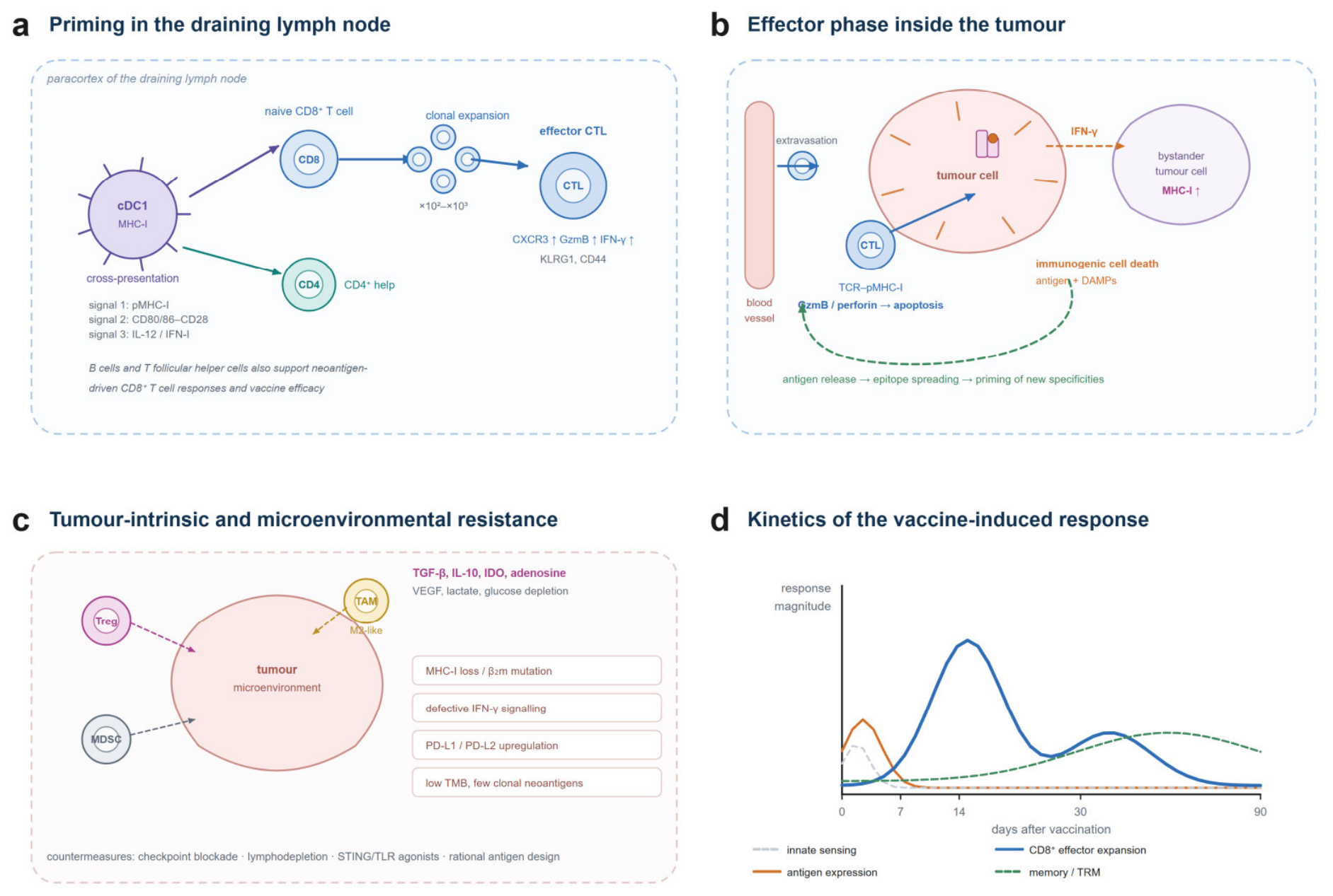
For a vaccine to generate CD8+ cytotoxic T lymphocytes against a non-haematopoietic tumour, antigen must be cross-presented on MHC class I by conventional type 1 dendritic cells (cDC1). This requirement is not bypassable: cDC1 are the dominant cross-presenting population in murine tumour models and in human tumours, and their abundance correlates with response to immunotherapy [52,53] (Fig. 3a). In pancreatic cancer-a tumour with profound immune exclusion-cDC1 are required for the efficacy of immunotherapy combinations, and their paucity is a principal barrier to vaccination [54].
mRNA-LNP vaccines engage this pathway in two ways. Direct transfection of cDC1 leads to endogenous antigen synthesis and canonical MHC class I presentation. Indirect transfer of antigen from transfected non-haematopoietic cells to cDC1-through protein transfer, exosome uptake or cross-dressing-extends the reach of the vaccine beyond the small number of cells that are actually transfected [52]. Both routes require dendritic cell maturation, which is supplied by the innate signals described above.
An underappreciated constraint is imposed by suppressive myeloid cells. Polymorphonuclear MDSC limit antigen cross-presentation by dendritic cells, and their removal or functional inhibition restores vaccine efficacy in preclinical models [55]. In patients with advanced cancer, where myeloid suppression is often dominant, this may explain the discrepancy between immunogenicity in peripheral blood and lack of tumour control.
4.2. Priming, Help and the Persistence Problem
Productive priming requires three signals: peptide-MHC engagement of the T cell receptor, costimulation through CD80/CD86-CD28, and a polarising cytokine signal-principally IL-12 and IFN-I-that drives TH1 differentiation and effector programming. mRNA-LNP-activated cDC1 upregulate all three, together with CCR7, which directs them to the paracortex of the dLN where naive T cells are encountered [12].
CD4+ help is required for the generation of durable CD8+ memory and for the maintenance of stem-like progenitor populations that sustain effector responses within tumours [21,56]. This is one reason why antigen formats that present class II epitopes-full-length ORFs, or constructs engineered for endosomal localisation-may outperform minimal class I epitope vaccines despite lower nominal epitope density.
The single most important qualitative property of a therapeutic vaccine is not the peak magnitude of the response but its persistence. In pancreatic cancer, autogene cevumeran expanded neoantigen-specific CD8+ T cells that persisted for years and included clones with stem-like features, and the presence of these cells was associated with delayed recurrence [41,57]. Memory CD8+ T cells with a stem cell memory phenotype are generated by mRNA vaccination and can be recalled by boosting [40]. The implication for trial design is that immunogenicity should be assessed months, not weeks, after vaccination.
4.3. Effector Function Within the Tumour
A primed CD8+ T cell must still reach and kill the tumour cell. Effector CTLs exit the dLN, extravasate through tumour endothelium guided by CXCR3 ligands, and engage tumour cells through TCR-pMHC-I interactions, killing via granzyme B and perforin [52]. IFN-γ released by CTLs acts on neighbouring tumour cells and on stromal cells, upregulating MHC class I and CXCL9/10 and thereby propagating further T cell recruitment-a positive feedback loop that is essential for the abscopal-like effects seen when a single lesion is vaccinated [58].
Killing is immunologically amplifiable. Immunogenic cell death releases tumour antigen and damage-associated molecular patterns, which are captured by dendritic cells and drive priming of additional specificities-epitope spreading-so that the response broadens beyond the encoded epitopes [59,60]. Persistent memory responses with epitope spreading have been documented years after neoantigen vaccination in melanoma, indicating that this amplification is clinically operative rather than theoretical [60].
4.4. Resistance: What Limits Efficacy
Vaccine failure in the clinic is more often a failure of the effector phase than of immunogenicity. Three classes of resistance dominate.
Tumour-intrinsic lesions include loss of MHC class I or β2-microglobulin, which renders tumour cells invisible to CTLs; defective IFN-γ signalling through JAK1/JAK2 mutations; and low clonal neoantigen burden in tumours with low tumour mutational burden [61,62]. Somatic HLA class I loss is common across tumour types and refines the interpretation of TMB as a predictive biomarker [62].
Microenvironmental suppression includes regulatory T cells, myeloid-derived suppressor cells and M2-like tumour-associated macrophages, together with soluble mediators such as TGF-β, IL-10, indoleamine 2,3-dioxygenase and adenosine [63,64]. These populations are themselves targets: lymphodepletion, CSF1R blockade, and chemotherapeutic regimens that reduce MDSC can convert a vaccine-refractory tumour into a responsive one.
The principal resistance mechanisms and their countermeasures are summarised in Table 3. T cell dysfunction includes exhaustion programmes driven by chronic antigen exposure, and the upregulation of PD-L1 within the tumour. The presence of stem-like progenitor CD8+ T cells, which respond to checkpoint blockade by proliferating and differentiating, is a strong predictor of response and may be the population that a vaccine is best positioned to expand [56].
Figure 3.
From priming to tumour control, and the mechanisms that limit it. a, Cross-presentation by cDC1 in the draining lymph node and the three signals required for productive priming of CD8+ T cells, with CD4+ help. b, Effector phase within the tumour: extravasation, TCR engagement, granzyme B- and perforin-mediated killing, IFN-γ-driven upregulation of MHC class I on bystander tumour cells, and immunogenic cell death feeding epitope spreading. c, Tumour-intrinsic and microenvironmental resistance mechanisms. d, Kinetics of innate activation, antigen expression, CD8+ effector expansion and memory formation after vaccination, with the readouts used to monitor each phase.
Figure 3.
From priming to tumour control, and the mechanisms that limit it. a, Cross-presentation by cDC1 in the draining lymph node and the three signals required for productive priming of CD8+ T cells, with CD4+ help. b, Effector phase within the tumour: extravasation, TCR engagement, granzyme B- and perforin-mediated killing, IFN-γ-driven upregulation of MHC class I on bystander tumour cells, and immunogenic cell death feeding epitope spreading. c, Tumour-intrinsic and microenvironmental resistance mechanisms. d, Kinetics of innate activation, antigen expression, CD8+ effector expansion and memory formation after vaccination, with the readouts used to monitor each phase.
5. Choosing What to Encode
5.1. The Neoantigen Discovery Pipeline
Neoantigens arise from somatic mutations, frameshifts, splice variants and aberrant translation, and only a small minority of predicted peptides are genuinely immunogenic. The factors that determine immunogenicity have been systematically mapped by consortium efforts and can be grouped into four categories: peptide-HLA binding affinity and stability, proteasomal processing and transport efficiency, abundance of the source transcript and protein, and the foreignness of the peptide relative to its wild-type counterpart-the agretopicity that determines whether a T cell repertoire exists [1,65]. Machine-learning models trained on harmonised immunogenicity datasets have improved prediction accuracy substantially over position-weight-matrix approaches, and are now embedded in clinical pipelines [2,66].
Direct identification of presented peptides by mass spectrometry provides a complementary and less model-dependent route. Immunopeptidomics can confirm that a predicted neoepitope is actually displayed on the tumour surface, and recent studies have used it to guide antigen selection for personalised vaccines [3,67]. The trade-off is sensitivity: immunopeptidomics requires substantial tumour material and deep sequencing depth, and low-abundance neoepitopes may escape detection.
Two biological filters are often neglected. First, clonality: truncal mutations present in every tumour cell are more attractive targets than subclonal mutations, because the latter invite outgrowth of antigen-negative subclones [68]. Second, HLA loss: tumours that have lost the restricting HLA allele cannot present the corresponding epitopes, and this is a frequent mechanism of acquired resistance that should inform antigen selection [61,62].
5.2. Shared Versus Individualised Antigens
Individualised vaccines dominate current clinical development because they exploit the unique mutational repertoire of a tumour and minimise central tolerance. Their limitations are equally clear: cost, turnaround time, dependence on sufficient tumour material, and the impossibility of use in patients whose tumours have low mutational burden or who lack accessible tissue.
Shared antigens offer an off-the-shelf alternative. These include hotspot mutations (KRAS G12C/G12D/G12V, BRAF V600E, IDH1 R132H), cancer-testis antigens and tumour-associated antigens such as NY-ESO-1, MAGE-A and WT1 [69,70]. Shared neoantigen vaccines can be manufactured at scale and administered immediately, which matters in rapidly progressing disease, and they can be combined with HLA-matching to enrich for responders. Their weakness is immunological: epitopes shared across patients are subject to central tolerance, and tumours can downregulate them under selective pressure. A recent case report of a KRAS G12V mRNA vaccine combined with pembrolizumab provides proof of principle that a shared hotspot vaccine can produce clinical benefit [69].
A pragmatic hybrid is emerging in which a fixed backbone of shared, clonal epitopes is combined with a small individualised component. This retains the logistics of an off-the-shelf product while preserving the specificity that gives individualised vaccines their potency [71].
5.3. Design Choices Beyond the Epitope
The physical format of the encoded antigen affects processing. Full-length ORFs allow presentation of both class I and class II epitopes and permit the immune system to select the dominant epitope, but they also risk tolerance and inefficiency. Poly-epitope constructs concentrate the response and allow removal of self-epitopes, but junctional sequences can create unintended neoepitopes and epitope processing is uneven 1. Incorporating signals that route the antigen into the endosomal compartment enhances class II presentation and CD4+ help [22].
Finally, the delivered mRNA can encode more than antigen. Constructs encoding cytokines (IL-12, IL-2, IL-15), costimulatory ligands (OX40L, 4-1BBL) or even chimeric antigen receptors convert the vaccine into a local immunomodulatory agent, a strategy now being tested clinically in the intratumoral setting [72,73,74].
6. Clinical Development
6.1. Individualised Neoantigen Therapy in Melanoma and Beyond
The first randomised evidence that an mRNA-LNP cancer vaccine can alter clinical outcomes came from KEYNOTE-942, a phase 2b trial in which 157 patients with resected stage IIIB-IV melanoma were randomised to mRNA-4157 (V940) plus pembrolizumab or pembrolizumab alone [9]. The individualised neoantigen therapy (INT) encodes up to 34 neoantigens selected from tumour sequencing. At a median follow-up of approximately two years, recurrence-free survival favoured the combination (hazard ratio 0.561, 95% CI 0.309-1.017), with a more pronounced effect on distant metastasis-free survival (hazard ratio 0.347). T cell responses to vaccine-encoded neoantigens were detected in the majority of vaccinated patients and were associated with clinical benefit [75] (Table 1).
These results should be interpreted with appropriate caution: the trial was not powered for definitive superiority, the confidence interval crosses unity, and the control arm received an active standard of care. Nevertheless, the consistency between immunological and clinical readouts, and the magnitude of the distant metastasis-free survival effect, provided the rationale for a phase 3 programme. The INTerpath studies extend the approach to the adjuvant and neoadjuvant settings in melanoma, non-small-cell lung cancer, bladder cancer and renal cell carcinoma [76]. Whether the benefit replicates at scale is the single most important open question in the field.
6.2. Autogene Cevumeran and the Pancreatic Cancer Proof of Concept
Pancreatic ductal adenocarcinoma (PDAC) has been regarded as a paradigm of vaccine resistance: it is poorly vascularised, densely stromal, and contains few cDC1 [54]. The phase 1 study of autogene cevumeran-an mRNA-lipoplex vaccine encoding up to 20 neoantigens, administered with atezolizumab and mFOLFIRINOX after resection-challenged that assumption [41]. Vaccine-expanded neoantigen-specific T cells were detected in half of the patients, and their presence was associated with a substantially prolonged recurrence-free survival. Subsequent analysis showed that these cells persisted as long-lived, stem-like CD8+ clones for years after vaccination, and that vaccine boosting expanded pre-existing clones rather than generating entirely new ones [57].
This work established three principles. First, immunological non-responsiveness in PDAC is not absolute and can be overcome by combining vaccination with chemotherapy-induced lymphodepletion and checkpoint blockade. Second, the quality of the induced T cell-its persistence and stemness-predicts outcome better than peak frequency. Third, the vaccine can amplify a pre-existing, spontaneously generated anti-tumour response, which reframes the therapeutic objective from de novo priming to expansion and diversification.
A phase 1 study of autogene cevumeran with or without atezolizumab in advanced solid tumours has since reported manageable safety and reproducible immunogenicity, supporting the ongoing phase 2 programme in colorectal cancer and pancreatic cancer [77].
6.3. Intratumoral Vaccination and In Situ Approaches
An alternative to systemic vaccination is direct intratumoral injection, which converts the tumour itself into the antigen source and draining lymph node into the site of priming. mRNA-2752 encodes three immunomodulators-OX40L, IL-23 and IL-36γ-and was tested intratumorally with pembrolizumab in patients with high-risk ductal carcinoma in situ of the breast, where it was well tolerated and produced immunological changes in the tumour microenvironment [42]. A related study combining intratumoral mRNA-2752 with durvalumab reported induction of pro-inflammatory remodelling in injected and non-injected lesions [72].
Intratumoral delivery has attractive properties: low systemic exposure, exploitation of the full autologous antigen repertoire rather than a predicted subset, and the induction of epitope spreading. It also has clear limitations-accessibility of injectable lesions, heterogeneity of injection, and the fact that visceral tumours are often not amenable. Preclinical work combining intratumoral mRNA encoding cytokines with checkpoint blockade or with oncolytic viruses suggests that the approach is most effective when the injected lesion is used to prime a systemic response that then acts on distant disease [78,79,80].
A third strategy targets antigens that are not tumour-specific at all: mRNA-4359 encodes IDO1 and PD-L1 epitopes and aims to eliminate immunosuppressive cell populations that overexpress these proteins. It is in early-phase testing both as monotherapy and with pembrolizumab, and represents a conceptual shift from vaccinating against tumour cells to vaccinating against the immunosuppressive stroma [81].
6.6. What the Negative Trials Teach
The recent record also includes instructive failures. Vaccines directed at single tumour-associated antigens have repeatedly failed to improve survival despite inducing measurable T cell responses, largely because the targeted antigen was not required for tumour growth and could be lost or downregulated [16]. Vaccination in the setting of bulky, advanced disease has consistently produced weaker and less durable responses than vaccination in the adjuvant or neoadjuvant setting, where tumour burden and immunosuppression are lower [82]. And trials that combined a vaccine with chemotherapy without attention to schedule have sometimes cancelled out the immunological effect they were designed to measure [41].
Three design principles follow. First, the antigen must be a driver of tumour fitness rather than a passenger, which is the strongest argument for clonal neoantigens [68]. Second, the setting should be one of minimal residual disease; this is the rationale for the adjuvant and neoadjuvant programmes now underway [76]. Third, the combination partner and its timing should be chosen to amplify rather than suppress the vaccine-induced response, which generally means sequencing checkpoint blockade after priming rather than concurrently with it [75].
Figure 4.
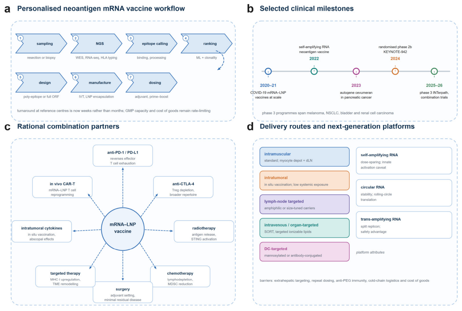
From design to clinic. a, Workflow for a personalised neoantigen mRNA vaccine from tumour sampling to dosing. b, Selected clinical milestones of the past five years. c, Rational combination partners and their mechanisms of action. d, Delivery routes and next-generation RNA platforms, with the principal remaining barriers.
Figure 4.
From design to clinic. a, Workflow for a personalised neoantigen mRNA vaccine from tumour sampling to dosing. b, Selected clinical milestones of the past five years. c, Rational combination partners and their mechanisms of action. d, Delivery routes and next-generation RNA platforms, with the principal remaining barriers.
6.4. Other Tumour Types and Alternative RNA Delivery Formats
Beyond melanoma and pancreatic cancer, mRNA-LNP vaccines are being explored in non-small-cell lung cancer, colorectal cancer, bladder cancer, renal cell carcinoma, glioblastoma, prostate cancer and hepatocellular carcinoma [71,83,84]. A neoantigen vaccine has recently been shown to generate antitumour immunity in renal cell carcinoma, a tumour with an intrinsically immunogenic microenvironment, supporting the generalisability of the approach [85] (Table 2 summarises the RNA formats in clinical use).
Two alternative architectures are advancing in parallel. Self-amplifying mRNA (saRNA) retains the alphaviral replicase and amplifies the transcript intracellularly, allowing dose sparing; a heterologous chimpanzee adenovirus prime followed by saRNA boost was immunogenic in patients with advanced cancer, and single immunisations of saRNA-LNP controlled HPV-associated tumours in preclinical models [39,86,87]. Circular RNA offers greater stability and sustained translation, and has shown activity in hard-to-treat tumour models [88,89]. Trans-amplifying RNA, which splits the replicon between a replicase-encoding and an antigen-encoding molecule, adds a safety layer by requiring co-delivery [90].
6.5. Safety in Patients with Cancer
The prophylactic experience with mRNA-LNP vaccines provides a large safety database, and prospective studies in patients receiving active anticancer therapy show acceptable immunogenicity and safety, albeit with attenuated responses in those receiving cytotoxic chemotherapy or anti-CD20 antibodies [93,94]. For therapeutic cancer vaccines, the principal safety considerations are different: repeated dosing with a pro-inflammatory agent in patients with autoimmune predisposition, cumulative reactogenicity, and the theoretical risk of vaccinating against self-antigens. A meta-analysis of mRNA vaccines in advanced solid tumours found the toxicity profile to be dominated by transient local and systemic reactions consistent with innate immune activation, with no unexpected safety signal at the doses tested [95].
7. Combination Strategies and Biomarkers
7.1. Rational Combinations
Vaccination generates and expands T cells; it does not by itself remove the brakes that tumours impose on them. The most obvious partner is therefore checkpoint blockade. Preclinical and clinical data indicate that the combination is more than additive: vaccination increases the pool of tumour-specific T cells, including stem-like progenitors, which checkpoint blockade can then mobilise [56,75]. Anti-CTLA-4 may contribute additionally through Treg depletion and broadening of the T cell repertoire [63].
Chemotherapy and radiotherapy contribute in ways that are often described as immunosuppressive but are, in the vaccine context, frequently facilitative. Lymphodepletion removes regulatory and suppressor populations and creates space for vaccine-induced T cells, which is the rationale for the sequencing used in the autogene cevumeran studies [41]. Radiotherapy and selected cytotoxics induce immunogenic cell death and increase antigen availability, and their combination with vaccination can generate abscopal responses [80,96]. Targeted agents that restore MHC class I expression or remodel the myeloid compartment are also rational partners [58].
Finally, vaccination can be combined with cell therapies. mRNA-LNP platforms can engineer T cells in vivo by delivering a CAR-encoding transcript to T cells, transiently reprogramming them without ex vivo manufacture; this has been demonstrated in preclinical models of cancer and is now entering early clinical testing [97,98].
7.2. Measuring the Right Thing
Trial design in this field has been hampered by the absence of validated surrogate endpoints. IFN-γ ELISpot remains the workhorse but measures frequency, not function, and correlates imperfectly with outcome [75]. Peptide-MHC multimer staining and TCR sequencing provide clonal resolution and can demonstrate expansion of pre-existing clones, which appears to be the mechanism of action of at least some vaccines [57]. Single-cell transcriptomics of blood and tumour informs on the differentiation state of vaccine-induced cells and on the emergence of exhaustion programmes.
Three additional readouts deserve routine incorporation. Circulating tumour DNA (ctDNA) provides a quantitative measure of molecular residual disease and can distinguish immunological effects from effects on tumour burden, particularly in the adjuvant setting where imaging is insensitive [99]. Immunopeptidomics of the tumour can confirm that encoded epitopes are actually presented. And paired tumour biopsies before and after vaccination can quantify the influx of cDC1 and effector T cells, which is the proximate biological effect of the vaccine [100].
A practical framework is to define a co-primary immunological endpoint-expansion and persistence of neoantigen-specific CD8+ T cells at a pre-specified late time point-alongside the clinical endpoint. The pancreatic cancer experience suggests that persistence, rather than peak magnitude, is the immunological variable most tightly linked to outcome [57].

Table 1.
Selected clinical trials of mRNA-LNP cancer vaccines.
| Programme (construct) | Setting | Phase / design | Principal immunological and clinical findings | Ref. |
| mRNA-4157 / V940 (up to 34 neoantigens) | Resected stage IIIB-IV melanoma | Phase 2b, randomised 2:1 (KEYNOTE-942); + pembrolizumab vs pembrolizumab; n = 157 | Recurrence-free survival HR 0.561 (95% CI 0.309-1.017); distant metastasis-free survival HR 0.347; neoantigen-specific T cell responses in most vaccinees | [9,75] |
| Intismeran autogene (mRNA-4157) | Adjuvant melanoma | Phase 3, randomised (INTerpath-001); + pembrolizumab | Ongoing; primary endpoint recurrence-free survival | [76] |
| Intismeran autogene (mRNA-4157) | Perioperative NSCLC | Phase 3, randomised (INTerpath-002); + pembrolizumab | Ongoing | [71] |
| Autogene cevumeran (BNT122/RO7198457; up to 20 neoantigens) | Resected pancreatic ductal adenocarcinoma | Phase 1; + atezolizumab + mFOLFIRINOX | Vaccine-expanded neoantigen-specific CD8⁺ T cells in 8 of 16 patients; prolonged recurrence-free survival in responders; persistence of stem-like clones for years | [41,57] |
| Autogene cevumeran | Advanced solid tumours | Phase 1; ± atezolizumab | Manageable safety; reproducible induction of neoantigen-specific T cells | [77] |
| Autogene cevumeran | Resected colorectal cancer | Phase 2; + atezolizumab + FOLFOX | Ongoing | [71] |
| mRNA-2752 (OX40L, IL-23, IL-36γ) | High-risk ductal carcinoma in situ | Phase 1; intratumoral + pembrolizumab | Well tolerated; remodelling of the tumour immune microenvironment | [42] |
| mRNA-2752 | Advanced solid tumours | Phase 1; intratumoral + durvalumab | Pro-inflammatory changes in injected and non-injected lesions | [72] |
| mRNA-4359 (IDO1 and PD-L1 epitopes) | Advanced solid tumours | Phase 1/2; ± pembrolizumab | Dose selection and immunogenicity under evaluation | [81] |
HR, hazard ratio; NSCLC, non-small-cell lung cancer. Phase 3 studies are ongoing at the time of writing.
Table 2.
RNA formats used in cancer vaccines.
| Format | Payload and mechanism | Innate activation | Clinical stage | Main limitation |
| Unmodified mRNA | Conventional IVT mRNA; strong TLR7/8 and RIG-I/MDA5 engagement | High (adjuvant-intrinsic) | Preclinical; selected clinical constructs | Translational repression; reactogenicity |
| Nucleoside-modified mRNA (m¹Ψ) | Modified nucleosides suppress RNA sensing; high protein output | Low to moderate | Basis of most clinical cancer vaccines | Reduced intrinsic adjuvanticity; carrier dependence |
| Self-amplifying mRNA | Alphaviral replicon amplifies the transcript intracellularly; antigen encoded in trans | Moderate to high (dsRNA replication intermediates) | Phase 1-2 | Replicon immunogenicity; larger particle |
| Trans-amplifying RNA | Replicon split between two molecules; requires co-delivery | Moderate | Preclinical | Co-delivery efficiency |
| Circular RNA | Covalently closed RNA; rolling-circle translation, no cap | Low to moderate | Preclinical | Manufacturing; intrinsic immunogenicity |
IVT, in vitro transcription; dsRNA, double-stranded RNA.
Table 3.
Mechanisms of resistance to mRNA-LNP cancer vaccines and mitigation strategies.
| Mechanism | Compartment | Functional consequence | Mitigation strategy |
| MHC class I / β₂m loss | Tumour cell | Loss of CTL recognition; resistance to vaccination and checkpoint blockade | Selection of epitopes restricted by retained HLA alleles; NK-cell-engaging combinations |
| Defective IFN-γ signalling (JAK1/2) | Tumour cell | Failure to upregulate MHC-I and CXCL9/10 | Epigenetic priming; alternative effector modalities |
| Low clonal neoantigen burden | Tumour | Fewer targetable epitopes; narrow response | Shared-antigen backbone; combination with radiotherapy or chemotherapy |
| Treg, MDSC and M2 macrophage dominance | Microenvironment | Suppression of priming and effector function | Lymphodepletion, anti-CTLA-4, CSF1R or MDSC-directed agents |
| PD-L1/PD-L2 upregulation and exhaustion | T cell / tumour | Effector dysfunction; limited persistence | Checkpoint blockade sequenced after vaccination |
| Poor cDC1 abundance | Draining lymph node | Inefficient cross-presentation | Route optimisation, FLT3L, intratumoral delivery |
| Anti-PEG antibodies and carrier immunity | Systemic | Accelerated clearance at later doses | PEG-free or brush-polymer lipids; alternating carriers |
CTL, cytotoxic T lymphocyte; cDC1, conventional type 1 dendritic cell.
8. Manufacturing, Regulation and Access
The speed of mRNA-LNP manufacturing is often overstated by conflating the drug substance with the drug product. In vitro transcription of a template is fast-days-but the release of a GMP batch, including purification from dsRNA, encapsulation, physicochemical characterisation and sterility testing, is a matter of weeks, and for an individualised product each batch is a separate regulatory dossier [101]. Continuous and design-of-experiments-based processes have improved consistency and scalability of LNP formation, and microfluidic mixing is now the industrial standard [24,102].
For individualised neoantigen therapy the logistical challenge is not the chemistry but the chain from tumour sample to released product. Each step-sequencing, epitope calling, template synthesis, IVT, encapsulation, release-must be completed within a clinically meaningful window, and each is a potential point of failure when tumour material is limited or of low cellularity [4]. The turnaround achieved by the current generation of programmes is measured in weeks to a few months, which is compatible with adjuvant settings but not with rapidly progressive disease.
Regulatory pathways for individualised products remain unsettled. The platform is regulated as a new biological entity for each patient, which raises questions about how much analytical and non-clinical data can be bridged across batches, and how comparability is established when the manufacturing process evolves [103]. Regulators have shown willingness to accommodate individualised products through expedited pathways, but the framework is still being defined.
Access is the least discussed and most consequential constraint. A bespoke product with a complex supply chain, cold-chain requirements and per-patient manufacturing will be expensive, and the cost of goods is dominated by fixed rather than scale-dependent costs [102]. Unless manufacturing is decentralised or the platform is adapted to shared-antigen formats, the therapy risks remaining unavailable in the settings where cancer burden is growing fastest.
9. Open Questions
Is the adjuvant effect separable from the translational brake? The evidence reviewed above suggests that the IFN-I-PKR axis couples them, but the coupling is not absolute. Lipids with reduced inflammatory potential, purified mRNA and deliberate co-adjuvants each shift the relationship [19,20,49]. Whether a formulation can be designed that maximises cDC1 maturation while minimising systemic cytokine release and translational repression is the central question in formulation science for this platform.
Can delivery be made truly tumour-selective? Current systemic LNPs are liver-tropic, and the SORT and targeted-lipid strategies that redirect them have been validated principally in mice [36,37]. Extrapolation to humans is complicated by differences in lipoprotein biology, protein corona composition and myeloid cell distribution [32,104]. For a therapeutic vaccine, the relevant target may not be the tumour cell at all but the cDC1 in the draining node or the myeloid cell in the tumour-an objective that requires different design rules [105].
How should repeat dosing be handled? Therapeutic vaccination requires multiple doses, and anti-PEG antibodies, carrier-specific immunity and innate desensitisation all attenuate the response to later doses [47,48]. Alternating carriers, removing PEG, or using saRNA to reduce dose may mitigate this, but the optimal schedule-and whether boosting is more effective early or late-remains undefined [57].
Which patients benefit? The clinical data suggest that benefit concentrates in patients with low residual disease burden, an immunologically competent microenvironment and sufficient neoantigen load [9,41]. Biomarkers that identify these patients before treatment-cDC1 abundance, HLA integrity, ctDNA dynamics-are more likely to be useful than TMB alone [62,99].
What is the right combination and sequence? The interaction between vaccination and checkpoint blockade, chemotherapy and surgery is schedule-dependent, and preclinical studies rarely model the clinical sequence. Neoadjuvant administration, where the primary tumour is still in place and provides an antigen source, is theoretically attractive and is being tested [76,82].
Do we need to vaccinate against the stroma? Constructs targeting IDO1 and PD-L1 rather than tumour antigens represent a different therapeutic logic, in which the objective is to remodel the immunosuppressive microenvironment rather than to kill tumour cells directly [81]. Whether this generates durable benefit, and how it should be combined with conventional vaccination, is unknown.
10. Conclusions
The mRNA-LNP platform has moved cancer vaccination from an repeatedly disappointed hypothesis to a clinically validated approach with a plausible mechanism. Three findings from the past five years carry most of the weight. First, the LNP is not a passive carrier: its lipid composition and the RNA it encapsulates jointly determine the magnitude, location and quality of the immune response, and this can be engineered. Second, cross-presentation by cDC1 remains the non-negotiable requirement for CD8+ T cell induction, which means that antigen selection, delivery route and innate activation must all be optimised for that cell rather than for bulk protein expression. Third, the clinical signal-randomised benefit in resected melanoma and durable, stem-like CD8+ responses in pancreatic cancer-was obtained with vaccines that would now be considered first-generation, which implies that the ceiling has not been reached.
The next phase of the field will be decided less by new payloads than by three practical problems: making delivery selective in humans, making repeated dosing immunologically sustainable, and defining endpoints that capture persistence rather than peak response. If those are solved, the individualised vaccine becomes a component of standard adjuvant therapy rather than a bespoke experimental option; if they are not, the platform risks repeating the history of earlier cancer vaccine generations, in which immunological activity was abundant and clinical benefit was not.
References
- Wells, D.K.; van Buuren, M.M.; Dang, K.K.; Hubbard-Lucey, V.M.; Sheehan, K.C.F.; Campbell, K.M.; et al. Key Parameters of Tumor Epitope Immunogenicity Revealed Through a Consortium Approach Improve Neoantigen Prediction. Cell 2020, 183(3), 818–834.e13. [Google Scholar] [CrossRef]
- Müller, M.; Huber, F.; Arnaud, M.; Kraemer, A.I.; Altimiras, E.R.; Michaux, J.; et al. Machine learning methods and harmonized datasets improve immunogenic neoantigen prediction. Immunity 2023, 56(11), 2650–2663.e6. [Google Scholar] [CrossRef]
- Cai, Y.; Gong, M.; Zeng, M.; Leng, F.; Lv, D.; Guo, J.; et al. Immunopeptidomics-guided discovery and characterization of neoantigens for personalized cancer immunotherapy. Sci. Adv. 2025, 11(21), eadv6445. [Google Scholar] [CrossRef]
- Whitley, J.; Zwolinski, C.; Denis, C.; Maughan, M.; Hayles, L.; Clarke, D.; et al. Development of mRNA manufacturing for vaccines and therapeutics: mRNA platform requirements and development of a scalable production process to support early phase clinical trials. Transl. Res. 2022, 242, 38–55. [Google Scholar] [CrossRef]
- Rohner, E.; Yang, R.; Foo, K.S.; Goedel, A.; Chien, K.R. Unlocking the promise of mRNA therapeutics. Nat. Biotechnol. 2022, 40(11), 1586–1600. [Google Scholar] [CrossRef]
- Chaudhary, N.; Weissman, D.; Whitehead, K.A. mRNA vaccines for infectious diseases: principles, delivery and clinical translation. Nat. Rev. Drug Discov. 2021, 20(11), 817–838. [Google Scholar] [CrossRef]
- Polack, F.P.; Thomas, S.J.; Kitchin, N.; Absalon, J.; Gurtman, A.; Lockhart, S.; et al. Safety and Efficacy of the BNT162b2 mRNA Covid-19 Vaccine. N Engl. J. Med. 2020, 383(27), 2603–2615. [Google Scholar] [CrossRef]
- Blass, E.; Ott, P.A. Advances in the development of personalized neoantigen-based therapeutic cancer vaccines. Nat. Rev. Clin. Oncol. 2021, 18(4), 215–229. [Google Scholar] [CrossRef]
- Weber, J.S.; Carlino, M.S.; Khattak, A.; Meniawy, T.; Ansstas, G.; Taylor, M.H.; et al. Individualised neoantigen therapy mRNA-4157 (V940) plus pembrolizumab versus pembrolizumab monotherapy in resected melanoma (KEYNOTE-942): a randomised, phase 2b study. Lancet 2024, 403(10427), 632–644. [Google Scholar] [CrossRef]
- Sayour, E.J.; Boczkowski, D.; Mitchell, D.A.; Nair, S.K. Cancer mRNA vaccines: clinical advances and future opportunities. Nat. Rev. Clin. Oncol. 2024, 21(7), 489–500. [Google Scholar] [CrossRef]
- Lorentzen, C.L.; Haanen, J.B.; Met, Ö.; Svane, I.M. Clinical advances and ongoing trials on mRNA vaccines for cancer treatment. Lancet Oncol. 2022, 23(10), e450–e458. [Google Scholar] [CrossRef]
- Verbeke, R.; Hogan, M.J.; Loré, K.; Pardi, N. Innate immune mechanisms of mRNA vaccines. Immunity 2022, 55(11), 1993–2005. [Google Scholar] [CrossRef]
- Zelkoski, A.E.; Lu, Z.; Sukumar, G.; Dalgard, C.; Said, H.; Alameh, M.G.; et al. Ionizable lipid nanoparticles of mRNA vaccines elicit NF-κB and IRF responses through toll-like receptor 4. npj Vaccines 2025, 10(1), 73. [Google Scholar] [CrossRef]
- Sittplangkoon, C.; Alameh, M.G.; Weissman, D.; Lin, P.J.C.; Tam, Y.K.; Prompetchara, E.; et al. mRNA vaccine with unmodified uridine induces robust type I interferon-dependent anti-tumor immunity in a melanoma model. Front Immunol. 2022, 13, 983000. [Google Scholar] [CrossRef]
- Sobhani, N.; Scaggiante, B.; Morris, R.; Chai, D.; Catalano, M.; Tardiel-Cyril, D.R.; et al. Therapeutic cancer vaccines: From biological mechanisms and engineering to ongoing clinical trials. Cancer Treat. Rev. 2022, 109, 102429. [Google Scholar] [CrossRef]
- Vlachostergios, P.J. Cancer vaccines: advances, hurdles, and future directions. Explor Target Antitumor Ther. 2025, 6, 1002350. [Google Scholar] [CrossRef]
- Sikorski, P.J.; Warminski, M.; Kubacka, D.; Ratajczak, T.; Nowis, D.; Kowalska, J.; et al. The identity and methylation status of the first transcribed nucleotide in eukaryotic mRNA 5' cap modulates protein expression in living cells. Nucleic Acids Res. 2020, 48(4), 1607–1626. [Google Scholar] [CrossRef]
- Passmore, L.A.; Coller, J. Roles of mRNA poly(A) tails in regulation of eukaryotic gene expression. Nat. Rev. Mol. Cell Biol. 2022, 23(2), 93–106. [Google Scholar] [CrossRef]
- Danz, H.; Dauner, A.; Sharma, S.; Babiceanu, M.; Clark, N.; Ontiveros, R.J.; et al. Synergistic effect of nucleoside modification and ionizable lipid composition on translation and immune responses to mRNA vaccines. npj Vaccines 2025, 10(1), 212. [Google Scholar] [CrossRef]
- Nelson, J.; Sorensen, E.W.; Mintri, S.; Rabideau, A.E.; Zheng, W.; Besin, G.; et al. Impact of mRNA chemistry and manufacturing process on innate immune activation. Sci. Adv. 2020, 6(26), eaaz6893. [Google Scholar] [CrossRef]
- Cui, C.; Wang, J.; Fagerberg, E.; Chen, P.M.; Connolly, K.A.; Damo, M.; et al. Neoantigen-driven B cell and CD4 T follicular helper cell collaboration promotes anti-tumor CD8 T cell responses. Cell 2021, 184(25), 6101–6118.e13. [Google Scholar] [CrossRef]
- Lei, J.; Qi, S.; Yu, X.; Gao, X.; Yang, K.; Zhang, X.; et al. Development of Mannosylated Lipid Nanoparticles for mRNA Cancer Vaccine with High Antigen Presentation Efficiency and Immunomodulatory Capability. Angew. Chem. Int. Ed. Engl. 2024, 63(13), e202318515. [Google Scholar] [CrossRef]
- Hou, X.; Zaks, T.; Langer, R.; Dong, Y. Lipid nanoparticles for mRNA delivery. Nat. Rev. Mater. 2021, 6(12), 1078–1094. [Google Scholar] [CrossRef]
- Hwang, Y.H.; Shepherd, S.J.; Kim, D.; Mukalel, A.J.; Mitchell, M.J.; Issadore, D.A.; et al. Robust, Scalable Microfluidic Manufacturing of RNA-Lipid Nanoparticles Using Immobilized Antifouling Lubricant Coating. ACS Nano 2025, 19(1), 1090–1102. [Google Scholar] [CrossRef]
- Chen, J.; Xu, Y.; Zhou, M.; Xu, S.; Varley, A.J.; Golubovic, A.; et al. Combinatorial design of ionizable lipid nanoparticles for muscle-selective mRNA delivery with minimized off-target effects. Proc. Natl. Acad. Sci. U S A 2023, 120(50), e2309472120. [Google Scholar] [CrossRef]
- Lv, K.; Yu, Z.; Wang, J.; Li, N.; Wang, A.; Xue, T.; et al. Discovery of Ketal-Ester Ionizable Lipid Nanoparticle with Reduced Hepatotoxicity, Enhanced Spleen Tropism for mRNA Vaccine Delivery. Adv. Sci. (Weinh) 2024, 11(45), e2404684. [Google Scholar] [CrossRef]
- Li, B.; Raji, I.O.; Gordon, A.G.R.; Sun, L.; Raimondo, T.M.; Oladimeji, F.A.; et al. Accelerating ionizable lipid discovery for mRNA delivery using machine learning and combinatorial chemistry. Nat. Mater. 2024, 23(7), 1002–1008. [Google Scholar] [CrossRef]
- Chatterjee, S.; Kon, E.; Sharma, P.; Peer, D. Endosomal escape: A bottleneck for LNP-mediated therapeutics. Proc. Natl. Acad. Sci. U S A 2024, 121(11), e2307800120. [Google Scholar] [CrossRef]
- Paramasivam, P.; Franke, C.; Stöter, M.; Höijer, A.; Bartesaghi, S.; Sabirsh, A.; et al. Endosomal escape of delivered mRNA from endosomal recycling tubules visualized at the nanoscale. J. Cell Biol. 2022, 221(2), e202110137. [Google Scholar] [CrossRef]
- LoPresti, S.T.; Arral, M.L.; Chaudhary, N.; Whitehead, K.A. The replacement of helper lipids with charged alternatives in lipid nanoparticles facilitates targeted mRNA delivery to the spleen and lungs. J. Control Release 2022, 345, 819–831. [Google Scholar] [CrossRef]
- Repellin, M.; Ugel, S.; De Sanctis, F.; Coudert, L.; Wang, T.; Anselmi, C.; et al. Muscle contribution in lipid nanoparticle mediated mRNA vaccine delivery and efficacy. J. Control Release 2026, 397, 115128. [Google Scholar] [CrossRef]
- Amici, A.; Pozzi, D.; Marchini, C.; Caracciolo, G. The Transformative Potential of Lipid Nanoparticle-Protein Corona for Next-Generation Vaccines and Therapeutics. Mol. Pharm. 2023, 20(11), 5247–5253. [Google Scholar] [CrossRef]
- Ren, Y.; Lin, L.; Abdallah, M.; Zhu, X.; Liu, H.; Fabb, S.A.; et al. Impact of ionizable lipid type on the pharmacokinetics and biodistribution of mRNA-lipid nanoparticles after intravenous and subcutaneous injection. J. Control Release 2025, 384, 113945. [Google Scholar] [CrossRef]
- Kubara, K.; Yamazaki, K.; Miyazaki, T.; Kondo, K.; Kurotaki, D.; Tamura, T.; et al. Lymph node macrophages drive innate immune responses to enhance the anti-tumor efficacy of mRNA vaccines. Mol. Ther. 2024, 32(3), 704–721. [Google Scholar] [CrossRef]
- Castaño, D.; Bettini, E.; Kumar, B.; Chudnovskiy, A.; Siv, A.; Protti, G.; et al. Distinct components of mRNA vaccines cooperate to instruct efficient germinal center responses. Cell 2025, 188(26), 7461–7480.e23. [Google Scholar] [CrossRef]
- Cheng, Q.; Wei, T.; Farbiak, L.; Johnson, L.T.; Dilliard, S.A.; Siegwart, D.J. Selective organ targeting (SORT) nanoparticles for tissue-specific mRNA delivery and CRISPR-Cas gene editing. Nat. Nanotechnol. 2020, 15(4), 313–320. [Google Scholar] [CrossRef]
- Dilliard, S.A.; Cheng, Q.; Siegwart, D.J. On the mechanism of tissue-specific mRNA delivery by selective organ targeting nanoparticles. Proc. Natl. Acad. Sci. U S A 2021, 118(52), e2109256118. [Google Scholar] [CrossRef]
- Billingsley, M.M.; Gong, N.; Mukalel, A.J.; Thatte, A.S.; El-Mayta, R.; Patel, S.K.; et al. In Vivo mRNA CAR T Cell Engineering via Targeted Ionizable Lipid Nanoparticles with Extrahepatic Tropism. Small 2024, 20(11), e2304378. [Google Scholar] [CrossRef]
- Ramos da Silva, J.; Bitencourt Rodrigues, K.; Formoso Pelegrin, G.; Silva Sales, N.; Muramatsu, H.; de Oliveira Silva, M.; et al. Single immunizations of self-amplifying or non-replicating mRNA-LNP vaccines control HPV-associated tumors in mice. Sci. Transl. Med. 2023, 15(686), eabn3464. [Google Scholar] [CrossRef]
- Guerrera, G.; Picozza, M.; D'Orso, S.; Placido, R.; Pirronello, M.; Verdiani, A.; et al. BNT162b2 vaccination induces durable SARS-CoV-2-specific T cells with a stem cell memory phenotype. Sci. Immunol. 2021, 6(66), eabl5344. [Google Scholar] [CrossRef]
- Rojas, L.A.; Sethna, Z.; Soares, K.C.; Olcese, C.; Pang, N.; Patterson, E.; et al. Personalized RNA neoantigen vaccines stimulate T cells in pancreatic cancer. Nature 2023, 618(7963), 144–150. [Google Scholar] [CrossRef]
- Ramalingam, K.; Woody, R.; Glencer, A.; Schwartz, C.J.; Mori, H.; Wong, J.; et al. Intratumoral Injection of mRNA-2752 and Pembrolizumab for High-Risk Ductal Carcinoma In Situ: A Phase 1 Nonrandomized Clinical Trial. JAMA Oncol. 2025, 11(3), 288–292. [Google Scholar] [CrossRef]
- Chen, J.; Ye, Z.; Huang, C.; Qiu, M.; Song, D.; Li, Y.; et al. Lipid nanoparticle-mediated lymph node-targeting delivery of mRNA cancer vaccine elicits robust CD8+ T cell response. Proc. Natl. Acad. Sci. U S A 2022, 119(34), e2207841119. [Google Scholar] [CrossRef]
- Kim, S.; Jeon, J.H.; Kim, M.; Lee, Y.; Hwang, Y.H.; Park, M.; et al. Innate immune responses against mRNA vaccine promote cellular immunity through IFN-β at the injection site. Nat. Commun. 2024, 15(1), 7226. [Google Scholar] [CrossRef]
- Bakos, T.; Mészáros, T.; Kozma, G.T.; Berényi, P.; Facskó, R.; Farkas, H.; et al. mRNA-LNP COVID-19 Vaccine Lipids Induce Complement Activation and Production of Proinflammatory Cytokines: Mechanisms, Effects of Complement Inhibitors, and Relevance to Adverse Reactions. Int. J. Mol. Sci. 2024, 25(7), 3595. [Google Scholar] [CrossRef]
- Ju, Y.; Lee, W.S.; Pilkington, E.H.; Kelly, H.G.; Li, S.; Selva, K.J.; et al. Anti-PEG Antibodies Boosted in Humans by SARS-CoV-2 Lipid Nanoparticle mRNA Vaccine. ACS Nano 2022, 16(8), 11769–11780. [Google Scholar] [CrossRef]
- Ju, Y.; Carreño, J.M.; Simon, V.; Dawson, K.; Krammer, F.; Kent, S.J. Impact of anti-PEG antibodies induced by SARS-CoV-2 mRNA vaccines. Nat. Rev. Immunol. 2023, 23(3), 135–136. [Google Scholar] [CrossRef]
- Xiao, Y.; Lian, X.; Sun, Y.; Sung, Y.C.; Vaidya, A.; Chen, Z.; et al. High-density brush-shaped polymer lipids reduce anti-PEG antibody binding for repeated administration of mRNA therapeutics. Nat. Mater. 2025, 24(11), 1840–1851. [Google Scholar] [CrossRef]
- Tockary, T.A.; Abbasi, S.; Matsui-Masai, M.; Hayashi, A.; Yoshinaga, N.; Boonstra, E.; et al. Comb-structured mRNA vaccine tethered with short double-stranded RNA adjuvants maximizes cellular immunity for cancer treatment. Proc. Natl. Acad. Sci. U S A 2023, 120(29), e2214320120. [Google Scholar] [CrossRef]
- Chen, Z.; Meng, C.; Mai, J.; Liu, Y.; Li, H.; Shen, H. An mRNA vaccine elicits STING-dependent antitumor immune responses. Acta Pharm. Sin. B 2023, 13(3), 1274–1286. [Google Scholar] [CrossRef]
- Shirai, S.; Kawai, A.; Shibuya, M.; Munakata, L.; Omata, D.; Suzuki, R.; et al. Lipid Nanoparticle Acts as a Potential Adjuvant for Influenza Split Vaccine without Inducing Inflammatory Responses. Vaccines 2020, 8(3), E433. [Google Scholar] [CrossRef]
- Luri-Rey, C.; Teijeira, Á.; Wculek, S.K.; de Andrea, C.; Herrero, C.; Lopez-Janeiro, A.; et al. Cross-priming in cancer immunology and immunotherapy. Nat. Rev. Cancer 2025, 25(4), 249–273. [Google Scholar] [CrossRef]
- Hubert, M.; Gobbini, E.; Couillault, C.; Manh, T.V.; Doffin, A.C.; Berthet, J.; et al. IFN-III is selectively produced by cDC1 and predicts good clinical outcome in breast cancer. Sci. Immunol. 2020, 5(46), eaav3942. [Google Scholar] [CrossRef]
- Mahadevan, K.K.; Dyevoich, A.M.; Chen, Y.; Li, B.; Sugimoto, H.; Sockwell, A.M.; et al. Type I conventional dendritic cells facilitate immunotherapy in pancreatic cancer. Science 2024, 384(6703), eadh4567. [Google Scholar] [CrossRef]
- Ugolini, A.; Tyurin, V.A.; Tyurina, Y.Y.; Tcyganov, E.N.; Donthireddy, L.; Kagan, V.E.; et al. Polymorphonuclear myeloid-derived suppressor cells limit antigen cross-presentation by dendritic cells in cancer. JCI Insight 2020, 5(15), 138581. [Google Scholar] [CrossRef]
- Sun, Q.; Cai, D.; Liu, D.; Zhao, X.; Li, R.; Xu, W.; et al. BCL6 promotes a stem-like CD8+ T cell program in cancer via antagonizing BLIMP1. Sci. Immunol. 2023, 8(88), eadh1306. [Google Scholar] [CrossRef]
- Sethna, Z.; Guasp, P.; Reiche, C.; Milighetti, M.; Ceglia, N.; Patterson, E.; et al. RNA neoantigen vaccines prime long-lived CD8+ T cells in pancreatic cancer. Nature 2025, 639(8056), 1042–1051. [Google Scholar] [CrossRef]
- Wu, B.; Zhang, B.; Li, B.; Wu, H.; Jiang, M. Cold and hot tumors: from molecular mechanisms to targeted therapy. Signal Transduct. Target Ther. 2024, 9(1), 274. [Google Scholar] [CrossRef]
- Ahmed, A.; Tait, S.W.G. Targeting immunogenic cell death in cancer. Mol. Oncol. 2020, 14(12), 2994–3006. [Google Scholar] [CrossRef]
- Hu, Z.; Leet, D.E.; Allesøe, R.L.; Oliveira, G.; Li, S.; Luoma, A.M.; et al. Personal neoantigen vaccines induce persistent memory T cell responses and epitope spreading in patients with melanoma. Nat. Med. 2021, 27(3), 515–525. [Google Scholar] [CrossRef]
- Aptsiauri, N.; Garrido, F. The Challenges of HLA Class I Loss in Cancer Immunotherapy: Facts and Hopes. Clin. Cancer Res. 2022, 28(23), 5021–5029. [Google Scholar] [CrossRef]
- Montesion, M.; Murugesan, K.; Jin, D.X.; Sharaf, R.; Sanchez, N.; Guria, A.; et al. Somatic HLA Class I Loss Is a Widespread Mechanism of Immune Evasion Which Refines the Use of Tumor Mutational Burden as a Biomarker of Checkpoint Inhibitor Response. Cancer Discov. 2021, 11(2), 282–292. [Google Scholar] [CrossRef]
- Tay, C.; Tanaka, A.; Sakaguchi, S. Tumor-infiltrating regulatory T cells as targets of cancer immunotherapy. Cancer Cell 2023, 41(3), 450–465. [Google Scholar] [CrossRef]
- Veglia, F.; Sanseviero, E.; Gabrilovich, D.I. Myeloid-derived suppressor cells in the era of increasing myeloid cell diversity. Nat. Rev. Immunol. 2021, 21(8), 485–498. [Google Scholar] [CrossRef]
- De Mattos-Arruda, L.; Vazquez, M.; Finotello, F.; Lepore, R.; Porta, E.; Hundal, J.; et al. Neoantigen prediction and computational perspectives towards clinical benefit: recommendations from the ESMO Precision Medicine Working Group. Ann. Oncol. 2020, 31(8), 978–990. [Google Scholar] [CrossRef]
- Castillo-Hair, S.M.; Seelig, G. Machine Learning for Designing Next-Generation mRNA Therapeutics. Acc. Chem. Res. 2022, 55(1), 24–34. [Google Scholar] [CrossRef]
- Pak, H.; Michaux, J.; Huber, F.; Chong, C.; Stevenson, B.J.; Müller, M.; et al. Sensitive Immunopeptidomics by Leveraging Available Large-Scale Multi-HLA Spectral Libraries, Data-Independent Acquisition, and MS/MS Prediction. Mol. Cell Proteom. 2021, 20, 100080. [Google Scholar] [CrossRef]
- Sun, S.; Liu, L.; Zhang, J.; Sun, L.; Shu, W.; Yang, Z.; et al. The role of neoantigens and tumor mutational burden in cancer immunotherapy: advances, mechanisms, and perspectives. J. Hematol. Oncol. 2025, 18(1), 84. [Google Scholar] [CrossRef]
- Wang, X.; Wang, W.; Zou, S.; Xu, Z.; Cao, D.; Zhang, S.; et al. Combination therapy of KRAS G12V mRNA vaccine and pembrolizumab: clinical benefit in patients with advanced solid tumors. Cell Res. 2024, 34(9), 661–664. [Google Scholar] [CrossRef]
- Meng, X.; Sun, X.; Liu, Z.; He, Y. A novel era of cancer/testis antigen in cancer immunotherapy. Int. Immunopharmacol. 2021, 98, 107889. [Google Scholar] [CrossRef]
- Fritah, H.; Rovelli, R.; Chiang, C.L.; Kandalaft, L.E. The current clinical landscape of personalized cancer vaccines. Cancer Treat. Rev. 2022, 106, 102383. [Google Scholar] [CrossRef]
- Sweis, R.F.; Long, G.V.; Daud, A.; Stemmer, S.M.; Jimeno, A.; Perets, R.; et al. Lipid Nanoparticle-Encapsulated mRNA-2752 Encoding Human OX40L, IL-23, and IL-36γ plus Durvalumab Induces an Immunostimulatory Effect within the Tumor Microenvironment in Patients with Advanced Solid Tumors. Clin. Cancer Res. 2026, 32(16), 3493–3505. [Google Scholar] [CrossRef]
- Wang, B.; Tang, M.; Chen, Q.; Ho, W.; Teng, Y.; Xiong, X.; et al. Delivery of mRNA Encoding Interleukin-12 and a Stimulator of Interferon Genes Agonist Potentiates Antitumor Efficacy through Reversing T Cell Exhaustion. ACS Nano 2024, 18(24), 15499–15516. [Google Scholar] [CrossRef]
- Jiang, Y.; Zhang, Y.; Liu, C.; Liu, J.; Xue, W.; Wang, Z.; et al. Tumor-activated IL-2 mRNA delivered by lipid nanoparticles for cancer immunotherapy. J. Control Release 2024, 368, 663–675. [Google Scholar] [CrossRef]
- Gainor, J.F.; Patel, M.R.; Weber, J.S.; Gutierrez, M.; Bauman, J.E.; Clarke, J.M.; et al. T-cell Responses to Individualized Neoantigen Therapy mRNA-4157 (V940) Alone or in Combination with Pembrolizumab in the Phase 1 KEYNOTE-603 Study. Cancer Discov. 2024, 14(11), 2209–2223. [Google Scholar] [CrossRef]
- Spicer, J.D.; Peters, S.; Gainor, J.F.; Chaft, J.E.; Bestvina, C.M.; Lee, J.M.; et al. Study Designs for INTerpath-002 and INTerpath-009 of Adjuvant Intismeran Autogene Plus Pembrolizumab for Non-Small Cell Lung Cancer. Ann. Thorac. Surg. Short. Rep. 2026, 4(3), 903–908. [Google Scholar] [CrossRef]
- Lopez, J.; Powles, T.; Braiteh, F.; Siu, L.L.; LoRusso, P.; Friedman, C.F.; et al. Autogene cevumeran with or without atezolizumab in advanced solid tumors: a phase 1 trial. Nat. Med. 2025, 31(1), 152–164. [Google Scholar] [CrossRef]
- Hamouda, A.E.I.; Filtjens, J.; Brabants, E.; Kancheva, D.; Debraekeleer, A.; Brughmans, J.; et al. Intratumoral delivery of lipid nanoparticle-formulated mRNA encoding IL-21, IL-7, and 4-1BBL induces systemic anti-tumor immunity. Nat. Commun. 2024, 15(1), 10635. [Google Scholar] [CrossRef]
- Fu, R.; Qi, R.; Xiong, H.; Lei, X.; Jiang, Y.; He, J.; et al. Combination therapy with oncolytic virus and T cells or mRNA vaccine amplifies antitumor effects. Signal Transduct. Target Ther. 2024, 9(1), 118. [Google Scholar] [CrossRef]
- Golden, E.B.; Marciscano, A.E.; Formenti, S.C. Radiation Therapy and the In Situ Vaccination Approach. Int. J. Radiat. Oncol. Biol. Phys. 2020, 108(4), 891–898. [Google Scholar] [CrossRef]
- Greene, S.A.; Channavazzala, M.; Paleja, B.; Sandhu, H.S.; Kumar, R.; Attarwala, H.; et al. Immunostimulatory and Immunodynamic Modeling Analysis to Determine a Plausible Starting Dose of mRNA-4359 for Use in First-In-Human Trials. CPT Pharmacomet. Syst. Pharmacol. 2026, 15(2), e70188. [Google Scholar] [CrossRef]
- He, T.; Hu, M.; Zhu, S.; Shen, M.; Kou, X.; Liang, X.; et al. A tactical nanomissile mobilizing antitumor immunity enables neoadjuvant chemo-immunotherapy to minimize postsurgical tumor metastasis and recurrence. Acta Pharm. Sin. B 2023, 13(2), 804–818. [Google Scholar] [CrossRef]
- Cao, F.; Xu, Y.; Guan, Y.; Zhang, K.; Qiu, H.; Xu, Z.; et al. Enhancing the potency of 5T4 mRNA vaccine by CD70 mRNA-LNPs through ADCC and T cell boosting in prostate cancer therapy. J. Nanobiotechnology 2025, 23(1), 523. [Google Scholar] [CrossRef]
- Saxena, M.; Marron, T.U.; Kodysh, J.; Finnigan, J.P.; Onkar, S.; Kaminska, A.; et al. PGV001, a Multi-Peptide Personalized Neoantigen Vaccine Platform: Phase I Study in Patients with Solid and Hematologic Malignancies in the Adjuvant Setting. Cancer Discov. 2025, 15(5), 930–947. [Google Scholar] [CrossRef]
- Braun, D.A.; Moranzoni, G.; Chea, V.; McGregor, B.A.; Blass, E.; Tu, C.R.; et al. A neoantigen vaccine generates antitumour immunity in renal cell carcinoma. Nature 2025, 639(8054), 474–482. [Google Scholar] [CrossRef]
- Palmer, C.D.; Rappaport, A.R.; Davis, M.J.; Hart, M.G.; Scallan, C.D.; Hong, S.J.; et al. Individualized, heterologous chimpanzee adenovirus and self-amplifying mRNA neoantigen vaccine for advanced metastatic solid tumors: phase 1 trial interim results. Nat. Med. 2022, 28(8), 1619–1629. [Google Scholar] [CrossRef]
- Bloom, K.; van den Berg, F.; Arbuthnot, P. Self-amplifying RNA vaccines for infectious diseases. Gene Ther. 2021, 28(3-4), 117–129. [Google Scholar] [CrossRef]
- Amaya, L.; Grigoryan, L.; Li, Z.; Lee, A.; Wender, P.A.; Pulendran, B.; et al. Circular RNA vaccine induces potent T cell responses. Proc. Natl. Acad. Sci. U S A 2023, 120(20), e2302191120. [Google Scholar] [CrossRef]
- Qu, L.; Yi, Z.; Shen, Y.; Lin, L.; Chen, F.; Xu, Y.; et al. Circular RNA vaccines against SARS-CoV-2 and emerging variants. Cell 2022, 185(10), 1728–1744.e16. [Google Scholar] [CrossRef]
- Perkovic, M.; Gawletta, S.; Hempel, T.; Brill, S.; Nett, E.; Sahin, U.; et al. A trans-amplifying RNA simplified to essential elements is highly replicative and robustly immunogenic in mice. Mol. Ther. 2023, 31(6), 1636–1646. [Google Scholar] [CrossRef]
- Chung, D.J.; Sharma, S.; Rangesa, M.; DeWolf, S.; Elhanati, Y.; Perica, K.; et al. Langerhans dendritic cell vaccine bearing mRNA-encoded tumor antigens induces antimyeloma immunity after autotransplant. Blood Adv. 2022, 6(5), 1547–1558. [Google Scholar] [CrossRef]
- Berneman, Z.N.; De Laere, M.; Germonpré, P.; Huizing, M.T.; Willemen, Y.; Lion, E.; et al. WT1-mRNA dendritic cell vaccination of patients with glioblastoma multiforme, malignant pleural mesothelioma, metastatic breast cancer, and other solid tumors: type 1 T-lymphocyte responses are associated with clinical outcome. J. Hematol. Oncol. 2025, 18(1), 9. [Google Scholar] [CrossRef]
- Ligumsky, H.; Safadi, E.; Etan, T.; Vaknin, N.; Waller, M.; Croll, A.; et al. Immunogenicity and Safety of the BNT162b2 mRNA COVID-19 Vaccine Among Actively Treated Cancer Patients. J. Natl. Cancer Inst. 2022, 114(2), 203–209. [Google Scholar] [CrossRef]
- Nakashima, K.; Ishida, M.; Matsui, H.; Yoshida, C.; Nagai, T.; Shiraga, M.; et al. Immunogenicity and safety of COVID-19 vaccine in lung cancer patients receiving anticancer treatment: A prospective multicenter cohort study. Hum. Vaccin Immunother. 2022, 18(6), 2140549. [Google Scholar] [CrossRef]
- Zhang, T.Y.; Xu, H.; Zheng, X.N.; Xiong, X.Y.; Zhang, S.Y.; Yi, X.Y.; et al. Clinical benefit and safety associated with mRNA vaccines for advanced solid tumors: A meta-analysis. MedComm (2020) 2023, 4(4), e286. [Google Scholar] [CrossRef]
- Jin, M.Z.; Wang, X.P. Immunogenic Cell Death-Based Cancer Vaccines. Front Immunol. 2021, 12, 697964. [Google Scholar] [CrossRef]
- Rurik, J.G.; Tombácz, I.; Yadegari, A.; Méndez Fernández, P.O.; Shewale, S.V.; Li, L.; et al. CAR T cells produced in vivo to treat cardiac injury. Science 2022, 375(6576), 91–96. [Google Scholar] [CrossRef]
- Bimbo, J.F.; van Diest, E.; Murphy, D.E.; Ashoti, A.; Evers, M.J.W.; Narayanavari, S.A.; et al. T cell-specific non-viral DNA delivery and in vivo CAR-T generation using targeted lipid nanoparticles. J. Immunother. Cancer 2025, 13(7), e011759. [Google Scholar] [CrossRef]
- Semenkovich, N.P.; Szymanski, J.J.; Earland, N.; Chauhan, P.S.; Pellini, B.; Chaudhuri, A.A. Genomic approaches to cancer and minimal residual disease detection using circulating tumor DNA. J. Immunother. Cancer 2023, 11(6), e006284. [Google Scholar] [CrossRef]
- Strum, S.; Andersen, M.H.; Svane, I.M.; Siu, L.L.; Weber, J.S. State-Of-The-Art Advancements on Cancer Vaccines and Biomarkers. Am. Soc. Clin. Oncol. Educ. Book. 2024, 44(3), e438592. [Google Scholar] [CrossRef]
- Webb, C.; Ip, S.; Bathula, N.V.; Popova, P.; Soriano, S.K.V.; Ly, H.H.; et al. Current Status and Future Perspectives on MRNA Drug Manufacturing. Mol. Pharm. 2022, 19(4), 1047–1058. [Google Scholar] [CrossRef]
- Nag, K.; Sarker, M.E.H.; Kumar, S.; Khan, H.; Chakraborty, S.; Islam, M.J.; et al. DoE-derived continuous and robust process for manufacturing of pharmaceutical-grade wide-range LNPs for RNA-vaccine/drug delivery. Sci. Rep. 2022, 12(1), 9394. [Google Scholar] [CrossRef]
- Knezevic, I.; Liu, M.A.; Peden, K.; Zhou, T.; Kang, H.N. Development of mRNA Vaccines: Scientific and Regulatory Issues. Vaccines 2021, 9(2), 81. [Google Scholar] [CrossRef]
- Simonsen, J.B. Lipid nanoparticle-based strategies for extrahepatic delivery of nucleic acid therapies - challenges and opportunities. J. Control Release 2024, 370, 763–772. [Google Scholar] [CrossRef]
- Xu, X.; An, J.; Bai, D.; Xu, Y.; Wu, T.; Yang, H.; et al. Large and low-PEG lipid nanoparticles enable efficient dendritic cell targeting for potent mRNA Cancer vaccines. J. Control Release 2026, 392, 114680. [Google Scholar] [CrossRef]
Disclaimer/Publisher’s Note: The statements, opinions and data contained in all publications are solely those of the individual author(s) and contributor(s) and not of MDPI and/or the editor(s). MDPI and/or the editor(s) disclaim responsibility for any injury to people or property resulting from any ideas, methods, instructions or products referred to in the content. |
© 2026 by the authors. Licensee MDPI, Basel, Switzerland. This article is an open access article distributed under the terms and conditions of the Creative Commons Attribution (CC BY) license.
Copyright: This open access article is published under a Creative Commons CC BY 4.0 license, which permit the free download, distribution, and reuse, provided that the author and preprint are cited in any reuse.