
Submitted:
16 November 2026
Posted:
18 November 2026
You are already at the latest version
Abstract
Human induced pluripotent stem cells (iPSCs) provide a renewable source of cells for disease modeling, genome engineering, and the development of differentiated cell products. Here, we report the establishment and characterization of the human iPSC line RCMGi021-A, derived from dermal fibroblasts of an apparently healthy female donor, and evaluate its potential for differentiation into natural killer (NK) cells. The established line exhibited characteristic pluripotent stem cell morphology, expressed pluripotency-associated markers, maintained a normal female karyotype (46,XX), retained the donor-specific short tandem repeat profile, and demonstrated the capacity to differentiate into cells expressing markers associated with all three embryonic germ layers. Whole-exome sequencing additionally identified a heterozygous likely pathogenic loss-of-function variant in ABCC9, providing important information about the genetic background of the line. Feeder-free differentiation generated an NK-like population that was subsequently expanded using genetically modified K562 feeder cells. The expanded iPSC-derived NK (iNK) cells expressed NK-associated receptors, including NKp30, NKp44, NKp46, and CD94, and displayed a predominantly CD56⁺CD16⁻ phenotype. Intracellular cytokine analysis demonstrated stimulus-dependent TNF-α and IFN-γ positivity within the CD56⁺CD107a⁺ population. In a luciferase-based assay, iNK cells exhibited cytotoxic activity against SKOV3-Luc ovarian cancer cells, reaching 50.7% at an effector-to-target ratio of 10:1. These findings establish RCMGi021-A as a characterized iPSC resource for further differentiation and functional studies, while highlighting the importance of its genetic profile when selecting applications and interpreting experimental results.
Keywords:
human induced pluripotent stem cells
; iPSC line
; RCMGi021-A
; genetic characterization
; ABCC9
; natural killer cells
; NK-cell differentiation
; cytotoxicity
1. Introduction
Human induced pluripotent stem cells (iPSCs) have transformed biomedical research by providing a renewable source of human cells capable of differentiating into derivatives of all three embryonic germ layers [1]. These properties have established iPSCs as valuable tools for studying human development, modeling diseases in vitro, screening potential therapeutics, and investigating approaches to regenerative medicine [2]. Their capacity for targeted genetic modification further enables the generation of cellular models with defined genetic backgrounds and the investigation of gene function in human cells.
Although considerable attention has been directed toward patient-specific iPSC lines carrying disease-associated variants, well-characterized lines derived from healthy donors remain equally important for biomedical research [3]. Such lines can serve as reference controls in disease-modeling studies and provide a genetic background for genome-engineering approaches [4]. The introduction of specific pathogenic variants into a control line can facilitate the generation of isogenic models, helping to investigate genotype–phenotype relationships while minimizing confounding effects arising from differences in genetic background [5]. Healthy donor-derived iPSCs can also support the development and evaluation of standardized differentiation protocols and engineered cell products for regenerative medicine and immune-cell-based applications [6]. In parallel, advances in genome editing have expanded the potential of iPSCs for generating hypoimmunogenic cells and developing universal donor cell sources for allogeneic cell therapies [7,8,9]. The utility of any iPSC line for these applications, however, depends on its characterization, including the assessment of pluripotency, genomic stability, identity, differentiation potential, and relevant genetic variants.
Natural killer (NK) cells are innate immune effector cells with the capacity to recognize and eliminate susceptible target cells, including tumor cells. The generation of NK cells from iPSCs offers a potential route to renewable, scalable cell sources for studying NK-cell biology and developing cell-based immunotherapeutic strategies. However, the suitability of a particular iPSC line for these applications cannot be inferred from pluripotency alone; it requires direct assessment of differentiation outcomes, NK-associated surface-marker expression, and functional responses. Characterizing both the parental iPSC line and the NK cells derived from it therefore provides a foundation for evaluating the line as a research resource.
In this study, we report the establishment and characterization of the human iPSC line RCMGi021-A, derived from dermal fibroblasts of an apparently healthy female donor using a non-integrating, self-replicating RNA-based reprogramming system. We assessed its morphology, expression of pluripotency-associated markers, karyotype, genetic identity, mycoplasma status, and capacity for differentiation into derivatives of all three embryonic germ layers. Whole-exome sequencing was also performed to characterize the donor's genetic background and identify potentially relevant variants. To further evaluate the utility of RCMGi021-A, we differentiated the line into NK cells and characterized the expanded cells by flow cytometry, assessed their intracellular cytokine responses following cellular and pharmacological stimulation, and evaluated their cytotoxic activity against SKOV3-Luc ovarian cancer cells. Together, these analyses provide an integrated characterization of RCMGi021-A and assess its suitability as a renewable source for generating NK cells for further research and the development of cell-based therapeutic strategies.
2. Results
2.1. Generation and Characterization of the RCMGi021-A iPSC Line Derived from a Healthy Female Donor
A primary dermal fibroblast culture was established from a skin punch biopsy specimen obtained from a 25-year-old apparently healthy female donor. iPSCs were generated using a non-integrating RNA-based reprogramming system encoding the reprogramming factors OCT4, KLF4, SOX2, GLIS1, and c-MYC and carrying a puromycin resistance gene. Before transfection, the optimal puromycin concentration was determined by serial dilution. The concentration resulting in approximately 80% cell death after 5 days of treatment (IC80) was 0.25 µg/mL. Following transfection, fibroblasts were subjected to puromycin selection.
Approximately 3 weeks after transfection, dozens of colonies with morphology characteristic of human embryonic stem cells (ESCs) were observed. Reprogramming efficiency was estimated at approximately 0.04% based on colony counts. Individual colonies were manually picked, expanded, and subjected to further characterization. The established iPSCs displayed typical ESC-like morphology, with compact colonies composed of small, tightly packed cells with a high nucleus-to-cytoplasm ratio and prominent nucleoli (Figure 1a).
Immunofluorescence analysis confirmed the expression of the pluripotency-associated transcription factors OCT4 and SOX2 and the surface markers SSEA-4 and TRA-1-60 (Figure 1b). SSEA-4 expression was further quantified by flow cytometry, with the positive population defined relative to the fluorescence signal of the unstained control. Flow cytometric analysis demonstrated that the established iPSC culture consisted predominantly of SSEA-4-positive cells (Figure 1c).
To assess the differentiation potential of the RCMGi021-A iPSC line, directed differentiation into derivatives of the three embryonic germ layers was performed. Immunofluorescence analysis demonstrated lineage-associated marker expression following endodermal, mesodermal, and ectodermal induction. Cells differentiated under endoderm-inducing conditions expressed FOXA2 and SOX17, whereas mesoderm-induced cultures were positive for Brachyury and vimentin. Cells subjected to ectodermal induction expressed nestin and cytokeratin 18 (CK18) (Figure 1d). These findings support the capacity of the RCMGi021-A iPSC line to differentiate into cells expressing markers associated with all three embryonic germ layers.
The genomic stability of the established iPSC line was assessed by G-banding analysis, which revealed a normal female karyotype (46,XX) in the metaphases examined (Figure 1e). STR profiling demonstrated complete concordance between the parental dermal fibroblasts and the RCMGi021-A iPSC line across all analyzed loci, confirming the identity and parental origin of the established cell line (Table S1; Figure S1). Quantitative PCR (qPCR) analysis detected no evidence of mycoplasma contamination in the RCMGi021-A iPSC culture (Figure 1f).
To further characterize the genetic background of the donor, whole-exome sequencing (WES) was performed to screen for pathogenic and likely pathogenic variants in disease-associated genes. WES identified a heterozygous variant in ABCC9 (NM_020297.4: c.146G>A, p.(Trp49Ter); chr12:21933920C>T), corresponding to a previously reported variant associated with cardiomyopathy [10]. Based on the available evidence and current variant interpretation criteria, this variant was classified as likely pathogenic. The identified variant was present in the heterozygous state, and no second pathogenic, likely pathogenic, or variant of uncertain significance was detected in ABCC9. The identified variant was not reported in the gnomAD v3.1.2 reference population [11].
The detected ABCC9 variant is associated with a loss-of-function effect and has been described in relation to cardiomyopathy in the heterozygous state, whereas biallelic loss-of-function variants in ABCC9 have been associated with autosomal recessive intellectual disability and myopathy syndrome [12,13]. The donor was apparently healthy at the time of sample collection. Nevertheless, the identified variant may be relevant to the genetic background of the RCMGi021-A line and should be considered when selecting research applications and interpreting experimental findings. The present study does not establish a clinical diagnosis in the donor or predict a specific phenotype in iPSC-derived cells.
2.2. Differentiation of RCMGi021-A iPSCs into NK cells
Directed differentiation of the RCMGi021-A iPSC line into NK cells was performed using the feeder-free protocol described by Zhu and Kaufman [14]. The differentiation strategy involved sequential induction of mesodermal commitment, hematopoietic specification, lymphoid differentiation, and NK-cell development, resulting in the generation of cells with an NK-like phenotype within 3–5 weeks (Figure 2).
Distinct morphological changes were observed throughout differentiation, consistent with the transition from PSCs to hematopoietic progenitors and subsequently to NK-like cells. Initially, undifferentiated iPSCs formed compact colonies with well-defined borders and a high nucleus-to-cytoplasm ratio (Figure 3a). Following the initiation of differentiation, the cells lost their characteristic colony morphology and formed three-dimensional embryoid bodies (EBs; Figure 3b). By day 7, cells with morphological features consistent with hematopoietic progenitors emerged from the EBs (Figure 3c). Continued culture under NK-cell differentiation conditions resulted in the progressive accumulation of suspension cells exhibiting lymphocyte-like morphology (Figure 3d). After 5 weeks of differentiation, a population of cells with NK-like morphology was obtained.
After 23 days of differentiation, the phenotype of the developing cell population was assessed by flow cytometry based on CD56 and CD16 expression (Figure 4). The CD56/CD16 dot plot showed that 77.85% of cells were CD56⁺, comprising 32.80% CD56⁺CD16⁻ and 45.05% CD56⁺CD16⁺ cells. CD16⁺ cells accounted for 47.90% of the population, with most of these cells also expressing CD56. A minor fraction of cells was CD56⁻CD16⁺ (2.85%), whereas 19.30% were negative for both markers.
Following 35 days of differentiation, the generated RCMGi021-A iPSC-derived NK (iNK) cells were expanded using genetically modified K562 feeder cells expressing membrane-bound IL-21 (mbIL-21), an IL-15 receptor fusion protein (IL-15RF), 4-1BB ligand (4-1BBL; CD137L), and CD86. Before co-culture, the K562 feeder cells were γ-irradiated to prevent their proliferation. The iNK cells were co-cultured with the irradiated feeder cells at a 1:1 ratio in medium supplemented with IL-2 and maintained under expansion conditions with regular passaging. The expanded iNK cells were subsequently used for phenotypic and functional characterization.
2.3. Phenotypic Characterization of iPSC-Derived NK Cells
To further characterize the phenotype of iNK cells, the expanded population was analyzed by flow cytometry using a panel of NK-cell-associated surface markers. Expanded peripheral blood-derived NK (PB-NK) cells were analyzed in parallel for comparison. PB-NK cells had been expanded using the same K562-based culture conditions as the iNK cells. Representative flow cytometry plots for the analyzed markers are shown in Figure 5.
The expanded iNK-cell population was predominantly CD56⁺CD16⁻, accounting for 71.73% of the analyzed cells, whereas 28.21% were CD56⁺CD16⁺. CD56⁻CD16⁺ and CD56⁻CD16⁻ cells represented 0.00% and 0.06% of the population, respectively. In contrast, expanded PB-NK cells were predominantly CD56⁺CD16⁺ (98.85%), while 0.93% displayed a CD56⁺CD16⁻ phenotype (Figure 5a). CD3 expression was not detected in the iNK-cell population (0.00%) and was low in PB-NK cells (2.55%) (Figure 5b).
Analysis of NK-cell-associated receptors revealed that both populations expressed several receptors characteristic of NK cells. NKp46 was detected in 85.50% of iNK cells and 86.16% of PB-NK cells (Figure 5c). NKp44 was detected in 67.28% of iNK cells and 82.90% of PB-NK cells (Figure 5d), whereas NKp30 was detected in 99.81% and 99.18% of the respective populations (Figure 5e). CD94 was detected in 78.25% of iNK cells, compared with 99.93% of PB-NK cells (Figure 5f). CD158-positive cells accounted for 0.30% of the iNK-cell population and 28.12% of the PB-NK population (Figure 5g).
Overall, the expanded iNK cells displayed a predominantly CD56⁺CD16⁻ phenotype, with high frequencies of NKp46-, NKp44-, and NKp30-positive cells and substantial CD94 expression. CD3 and CD158 positivity were negligible in the iNK-cell population. Compared with expanded PB-NK cells, iNK cells showed lower frequencies of CD56⁺CD16⁺, CD94-positive, and CD158-positive cells, whereas the frequencies of NKp46- and NKp30-positive cells were similar between the populations.
2.4. Functional Characterization of iPSC-Derived NK Cells
Following phenotypic characterization, the functional capacity of iNK cells was assessed by measuring intracellular TNF-α and IFN-γ under basal conditions and following stimulation with K562 target cells or the pharmacological activators phorbol 12-myristate 13-acetate (PMA) and ionomycin. Cytokine positivity was evaluated within the CD56⁺CD107a⁺ population.
Under basal conditions, the vast majority of iNK cells were negative for both TNF-α and IFN-γ (99.7%), whereas 0.16% and 0.13% of cells were positive for TNF-α and IFN-γ, respectively (Figure 6a). These findings indicate that spontaneous intracellular cytokine positivity was minimal under the conditions tested.
Stimulation with K562 target cells induced a marked increase in cytokine-positive cells. TNF-α positivity increased to 42.6%, comprising 36.2% TNF-α single-positive and 6.37% TNF-α/IFN-γ double-positive cells. IFN-γ positivity was detected in 6.68% of the cells, including 0.31% IFN-γ single-positive cells. Following stimulation, 57.2% of the cells remained negative for both cytokines (Figure 6b). Thus, the response to K562 was predominantly associated with TNF-α positivity, while a smaller fraction of cells exhibited IFN-γ positivity.
Pharmacological stimulation with PMA and ionomycin elicited a more pronounced cytokine response. Under these conditions, 76.5% of iNK cells were TNF-α-positive, including 55.4% TNF-α single-positive and 21.1% TNF-α/IFN-γ double-positive cells. IFN-γ positivity was detected in 21.8% of the cells, with 21.1% positive for both cytokines. The proportion of cells negative for both cytokines decreased to 22.8% (Figure 6c).
Overall, the results demonstrate that the CD56⁺CD107a⁺ population of RCMGi021-A iNK cells exhibited minimal spontaneous TNF-α and IFN-γ positivity and showed an inducible intracellular cytokine response following stimulation. The response varied according to the stimulation condition: K562 induced a predominantly TNF-α-associated response, whereas PMA/ionomycin elicited a broader response involving both cytokines. Together with the phenotypic findings, these results provide evidence of cytokine responsiveness in the analyzed iNK-cell population.
2.5. Cytotoxicity Assay
The cytotoxic activity of iNK cells was evaluated using a luciferase-based cytotoxicity assay with SKOV3-Luc ovarian cancer cells as targets. PB-NK cells were analyzed in parallel as a reference population. Cytotoxicity was assessed at effector-to-target (E:T) ratios of 10:1, 5:1, 2:1, and 1:1 (Figure 7, Table S4).
iNK cells exhibited measurable cytotoxic activity against SKOV3-Luc cells at all E:T ratios tested. The highest cytotoxicity was observed at an E:T ratio of 10:1, reaching 50.7%, and decreased to 43.1% and 29.3% at ratios of 5:1 and 2:1, respectively. At the lowest E:T ratio of 1:1, cytotoxicity remained at 30.7%. Thus, although cytotoxicity was generally higher at greater E:T ratios, the relationship between the E:T ratio and the measured cytotoxic response was not strictly monotonic.
PB-NK cells also exhibited cytotoxic activity against SKOV3-Luc cells, with values of 28.6%, 16.7%, 21.4%, and 11.9% at E:T ratios of 10:1, 5:1, 2:1, and 1:1, respectively. The cytotoxicity values obtained for iNK cells were numerically higher than those obtained for PB-NK cells at all E:T ratios tested. The difference was most pronounced at ratios of 10:1 and 5:1, while both populations exhibited lower cytotoxicity at reduced E:T ratios.
Overall, these findings demonstrate that iNK cells exhibited direct cytotoxic activity against SKOV3-Luc ovarian cancer cells across the range of E:T ratios tested. Together with the intracellular cytokine-response data, these results provide complementary evidence of cytokine-associated effector activity and direct tumor-cell killing by the differentiated iNK cells.
3. Discussion
The generation and comprehensive characterization of the RCMGi021-A iPSC line established a PSC resource for subsequent differentiation and functional studies. Reprogramming of dermal fibroblasts using a non-integrating RNA-based system yielded colonies with characteristic ESC-like morphology and expression of established pluripotency-associated markers, including OCT4, SOX2, SSEA-4, and TRA-1-60. The predominantly SSEA-4-positive population observed by flow cytometry further supported the successful establishment of the iPSC culture. Importantly, directed differentiation resulted in the expression of lineage-associated markers representing all three embryonic germ layers, supporting the differentiation potential of the established line.
Several complementary analyses further established the identity and baseline quality of RCMGi021-A. G-banding revealed a normal female karyotype (46,XX) in the metaphases examined, STR profiling confirmed concordance with the parental dermal fibroblasts, and qPCR testing detected no evidence of mycoplasma contamination. Together, these findings support the use of RCMGi021-A as a characterized cellular resource for downstream differentiation studies. Nevertheless, a normal karyotype does not exclude submicroscopic genetic alterations or sequence-level variants, highlighting the importance of considering genomic findings alongside conventional cytogenetic characterization.
WES provided additional information about the donor's genetic background by identifying a heterozygous likely pathogenic loss-of-function variant in ABCC9. This finding is particularly relevant when considering the potential applications of the line in disease modeling and the development of differentiated cell products. Although the donor was apparently healthy at the time of sample collection, the variant warrants consideration when interpreting studies in which ABCC9-associated biology may be relevant. Its identification also illustrates the value of including genetic characterization in the assessment of iPSC resources, as clinically relevant variants may be present even in donors without an apparent disease phenotype. The presence of this variant does not, by itself, establish a clinical diagnosis in the donor or predict a specific phenotype in iPSC-derived cells.
Taken together, the morphological, molecular, cytogenetic, identity, and microbiological assessments support the establishment of the RCMGi021-A iPSC line as a characterized resource for differentiation and functional studies. Its demonstrated capacity to generate cells expressing markers associated with the three embryonic germ layers provided a basis for investigating its suitability for NK-cell differentiation and subsequent evaluation of NK-cell phenotype and function.
The generation of NK cells from human iPSCs provides a potential route to renewable cell sources for studying NK-cell biology and developing cell-based immunotherapeutic strategies. In the present study, RCMGi021-A iPSCs were differentiated using the feeder-free protocol described by Zhu and Kaufman [14]. This approach enables NK-cell differentiation without the use of stromal feeder cells during the induction of hematopoietic and NK-cell development. The resulting cells were subsequently expanded using genetically modified K562 feeder cells before phenotypic and functional characterization. This workflow separates the differentiation and expansion stages and provides a practical framework for generating iPSC-derived NK cells for downstream research.
The differentiation of RCMGi021-A iPSCs resulted in a progressive sequence of morphological changes, beginning with the formation of EBs and followed by the emergence of cells with hematopoietic progenitor-like morphology and the subsequent accumulation of suspension cells with lymphocyte-like features. Flow cytometric analysis on day 23 further demonstrated the emergence of a CD56-positive population, with both CD56⁺CD16⁻ and CD56⁺CD16⁺ subsets represented. These findings support the successful progression of the differentiation process toward an NK-like phenotype. However, CD56 and CD16 expression alone does not fully establish NK-cell identity or functional maturity, which is why further phenotypic and functional characterization of the expanded cells was performed.
Following 35 days of differentiation, the generated iNK cells were expanded using γ-irradiated K562 feeder cells expressing mbIL-21, IL-15RF, 4-1BBL, and CD86 in the presence of IL-2. K562-based feeder systems expressing membrane-bound cytokines and co-stimulatory molecules have been used to support NK-cell expansion, including that of iPSC-derived NK cells [15,16]. In this study, genetically modified K562 cells were introduced only after the feeder-free differentiation phase to support expansion before downstream analyses. Thus, the workflow combined feeder-free NK-cell differentiation with feeder-supported expansion, enabling the generation of cells for subsequent phenotypic and functional characterization.
Phenotypic characterization demonstrated that the iNK cells expressed several surface receptors associated with NK-cell identity, including the natural cytotoxicity receptors NKp30, NKp44, and NKp46, as well as CD94. CD3 expression was undetectable in the iNK-cell population, supporting its NK-cell rather than conventional T-cell phenotype. NKp30, NKp44, and NKp46 contribute to the recognition of cellular stress and transformed cells and participate in NK-cell activation and target-cell elimination [17,18,19]. The expression of these receptors in the majority of the generated cells therefore supports the acquisition of an NK-associated phenotype. The observed receptor profile is also broadly consistent with previous reports describing the expression of NKp30, NKp44, NKp46, and CD94 in iPSC-derived NK cells [20,21,22].
A prominent feature of the iNK-cell population was the predominance of CD56⁺CD16⁻ cells, whereas expanded PB-NK cells were predominantly CD56⁺CD16⁺. In human peripheral blood, CD56^bright CD16⁻/dim NK cells are generally enriched for cytokine-producing cells, while CD56^dim CD16⁺ cells constitute the major circulating NK-cell subset and are associated with antibody-dependent cellular cytotoxicity [23,24,25]. However, CD56 and CD16 positivity alone does not establish the CD56^bright or CD56^dim status of a population, which requires assessment of CD56 expression intensity. Accordingly, the predominance of CD56⁺CD16⁻ cells in the present iNK-cell population is consistent with a phenotype frequently reported for iPSC-derived NK cells, but does not by itself establish their maturation state. For example, Thongsin et al. reported a predominance of CD56^brightCD16⁻ cells among iPSC-derived NK cells, in contrast to the CD56^dimCD16⁺ phenotype prevalent among PB-NK cells [26]. Other studies have likewise described CD56^bright-like phenotypes in iPSC-derived NK-cell populations, although CD16 expression varies with the differentiation and expansion protocols used [27,28,29,30]. Thus, the different CD56/CD16 distributions observed here indicate a phenotypic distinction between the expanded iNK and PB-NK populations, without independently establishing a difference in their functional maturity.
The markedly lower frequency of CD158-positive cells in iNK cells compared with expanded PB-NK cells indicates a difference in the expression of the KIR receptors recognized by the antibody used in this study. The anti-CD158 antibody (clone HP-MA4) recognizes KIR2DL1, KIR2DS1, KIR2DS3, and KIR2DS5, and therefore does not provide a comprehensive assessment of the KIR repertoire [31]. In the present study, only 0.30% of iNK cells were CD158-positive, compared with 28.12% of expanded PB-NK cells. This finding is consistent with previous reports describing low or heterogeneous expression of selected KIRs in iPSC-derived NK cells. For example, Euchner et al. reported limited expression of KIR2DL2/L3 and KIR3DL1 in iPSC-derived NK cells, although the specific receptors examined differed from those assessed in the present study [32]. The low frequency of cells positive for the KIRs recognized by the antibody, together with the predominant CD56⁺CD16⁻ phenotype of the iNK population, suggests that the receptor-expression profile of these cells differs from that of expanded PB-NK cells. However, because only a subset of KIRs was assessed, these results do not establish the overall frequency of KIR-negative cells or permit a comprehensive evaluation of NK-cell maturation. Importantly, limited expression of selected KIRs does not necessarily imply impaired effector function, as iPSC-derived NK cells with low or heterogeneous KIR expression have been reported to retain cytokine-producing and cytotoxic activities [21,33].
The phenotypic analysis therefore established that the expanded RCMGi021-A-derived cells expressed multiple NK-associated receptors, while also revealing differences from expanded PB-NK cells in CD56/CD16 distribution and the frequencies of CD94- and CD158-positive cells. To determine whether these phenotypic characteristics were accompanied by functional responses, we next assessed intracellular cytokine positivity by the iNK cells following cellular and pharmacological stimulation.
The functional analysis demonstrated a stimulus-dependent intracellular cytokine response within the CD56⁺CD107a⁺ population of iNK cells. Under basal conditions, TNF-α- and IFN-γ-positive cells were detected at low frequencies, whereas stimulation with K562 cells induced a pronounced increase in cytokine positivity, predominantly involving TNF-α. Pharmacological stimulation with PMA/ionomycin elicited a broader response involving both TNF-α and IFN-γ. These findings provide evidence of stimulus-responsive cytokine-associated effector activity within the analyzed population. However, because cytokine expression was assessed within a pre-gated CD56⁺CD107a⁺ population, the observed frequencies should not be interpreted as representative of the entire iNK-cell population.
IFN-γ and TNF-α are important mediators of NK-cell effector responses and contribute to antitumor immunity through complementary mechanisms [34,35]. IFN-γ promotes immune activation and can influence the tumor microenvironment by modulating tumor-cell phenotype, antigen presentation, and chemokine signaling [36]. TNF-α contributes to inflammatory signaling and can exert context-dependent effects on tumor cells and other components of the tumor microenvironment [37]. The detection of both cytokines following activation therefore provides functional evidence that differentiation was accompanied not only by the acquisition of characteristic NK-cell surface markers but also by the development of cytokine-associated effector activity.
The response to K562 cells was predominantly associated with TNF-α positivity, whereas IFN-γ-positive cells represented a smaller fraction of the analyzed population. This pattern is consistent with the capacity of K562 cells to activate NK cells through interactions involving multiple activating receptors and their ligands, although the magnitude and composition of the resulting response depend on the characteristics of both the effector and target cells [38,39]. The predominance of TNF-α positivity in the present study highlights that the cytokine response of iNK cells was not uniform across the two measured effector mediators. However, the present data do not establish the molecular mechanisms responsible for this difference.
Pharmacological stimulation with PMA/ionomycin elicited a broader intracellular cytokine response than stimulation with K562 cells, with increased frequencies of both TNF-α-positive and IFN-γ-positive cells. PMA and ionomycin activate intracellular signaling pathways downstream of membrane-proximal receptor engagement, thereby providing a means of assessing cytokine responsiveness under strong pharmacological stimulation. The substantial fraction of TNF-α/IFN-γ double-positive cells observed under these conditions further indicates that a proportion of the analyzed population was capable of expressing both cytokines simultaneously. Nevertheless, this response should be interpreted in the context of pharmacological activation and does not necessarily predict the magnitude or profile of responses to physiological target-cell recognition.
Taken together, the intracellular cytokine analysis provides evidence of stimulus-responsive effector activity in iNK cells. The low frequency of cytokine-positive cells under basal conditions, the TNF-α-dominant response to K562 cells, and the broader response to PMA/ionomycin demonstrate that the observed cytokine profile depended on the mode of stimulation. These findings complement the phenotypic characterization and provide a basis for further evaluating the functional properties of the differentiated cells. In addition, because brefeldin A was not included during stimulation, the measured intracellular cytokine positivity should be interpreted as an endpoint readout under the conditions used rather than as a comprehensive quantitative assessment of cytokine production.
Although intracellular cytokine analysis provides insight into the activation-associated effector responses of NK cells, it does not directly establish their capacity to kill target cells. Therefore, the functional characterization was complemented by a cytotoxicity assay to assess the ability of iNK cells to mediate direct tumor-cell lysis.
The luciferase-based cytotoxicity assay provided complementary evidence of the functional activity of iNK cells by demonstrating their ability to reduce the viability-associated signal of SKOV3-Luc ovarian cancer cells. The highest measured cytotoxicity was observed at an E:T ratio of 10:1 and generally decreased with reducing effector-cell numbers. However, the relationship was not strictly monotonic, as cytotoxicity at an E:T ratio of 1:1 was slightly higher than that at 2:1. Such variation may reflect biological and experimental variability and warrants caution when interpreting the relationship between E:T ratio and cytotoxicity. The present data do not establish a strictly dose dependent response.
At all E:T ratios examined, the cytotoxicity values obtained for iNK cells were numerically higher than those obtained for PB-NK cells. This observation suggests that the differentiated iNK population retained substantial direct cytotoxic activity under the conditions tested. However, in the absence of evidence establishing statistically significant differences, the numerical separation between the two populations should not be interpreted as demonstrating the superiority of iNK cells over PB-NK cells. The observed activity is consistent with previous reports showing that iPSC-derived NK cells can exert cytotoxic effects against hematological and solid tumor targets, including ovarian cancer models [33,40].
SKOV3 cells provide a relevant in vitro model for assessing NK-cell activity against ovarian cancer targets. However, their susceptibility to NK-cell-mediated killing is variable and depends, at least in part, on the balance between inhibitory signals associated with MHC class I expression and activating signals mediated by ligands recognized by NK-cell receptors, including NKG2D and natural cytotoxicity receptors [41,42,43]. The use of SKOV3-Luc cells enabled the cytotoxic response to be evaluated through a viability-associated luminescence readout. As with other reporter-based assays, interpretation depends on the relationship between luciferase signal and target-cell viability under the experimental conditions used.
Taken together with the intracellular cytokine analysis, the cytotoxicity assay provides complementary evidence of two distinct aspects of iNK-cell effector activity: an inducible intracellular cytokine response and direct cytotoxic activity against tumor cells. Whereas the cytokine analysis demonstrated stimulus-associated TNF-α and IFN-γ positivity within the analyzed CD56⁺CD107a⁺ population, the cytotoxicity assay independently demonstrated a reduction in the viability-associated signal of SKOV3-Luc target cells during co-culture with iNK cells. These findings support the functional characterization of RCMGi021-A as an iPSC line capable of generating NK cells with measurable cytokine-associated and direct cytotoxic effector activities. The line may therefore serve as a renewable resource for further studies of NK-cell biology, genome engineering, and the development of cell-based therapeutic strategies.
4. Materials and Methods
4.1. Isolation and Maintenance of Human Dermal Fibroblasts
Human dermal fibroblasts (HDFs) were isolated from a 2-mm-diameter skin punch biopsy specimen. The biopsy was dissected into small fragments and cultured in DMEM (Gibco, USA) supplemented with 10% FBS (Cytiva, USA), 1× GlutaMAX (Gibco), and 1% MEM Non-Essential Amino Acids (NEAA; Gibco) in 6-well plates containing sterile glass coverslips at 37°C in a humidified atmosphere with 5% CO₂. Once fibroblast outgrowth from the explants was observed, the culture medium was replaced every 3 days. After approximately 2 weeks, a confluent fibroblast monolayer had formed. Cells were passaged using 0.25% Trypsin-EDTA (Gibco). Fibroblasts were cryopreserved in DMEM supplemented with 20% FBS (Cytiva) and 10% DMSO (Paneco, Russia) and stored in liquid nitrogen. The absence of mycoplasma contamination was confirmed by quantitative PCR (qPCR) using the Myco Real-Time kit (Evrogen, Russia).
4.2. Puromycin Titration
HDFs were seeded at a density of 3,000 cells/cm² in 24-well plates and cultured in DMEM supplemented with 10% FBS (Cytiva), 1× GlutaMAX (Gibco), and 1% NEAA (Gibco). Upon reaching 50–60% confluence, cells were treated with increasing concentrations of puromycin dihydrochloride (Thermo Fisher Scientific, USA) for 5 days, with daily medium replacement to maintain the antibiotic concentration. Puromycin concentrations ranged from 0.20 to 0.48 μg/mL in increments of 0.02 μg/mL. Cell viability was assessed after five complete days of selection, and the concentration resulting in approximately 80% cell death (IC80) was determined.
4.3. iPSCs Reprogramming and Cell Culture
HDFs were transfected with the self-replicating RNA vector ReproRNA™-OKSGM (STEMCELL Technologies, Canada) according to the manufacturer's instructions, followed by selection with puromycin (Thermo Fisher Scientific). Twenty-one days after transfection, individual colonies were manually picked and transferred to Matrigel-coated 96-well plates (Corning, USA). Long-term feeder-free culture of the RCMGi021-A iPSC line was maintained in mTeSR™8 medium (STEMCELL Technologies) at 37°C in a humidified atmosphere containing 5% CO₂. Cells were passaged every 4–5 days using Versene solution (Paneco) at a split ratio of 1:12–1:15 in the presence of a ROCK inhibitor (STEMCELL Technologies).
4.4. Immunocytochemistry
Immunocytochemistry was performed to assess the pluripotency of the RCMGi021-A iPSC line at passage 10. Cells were fixed with 4% paraformaldehyde (Thermo Fisher Scientific) for 15 min at room temperature (RT), washed twice with DPBS (Paneco), permeabilized with 0.25% Triton X-100 for 10 min, and blocked with PBST containing 5% FBS (Cytiva) for 2 h. Cells were then incubated overnight at 4°C with primary antibodies diluted in PBST containing 2.5% FBS (Cytiva) at the concentrations listed in Table S2. On the following day, cells were washed three times with DPBS and incubated with the appropriate secondary antibodies for 1 h at RT. Nuclei were counterstained with DAPI (Thermo Fisher Scientific; 1 μg/mL). Fluorescence images were acquired using a BioTek Lionheart FX imaging system (Agilent Technologies, USA).
4.5. Karyotyping
Cells at passage 16 and approximately 70% confluence were arrested with 0.1 μg/mL demecolcine (Sigma-Aldrich, Germany), harvested by trypsinization, subjected to hypotonic treatment in 0.075 M KCl for 14 min at 37°C, and fixed according to standard cytogenetic procedures. Chromosome preparations were mounted with VECTASHIELD mounting medium containing DAPI (Thermo Fisher Scientific) and counterstained with 0.3 mg/mL actinomycin D (Serva, Germany). At least 20 metaphase spreads were analyzed according to the International System for Human Cytogenomic Nomenclature (ISCN) 2020.
4.6. Trilineage Differentiation Assay
To functionally confirm the pluripotency of the RCMGi021-A iPSC line, directed differentiation into derivatives of the three embryonic germ layers was performed. iPSCs cultured on Matrigel (Corning) were differentiated under lineage-specific induction conditions.
For endoderm induction, cells were cultured in RPMI 1640 (Paneco) supplemented with 1% B27 (Gibco), 1× GlutaMAX (Gibco), 110 μg/mL sodium pyruvate (Paneco), and 100 ng/mL Activin A (STEMCELL Technologies). CHIR99021 (3 μM; STEMCELL Technologies) was added during the first 24 h, followed by supplementation with 10 ng/mL bFGF (ProSpec) on days 2 and 3.
For mesoderm induction, cells were cultured in RPMI 1640 supplemented with 1% B27 (Gibco), 1× GlutaMAX (Gibco), and 110 μg/mL sodium pyruvate (Paneco). Cells were treated with 3 μM CHIR99021 (STEMCELL Technologies) for the first 24 h and cultured for an additional 2 days in CHIR99021-free medium.
For ectoderm induction, cells were cultured for 3 days in DMEM/F12 supplemented with 1× GlutaMAX (Gibco), 110 μg/mL sodium pyruvate (Paneco), 1% NEAA (Gibco), 1% B27 supplement (Gibco), 1% N2 supplement (Paneco), 50 μM DAPT (Sigma-Aldrich), 35 ng/mL BMP4 (R&D Systems, USA), and 5 μM SB431542 (STEMCELL Technologies).
Following differentiation, lineage-associated marker expression was evaluated by immunofluorescence under endoderm-, mesoderm-, and ectoderm-inducing conditions using the antibodies listed in Table S2.
4.7. STR Profiling
STR analysis was performed to verify the genetic identity of the RCMGi021-A iPSC line and its parental dermal fibroblasts. Genomic DNA was analyzed from RCMGi021-A iPSCs at passage 10 and the corresponding fibroblasts using the COrDIS mini 2 system (GORDIZ, Russia). The kit is designed for molecular genetic identification based on multiplex PCR amplification of 10 autosomal STR loci and the amelogenin (AMEL) sex-determination locus. The analyzed STR loci were D3S1358, TH01, D12S391, D2S441, D7S820, D13S317, TPOX, D18S51, vWA, and D21S11. Amplified products were analyzed according to the manufacturer's instructions, and the resulting STR profiles of the iPSC line and parental HDFs were compared to assess their genetic concordance.
4.8. Whole-Exome Sequencing
WES was performed on genomic DNA isolated from RCMGi021-A iPSCs using the SURFSeq 5000 next-generation sequencing platform with paired-end sequencing (2 × 150 bp). Library preparation and target enrichment were performed using the NadPrep EZ DNA Library Preparation Kit (for Illumina) v2, NadPrep ES Hybrid Capture Reagents, and the NEXome Plus Panel v1.0 (Nanodigmbio), which targets coding regions of approximately 20,000 genes. Sequencing data were processed using NGSData, an online platform for automated bioinformatic analysis of high-throughput sequencing data [44].
The sequencing run generated 39,590,473 reads, with a mean coverage depth of 77×; 1.88% of fragments had a coverage depth below 10×. A total of 269,730 variants were initially identified. Following filtering based on pathogenicity-related criteria and clinical assessment, one variant was retained for further interpretation.
Variant nomenclature followed the recommendations of the Human Genome Variation Society (HGVS; version 21.1.3). Population allele frequencies were assessed using data from the 1000 Genomes Project and the Genome Aggregation Database (gnomAD v3.1.2). Clinical interpretation was based on Russian recommendations for the interpretation of data obtained by massively parallel sequencing [45], with reference to Online Mendelian Inheritance in Man (OMIM), HGMD® Professional (version 2025.1), disease-specific databases where applicable, and published literature.
The analysis identified a heterozygous ABCC9 variant, NM_020297.4.146G>A, p.(Trp49Ter), corresponding to chr12:21933920C>T in the GRCh38/hg38 reference genome. The variant was detected at a read depth of 33× and was classified by the diagnostic laboratory as likely pathogenic. The variant was absent from the gnomAD v3.1.2 population database. No additional pathogenic, likely pathogenic, or variant-of-uncertain-significance variants were reported in ABCC9.
The assay had several limitations. It was not designed to reliably detect insertions or deletions longer than 10 bp, intronic variants outside canonical splice sites, repeat-length variations (including trinucleotide repeat expansions), or variants in genes with highly similar paralogous sequences or pseudogenes. The method was also not designed to determine the phase (cis/trans) of heterozygous variants, assess DNA methylation, detect chromosomal rearrangements or polyploidy, or identify mosaic variants.
4.9. iPSCs Mycoplasma Testing
Mycoplasma contamination was assessed at passage 7 by real-time PCR using the Myco Real-Time kit (Evrogen, Russia) according to the manufacturer’s instructions.
4.10. Directed Differentiation into NK Cells
RCMGi021-A iPSCs at passage 25 were used for directed differentiation into NK cells. Five to six days before EBs formation, iPSCs were seeded onto Matrigel-coated wells (Corning) of a 6-well plate. At the initiation of differentiation, the cells reached approximately 70–80% confluence. The cells were washed once with Ca²⁺/Mg²⁺-free PBS (Paneco) and incubated with Versene solution (Paneco) for 5–7 min at 37°C, without allowing the cells to detach from the substrate. After removal of the Versene solution, the cell aggregates were gently resuspended in 1 mL of DMEM/F12 supplemented with 10% FBS (Cytiva) and dissociated into a single-cell suspension. The suspension was transferred to a 15-mL tube, supplemented with an additional 5 mL of DMEM/F12 containing 10% FBS (Cytiva), and passed through a 70-μm cell strainer. The cells were centrifuged at 290 × g for 5 min at RT, washed once with 5 mL of Ca²⁺/Mg²⁺-free PBS, and gently resuspended in 1 mL of STEMdiff™ APEL™2 Medium (STEMCELL Technologies).
The cells were counted and adjusted to the required concentration in STEMdiff™ APEL™2 Medium supplemented with 40 ng/mL stem cell factor (SCF; PeproTech, Germany), 20 ng/mL bone morphogenetic protein 4 (BMP4; PeproTech), 20 ng/mL vascular endothelial growth factor (VEGF; PeproTech), and 10 μM Y-27632 (STEMCELL Technologies). Cells were seeded at a density of 8,000 cells per well in 100 μL of medium (80,000 cells/mL) in 96-well plates using a multichannel pipette. The plates were centrifuged at 300 × g for 5 min at RT and incubated at 37°C in a humidified atmosphere containing 5% CO₂. On day 6 of differentiation, the resulting EBs were gently transferred from the 96-well plates to a 10-cm Petri dish using a 10-mL serological pipette, and most of the medium was removed.
EBs were then transferred at a density of 14–16 EBs per well into 6-well plates pre-coated with 0.1% gelatin (Paneco, Russia). Each well was supplemented with 3 mL of NK-cell differentiation medium consisting of 56.6% DMEM + GlutaMAX™-I (Gibco), 28.3% F12 (Gibco), 15% heat-inactivated human AB serum (prepared in-house from non-anticoagulated human AB blood), 1× penicillin–streptomycin (50 U/mL penicillin and 50 μg/mL streptomycin; Paneco), 2 mM L-glutamine (Paneco), 1 μM β-mercaptoethanol (Roth, Germany), 5 ng/mL sodium selenite (Paneco), 50 μM ethanolamine (Sigma-Aldrich), 20 μg/mL ascorbic acid (Sigma-Aldrich), 5 ng/mL IL-3 (PeproTech), 20 ng/mL SCF (PeproTech), 20 ng/mL IL-7 (PeproTech), 10 ng/mL IL-15 (PeproTech), and 10 ng/mL Flt3 ligand (PeproTech). The cells were maintained at 37°C in a humidified atmosphere containing 5% CO₂ for 3–5 weeks. The medium was replaced every 5–7 days during the first 14 days of differentiation and every 3 days thereafter. IL-3 was omitted from the medium after day 7 of differentiation.
4.11. Expansion of iNK Cells Using Genetically Modified K562 Cells
For NK-cell expansion, a genetically modified K562 cell line, previously generated in our laboratory, stably expressing mbIL-21, an IL-15RF, 4-1BBL and CD86 was used. K562 cells were maintained in RPMI 1640 medium (Paneco) supplemented with 2.1 mM L-glutamine, 10% FBS (Cytiva), and 1× penicillin-streptomycin (50 U/mL penicillin and 50 μg/mL streptomycin; Paneco) at 37°C in a humidified atmosphere containing 5% CO₂. Cells were passaged every 2–3 days and maintained at a density of 1 × 10⁵–1 × 10⁶ cells/mL. Cell viability was determined by trypan blue exclusion using a hemocytometer.
For use as feeder cells, genetically modified K562 cells were γ-irradiated at a dose of 100 Gy using a Gammacell® 3000 irradiator (Best Theratronics, Canada) and subsequently cryopreserved in a medium containing 52.5% RPMI 1640 (Paneco), 40% FBS (Cytiva), and 7.5% DMSO (Paneco). Following irradiation and cryopreservation, the feeder cells retained expression of the introduced transgenes and were used as non-proliferating feeder cells.
On day 35 of differentiation, iNK cells were co-cultured with thawed, γ-irradiated K562 feeder cells at a 1:1 ratio in culture medium supplemented with 200 IU/mL recombinant human IL-2 (BIOKHIMTEKH NPK LLC, Russia). Cultures were passaged every 2–3 days and maintained at a cell density of 1 × 10⁶–2.5 × 10⁶ cells/mL. IL-2 was maintained at a final concentration of 200 IU/mL throughout the expansion period.
4.12. Isolation of PB-NK Cells
Peripheral blood mononuclear cells were isolated from peripheral blood obtained from healthy donors by density gradient centrifugation using Ficoll-Paque™ PLUS (Cytiva), according to the manufacturer's instructions. NK cells were isolated by negative selection using the NK Cell Isolation Kit, human (Miltenyi Biotec), following the manufacturer's instructions.
The isolated PB-NK cells were expanded using genetically modified K562 feeder cells and recombinant human IL-2 under the same culture conditions as described for iNK cells in Section 4.10.
4.13. Flow Cytometry
To characterize iPSCs and iNK cells, flow cytometry was employed for both phenotypic profiling and functional assessments (specifically TNF-α and IFN-γ expression). All antibodies used in these assays are listed in Table S3.
4.13.1. iPSC
iPSC colonies were collected at a high confluence of up to 80% at passage 23. Cells were dissociated into single-cell suspension using TrypLE Express (Thermo Fisher Scientific). The cells were incubated with 2% FBS (Cytiva) for 30 min at RT to reduce nonspecific antibody binding, followed by staining with an Alexa Fluor 647-conjugated SSEA-4 antibody (BD Biosciences, USA) for extracellular staining. The cells were subsequently washed with DPBS. Flow cytometry data were acquired using a DxFLEX flow cytometer (Beckman Coulter Biotechnology, USA). The gating strategy included selection of the main cell population based on forward and side scatter (FSC-A/SSC-A), followed by exclusion of doublets using FSC-A/FSC-H. SSEA-4 expression was then assessed within the gated single-cell population. An unstained control was used to define the negative population and establish the positive gate for SSEA-4 expression.
4.13.2. NK Cells
Cells were harvested, washed with DPBS (Paneco), and incubated with 2% FBS (Cytiva) for 30 min at RT to reduce nonspecific antibody binding. The cells were then stained with fluorochrome-conjugated antibodies against the surface markers CD56, CD16, NKp46, NKp44, NKp30, CD94, CD158, and CD3 for 20 min at 4°C in the dark. After staining, the cells were washed with DPBS (Paneco). DAPI (Thermo Fisher Scientific) was used as a viability dye to exclude dead cells from the analysis. Flow cytometry data were acquired using a CytoFLEX flow cytometer (Beckman Coulter Biotechnology). The gating strategy included selection of the main cell population based on forward and side scatter (FSC-A/SSC-A), followed by exclusion of doublets using FSC-A/FSC-H and selection of viable cells based on DAPI negativity. Surface marker expression was assessed within the gated live single-cell population. Unstained controls were used to define the negative population and establish the positivity thresholds for surface marker expression.
4.13.3. Functional Characterization of iPSC-Derived NK Cells
For functional characterization, iNK cells were co-cultured with K562 target cells at an E:T ratio of 10:1 or stimulated with a combination of PMA (50 ng/mL; Sigma-Aldrich) and ionomycin (1 µg/mL; Sigma-Aldrich) for 6 h. In parallel, unstimulated iNK cells were maintained under identical culture conditions to determine the basal levels of cytokine production. Brefeldin A was not used during stimulation.
Following stimulation, cells were harvested and washed with DPBS (Gibco). Cells were fixed and permeabilized using the BD Cytofix/Cytoperm kit (BD Biosciences) according to the manufacturer’s instructions. Antibodies against CD107a were added after fixation, whereas fluorochrome-conjugated antibodies against intracellular IFN-γ and TNF-α were used for cytokine staining. Antibody staining was performed for 20 min at 4°C in the dark. Flow cytometry data were acquired using a BD FACSFortessa flow cytometer (BD Biosciences) and analyzed using FlowJo software (BD Biosciences).
The gating strategy included selection of the main cell population based on forward and side scatter (FSC-A/SSC-A), followed by exclusion of doublets using FSC-A/FSC-H. Cells positive for both CD56 and CD107a were then selected for downstream functional analysis. Within this gated subpopulation, TNF-α and IFN-γ positivity was assessed using CD56/cytokine plots, and the distribution of cells producing both cytokines was evaluated using an IFN-γ/TNF-α plot. Unstained and fluorescence-minus-one controls were used to define negative populations and establish positivity thresholds.
4.14. Culture of SKOV3-Luc Cells
The luciferase-expressing SKOV3-Luc derivative was generated in-house via lentiviral transduction of the parental SKOV3 cell line to introduce the luc2 reporter gene encoding firefly luciferase. Both parental and modified cells were maintained in McCoy’s 5A medium (Thermo Fisher Scientific) supplemented with 10% FBS (Cytiva) and 1× penicillin-streptomycin (50 U/mL penicillin and 50 μg/mL streptomycin; Paneco). Cultures were incubated at 37°C in a humidified atmosphere containing 5% CO₂ and passaged at 80–90% confluence. Cell detachment was performed using 0.25% Trypsin-EDTA (Gibco) for 3–5 min at 37°C. SKOV3-Luc cells in the logarithmic growth phase were utilized for all downstream experiments.
4.15. Cytotoxicity Assay
The cytotoxic activity of iNK cells was evaluated using a luciferase-based cytotoxicity assay with SKOV3-Luc ovarian cancer cells as targets. PB-NK cells were analyzed in parallel as a reference population. Target cells (10,000 cells per well) were co-cultured with iNK or PB-NK cells at E:T ratios of 10:1, 5:1, 2:1, and 1:1 for 6 h.
Following co-culture, luciferase activity was measured using the Bio-Lite Luciferase Assay System (Vazyme, China), according to the manufacturer's instructions. Luminescence was recorded using a Varioskan LUX multimode microplate reader (Thermo Fisher Scientific).
Target cells cultured in the absence of effector cells served as the control for spontaneous changes in the luminescence signal. Cytotoxicity was calculated as the percentage reduction in luminescence in co-cultures relative to the target-cell control, using the following formula:
Graphical representation and statistical analysis were performed using SigmaPlot version 12.5 (Systat Software, USA).
5. Conclusions
In this study, we established and characterized the human iPSC line RCMGi021-A derived from dermal fibroblasts of an apparently healthy female donor. The line exhibited characteristic pluripotency-associated features, a normal female karyotype (46,XX), a STR profile fully concordant with that of the parental fibroblasts, and the capacity to differentiate into cells expressing markers associated with all three embryonic germ layers. Whole-exome sequencing additionally identified a heterozygous likely pathogenic variant in ABCC9, providing relevant information for the selection of research applications and interpretation of findings obtained using this line.
RCMGi021-A iPSCs were successfully differentiated into NK-like cells that expressed multiple NK-associated surface receptors and displayed stimulus-dependent intracellular TNF-α and IFN-γ positivity within the analyzed CD56⁺CD107a⁺ population. The differentiated cells also exhibited cytotoxic activity against SKOV3-Luc ovarian cancer cells in a luciferase-based assay. Together, these findings demonstrate that RCMGi021-A can serve as a renewable source for generating NK cells with measurable cytokine-associated responses and direct cytotoxic activity in vitro.
The RCMGi021-A line therefore represents a characterized resource for further studies of NK-cell differentiation and function, disease modeling, and genome engineering. Its genetic background, including the identified ABCC9 variant, should be considered when selecting specific applications. Further studies will be required to assess the reproducibility and scalability of NK-cell generation from this line and to characterize its functional properties across additional target-cell models and experimental conditions.
Supplementary Materials
The following supporting information can be downloaded at the website of
this paper posted on Preprints.org
Author Contributions
Conceptualization, D.G., E.S.P., B.Z., A.E.; Methodology, E.S.P., B.Z., A.E.; Investigation, A.B., D.A., E.K., K.V., D.M., V.P.; Resources, E.S.P., B.Z.; Formal analysis, D.G.; Visualization, T.P.; Supervision, E.S.P., B.Z.; Project administration, D.G.; Writing – original draft, A.B.; Writing – review & editing, D.G. All authors have read and agreed to the published version of the manuscript.
Funding
This research was funded by the state assignment of the Federal State Budgetary Scientific Institution “Research Centre for Medical Genetics” of the Ministry of Science and Higher Education of the Russian Federation.
Institutional Review Board Statement
The study was conducted in accordance with the Declaration of Helsinki, and approved by the Ethics Committee of the Research Centre for Medical Genetics (Moscow, Russia), protocol No. 5/17.10.2024.
Informed Consent Statement
Informed consent was obtained from all subjects involved in the study.
Conflicts of Interest
The authors declare no conflicts of interest.
Abbreviations
The following abbreviations are used in this manuscript:
| BMP4 | Bone morphogenetic protein 4 |
| CK18 | Cytokeratin 18 |
| EB | Embryoid body |
| ESC | Embryonic stem cell |
| E:T | Effector-to-target |
| HDFs | Human dermal fibroblasts |
| iNK = | iPSC-derived natural killer |
| iPSC | Induced pluripotent stem cell |
| ISCN | International System for Human Cytogenomic Nomenclature |
| NEAA | Non-essential amino acids |
| NK | Natural killer |
| PB-NK | Peripheral blood-derived NK cell |
| PMA | Phorbol 12-myristate 13-acetate |
| PSC | Pluripotent stem cell |
| RT | Room temperature |
| SCF | Stem cell factor |
| STR | Short tandem repeat |
| WES | Whole-exome sequencing |
| qPCR | Quantitative PCR |
| VEGF | Vascular endothelial growth factor |
References
- Yamanaka, S. Pluripotent Stem Cell-Based Cell Therapy—Promise and Challenges. Cell Stem Cell 2020, 27(4), 523–531. [Google Scholar] [CrossRef]
- Cerneckis, J.; Cai, H.; Shi, Y. Induced Pluripotent Stem Cells (IPSCs): Molecular Mechanisms of Induction and Applications. Signal Transduct. Target. Ther. 2024, 9(1), 112. [Google Scholar] [CrossRef]
- Schaniel, C.; Dhanan, P.; Hu, B.; Xiong, Y.; Raghunandan, T.; Gonzalez, D. M.; Dariolli, R.; D’Souza, S. L.; Yadaw, A. S.; Hansen, J.; Jayaraman, G.; Mathew, B.; Machado, M.; Berger, S. I.; Tripodi, J.; Najfeld, V.; Garg, J.; Miller, M.; Surlyn, C. S.; Michelis, K. C.; Tangirala, N. C.; Weerahandi, H.; Thomas, D. C.; Beaumont, K. G.; Sebra, R.; Mahajan, M.; Schadt, E.; Vidovic, D.; Schürer, S. C.; Goldfarb, J.; Azeloglu, E. U.; Birtwistle, M. R.; Sobie, E. A.; Kovacic, J. C.; Dubois, N. C.; Iyengar, R. A Library of Induced Pluripotent Stem Cells from Clinically Well-Characterized, Diverse Healthy Human Individuals. Stem Cell Rep. 2021, 16(12), 3036–3049. [Google Scholar] [CrossRef]
- Hildebrandt, M. R.; Reuter, M. S.; Wei, W.; Tayebi, N.; Liu, J.; Sharmin, S.; Mulder, J.; Lesperance, L. S.; Brauer, P. M.; Mok, R. S. F.; Kinnear, C.; Piekna, A.; Romm, A.; Howe, J.; Pasceri, P.; Meng, G.; Rozycki, M.; Rodrigues, D. C.; Martinez, E. C.; Szego, M. J.; Zúñiga-Pflücker, J. C.; Anderson, M. K.; Prescott, S. A.; Rosenblum, N. D.; Kamath, B. M.; Mital, S.; Scherer, S. W.; Ellis, J. Precision Health Resource of Control IPSC Lines for Versatile Multilineage Differentiation. Stem Cell Rep. 2019, 13(6), 1126–1141. [Google Scholar] [CrossRef]
- Pantazis, C. B.; Yang, A.; Lara, E.; McDonough, J. A.; Blauwendraat, C.; Peng, L.; Oguro, H.; Kanaujiya, J.; Zou, J.; Sebesta, D.; Pratt, G.; Cross, E.; Blockwick, J.; Buxton, P.; Kinner-Bibeau, L.; Medura, C.; Tompkins, C.; Hughes, S.; Santiana, M.; Faghri, F.; Nalls, M. A.; Vitale, D.; Ballard, S.; Qi, Y. A.; Ramos, D. M.; Anderson, K. M.; Stadler, J.; Narayan, P.; Papademetriou, J.; Reilly, L.; Nelson, M. P.; Aggarwal, S.; Rosen, L. U.; Kirwan, P.; Pisupati, V.; Coon, S. L.; Scholz, S. W.; Priebe, T.; Öttl, M.; Dong, J.; Meijer, M.; Janssen, L. J. M.; Lourenco, V. S.; van der Kant, R.; Crusius, D.; Paquet, D.; Raulin, A.-C.; Bu, G.; Held, A.; Wainger, B. J.; Gabriele, R. M. C.; Casey, J. M.; Wray, S.; Abu-Bonsrah, D.; Parish, C. L.; Beccari, M. S.; Cleveland, D. W.; Li, E.; Rose, I. V. L.; Kampmann, M.; Calatayud Aristoy, C.; Verstreken, P.; Heinrich, L.; Chen, M. Y.; Schüle, B.; Dou, D.; Holzbaur, E. L. F.; Zanellati, M. C.; Basundra, R.; Deshmukh, M.; Cohen, S.; Khanna, R.; Raman, M.; Nevin, Z. S.; Matia, M.; Van Lent, J.; Timmerman, V.; Conklin, B. R.; Johnson Chase, K.; Zhang, K.; Funes, S.; Bosco, D. A.; Erlebach, L.; Welzer, M.; Kronenberg-Versteeg, D.; Lyu, G.; Arenas, E.; Coccia, E.; Sarrafha, L.; Ahfeldt, T.; Marioni, J. C.; Skarnes, W. C.; Cookson, M. R.; Ward, M. E.; Merkle, F. T. A Reference Human Induced Pluripotent Stem Cell Line for Large-Scale Collaborative Studies. Cell Stem Cell 2022, 29(12), 1685–1702.e22. [Google Scholar] [CrossRef]
- Madrid, M.; Lakshmipathy, U.; Zhang, X.; Bharti, K.; Wall, D. M.; Sato, Y.; Muschler, G.; Ting, A.; Smith, N.; Deguchi, S.; Kawamata, S.; Moore, J. C.; Makovoz, B. Considerations for the Development of IPSC-Derived Cell Therapies: A Review of Key Challenges by the JSRM-ISCT IPSC Committee. Cytotherapy 2024, 26(11), 1382–1399. [Google Scholar] [CrossRef]
- Han, X.; Wang, M.; Duan, S.; Franco, P. J.; Kenty, J. H.-R.; Hedrick, P.; Xia, Y.; Allen, A.; Ferreira, L. M. R.; Strominger, J. L.; Melton, D. A.; Meissner, T. B.; Cowan, C. A. Generation of Hypoimmunogenic Human Pluripotent Stem Cells. Proc. Natl. Acad. Sci. U. S. A. 2019, 116(21), 10441–10446. [Google Scholar] [CrossRef]
- Asadi-Sarabi, P.; Rismani, E.; Shabanpouremam, M.; Hendi, Z.; Nikoubin, B.; Rahimi, S.; Taleb, M.; Khosravi, A.; Zarrabi, A.; Hassan, M.; Vosough, M. Hypoimmunogenic Pluripotent Stem Cells: A Game-Changer in Cell-Based Regenerative Medicine. Int. Immunopharmacol. 2025, 162, 115134. [Google Scholar] [CrossRef]
- Fang, Y.; Chen, Y.; Li, Y.-R. Engineering the next Generation of Allogeneic CAR Cells: IPSCs as a Scalable and Editable Platform. Stem Cell Rep. 2025, 20(7), 102515. [Google Scholar] [CrossRef]
- Forrest, I. S.; Duffy, Á.; Park, J. K.; Vy, H. M. T.; Pasquale, L. R.; Nadkarni, G. N.; Cho, J. H.; Do, R. Genome-First Evaluation with Exome Sequence and Clinical Data Uncovers Underdiagnosed Genetic Disorders in a Large Healthcare System. Cell Reports. Med. 2024, 5(5), 101518. [Google Scholar] [CrossRef]
- Genome Aggregation Database . Available online: https://gnomad.broadinstitute.org.
- Efthymiou, S.; Scala, M.; Nagaraj, V.; Ochenkowska, K.; Komdeur, F. L.; Liang, R. A.; Abdel-Hamid, M. S.; Sultan, T.; Barøy, T.; Van Ghelue, M.; Vona, B.; Maroofian, R.; Zafar, F.; Alkuraya, F. S.; Zaki, M. S.; Severino, M.; Duru, K. C.; Tryon, R. C.; Brauteset, L. V.; Ansari, M.; Hamilton, M.; van Haelst, M. M.; van Haaften, G.; Zara, F.; Houlden, H.; Samarut, É.; Nichols, C. G.; Smeland, M. F.; McClenaghan, C. Novel Loss-of-Function Variants Expand ABCC9-Related Intellectual Disability and Myopathy Syndrome. Brain 2024, 147(5), 1822–1836. [Google Scholar] [CrossRef]
- Smeland, M. F.; McClenaghan, C.; Roessler, H. I.; Savelberg, S.; Hansen, G. Å. M.; Hjellnes, H.; Arntzen, K. A.; Müller, K. I.; Dybesland, A. R.; Harter, T.; Sala-Rabanal, M.; Emfinger, C. H.; Huang, Y.; Singareddy, S. S.; Gunn, J.; Wozniak, D. F.; Kovacs, A.; Massink, M.; Tessadori, F.; Kamel, S. M.; Bakkers, J.; Remedi, M. S.; Van Ghelue, M.; Nichols, C. G.; van Haaften, G. ABCC9-Related Intellectual Disability Myopathy Syndrome Is a KATP Channelopathy with Loss-of-Function Mutations in ABCC9. Nat. Commun. 2019, 10(1), 4457. [Google Scholar] [CrossRef]
- Zhu, H.; Kaufman, D. S. An Improved Method to Produce Clinical-Scale Natural Killer Cells from Human Pluripotent Stem Cells. In In Vitro Differentiation of T-Cells. Methods in Molecular Biology; Kaneko, S., Ed.; Humana, New York, NY, 2019; Vol. 2048, pp. 107–119. [Google Scholar] [CrossRef]
- Thangaraj, J. L.; Lopez, E.; Kaufman, D. S. Improved Expansion of Peripheral Blood and IPSC-Derived Natural Killer Cells for Clinical Applications. Cell Rep. Methods 2026, 6(6), 101422. [Google Scholar] [CrossRef]
- van Hees, E. P.; Pothast, C. R.; Pool, E. S.; Falkenburg, J. H. F.; Melsen, J. E.; Heemskerk, M. H. M. Optimal Genetic Feeder Cell-Expanded and Engineered NK Cell Products Are Composed of CD56(Bright) and CD56(Dim) NK Cells. Cancer Immunol. Immunother. 2026, 75(2), 57. [Google Scholar] [CrossRef]
- Kim, J.-T.; Park, E. S.; Hwang, Y. S.; Yoon, H. R.; Yoon, S. R.; Cho, H. J.; Lee, H. G. Attenuation of Natural Killer Cell Cytotoxicity by Interaction between NKp30 of NK Cells and Dipeptidase 1 of Colon Cancer Cells. Sci. Rep. 2025, 15(1), 34777. [Google Scholar] [CrossRef]
- Qian, S.; Zhou, Y.; Jin, Z.; Li, X.; Tian, Y.; Chen, F.; Zhang, B.; Yan, Z. Advancements in the Study of the Immune Molecule NKp46 in Immune System-Related Diseases. Clin. Rev. Allergy Immunol. 2024, 67(1), 96–110. [Google Scholar] [CrossRef]
- Palmer, N.; Khakoo, S.; Sanchez-Elsner, T.; Vallejo, A.F. Enhancing Natural Killer Cell Anti-Tumour Activity through Macrophage Manipulation. Front. Immunol. 2025, 16-2025. [Google Scholar] [CrossRef]
- Thangaraj, J. L.; Lopez, E.; Tharp, K.; Kaufman, D. Engineered IPSC-Derived NK Cells Targeting the SIGLEC7-Glyco-Immune Checkpoint Improves Anti-Tumor Activity in Solid Cancers. Blood 2025, 146, 5913. [Google Scholar] [CrossRef]
- Goldenson, B. H.; Zhu, H.; Wang, Y. Z. M.; Heragu, N.; Bernareggi, D.; Ruiz-Cisneros, A.; Bahena, A.; Ask, E. H.; Hoel, H. J.; Malmberg, K. J.; Kaufman, D. S. Umbilical Cord Blood and IPSC-Derived Natural Killer Cells Demonstrate Key Differences in Cytotoxic Activity and KIR Profiles. Front. Immunol. 2020, 11 (October), 1–14. [Google Scholar] [CrossRef]
- Goldenson, B. H.; Hor, P.; Kaufman, D. S. IPSC-Derived Natural Killer Cell Therapies - Expansion and Targeting. Front. Immunol. 2022, 13-2022. [Google Scholar] [CrossRef]
- Smith, S. L.; Kennedy, P. R.; Stacey, K. B.; Worboys, J. D.; Yarwood, A.; Seo, S.; Solloa, E. H.; Mistretta, B.; Chatterjee, S. S.; Gunaratne, P.; Allette, K.; Wang, Y.-C.; Smith, M. L.; Sebra, R.; Mace, E. M.; Horowitz, A.; Thomson, W.; Martin, P.; Eyre, S.; Davis, D. M. Diversity of Peripheral Blood Human NK Cells Identified by Single-Cell RNA Sequencing. Blood Adv. 2020, 4(7), 1388–1406. [Google Scholar] [CrossRef]
- Poli, A.; Michel, T.; Thérésine, M.; Andrès, E.; Hentges, F.; Zimmer, J. CD56bright Natural Killer (NK) Cells: An Important NK Cell Subset. Immunology 2009, 126(4), 458–465. [Google Scholar] [CrossRef]
- Guo, X.; Chen, S.; Wang, R.; Ma, S.; Li, N. Natural Killer Cell Immunotherapies beyond Cancer. Cell Investig. 2025, 1(4), 100048. [Google Scholar] [CrossRef]
- Thongsin, N.; Suwanpitak, S.; Augsornworawat, P.; Srisantitham, J.; Saiprayong, K.; Jenjaroenpun, P.; Wattanapanitch, M. Phenotypic and Transcriptomic Profiling of Induced Pluripotent Stem Cell (IPSC)-Derived NK Cells and Their Cytotoxicity against Cancers. Stem Cell Res. Ther. 2024, 15(1), 418. [Google Scholar] [CrossRef]
- Huyghe, M.; Desterke, C.; Imeri, J.; Belliard, N.; Chaker, D.; Oudrirhi, N.; Bezerra, H.; Turhan, A. G.; Bennaceur-Griscelli, A.; Griscelli, F. Comparative Analysis of IPSC-Derived NK Cells from Two Differentiation Strategies Reveals Distinct Signatures and Cytotoxic Activities. Front. Immunol. 2024, 15, 1463736. [Google Scholar] [CrossRef]
- Knorr, D. A.; Ni, Z.; Hermanson, D.; Hexum, M. K.; Bendzick, L.; Cooper, L. J. N.; Lee, D. A.; Kaufman, D. S. Clinical-Scale Derivation of Natural Killer Cells From Human Pluripotent Stem Cells for Cancer Therapy. Stem Cells Transl. Med. 2013, 2(4), 274–283. [Google Scholar] [CrossRef]
- Zeng, J.; Tang, S. Y.; Toh, L. L.; Wang, S. Generation of “Off-the-Shelf” Natural Killer Cells from Peripheral Blood Cell-Derived Induced Pluripotent Stem Cells. Stem Cell Rep. 2017, 9(6), 1796–1812. [Google Scholar] [CrossRef]
- Lupo, K. B.; Moon, J.-I.; Chambers, A. M.; Matosevic, S. Differentiation of Natural Killer Cells from Induced Pluripotent Stem Cells under Defined, Serum- and Feeder-Free Conditions. Cytotherapy 2021, 23(10), 939–952. [Google Scholar] [CrossRef]
- Meazza, R.; Falco, M.; Canevali, P.; Loiacono, F.; Colomar-Carando, N.; Muntasell, A.; Rea, A.; Mingari, M. C.; Locatelli, F.; Moretta, L.; Lopez-Botet, M.; Pende, D. Characterization of KIR+ NK Cell Subsets with a Monoclonal Antibody Selectively Recognizing KIR2DL1 and Blocking the Specific Interaction with HLA-C. HLA 2022, 100(2), 119–132. [Google Scholar] [CrossRef]
- Euchner, J.; Sprissler, J.; Cathomen, T.; Fürst, D.; Schrezenmeier, H.; Debatin, K.-M.; Schwarz, K.; Felgentreff, K. Natural Killer Cells Generated From Human Induced Pluripotent Stem Cells Mature to CD56(Bright)CD16(+)NKp80(+/-)In-Vitro and Express KIR2DL2/DL3 and KIR3DL1. Front. Immunol. 2021, 12, 640672. [Google Scholar] [CrossRef]
- Cichocki, F.; Bjordahl, R.; Gaidarova, S.; Mahmood, S.; Abujarour, R.; Wang, H.; Tuininga, K.; Felices, M.; Davis, Z. B.; Bendzick, L.; Clarke, R.; Stokely, L.; Rogers, P.; Ge, M.; Robinson, M.; Rezner, B.; Robbins, D. L.; Lee, T. T.; Kaufman, D. S.; Blazar, B. R.; Valamehr, B.; Miller, J. S. IPSC-Derived NK Cells Maintain High Cytotoxicity and Enhance in Vivo Tumor Control in Concert with T Cells and Anti-PD-1 Therapy. Sci. Transl. Med. 2020, 12(568). [Google Scholar] [CrossRef]
- Karmakar, S.; Mishra, A.; Pal, P.; Lal, G. Effector and Cytolytic Function of Natural Killer Cells in Anticancer Immunity. J. Leukoc. Biol. 2024, 115(2), 235–252. [Google Scholar] [CrossRef]
- Gaggero, S.; Witt, K.; Carlsten, M.; Mitra, S. Cytokines Orchestrating the Natural Killer-Myeloid Cell Crosstalk in the Tumor Microenvironment: Implications for Natural Killer Cell-Based Cancer Immunotherapy. Front. Immunol. 2020, 11, 621225. [Google Scholar] [CrossRef]
- Thibaut, R.; Bost, P.; Milo, I.; Cazaux, M.; Lemaître, F.; Garcia, Z.; Amit, I.; Breart, B.; Cornuot, C.; Schwikowski, B.; Bousso, P. Bystander IFN-γ Activity Promotes Widespread and Sustained Cytokine Signaling Altering the Tumor Microenvironment. Nat. Cancer 2020, 1(3), 302–314. [Google Scholar] [CrossRef]
- Charles, K. A.; Kulbe, H.; Soper, R.; Escorcio-Correia, M.; Lawrence, T.; Schultheis, A.; Chakravarty, P.; Thompson, R. G.; Kollias, G.; Smyth, J. F.; Balkwill, F. R.; Hagemann, T. The Tumor-Promoting Actions of TNF-Alpha Involve TNFR1 and IL-17 in Ovarian Cancer in Mice and Humans. J. Clin. Invest. 2009, 119(10), 3011–3023. [Google Scholar] [CrossRef]
- Tremblay-McLean, A.; Coenraads, S.; Kiani, Z.; Dupuy, F. P.; Bernard, N. F. Expression of Ligands for Activating Natural Killer Cell Receptors on Cell Lines Commonly Used to Assess Natural Killer Cell Function. BMC Immunol. 2019, 20(1), 8. [Google Scholar] [CrossRef]
- Fauriat, C.; Long, E. O.; Ljunggren, H.-G.; Bryceson, Y. T. Regulation of Human NK-Cell Cytokine and Chemokine Production by Target Cell Recognition. Blood 2010, 115(11), 2167–2176. [Google Scholar] [CrossRef]
- Hermanson, D. L.; Bendzick, L.; Pribyl, L.; McCullar, V.; Vogel, R. I.; Miller, J. S.; Geller, M. A.; Kaufman, D. S. Induced Pluripotent Stem Cell-Derived Natural Killer Cells for Treatment of Ovarian Cancer. Stem Cells 2016, 34(1), 93–101. [Google Scholar] [CrossRef]
- Chovatiya, N.; Kaur, K.; Huerta-Yepez, S.; Chen, P.-C.; Neal, A.; DiBernardo, G.; Gumrukcu, S.; Memarzadeh, S.; Jewett, A. Inability of Ovarian Cancers to Upregulate Their MHC-Class I Surface Expression Marks Their Aggressiveness and Increased Susceptibility to NK Cell-Mediated Cytotoxicity. Cancer Immunol. Immunother. 2022, 71(12), 2929–2941. [Google Scholar] [CrossRef]
- Mallmann-Gottschalk, N.; Sax, Y.; Kimmig, R.; Lang, S.; Brandau, S. EGFR-Specific Tyrosine Kinase Inhibitor Modifies NK Cell-Mediated Antitumoral Activity against Ovarian Cancer Cells. Int. J. Mol. Sci. 2019, 20(19). [Google Scholar] [CrossRef]
- Tarannum, M.; Dinh, K.; Vergara, J.; Birch, G.; Abdulhamid, Y. Z.; Kaplan, I. E.; Ay, O.; Maia, A.; Beaver, O.; Sheffer, M.; Shapiro, R.; Ali, A. K.; Dong, H.; Ham, J. D.; Bobilev, E.; James, S.; Cameron, A. B.; Nguyen, Q.-D.; Ganapathy, S.; Chayawatto, C.; Koreth, J.; Paweletz, C. P.; Gokhale, P. C.; Barbie, D. A.; Matulonis, U. A.; Soiffer, R. J.; Ritz, J.; Porter, R. L.; Chen, J.; Romee, R. CAR Memory-like NK Cells Targeting the Membrane Proximal Domain of Mesothelin Demonstrate Promising Activity in Ovarian Cancer. Sci. Adv. 2024, 10(28), eadn0881. [Google Scholar] [CrossRef]
- NGS-DATA . Available online: https://ngs-data-ccu.epigenetic.ru/main.
- Ryzhkova, O.P.; Kardymon, O.L.; Prohorchuk, E.B.; Konovalov, F.A.; Maslennikov, A.B.; Stepanov, V.A.; Afanasyev, A.A.; Zaklyazminskaya, E. V.; Rebrikov, D.V.; Savostianov, K.V.; Glotov, A.S.; Kostareva, A.A.; Pavlov, A.E.; Golubenko, M.V.; Polyakov, A.V.; Kutsev, S. I. Guidelines for the Interpretation of Massive Parallel Sequencing Variants (Update 2018, V2). Med. Genet. 2019, 18(2), 3–24. [Google Scholar] [CrossRef]
Figure 1.
Characterization of RCMGi021-A cell line derived from an apparently healthy female donor. (a) Phase-contrast microscopy image of an iPSC colony showing typical human ESC-like morphology. Scale bar, 200 µm; (b) Immunofluorescent staining for pluripotency markers: transcription factors OCT4 (green) and SOX2 (red), and surface antigens SSEA-4 (green) and TRA-1-60 (red). Nuclei were counterstained with DAPI (blue). Scale bars, 200 µm; (c) Flow cytometric analysis of SSEA-4 expression in the RCMGi021-A iPSC line. Overlay histograms of the negative control (blue) and SSEA-4-stained cells (red) are shown. The percentage of SSEA-4-positive cells (99.3%) was determined by gating relative to the fluorescence of the negative control. Histograms were normalized to the maximum signal (% of Max) for visual comparison of fluorescence intensity profiles; (d) Immunofluorescent staining of lineage-associated markers following directed differentiation. Endoderm induction: FOXA2 and SOX17; mesoderm induction: Brachyury and vimentin; ectoderm induction: nestin and the epithelial marker CK18. Nuclei were counterstained with DAPI (blue). Scale bars, 200 µm; (e) Representative G-banded karyotype of the RCMGi021-A iPSC line at passage 16 showing a normal female karyotype (46,XX); (f) qPCR analysis detected no evidence of mycoplasma contamination at passages 7.
Figure 1.
Characterization of RCMGi021-A cell line derived from an apparently healthy female donor. (a) Phase-contrast microscopy image of an iPSC colony showing typical human ESC-like morphology. Scale bar, 200 µm; (b) Immunofluorescent staining for pluripotency markers: transcription factors OCT4 (green) and SOX2 (red), and surface antigens SSEA-4 (green) and TRA-1-60 (red). Nuclei were counterstained with DAPI (blue). Scale bars, 200 µm; (c) Flow cytometric analysis of SSEA-4 expression in the RCMGi021-A iPSC line. Overlay histograms of the negative control (blue) and SSEA-4-stained cells (red) are shown. The percentage of SSEA-4-positive cells (99.3%) was determined by gating relative to the fluorescence of the negative control. Histograms were normalized to the maximum signal (% of Max) for visual comparison of fluorescence intensity profiles; (d) Immunofluorescent staining of lineage-associated markers following directed differentiation. Endoderm induction: FOXA2 and SOX17; mesoderm induction: Brachyury and vimentin; ectoderm induction: nestin and the epithelial marker CK18. Nuclei were counterstained with DAPI (blue). Scale bars, 200 µm; (e) Representative G-banded karyotype of the RCMGi021-A iPSC line at passage 16 showing a normal female karyotype (46,XX); (f) qPCR analysis detected no evidence of mycoplasma contamination at passages 7.
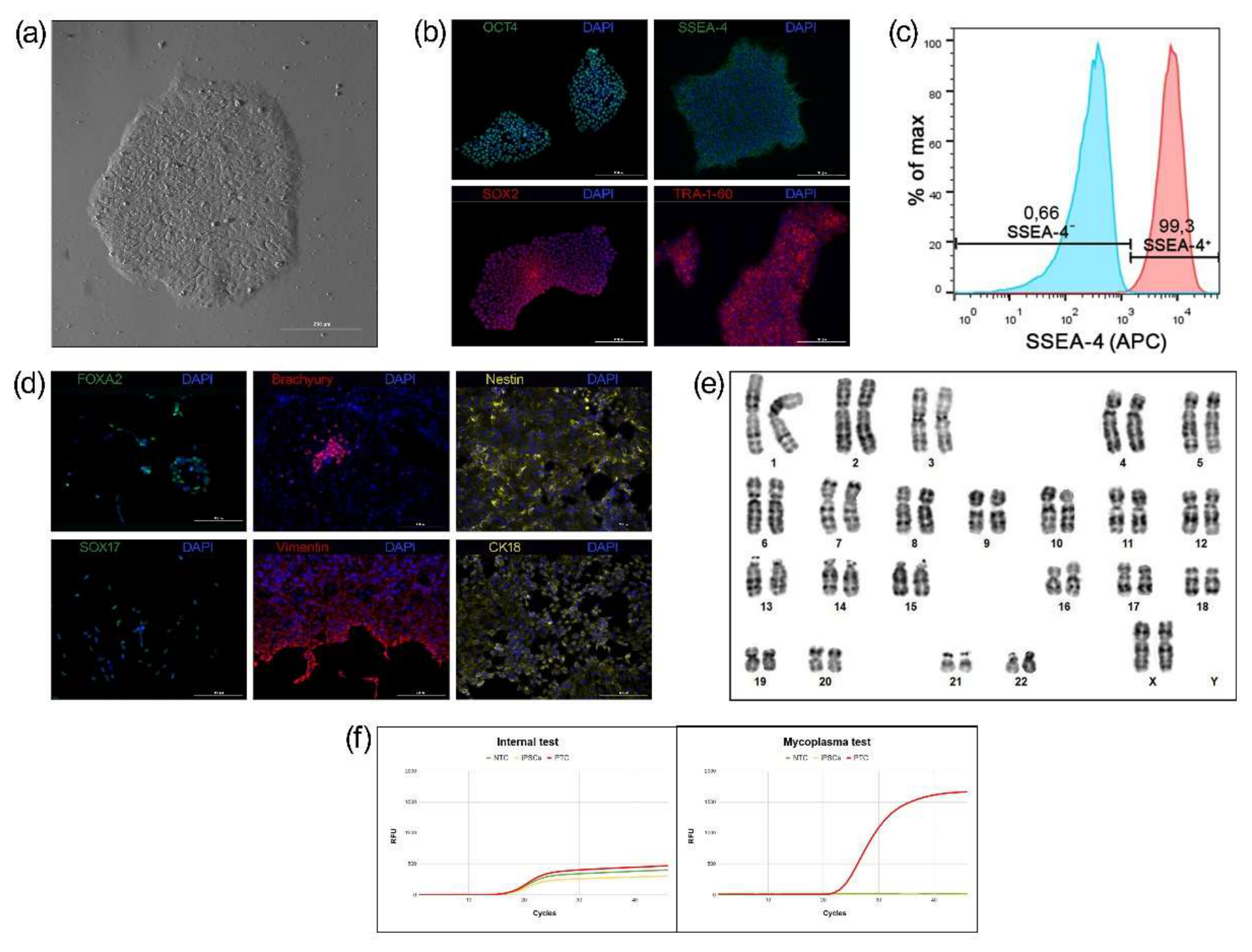
Figure 2.
Schematic representation of the differentiation protocol used to generate NK cells from RCMGi021-A iPSCs. The protocol comprises sequential stages of embryoid bodies formation, hematopoietic specification, lymphoid differentiation, NK-cell development, and subsequent expansion using genetically modified K562 cell line. Created in BioRender. Goldshtein, D. (2026). https://BioRender.com/wxyc5ah.
Figure 2.
Schematic representation of the differentiation protocol used to generate NK cells from RCMGi021-A iPSCs. The protocol comprises sequential stages of embryoid bodies formation, hematopoietic specification, lymphoid differentiation, NK-cell development, and subsequent expansion using genetically modified K562 cell line. Created in BioRender. Goldshtein, D. (2026). https://BioRender.com/wxyc5ah.
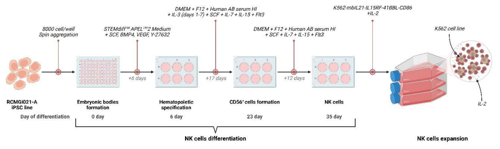
Figure 3.
Morphological changes observed during the differentiation of RCMGi021-A iPSCs into NK cells. (a) Undifferentiated iPSCs, imaged by phase-contrast microscopy. (b) EBs formed following the induction of differentiation. (c) Hematopoietic progenitor-like cells emerging from EBs at day 7 of differentiation. (d) Suspension cells with characteristic NK-cell morphology following continued culture under NK-cell differentiation conditions. Panels (b–d) were acquired by bright-field microscopy. Scale bars, 200 µm.
Figure 3.
Morphological changes observed during the differentiation of RCMGi021-A iPSCs into NK cells. (a) Undifferentiated iPSCs, imaged by phase-contrast microscopy. (b) EBs formed following the induction of differentiation. (c) Hematopoietic progenitor-like cells emerging from EBs at day 7 of differentiation. (d) Suspension cells with characteristic NK-cell morphology following continued culture under NK-cell differentiation conditions. Panels (b–d) were acquired by bright-field microscopy. Scale bars, 200 µm.
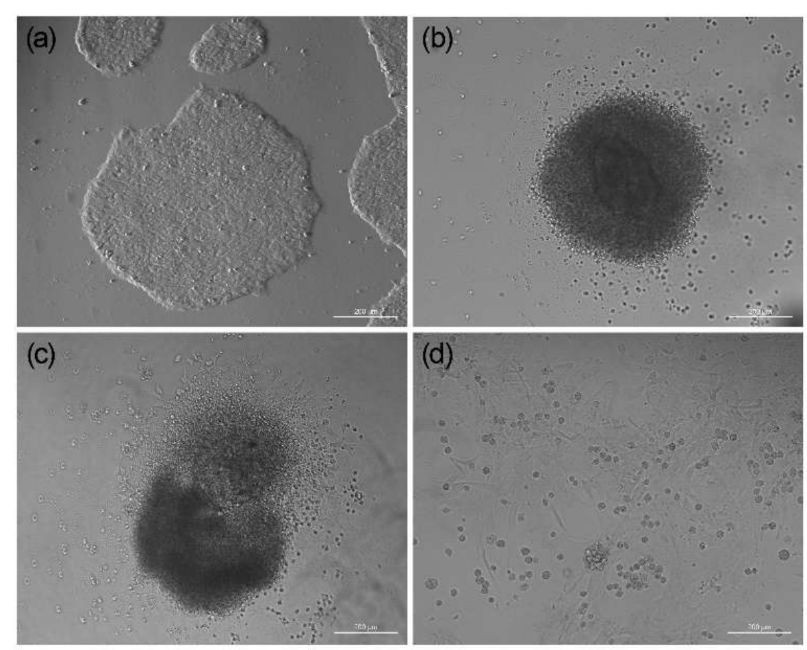
Figure 4.
Flow cytometric assessment of CD56 (FITC) and CD16 (APC) expression in RCMGi021-A iPSC-derived NK cells on day 23 of differentiation. Representative CD56/CD16 dot plot showing the distribution of CD56⁺CD16⁻ (32.80%), CD56⁺CD16⁺ (45.05%), CD56⁻CD16⁺ (2.85%), and CD56⁻CD16⁻ (19.30%) populations.
Figure 4.
Flow cytometric assessment of CD56 (FITC) and CD16 (APC) expression in RCMGi021-A iPSC-derived NK cells on day 23 of differentiation. Representative CD56/CD16 dot plot showing the distribution of CD56⁺CD16⁻ (32.80%), CD56⁺CD16⁺ (45.05%), CD56⁻CD16⁺ (2.85%), and CD56⁻CD16⁻ (19.30%) populations.
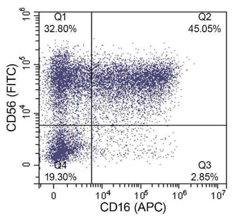
Figure 5.
Flow cytometry phenotypic characterization of expanded iNK cells compared to PB-NK cells. Representative dot plots and histograms illustrating the surface marker expression profiles of iNK cells (upper row) and PB-NK reference cells (lower row). (a) Bivariate dot plots showing the expression of CD56 (FITC) versus CD16 (APC) to define NK cell subpopulations. (b) Histograms depicting the evaluation of CD3 (APC) expression to confirm the absence of T-cell contamination. (c) Dot plots showing the co-expression of CD56 (FITC) and the natural cytotoxicity receptor NKp46 (PE). (d–g) Representative histograms demonstrating the expression frequencies of key NK-specific activating and inhibitory receptors, including (d) NKp44 (PE), (e) NKp30 (BV605), (f) CD94 (PE), and (g) CD158 (PE/Cyanine7). The percentages indicated within the gates represent the positive cell population for each respective marker.
Figure 5.
Flow cytometry phenotypic characterization of expanded iNK cells compared to PB-NK cells. Representative dot plots and histograms illustrating the surface marker expression profiles of iNK cells (upper row) and PB-NK reference cells (lower row). (a) Bivariate dot plots showing the expression of CD56 (FITC) versus CD16 (APC) to define NK cell subpopulations. (b) Histograms depicting the evaluation of CD3 (APC) expression to confirm the absence of T-cell contamination. (c) Dot plots showing the co-expression of CD56 (FITC) and the natural cytotoxicity receptor NKp46 (PE). (d–g) Representative histograms demonstrating the expression frequencies of key NK-specific activating and inhibitory receptors, including (d) NKp44 (PE), (e) NKp30 (BV605), (f) CD94 (PE), and (g) CD158 (PE/Cyanine7). The percentages indicated within the gates represent the positive cell population for each respective marker.
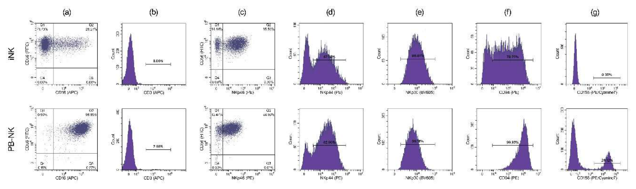
Figure 6.
Functional characterization of iNK cells by intracellular cytokine staining. Representative flow cytometry plots showing intracellular TNF-α (PE) and IFN-γ (APC) production by iNK cells under basal conditions (a), following co-culture with K562 cells (b), and following pharmacological stimulation with PMA/ionomycin (c). Numbers indicate the percentage of cells in each.
Figure 6.
Functional characterization of iNK cells by intracellular cytokine staining. Representative flow cytometry plots showing intracellular TNF-α (PE) and IFN-γ (APC) production by iNK cells under basal conditions (a), following co-culture with K562 cells (b), and following pharmacological stimulation with PMA/ionomycin (c). Numbers indicate the percentage of cells in each.
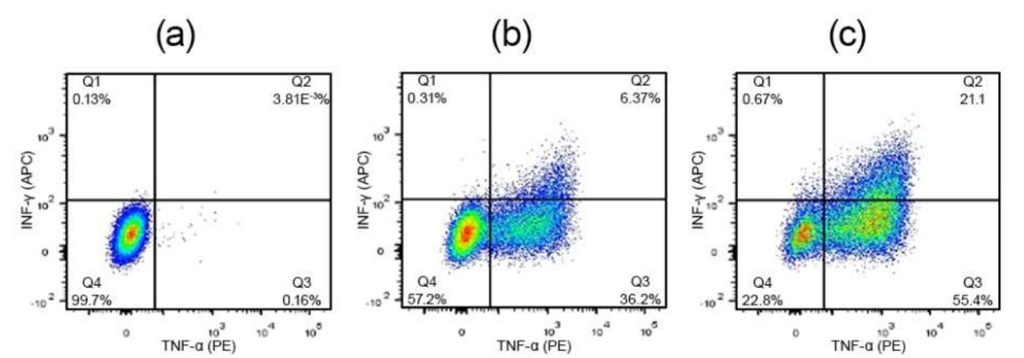
Figure 7.
Cytotoxic activity of iNK cells and PB-NK cells against SKOV3-Luc cells. Specific lysis was assessed at E:T ratios of 10:1, 5:1, 2:1, and 1:1 using a luciferase-based cytotoxicity assay. Data are presented as mean ± SD.
Figure 7.
Cytotoxic activity of iNK cells and PB-NK cells against SKOV3-Luc cells. Specific lysis was assessed at E:T ratios of 10:1, 5:1, 2:1, and 1:1 using a luciferase-based cytotoxicity assay. Data are presented as mean ± SD.
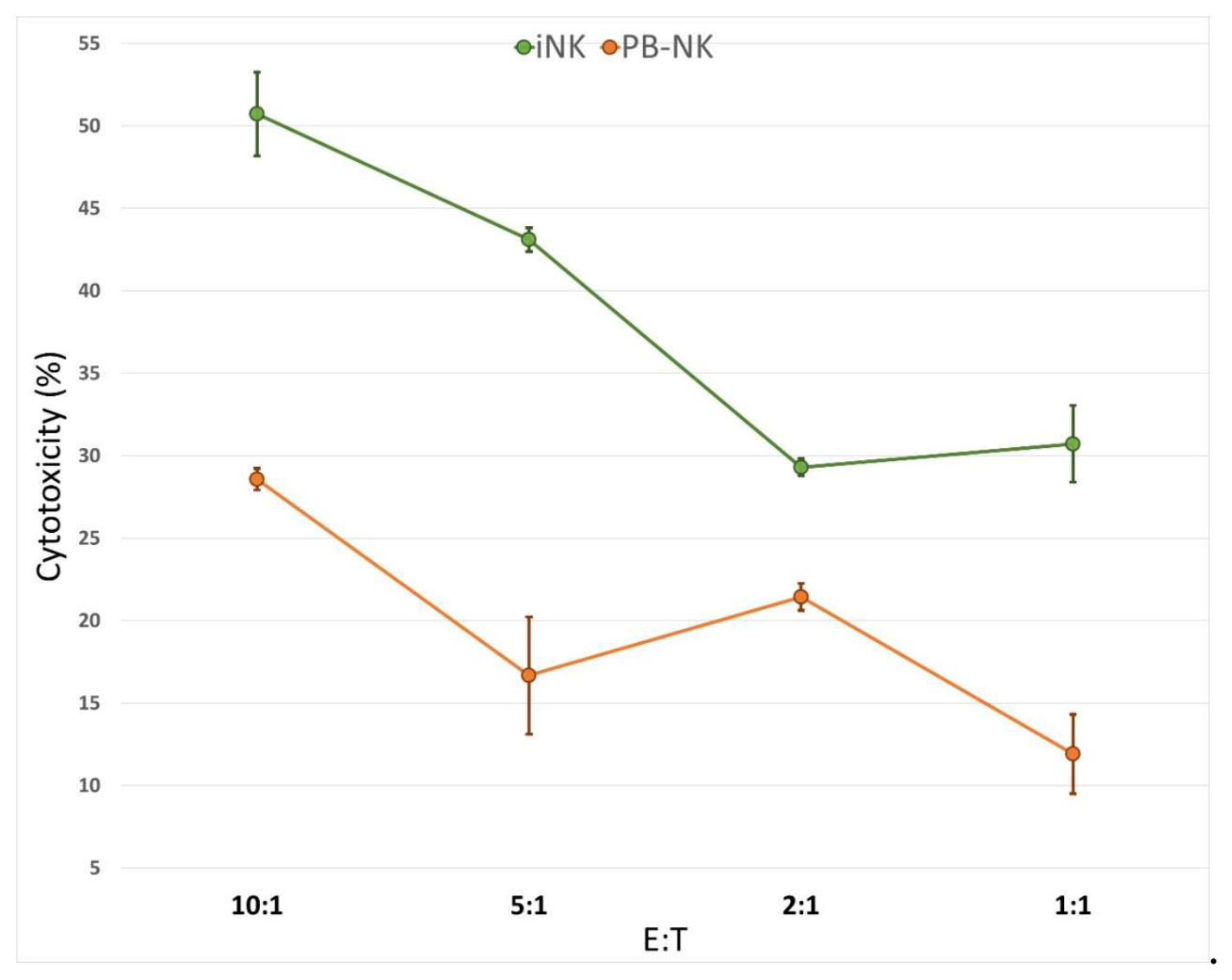

Table 1.
The RCMGi021-A cell line information.
| Characteristics | Description |
|---|---|
| Unique stem cell line identifier | RCMGi021-A |
| Institution | Research Centre for Medical Genetics |
| Approval of the Ethics Committee | Ethics Committee of Research Centre for Medical Genetics, Moscow, Russia Approval no. 5 of October 17, 2024 |
| Cell type | iPSC |
| Species of organisation | Human |
| Additional origin info | Age: 25 Sex: female Ethnicity if known: Russian |
| Initial cell type | Dermal fibroblasts |
| Date of biomaterial collection | January 22, 2025 |
| Reprogramming method | Non-integrating, self-replicating RNA-based reprogramming vector |
| Reprogramming factors | OCT4, KLF4, SOX2, GLIS1, c-MYC |
| Clonality | Clonal |
| Genetic modification | No |
| Type of genetic modification | No |
| Disease | N/A |
| Gene/locus | N/A |
| Morphology | Monolayer colonies similar to human PSCs |
| Pluripotency | Confirmed by in vitro trilineage differentiation |
| Karyotype | 46,XX |
| Contamination check | Bacteria, fungi and mycoplasma were not detected |
| Area of application | Human iPSC line derived from an apparently healthy donor for studies of human development, disease modeling, drug screening, genome engineering, and the generation of differentiated cell products for regenerative medicine and immune cell-based therapies |
| Cultivation method | Feeder free (Matrigel, Corning) |
| Culture medium | mTeSR™8 (STEMCELL Technologies) |
| Temperature, °C | 37 |
| Concentration CO2, % | 5 |
| Concentration O2, % | 20 |
| Passaging method | Non-enzymatic, Versene solution (Paneco) |
| Multiplicity of dilution during passaging | 1:12 – 1:15 |
| Cryopreservation | CryoStore (STEMCELL Technologies) |
| Storage conditions | Liquid nitrogen |
| Account in the registry | https://hpscreg.eu/cell-line/RCMGi021-A |
| Date archived/stock date | June 18, 2025 |
Table 2.
Comprehensive characterization of the RCMGi021-A cell line.
| Classification | Test | Result | Data |
|---|---|---|---|
| Morphology | Phase-contrast microscopy | Normal | Fig.1 panel a |
| Phenotype | Qualitative analysis: immunocytochemistry |
Positive for pluripotency markers: OCT4, SOX2, SSEA-4, TRA-1-60. | Fig.1 panel b |
| Quantitative analysis: flow cytometry |
Expression of pluripotency marker SSEA-4. Percentage of positive cells: 99.3% (n = 50.000) | Fig.1 panel c | |
| Differentiation potential | Directed differentiation into derivatives of the three embryonic germ layers |
Positive staining for characteristic germ layer markers: FOXA2 and SOX17 for endoderm; Brachyury and vimentin for mesodermal lineage; and Nestin as a neural ectoderm marker. | Fig.1 panel d |
| Genotype | Karyotype (G-banding) and resolution | 46,XX, Resolution 450-500 |
Fig.1 panel e |
| Microbiology and virology | Mycoplasma | Mycoplasma testing by qPCR. Negative | Fig.1 panel f |
| Identity | STR analysis | 11 loci analyzed, all matched |
Table S1; Figure S1 |
Disclaimer/Publisher’s Note: The statements, opinions and data contained in all publications are solely those of the individual author(s) and contributor(s) and not of MDPI and/or the editor(s). MDPI and/or the editor(s) disclaim responsibility for any injury to people or property resulting from any ideas, methods, instructions or products referred to in the content. |
© 2026 by the authors. Licensee MDPI, Basel, Switzerland. This article is an open access article distributed under the terms and conditions of the Creative Commons Attribution (CC BY) license.
Copyright: This open access article is published under a Creative Commons CC BY 4.0 license, which permit the free download, distribution, and reuse, provided that the author and preprint are cited in any reuse.