

Submitted:
15 November 2026
Posted:
17 November 2026
You are already at the latest version
Abstract
Background: Linezolid is an important treatment option for resistant gram-positive infections when conventional agents cannot be used. Reversible myelosuppression is well recognized, whereas hypersensitivity is rare and predominantly cutaneous, renal or systemic. Case: A 9-year-old boy with a right below-knee amputation was treated for stump cellulitis with possible early osteomyelitis. Because a previous stump isolate was methicillin-resistant Staphylococcus aureus, vancomycin had caused a diffuse rash and clindamycin was temporarily unavailable, intravenous linezolid 10 mg/kg every 8 hours was started. Approximately 5 days later, he developed tender right inguinal and submental lymphadenopathy and malaise near the end of each linezolid infusion. He remained afebrile and had no rash, facial edema, mucosal disease or renal, hepatic or pulmonary injury. The absolute eosinophil count increased from 480 to 702 cells/µL, the absolute neutrophil count decreased from 3,800 to 800 cells/µL, and the platelet count reached 139 × 10³/µL. Linezolid was withdrawn after approximately 9 days; lymph node tenderness improved within 24 hours and laboratory abnormalities improved within 3 days without corticosteroids. Clindamycin was reintroduced without recurrence. Conclusions: The Naranjo score was 5, indicating a probable adverse drug reaction, while RegiSCAR excluded DRESS. No previous report identified by a focused literature review described lymphadenopathy as the predominant manifestation of a linezolid-associated reaction. New multifocal lymphadenopathy arising during linezolid therapy should raise suspicion of a drug-related reaction even without rash or systemic organ involvement.
Keywords:
linezolid
; lymphadenopathy
; eosinophilia
; neutropenia
; adverse drug reaction
1. Introduction
Linezolid, the first clinically available oxazolidinone, is active against resistant gram-positive pathogens, including methicillin-resistant Staphylococcus aureus and vancomycin-resistant enterococci [1]. Its high oral bioavailability and tissue penetration make it a valuable alternative when conventional agents cannot be used [1]. Pediatric studies suggest that linezolid is generally effective and well tolerated; however, anemia, thrombocytopenia, leukopenia, and neutropenia remain clinically important adverse effects [1,2,3].
Myelosuppression is classically associated with treatments exceeding 14 days, although earlier onset has been reported [1,3,4]. Linezolid-associated hypersensitivity is less well characterized, with reported manifestations including drug eruptions, eosinophilia, acute interstitial nephritis, and drug reaction with eosinophilia and systemic symptoms (DRESS) [5,6].
We report the case of a pediatric patient who developed multifocal tender lymphadenopathy associated with eosinophilia, neutropenia and transient thrombocytopenia during linezolid therapy. Notably, these manifestations occurred without rash or biochemical evidence of hepatic or renal involvement. Clinical and hematological abnormalities improved after discontinuation of linezolid without the administration of corticosteroids. This case broadens the recognized clinical phenotype of linezolid-associated adverse reactions and underscores the importance of considering drug causality when new lymphadenopathy develops during antimicrobial treatment. This case report was prepared in accordance with the CARE reporting guidelines [7].
2. Case Presentation
A previously healthy 9-year-old boy sustained a severe blast injury in September 2024, resulting in a right below-knee amputation and a left distal femoral Salter–Harris II fracture requiring open reduction and internal fixation. His initial hospitalization was complicated by wound infection requiring repeated debridement. A right stump tissue culture in September 2024 grew carbapenem-resistant Pseudomonas aeruginosa that remained susceptible to cefepime; he improved after a 20-day course of cefepime. A right leg wound swab obtained in November 2024 grew methicillin-resistant S. aureus (MRSA) susceptible to clindamycin, ciprofloxacin, gentamicin, rifampin, tetracycline, trimethoprim-sulfamethoxazole and vancomycin.
After prosthetic fitting in January 2025, he developed recurrent stump ulceration attributed to limited soft-tissue padding, contracture and prosthetic pressure. On May 16, 2026, he stood directly on the stump, causing abrasions and skin breakdown, followed by progressive pain, erythema, warmth and fever reaching 39°C with rigors. He received intravenous cefepime and clindamycin. Magnetic resonance imaging (MRI) on May 20 showed stump cellulitis with mild distal tibial marrow signal interpreted as reactive osteitis from adjacent inflammation versus early osteomyelitis. A peripherally inserted central catheter (PICC) was placed, and after 6 days he was transferred to a peripheral hospital to continue antimicrobial therapy.
Continued coverage for MRSA was maintained because of the previous isolate. Vancomycin was avoided because it had caused a diffuse rash during an earlier exposure. When clindamycin became unavailable at the peripheral hospital, it was replaced on June 10, 2026, with intravenous linezolid 10 mg/kg every 8 hours, infused over 30 minutes. He had no previous linezolid exposure. Cefepime was continued concurrently.
Approximately 5 days later, on June 15, the child developed tender right inguinal and submental lymphadenopathy, prompting presentation to our emergency department. His father reported that the child appeared unwell and experienced diffuse discomfort toward the end of each linezolid infusion and briefly thereafter. These symptoms resolved between infusions and were not accompanied by rash, difficulty breathing, abdominal pain, vomiting or fever.
There was no recurrent fever or rigors and no worsening of stump pain, erythema, warmth, swelling or discharge. He reported no respiratory, pharyngeal, dental, gastrointestinal or urinary symptoms; nor did he have a rash, facial edema, oral or other mucosal lesions, weight loss, or night sweats. There were no sick contacts, animal exposures or consumption of raw meat or unpasteurized dairy products.
On admission, he was afebrile, comfortable, and well appearing. Two mobile, tender right submental lymph nodes and several tender, mobile right inguinal nodes, each greater than 1 cm, were palpable without overlying erythema, fluctuance or drainage. No other clinically significant lymphadenopathy was identified. The abdomen was soft and nontender without palpable hepatosplenomegaly. The stump was pink with 2 small dry ulcers and superficial abrasions at its tip but no swelling, fluctuance, crepitus, active drainage or palpable collection. The infectious diseases and surgical teams considered ongoing cellulitis unlikely, and repeat MRI was not indicated.
Serial laboratory findings are summarized in Table 1 and the clinical course in Figure 1. Mild eosinophilia preceded linezolid, with an absolute eosinophil count (AEC) of 480 cells/µL on May 23, and increased during exposure to 702 cells/µL on June 18. The absolute neutrophil count (ANC) fell to 800 cells/µL on June 19, and platelets transiently decreased to 139 × 10³/µL on June 18. C-reactive protein decreased from 95.0 mg/L during the stump infection to 11.5 mg/L on June 15 and 10.4 mg/L on June 18, consistent with improvement of the stump infection.
During the suspected reaction, alanine aminotransferase, aspartate aminotransferase, bilirubin, creatinine and lactate dehydrogenase were within their respective reference ranges. Abdominal ultrasonography showed mild hepatomegaly (15.5 cm) with homogeneous echotexture and a spleen at the upper limit of normal for age (11.4 cm). In the absence of biochemical injury, these findings were not considered visceral involvement. A peripheral blood film showed slight toxic granulation of neutrophils without atypical lymphocytes or blasts. Cytomegalovirus IgM and Epstein–Barr virus viral capsid antigen IgM were nonreactive; Brucella agglutination titers were below 1:20; and Bartonella henselae and B. quintana IgG and IgM were negative. Blood cultures were not obtained.
Linezolid was discontinued on June 19 after approximately 9 days of exposure. Cefepime was continued until the PICC was removed immediately before discharge later that day, completing approximately 4 weeks of therapy. He was transitioned to oral clindamycin and ciprofloxacin. No corticosteroids or antihistamines were administered. Infusion-associated discomfort ceased, and lymph-node tenderness improved within approximately 24 hours. By discharge, both nodal groups had decreased in size and the inguinal nodes were nontender.
Causality between linezolid exposure and the combined clinical and hematologic reaction—lymphadenopathy, infusion-associated discomfort, eosinophilia, neutropenia, and transient thrombocytopenia—was evaluated using the Naranjo algorithm (Table 2) [8]. The score was 5, indicating a probable linezolid-associated adverse drug reaction. The possibility of DRESS was considered but excluded by a RegiSCAR score of −1 (Table 3) [9].
On June 22, the WBC count was 5.8 × 10³/µL, ANC approximately 1,640 cells/µL, AEC approximately 389 cells/µL and platelet count 258 × 10³/µL, demonstrating recovery of neutrophils and platelets and resolution of eosinophilia. At follow-up on June 23, his general condition was markedly improved and the submental and inguinal nodes were substantially smaller, mobile and nontender. He remained afebrile without rash or systemic symptoms.
He subsequently completed a 6-week antimicrobial course for osteomyelitis, including clindamycin and ciprofloxacin after discharge, without further adverse effects or recurrence of lymphadenopathy, eosinophilia, or cytopenia. He remained clinically well at 3-month follow-up.
3. Discussion
This child developed multifocal tender lymphadenopathy approximately 5 days after starting linezolid, accompanied by infusion-associated discomfort, eosinophilia, neutropenia and transient thrombocytopenia. Clinical and hematological abnormalities improved promptly after linezolid withdrawal without corticosteroids. The temporal association and improvement following withdrawal suggest a probable linezolid-associated adverse reaction. This case highlights multifocal lymphadenopathy as a potential early manifestation of linezolid-associated hypersensitivity, even in the absence of rash, fever, or visceral involvement.
We conducted a focused literature search of PubMed/MEDLINE, Embase and Scopus from inception through September 20, 2026, combining “linezolid” with “lymphadenopathy,” “lymphadenitis,” “lymph node,” “hypersensitivity,” “eosinophilia,” “DRESS,” “drug eruption,” “angioedema,” “vasculitis” and “anaphylaxis.” Reference lists and citing articles of eligible publications were also screened. We included individual case reports or case letters describing a suspected immune-mediated reaction to linezolid with sufficient clinical detail to characterize the presentation, time to onset (latency), phenotype, and outcome. Seventeen reports were identified (Table 4).
The published reactions included purpuric eruptions or leukocytoclastic vasculitis [11,16,19,21], DRESS or drug rash with eosinophilia complicated by acute interstitial nephritis [6,12,22], immediate reactions including urticaria, angioedema and test-dose reactions managed by desensitization [13,14,17,24,25], macular exanthem [10], fixed drug eruption [18,23] and epidermal necrolysis [15,20]. The reported time to onset (latency) ranged from minutes to approximately 10 days. Reported clinical phenotypes were predominantly cutaneous, renal or systemic. Lymphadenopathy was explicitly absent in one report [21] and was not described in the remaining reports. By contrast, the predominant manifestation in our patient was multifocal tender lymphadenopathy without rash or evidence of visceral injury. To our knowledge, no previously published report identified through our focused search described lymphadenopathy as the predominant manifestation of a suspected linezolid-associated reaction.
Several features support a possible association with linezolid in this case. Lymphadenopathy appeared approximately 5 days after first exposure, a latency similar to that of several published delayed linezolid reactions [11,12,19,21,22]. Discomfort recurred near the end of infusions and ceased after linezolid withdrawal. Lymph node tenderness improved within approximately 24 hours without corticosteroids or antihistamines, and neither lymphadenopathy nor eosinophilia recurred after clindamycin was reintroduced. The Naranjo score of 5 classifies the reaction as a probable adverse drug reaction (Table 2) [8].
Alternative explanations were considered. Reactive lymphadenopathy associated with the stump infection remains a plausible explanation for the inguinal nodes; however, it does not readily account for the simultaneous submental lymphadenopathy, which lies outside the lymphatic drainage territory of the affected limb. Furthermore, recurrent infection was considered unlikely given the substantial decline in C-reactive protein, the clinically quiescent stump with only small dry ulcers and superficial abrasions, the absence of recurrent fever or local inflammatory progression, and assessment by both infectious diseases and surgical teams. A viral illness could cause lymphadenopathy and neutropenia, but there was no compatible clinical syndrome, no atypical lymphocytes and no serological evidence of acute cytomegalovirus or Epstein–Barr virus infection; other viral infections were not investigated. Brucella and Bartonella serologies were negative, and there were no relevant animal contacts or food exposure.
Cefepime was the principal alternative culprit, particularly for the hematological abnormalities. Prolonged β-lactam therapy, including cefepime, is a recognized cause of neutropenia and may be accompanied by eosinophilia and thrombocytopenia [26,27]. Nevertheless, the patient had tolerated cefepime for approximately 4 weeks before the onset of lymphadenopathy, whereas both the lymph node enlargement and infusion-associated discomfort developed only after linezolid was introduced. This temporal relationship favors linezolid as the principal cause of the overall reaction. However, a contribution from cefepime to the hematological abnormalities cannot be completely excluded.
Linezolid-associated myelosuppression is well recognized in children, is generally reversible and is more commonly associated with prolonged treatment [1,2,3]. The neutropenia and transient thrombocytopenia may therefore represent recognized hematological toxicity rather than hypersensitivity. The rise in eosinophils during treatment is more suggestive of an immune-mediated component, although mild eosinophilia preceded linezolid exposure, which limits its specificity. Multifocal tender lymphadenopathy, rather than the cytopenias, is thus the distinctive feature of this case.
Despite lymphadenopathy and eosinophilia, the presentation did not meet criteria for DRESS. The RegiSCAR score was −1, corresponding to “no case” (Table 3) [9]. A delayed hypersensitivity reaction not meeting DRESS criteria remains possible, although the underlying mechanism could not be established.
This report has several limitations. It describes a single case. Lymph-node biopsy was not performed, no drug-specific allergy testing was undertaken and linezolid concentrations were not measured. Rechallenge was not performed because it was not clinically justified. Mild eosinophilia preceded linezolid exposure. Concurrent cefepime and the recent infection limit causal certainty. Despite these limitations, the close exposure-response sequence and absence of a more persuasive explanation support a probable linezolid-associated reaction.
4. Conclusions
New multifocal lymphadenopathy shortly after linezolid initiation, particularly with concurrent eosinophilia, should prompt early consideration of a drug-related etiology alongside the evaluation of alternative infectious and hematological causes. Where clinically appropriate and an alternative agent is available, linezolid withdrawal may serve both diagnostic and therapeutic purposes. Further case reports and pharmacovigilance data are needed to better define the clinical spectrum of linezolid-associated hypersensitivity.
Author Contributions
Investigation, D.K., F.A., R.H.-W. and M.M.; data curation and analysis, D.K. and F.A.; writing—original draft preparation, D.K. and F.A.; writing—review and editing, R.H.-W. and M.M. All authors have read and agreed to the published version of the manuscript.
Funding
This research received no external funding.
Institutional Review Board Statement
Ethical review and approval were waived for this study because the Institutional Review Board of the American University of Beirut determined that formal ethics approval was not required for this single-patient case report.
Informed Consent Statement
Written informed consent has been obtained from the patient’s parent/legal guardian to publish this paper, including the patient’s deidentified clinical information.
Data Availability Statement
All data relevant to this case are included in the article. Additional patient-level data are not publicly available because of privacy and confidentiality considerations.
Acknowledgments
The authors thank the patient and his family for permitting publication of this case. During the preparation of this manuscript, the authors used ChatGPT (GPT-6, OpenAI) solely for language editing. The authors have reviewed and edited the output and take full responsibility for the content of this publication.
Conflicts of Interest
The authors declare no conflicts of interest.
References
- Garazzino, S.; Tovo, P.-A. Clinical experience with linezolid in infants and children. J. Antimicrob. Chemother. 2011, 66 (Suppl. 4), iv23–iv41. [Google Scholar] [CrossRef] [PubMed]
- Shi, Y.; Wu, H.-L.; Wu, Y.-H.; et al. Safety and clinical efficacy of linezolid in children: A systematic review and meta-analysis. World J. Pediatr. 2023, 19, 129–138. [Google Scholar] [CrossRef] [PubMed]
- Kato, H.; Hagihara, M.; Asai, N.; et al. A systematic review and meta-analysis of myelosuppression in pediatric patients treated with linezolid for Gram-positive bacterial infections. J. Infect. Chemother. 2021, 27, 1143–1150. [Google Scholar] [CrossRef] [PubMed]
- Zou, F.; Cui, Z.; Lou, S.; et al. Adverse drug events associated with linezolid administration: A real-world pharmacovigilance study from 2004 to 2023 using the FAERS database. Front. Pharmacol. 2024, 15, 1338902. [Google Scholar] [CrossRef] [PubMed]
- Dilley, M.; Geng, B. Immediate and delayed hypersensitivity reactions to antibiotics: Aminoglycosides, clindamycin, linezolid, and metronidazole. Clin. Rev. Allergy Immunol. 2022, 62, 463–475. [Google Scholar] [CrossRef] [PubMed]
- Savard, S.; Desmeules, S.; Riopel, J.; et al. Linezolid-associated acute interstitial nephritis and drug rash with eosinophilia and systemic symptoms (DRESS) syndrome. Am. J. Kidney Dis. 2009, 54, e17–e20. [Google Scholar] [CrossRef] [PubMed]
- Gagnier, J.J.; Kienle, G.; Altman, D.G.; et al. The CARE guidelines: Consensus-based clinical case reporting guideline development. Headache 2013, 53, 1541–1547. [Google Scholar] [CrossRef] [PubMed]
- Naranjo, C.A.; Busto, U.; Sellers, E.M.; et al. A method for estimating the probability of adverse drug reactions. Clin. Pharmacol. Ther. 1981, 30, 239–245. [Google Scholar] [CrossRef] [PubMed]
- Kardaun, S.H.; Sidoroff, A.; Valeyrie-Allanore, L.; et al. Variability in the clinical pattern of cutaneous side-effects of drugs with systemic symptoms: Does a DRESS syndrome really exist? Br. J. Dermatol. 2007, 156, 609–611. [Google Scholar] [CrossRef] [PubMed]
- Borrás-Blasco, J.; Verge, J.M.V.; Rosique-Robles, D.; et al. Possible macular exanthema associated with linezolid therapy. Int. J. Clin. Pharmacol. Ther. 2006, 44, 331–334. [Google Scholar] [CrossRef] [PubMed]
- Kim, F.S.; Kelley, W.; Resh, B.; et al. Linezolid-induced purpuric medication reaction. J. Cutan. Pathol. 2009, 36, 793–795. [Google Scholar] [CrossRef] [PubMed]
- Nayak, S.; Nandwani, A.; Rastogi, A.; et al. Acute interstitial nephritis and drug rash with secondary to linezolid. Indian J. Nephrol. 2012, 22, 367–369. [Google Scholar] [CrossRef] [PubMed]
- Yang, M.; Xu, M. Linezolid-induced angioedema and urticaria in a patient with renal failure. Braz. J. Infect. Dis. 2012, 16, 606–607. [Google Scholar] [CrossRef] [PubMed]
- Bagwell, A.D.; Stollings, J.L.; White, K.D.; et al. Linezolid desensitization for a patient with multiple medication hypersensitivity reactions. Ann. Pharmacother. 2013, 47, e30. [Google Scholar] [CrossRef] [PubMed]
- Ivić, D.; Ivić, J.; Došen, G.; et al. Toxic epidermal necrolysis after linezolid administration: Case report. Signa Vitae 2014, 9, 65–67. [Google Scholar] [CrossRef]
- Sathyanarayana, V.; Das, U.; Babu, K.G.; et al. Linezolid induced vasculitis: An unusual case report with review of the literature. J. Sci. Soc. 2015, 42, 27–30. [Google Scholar] [CrossRef]
- Guvenir, H.; Dibek Misirlioglu, E.; Toyran, M.; et al. Linezolid desensitization in a pediatric patient. Ann. Allergy Asthma Immunol. 2016, 117, 198. [Google Scholar] [CrossRef] [PubMed]
- Inamadar, A.C.; Palit, A. Fixed drug eruption due to linezolid. Indian J. Drugs Dermatol. 2017, 3, 38–39. [Google Scholar] [CrossRef] [PubMed]
- Kruzer, K.; Garner, W.; Honda, K.; et al. Linezolid-induced leukocytoclastic vasculitis. Ann. Pharmacother. 2018, 52, 1263–1264. [Google Scholar] [CrossRef] [PubMed]
- Memon, S.; Ahmed, N.; Nasir Memon, M.; et al. Linezolid and ciprofloxacin-induced SJS/TEN (Stevens-Johnson syndrome/toxic epidermolysis necrosis) overlap in a patient with borderline personality disorder during a single hospital stay: A difficult case to manage. Cureus 2023, 15, e39242. [Google Scholar] [CrossRef] [PubMed]
- Tirú-Vega, M.A.; Engel-Rodriguez, N.; Rivera-Bobé, N.; et al. A rare skin rash: Linezolid-induced purpuric drug eruption without vasculitis in a Puerto Rican man. Am. J. Case Rep. 2023, 24, e941725. [Google Scholar] [CrossRef] [PubMed]
- Velasco Hoyos, M.F.; Giraldo-Bahamón, G.; Díaz, Á.; et al. Síndrome de DRESS asociado al uso de linezolid. Rev. Asoc. Colomb. Dermatol. Cir. Dermatol. 2024, 32, 442–447. [Google Scholar] [CrossRef]
- Prathap, T.A.; Lokeshvaran, D.; Abinish, D.G.; et al. A rare case report on linezolid induced fixed drug eruption. Int. J. Res. Dermatol. 2025, 11, 167–170. [Google Scholar] [CrossRef]
- Ortiz-Fernández, P.; Almanchel-Rivadeneyra, M.; Urbieta-Sanz, E. Linezolid-induced eyelid angioedema: A case report. Eur. J. Hosp. Pharm. 2025. [Google Scholar] [CrossRef] [PubMed]
- Diwan, P.R.; Jarwani, B.S.; Malhotra, S.D. Immediate cutaneous reaction to linezolid. Natl. J. Pharmacol. Ther. 2026, 4, 34–37. [Google Scholar] [CrossRef] [PubMed]
- Foong, K.S.; Hsueh, K.; Bailey, T.C.; et al. A cluster of cefepime-induced neutropenia during outpatient parenteral antimicrobial therapy. Clin. Infect. Dis. 2019, 69, 534–537. [Google Scholar] [CrossRef] [PubMed]
- Koraysh, S.; Koleri, J.; Ali, M. Cefepime-induced neutropenia: A case report and literature review. Clin. Pharmacol. 2023, 15, 33–40. [Google Scholar] [CrossRef] [PubMed]
Figure 1.
Timeline of antimicrobial exposure, clinical events, and hematological changes. (A) Intravenous cefepime and clindamycin, intravenous linezolid (June 10–19) and oral clindamycin with ciprofloxacin after discharge. Dashed vertical lines indicate the onset of lymphadenopathy (June 15) and near-complete clinical resolution (June 23). (B) Absolute neutrophil count (ANC) and absolute eosinophil count (AEC) in cells/µL; the horizontal dashed line indicates an ANC of 1,500 cells/µL.
Figure 1.
Timeline of antimicrobial exposure, clinical events, and hematological changes. (A) Intravenous cefepime and clindamycin, intravenous linezolid (June 10–19) and oral clindamycin with ciprofloxacin after discharge. Dashed vertical lines indicate the onset of lymphadenopathy (June 15) and near-complete clinical resolution (June 23). (B) Absolute neutrophil count (ANC) and absolute eosinophil count (AEC) in cells/µL; the horizontal dashed line indicates an ANC of 1,500 cells/µL.
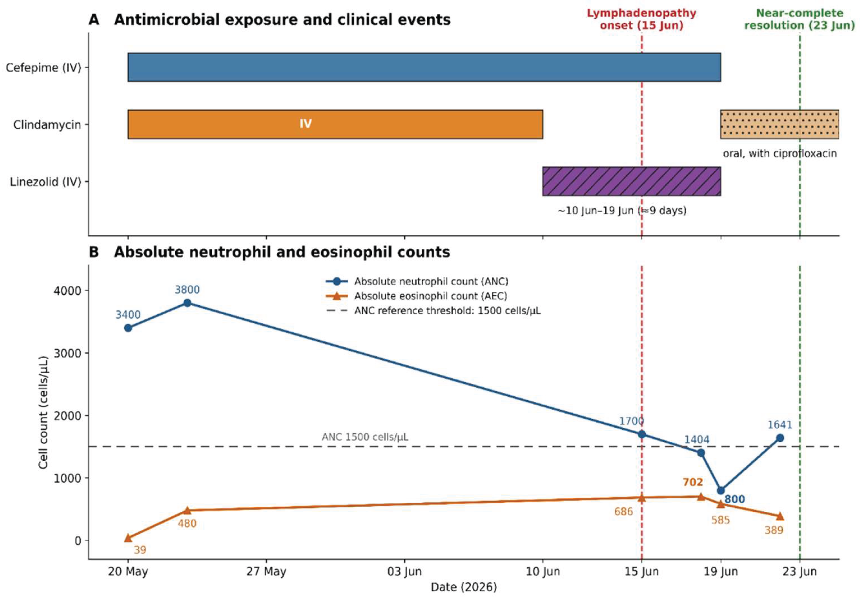

Table 1.
Antimicrobial exposure and relevant laboratory findings before, during and after linezolid treatment.
Table 1.
Antimicrobial exposure and relevant laboratory findings before, during and after linezolid treatment.
| Parameter | 20 May | 15 Jun | 18 Jun | 19 Jun | 22 Jun |
|---|---|---|---|---|---|
| Antimicrobial exposureᵃ | |||||
| Linezolid | ✕ | ✓ | ✓ | Discontinued | ✕ |
| Cefepime | ✓ | ✓ | ✓ | Discontinued | ✕ |
| Clindamycin | ✓ (IV) | ✕ | ✕ | Restarted (oral) | ✓ |
| Laboratory findings | |||||
| Hemoglobin, g/dL | 12.7 | 10.7 | 9.4 | 10.2 | 11.9 |
| WBC, ×10³/µL | 3.9 | 4.9 | 3.9 | 3.9 | 5.8 |
| ANC, cells/µL | 3,400 | 1,700 | 1,400 | 800 | 1,640 |
| AEC, cells/µLᵇ | 39 | 686 | 702 | 585 | 389 |
| Platelets, ×10³/µL | 212 | 176 | 139 | 167 | 258 |
| CRP, mg/L | 69.1 | 11.5 | 10.4 | — | 1.4 |
| ALT (SGPT), U/L | 85 | 29 | 39 | — | 19 |
| AST (SGOT), U/L | 164 | — | 41 | — | — |
| Alkaline phosphatase, U/L | 242 | — | 174 | — | — |
| GGT, U/L | 48 | — | 19 | — | — |
| Total/direct bilirubin, mg/dL | 0.4/0.2 | — | 0.3/0.1 | — | — |
| Creatinine, mg/dL | 0.41 | 0.25 | — | — | — |
| LDH, U/L | — | 248 | — | — | — |
Footnote: ᵃ ✓, received on that date; ✕, not received; —, not measured. Clindamycin was replaced by linezolid on June 10. ᵇ AEC values were calculated as WBC × eosinophil percentage when not directly reported. Bold values indicate the clinically relevant peak or nadir. Abbreviations: AEC, absolute eosinophil count; ALT, alanine aminotransferase; ANC, absolute neutrophil count; AST, aspartate aminotransferase; CRP, C-reactive protein; GGT, gamma-glutamyl transferase; IV, intravenous; LDH, lactate dehydrogenase; WBC, white blood cell count.
Table 2.
Naranjo adverse drug reaction probability assessment.
| No. | Naranjo item | Case assessment | Score |
|---|---|---|---|
| 1 | Are there previous conclusive reports of this reaction? | Yes. Linezolid-associated hypersensitivity and hematological toxicity have been reported [1,5], although the specific lymphadenopathy phenotype has not. | +1 |
| 2 | Did the adverse event occur after administration of the suspected drug? | Yes. Lymphadenopathy developed approximately 5 days after linezolid initiation. | +2 |
| 3 | Did the reaction improve after the drug was discontinued or a specific antagonist was administered? | Yes. Infusion-associated discomfort and lymph-node tenderness improved within approximately 24 hours. No corticosteroid or antihistamine was administered. | +1 |
| 4 | Did the reaction reappear on re-administration? | Not assessed; rechallenge was not clinically justified. | 0 |
| 5 | Are there alternative causes that could have caused the reaction? | No alternative cause was considered more likely: the stump infection was improving; C-reactive protein had fallen from 95.0 to 10.4 mg/L; the stump showed no progressive inflammation; inguinal drainage from the stump would not explain simultaneous submental lymphadenopathy; cefepime had been given for approximately 4 weeks before the reaction; and clindamycin was reintroduced without recurrence. | 0 |
| 6 | Did the reaction reappear after placebo administration? | Not assessed; placebo administration was not clinically justified. | 0 |
| 7 | Was the drug detected in blood at a toxic concentration? | Linezolid concentrations were not measured. | 0 |
| 8 | Was the reaction more severe with an increased dose or less severe with a decreased dose? | Not assessed. | 0 |
| 9 | Did the patient have a similar reaction to the same or a related drug previously? | No previous oxazolidinone exposure was reported. | 0 |
| 10 | Was the adverse event confirmed by objective evidence? | Yes. Lymphadenopathy was documented on serial examination, and eosinophilia, neutropenia and thrombocytopenia were demonstrated on serial blood counts. | +1 |
| Total score | Probable adverse drug reaction | 5 |
Footnote: Naranjo scores are interpreted as definite (≥9), probable (5–8), possible (1–4) or doubtful (≤0). Adapted from Naranjo et al. [8].
Table 3.
RegiSCAR assessment for drug reaction with eosinophilia and systemic symptoms.
| RegiSCAR criterion | Case assessment | Score |
|---|---|---|
| Fever ≥38.5°C during the suspected reaction (no/unknown −1; yes 0) | Absent | −1 |
| Enlarged lymph nodes at ≥2 sites and >1 cm (no/unknown 0; yes +1) | Submental and inguinal lymphadenopathy measuring >1 cm | +1 |
| Atypical lymphocytes (no/unknown 0; yes +1) | Not identified on peripheral blood film | 0 |
| Eosinophilia (700–1,499 cells/µL, or 10%–19.9% if leukocytes <4.0 × 10³/µL: +1; ≥1,500 cells/µL, or ≥20% if leukocytes <4.0 × 10³/µL: +2) | Peak eosinophils 18% with a leukocyte count of 3.9 × 10³/µL; AEC 702 cells/µL | +1 |
| Skin rash involving >50% of body surface area (no/unknown 0; yes +1) | No rash | 0 |
| Rash suggesting DRESS: ≥2 of edema, infiltration, purpura, scaling (no −1; unknown 0; yes +1) | No rash, facial edema, infiltration, purpura, or desquamation | −1 |
| Skin biopsy suggesting DRESS (no −1; yes/unknown 0) | Not performed | 0 |
| Internal-organ involvement (one organ +1; ≥2 organs +2) | None; mild hepatomegaly occurred without biochemical hepatic injury, and there was no renal or pulmonary involvement | 0 |
| Resolution requiring ≥15 days (no/unknown −1; yes 0) | No; substantial clinical and hematological improvement occurred within 3–8 days | −1 |
| Alternative diagnoses excluded by ≥3 biological investigations (yes +1) | Infectious investigations were negative, but the complete investigation set specified by RegiSCAR was not performed (antinuclear antibody, blood culture, hepatitis A/B/C serology, Chlamydia/Mycoplasma serology) | 0 |
| Total score | No case for DRESS | −1 |
Footnote: RegiSCAR scores are classified as no case (<2), possible (2–3), probable (4–5), or definite (>5) DRESS. Scored according to Kardaun et al. [9]. Abbreviations: DRESS, drug reaction with eosinophilia and systemic symptoms.
Table 4.
Published cases of linezolid-associated hypersensitivity reactions.
| Author, year | Age, sex | Latency | Clinical phenotype | Eosinophilia | Lymphadenopathy | Organ involvement | Management and outcome |
|---|---|---|---|---|---|---|---|
| Borrás-Blasco et al., 2006 [10] | 54, M | 5 days | Generalized erythematous macular eruption | Not reported | Not reported | None reported | Quick resolution with withdrawal |
| Kim et al., 2009 [11] | 64, M | 9 days | Confluent non-blanching petechiae and purpura; biopsy showed perivascular infiltrate without vasculitis | Not reported (Rare dermal eosinophils on biopsy) | Not reported | None reported | Resolved with withdrawal |
| Savard et al., 2009 [6] | 88, F | 7 days | DRESS: pruritus, macular rash, facial edema | Yes | Not reported | Acute interstitial nephritis (biopsy-proven), mild hepatitis | Resolved with withdrawal and prednisone |
| Nayak et al., 2012 [12] | 54, M | 3 days | Pruritic erythematous macular rash; reported as DRESS | Yes (AEC 2,125 cells/µL) | Not reported | Acute interstitial nephritis (biopsy-proven), requiring dialysis | Withdrawal and prednisolone; rash resolved, renal function improved |
| Yang and Xu, 2012 [13] | 48, M | 12 hours | Bilateral palpebral angioedema, generalized urticaria | Yes (34%) | Not reported | None reported | Resolved with withdrawal, cetirizine and single dexamethasone dose |
| Bagwell et al., 2013 [14] | 24, F | Immediate | Immediate hypersensitivity on test dose | Not reported | Not reported | None reported | Course of 2 weeks completed after 16-step IV desensitization |
| Ivić et al., 2014 [15] | 66, M | 6 hours | Purpuric eruption progressing to confluent purpuric macules, bullae, and erosions involving approximately 10% of body surface area; postmortem biopsy consistent with toxic epidermal necrolysis (TEN) | Not reported | Not reported | Progressive multiorgan failure, although confounded by severe pneumonia and sepsis | Linezolid withdrawn after the second dose; supportive intensive care provided; patient died 3 days later |
| Sathyanarayana et al., 2015 [16] | 17, M | 2 days | Generalized palpable erythematous rash over trunk and extremities, reported as leukocytoclastic vasculitis; no skin biopsy performed | Not reported | Not reported | Gastrointestinal involvement with abdominal pain, bloody stools, positive fecal occult blood, and bowel-wall changes suggestive of vascular injury; no renal involvement | Resolved with withdrawal and corticosteroids; prompt resolution of the rash and abdominal symptoms |
| Guvenir et al., 2016 [17] | 8, F | Immediate | Generalized urticaria involving the face, neck, and body | Not reported | Not reported | None reported | Initial infusions stopped and hydroxyzine administered; course completed after 13-step IV desensitization |
| Inamadar and Palit, 2017 [18] | 45, M | 10 days | Fixed drug eruption; recurred after self-administered re-exposure | Not reported | Not reported | None reported | Withdrawal and topical corticosteroid; final outcome not reported |
| Kruzer et al., 2018 [19] | 55, M | 6 days | Leukocytoclastic vasculitis (biopsy-confirmed) | Not reported | Not reported | None reported | Gradual improvement; substantial healing after withdrawal and 6 days of prednisone |
| Memon et al., 2023 [20] | 44, F | 4 hours | SJS/TEN overlap (14% BSA) | Not reported | Not reported | Oral, ocular and genital mucosal involvement | Resolved with withdrawal and IV hydrocortisone; discharged after 1 month |
| Tirú-Vega et al., 2023 [21] | 89, M | 5 days | Widespread purpuric eruption; superficial perivascular lymphocytic infiltrate with dermal eosinophils | Yes (10.7%; dermal eosinophils on biopsy) | Absent | None reported | Resolved with withdrawal, IV methylprednisolone, topical corticosteroid, cetirizine (Naranjo 6) |
| Velasco Hoyos et al., 2024 [22] | 36, F | 6 days | DRESS: erythematous macular eruption with later scaling | Yes (AEC 11,660 cells/µL) | Not reported | Acute interstitial nephritis | Resolved with withdrawal and methylprednisolone |
| Prathap et al., 2025 [23] | 48, F | 2 days | Multiple well defined central hyperpigmented patches with surrounding pale erythema over the thigh and axilla | Not reported | Not reported | None reported | Resolved with withdrawal, corticosteroids and antihistamines (Naranjo 8) |
| Ortiz-Fernández et al., 2025 [24] | 70, M | 1 hour after second dose | Bilateral palpebral angioedema and hyperemia | Not reported | Not reported | None reported | Complete resolution within 72 h of withdrawal and methylprednisolone |
| Diwan et al., 2026 [25] | 27, F | Immediate | Facial swelling, erythema, pruritus and papular eruption over the neck, right upper limb, and abdomen | Not reported | Not reported | None reported | Resolved with withdrawal and corticosteroids. |
| Present case | 9, M | 5 days | Multifocal tender lymphadenopathy and infusion-associated malaise; no rash | Yes (AEC 702 cells/µL, 18%) | Yes; inguinal and submental, > 1 cm | None | Withdrawal only; tenderness improved within 24 h; near-complete resolution by day 4 (Naranjo 5) |
Footnote: Reports are listed by year of publication. Abbreviations: AEC, absolute eosinophil count; BSA, body surface area; DRESS, drug reaction with eosinophilia and systemic symptoms; F, female; h, hours; IV, intravenous; M, male; SJS, Stevens–Johnson syndrome; TEN, toxic epidermal necrolysis.
Disclaimer/Publisher’s Note: The statements, opinions and data contained in all publications are solely those of the individual author(s) and contributor(s) and not of MDPI and/or the editor(s). MDPI and/or the editor(s) disclaim responsibility for any injury to people or property resulting from any ideas, methods, instructions or products referred to in the content. |
© 2026 by the authors. Licensee MDPI, Basel, Switzerland. This article is an open access article distributed under the terms and conditions of the Creative Commons Attribution (CC BY) license (http://creativecommons.org/licenses/by/4.0/).
Copyright: This open access article is published under a Creative Commons CC BY 4.0 license, which permit the free download, distribution, and reuse, provided that the author and preprint are cited in any reuse.