

Submitted:
15 November 2026
Posted:
17 November 2026
You are already at the latest version
Abstract
Background: Laser iris depigmentation presents distinct questions of initial patient suitability, eye-specific safety, pigmentary response, and authorization of subsequent staged exposure. These decisions are often described together, although their information requirements and consequences are not interchangeable. Objective: To specify an auditable clinical decision architecture that integrates existing Lumineyes™ program eligibility policy, four-domain anterior-segment reassessment, longitudinal response documentation, and independent physician authorization while retaining explicit boundaries between clinical practice, conceptual frameworks, and unvalidated mechanisms. Methods: This technical note synthesizes the publicly available Lumineyes™ Laser Iris Depigmentation Clinical Practice Standard v1.0 (Zenodo DOI: 10.5281/zenodo.24256204), previously proposed Selective Stromal Melanin Modulation (SSMM) and Lumineyes Anterior-Segment Safety Staging (L-SAFE) frameworks, and selected ophthalmic clinical and methodological literature. Four decision gates, a minimum linked record, withholding and sequence-closure rules, and a staged external-validation plan were specified. No new patient-level dataset, clinician-reliability experiment, or treatment-outcome analysis was performed. Proposed output: The architecture separates (A) patient- and eye-level eligibility, (B) a complete eye-specific safety checkpoint, (C) documentation of prior-stage pigmentary and non-pigmentary response, and (D) an explicit stage-specific authorization decision. An S0 (READY) safety state does not itself authorize treatment. Incomplete mandatory observations cannot establish S0; S1 and S2 withhold additional exposure, whereas S3 terminates the current treatment sequence. A linked record preserves prior events, medication context, clinical rationale, and follow-up disposition. Conclusion: The contribution is a traceable decision and recording specification, not a validated safety or effectiveness algorithm. Feasibility, inter-rater reproducibility, independent outcome association, and external utility must be evaluated before any clinical benefit is inferred.
Keywords:
laser iris depigmentation
; clinical decision architecture
; anterior-segment safety
; SSMM
; L-SAFE
; staged treatment
; longitudinal assessment
; clinical documentation
1. Introduction
The apparent color of the human iris reflects pigment distribution, stromal architecture, and optical interactions rather than a single scalar pigmentation variable [1,2]. Laser approaches intended to modify iris appearance have been described in a human case series and broader procedural reviews [3,4]. Reports of severe pigmentary glaucoma and other vision-threatening events after cosmetic iris-laser procedures underscore the need for procedure-specific follow-up and transparent documentation [5,6]. The published literature does not justify pooling dissimilar exposure protocols into a single efficacy or complication estimate.
An initial decision to accept a patient is logically different from the decision to deliver another exposure after observing a prior response. Intraocular pressure (IOP), anterior-chamber findings, corneal status, angle/outflow assessment, pigmentary appearance, visual function, and patient-reported symptoms are related yet non-substitutable observations. A reassuring value in one domain cannot cancel a clinically important concern in another.
Selective Stromal Melanin Modulation (SSMM) is a proposed integrative research framework that distinguishes intended optical interactions, accompanying tissue responses, and the clinical decisions made from successive observations [7]. The proposed L-SAFE framework formalizes four anterior-segment safety states at an eye-specific checkpoint [8]. Those publications define component concepts; they do not, by themselves, specify the complete linkage among program eligibility, longitudinal response, and stage-specific authorization.
The objective here is to make that linkage explicit. The resulting architecture is intended to be inspectable, transferable for independent testing, and resistant to ambiguous documentation. It is not proposed as a guideline endorsed by a professional society, a universal contraindication policy, a laser-dosing protocol, or evidence that the underlying intervention is safe or effective.
2. Development Approach and Evidentiary Position
2.1. Source and Design Process
The primary development input was the Lumineyes™ Laser Iris Depigmentation Clinical Practice Standard v1.0, deposited on Zenodo in November 2026 (DOI: 10.5281/zenodo.24256204) [9]. Its clinical structure was examined alongside the proposed SSMM, L-SAFE, LIPS and L-OAT frameworks [7,8,10,11], and selected external evidence relevant to iris anatomy, ophthalmic examination and complications [1,2,3,4,5,6,12]. This was a targeted methodological synthesis, not a systematic review, consensus exercise, derivation cohort or Delphi process.
The specification was organized by: (i) defining the unit of each decision; (ii) identifying information needed at that decision; (iii) separating clinical observation from classification and authorization; (iv) specifying behavior under missing, concerning, or historical findings; and (v) pairing each proposed component with a test capable of showing that it is unreliable or unhelpful. These are design outputs; no improvement in outcomes was measured. AI-assisted tools supported English-language drafting and editing, manuscript organization, reference cross-checking and Python-scripted preparation of conceptual figures.
2.2. Distinguishing Practice Policy, Recorded Findings, and Hypotheses
The clinical program’s eligibility classifications are institution- and method-specific selection policies, not globally established contraindications to every laser iris procedure. Standard ophthalmic measures, such as IOP and slit-lamp examination, are observations; assigning an L-SAFE state is a proposed classification; deciding on exposure remains a separate professional judgment. Mechanistic propositions about stromal preservation or durable aqueous-outflow changes belong to research hypotheses and are not prerequisites for the documentation architecture [7,8,13].
3. Proposed Four-Gate Clinical Architecture
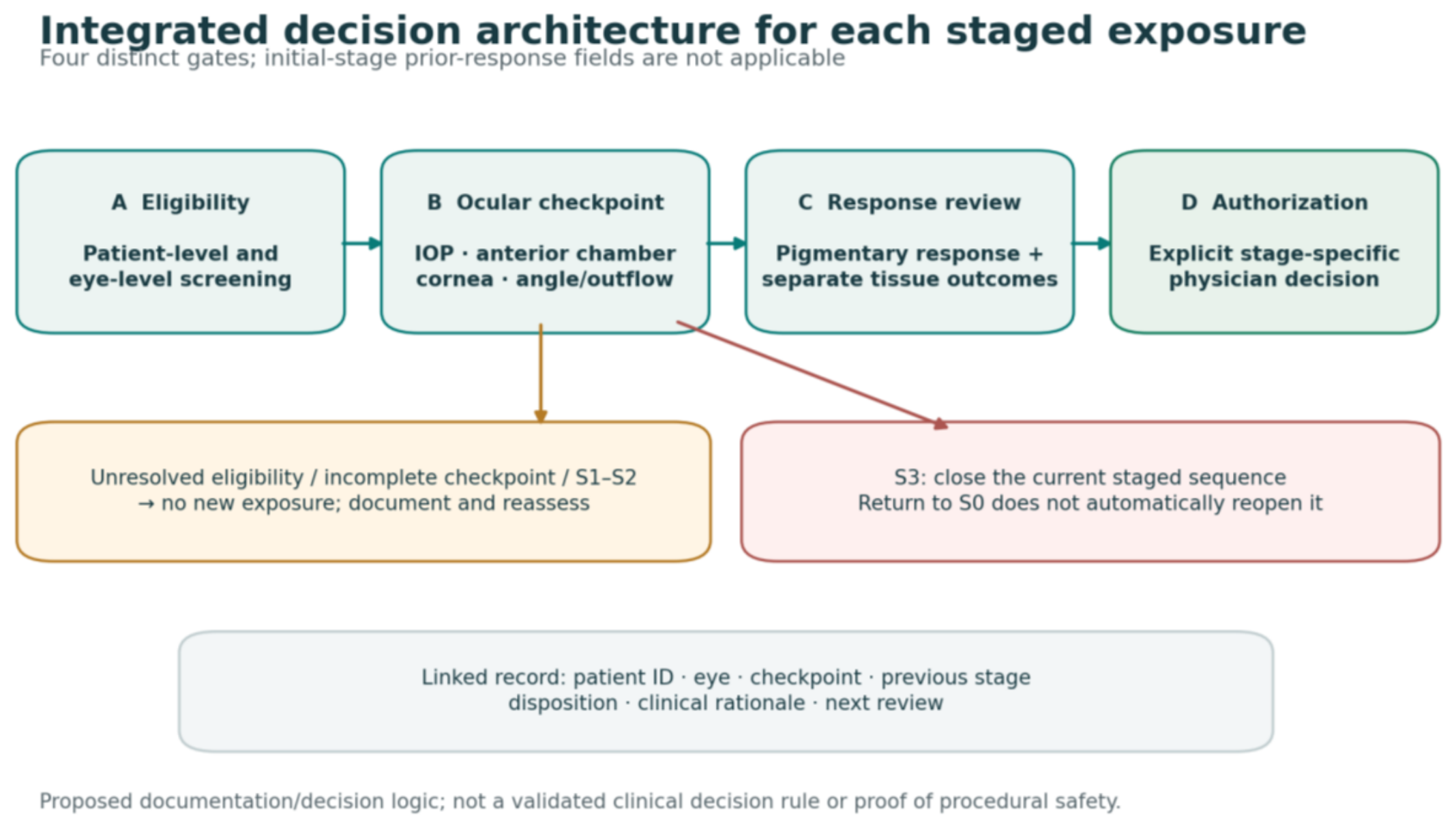
A patient-level identifier links bilateral information, systemic history, consent, and follow-up planning. Each eye has its own baseline, checkpoint, adverse-event history, and staged sequence. A prior completed treatment plan cannot substitute for a current eye-specific decision. For the initial stage, prior-exposure response and recovery-state fields are marked not applicable, not missing; baseline ocular examination and the first-stage decision still require documentation.
Figure 1.
Integrated four-gate, eye-specific architecture and its non-authorization pathways. Boxes and arrows denote proposed documentation logic, not a validated clinical decision rule.
Figure 1.
Integrated four-gate, eye-specific architecture and its non-authorization pathways. Boxes and arrows denote proposed documentation logic, not a validated clinical decision rule.
3.1. Gate A—Current Eligibility
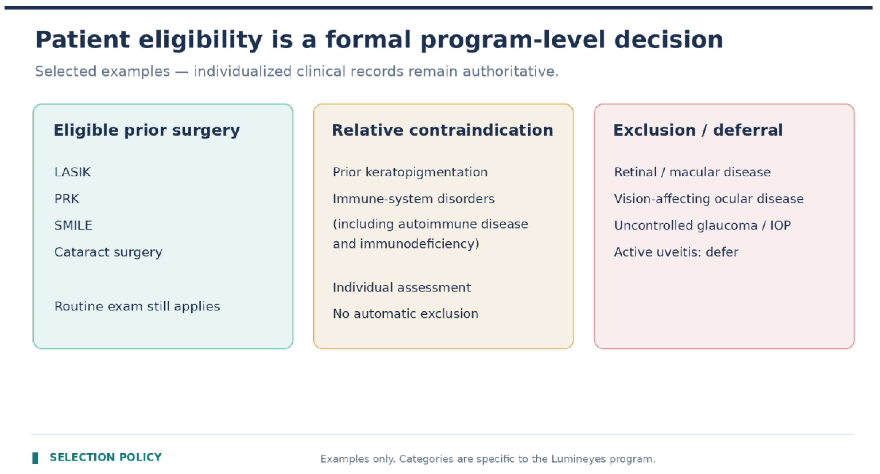
Before each proposed exposure, the clinician checks that the patient and the specific eye remain suitable under the current program selection policy, including newly detected ocular disease, medication or systemic changes, consent, expectations, and ability to return for follow-up. The policy distinguishes excluded states, conditions requiring postponement, relative contraindications requiring individual assessment, and eligible prior procedures. For example, the program regards prior LASIK, PRK, SMILE, or cataract surgery as compatible with eligibility, while prior keratopigmentation and heterogeneous immune-system disorders trigger individual review. Vision-affecting ocular disease is excluded under this specific program policy. These categories are not asserted to be universal medical rules.
Figure 2.
Illustrative Lumineyes™ selection-policy categories. Immune-system disorders, including autoimmune and inflammatory disorders, are classified as relative contraindications requiring individual assessment. Adapted from the author’s clinical standard [9]; this selection policy is not universal.
Figure 2.
Illustrative Lumineyes™ selection-policy categories. Immune-system disorders, including autoimmune and inflammatory disorders, are classified as relative contraindications requiring individual assessment. Adapted from the author’s clinical standard [9]; this selection policy is not universal.
3.2. Gate B—Complete Ocular Safety Checkpoint
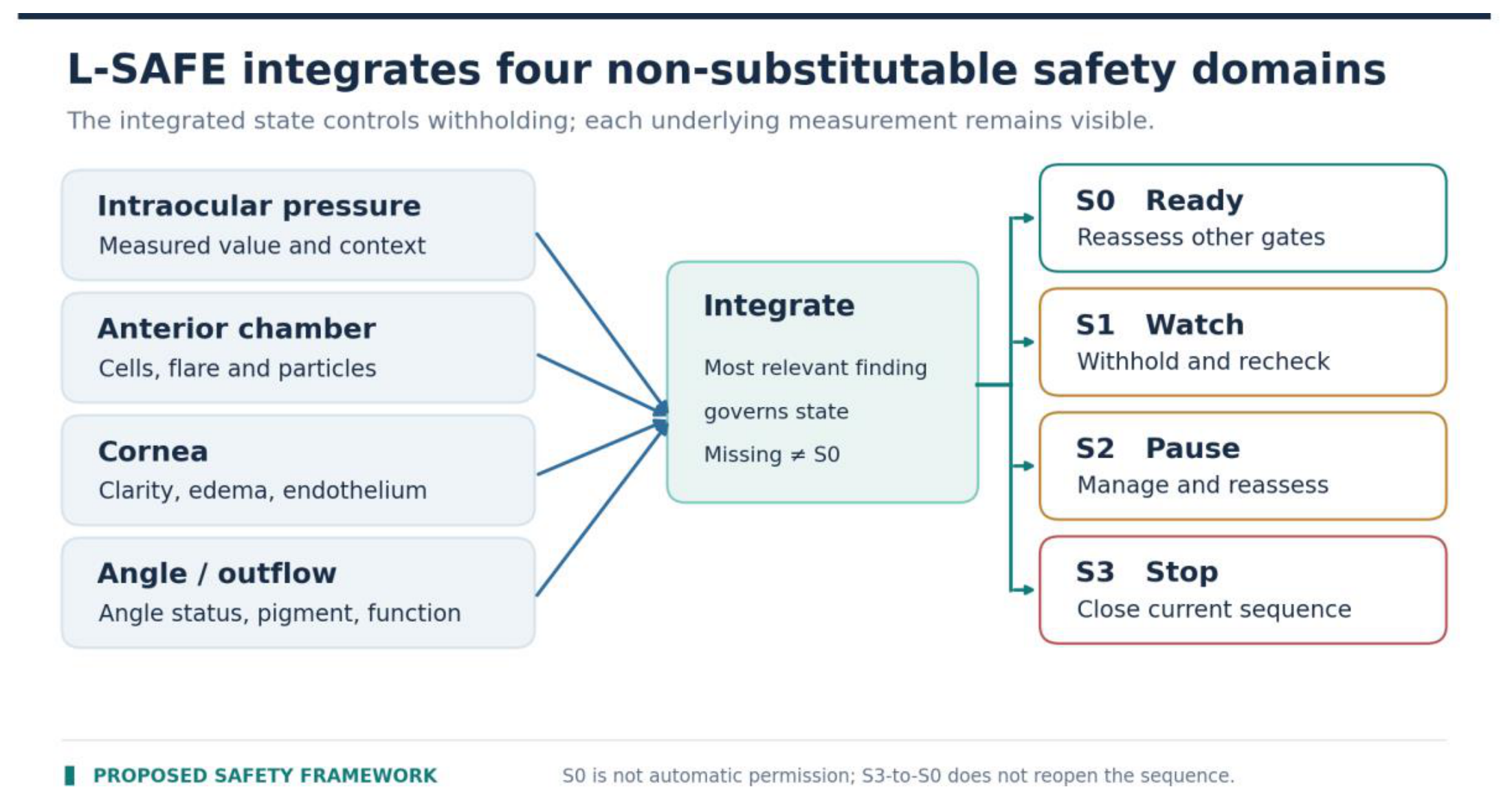
For consideration of a subsequent exposure, L-SAFE requires four recorded anterior-segment domains: measured IOP with baseline and medication context, anterior-chamber inflammatory response and particulate observations, corneal status, and angle/outflow findings [8]. At the initial stage, a baseline ocular examination documents the same clinically relevant domains without inventing a prior-exposure recovery state. The eye is assigned an integrated S0 (READY), S1 (WATCH), S2 (PAUSE), or S3 (STOP) state according to the clinically most relevant abnormality. L-SAFE is not an additive numerical score. A mandatory domain that was not assessed is recorded as incomplete rather than assumed normal. Important functional or posterior-segment pathology may independently preclude another exposure even if the four anterior-segment domains meet S0.
Figure 3.
Proposed L-SAFE integration of four anterior-segment domains into staged safety states. An S0 state is not itself authorization for additional exposure. Adapted from the clinical standard [9].
Figure 3.
Proposed L-SAFE integration of four anterior-segment domains into staged safety states. An S0 state is not itself authorization for additional exposure. Adapted from the clinical standard [9].
3.3. Gate C—Prior-Stage Response and Purpose
Where a prior stage exists, it is reviewed using dated, comparable pigmentary measures and separately captured tissue, symptom, and functional outcomes. At the initial stage, record the absence of a preceding exposure as not applicable and document the baseline rationale instead of treating this as missing follow-up. Apparent color change is not a proxy for an intact stroma, benign inflammation, or sustained physiological improvement. The record states the purpose of considering another stage and whether an outcome can be meaningfully interpreted in the presence of changing medications, examination conditions, or incomplete follow-up. The proposed Longitudinal Response Selectivity (LRS) construct motivates this separation of trajectories but has no validated score or clinical threshold [7].
3.4. Gate D—Physician Authorization and Disposition
Only after the preceding gates have been documented can the clinician independently record one of four dispositions: authorized for this stage, deferred, withheld, or current sequence terminated. For further staged exposure, an S0 eye may be considered but is not automatically cleared; a fresh decision and consent still apply. Initial-stage eligibility requires its own completed baseline assessment and authorization. S1 and S2 require withholding additional exposure pending reassessment, and an improving S1/S2 remains non-READY until a new integrated S0 assessment is supported. S3 terminates the current treatment sequence. A later S0 observation does not retroactively reopen a terminated sequence. These rules document the program’s decision architecture; they do not define universal numerical treatment limits.

Table 1.
Proposed clinical decision gates, minimum records, and conditions preventing automatic re-exposure.
Table 1.
Proposed clinical decision gates, minimum records, and conditions preventing automatic re-exposure.
| Decision gate | Required record | If unresolved or adverse |
|---|---|---|
| A. Eligibility | Patient/eye status; systemic and medication changes; consent and follow-up feasibility | Exclusion: no exposure. Relative/uncertain: individual review or defer. |
| B. Safety checkpoint | Initial stage: baseline assessment. Later stages: IOP, anterior chamber, cornea, angle/outflow; L-SAFE and rationale | Later-stage incomplete: no READY; S1/S2: withhold; S3: terminate sequence. |
| C. Response review | Initial stage: prior response N/A. Later: prior-stage dates; pigmentary and non-pigmentary observations | Later stage: insufficient response record requires review before reauthorization. |
| D. Authorization | Clinician; date; explicit disposition; reason; next action and record link | No documented authorization: do not infer permission from a planned stage. |
4. Minimum Data Model and Auditability
The proposed minimum trace uses a patient ID; laterality; eye-specific baseline; dated stage and checkpoint; the four safety-domain findings; pre-existing ocular conditions; medication and adherence context; standardized pigmentary/functional observations when available; prior S2/S3 history; and the explicit disposition with clinician rationale. Additional tests are recorded when indicated, but their absence must not silently convert an unassessed required domain into a normal result.
Documentation must distinguish “not present,” “not measured,” and “not interpretable.” Clinical records retain direct identifiers for patient care. A research extract, if separately approved and created, requires its own pseudonymization, date handling, linkage protection, and missing-data plan; a clinical form does not itself constitute a research dataset.
Table 2.
Proposed linked record. No patient-level dataset was assembled for this report.
| Level | Core fields | Interpretive constraint |
|---|---|---|
| Patient | Eligibility history; systemic status; medications; expectations; follow-up feasibility | Policy classification may change over time; record the policy version. |
| Eye / baseline | Laterality; IOP; visual function; anterior and posterior findings; imaging conditions | Comparisons are eye-specific; paired eyes are clustered within patients. |
| Checkpoint | Time since exposure; 4 L-SAFE domain findings; integrated state; missingness | State is a proposed classification, not a numerical risk estimate. |
| Stage / decision | Sequence and stage identifiers; response rationale; explicit authorization/withholding; author | S0 does not substitute for permission; S3 history persists. |
| Longitudinal follow-up | Repeated observations; medication changes; adverse events; missed visits | Change in IOP or color does not alone prove mechanism, efficacy, or safety. |
5. Relationship to Scientific Subframeworks
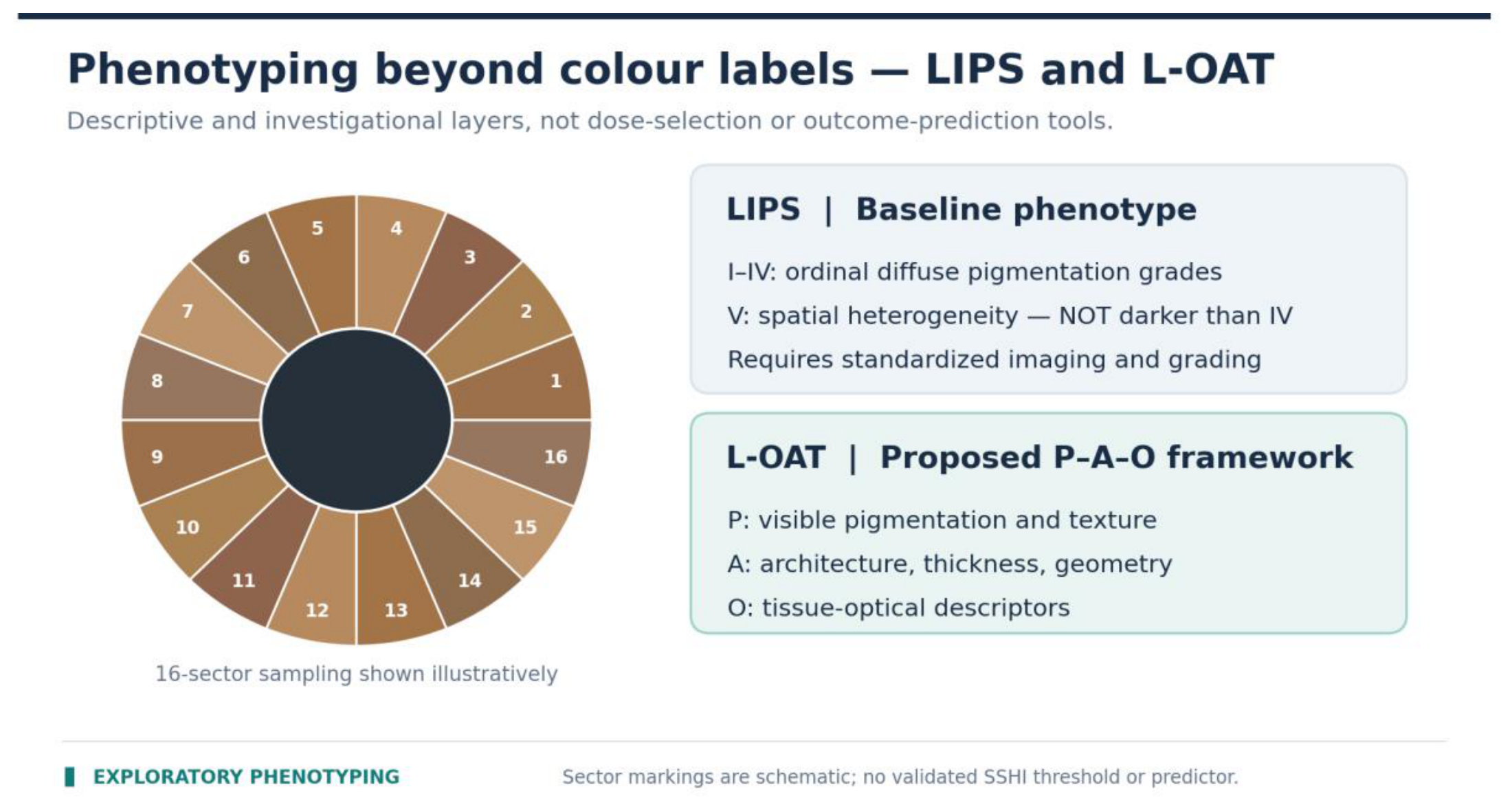
The clinical architecture uses specialized research constructs only where their contributions can be explicitly bounded. LIPS categorizes predominantly diffuse iris pigmentation from grades I–IV, while grade V denotes a spatially heterogeneous phenotype rather than the darkest end of a single severity axis [10]. L-OAT proposes multimodal appearance–architecture–optics descriptors but is not a validated selector of exposure parameters [11]. SSMM defines candidate dimensions of biophysical, anatomical, and functional selectivity; LRS asks whether pigmentary and accompanying tissue-response trajectories can be differentiated longitudinally [7].
Figure 4.
SSMM conceptual dimensions. Intended targeting, anatomic/functional findings and longitudinal response evaluation are distinct questions; LRS is not a fourth optical target. Adapted from the clinical standard [9].
Figure 4.
SSMM conceptual dimensions. Intended targeting, anatomic/functional findings and longitudinal response evaluation are distinct questions; LRS is not a fourth optical target. Adapted from the clinical standard [9].
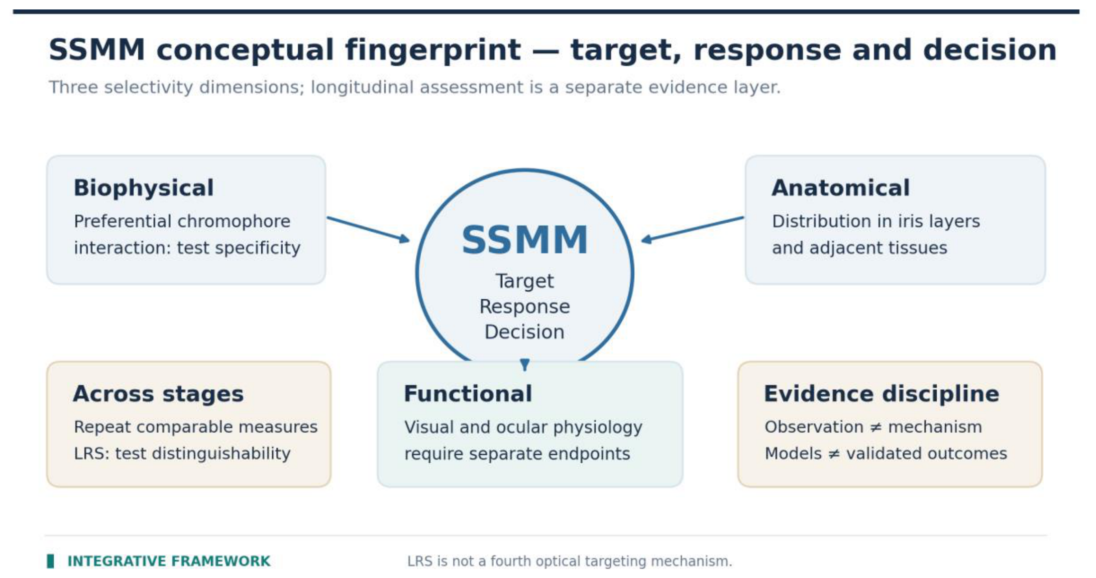
Figure 5.
LIPS and L-OAT are proposed research representations, not validated treatment selectors. LIPS grade V describes spatial heterogeneity rather than a severity level beyond grade IV. Adapted from the clinical standard [9].
Figure 5.
LIPS and L-OAT are proposed research representations, not validated treatment selectors. LIPS grade V describes spatial heterogeneity rather than a severity level beyond grade IV. Adapted from the clinical standard [9].
The SSMM–IOP hypothesis raises a separate physiological research question: whether iris-directed pigment modification can be associated with changes in conventional aqueous outflow beyond the direct optical target [13]. SLT-associated monocyte recruitment and outflow changes have been studied in a different laser-targeting context [14]; they do not prove that the same mechanism occurs after iris-directed exposure. Pigment overload may instead impair outflow [5,6]. Pressure trends alone cannot resolve these competing mechanisms.
Figure 6.
Competing candidate explanations for potential outflow responses after iris-directed exposure. SLT findings and pigment-burden analogues do not establish an SSMM-specific IOP effect. Adapted from the clinical standard [9].
Figure 6.
Competing candidate explanations for potential outflow responses after iris-directed exposure. SLT findings and pigment-burden analogues do not establish an SSMM-specific IOP effect. Adapted from the clinical standard [9].
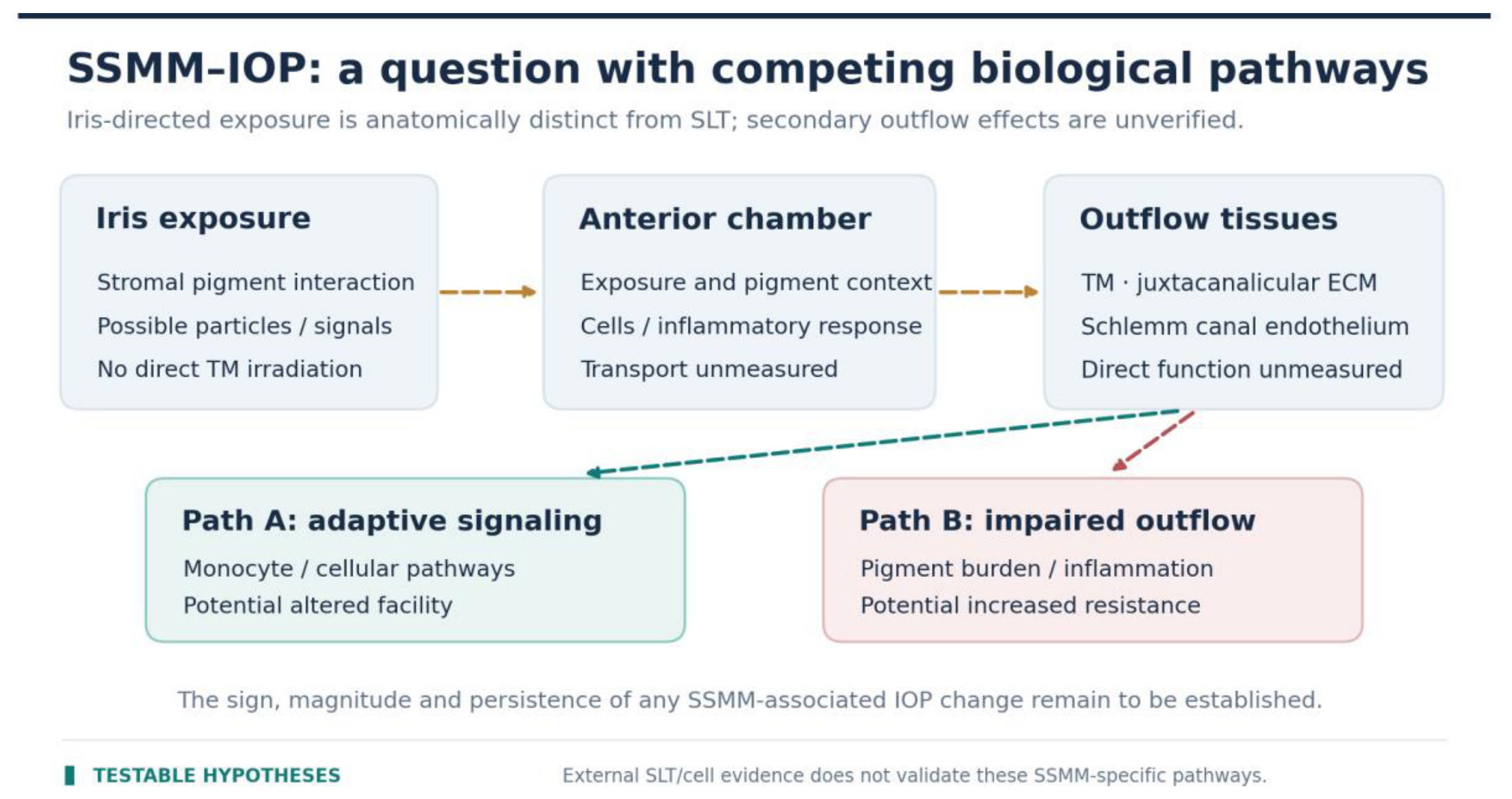
Table 3.
Proposed components are not interchangeable with validated biological findings.
| Component | What it contributes here | What is not established |
|---|---|---|
| SSMM / LRS | Distinct optical, anatomical, functional, and time-resolved observation targets | Selective stromal preservation or a validated LRS threshold. |
| L-SAFE | A four-domain, four-state withholding/recording architecture | Reliable risk prediction or improved clinical outcomes. |
| LIPS / L-OAT | Reproducible phenotype data as a proposed measurement aim | Validated response prediction or dosing decisions. |
| SSMM–IOP / outflow biology | Competing physiological questions and independent measurement requirements | A proven IOP-lowering effect or an established SSMM-specific pathway. |
6. Independent Evaluation and Falsification Plan
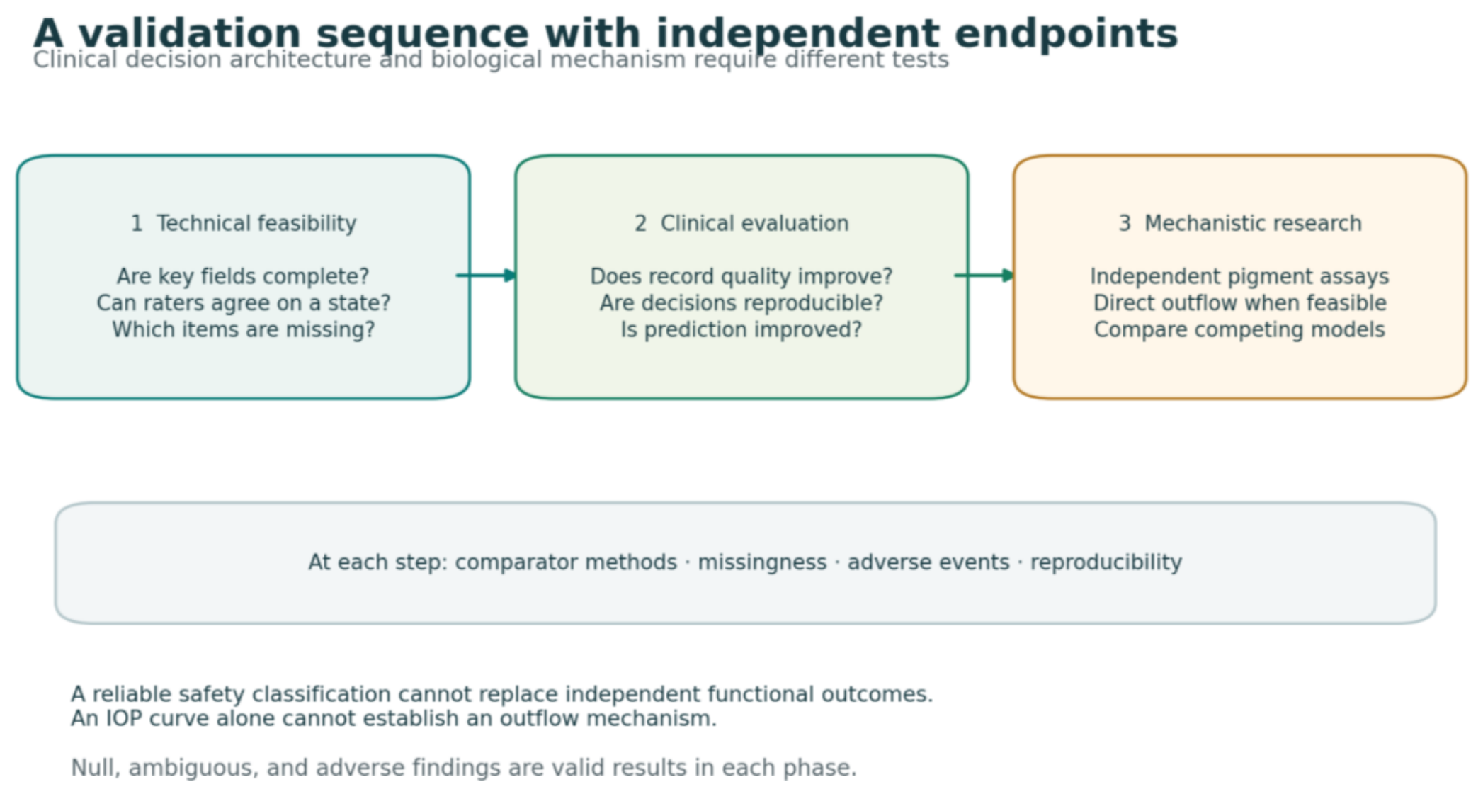
The architecture’s value is an empirical question. The sequence below prioritizes an assessment of what the framework measures before asking whether using it changes clinical outcomes. Existing guidance for reliability reporting and complex-intervention evaluation provides general methodological anchors, without conferring validation on this proposal [15,16].
Figure 7.
Proposed evaluation sequence. The clinical decision workflow, clinical endpoints, and mechanistic investigations constitute different evidence programs.
Figure 7.
Proposed evaluation sequence. The clinical decision workflow, clinical endpoints, and mechanistic investigations constitute different evidence programs.
6.1. Feasibility, Completeness, and Reproducibility
A prespecified audit should test completion rates for all four mandatory L-SAFE domains and the linked disposition fields across consecutive eligible records. Independent reviewers should rate de-identified standardized vignettes or patient records, with masked conditions appropriate to the task. Report category-specific agreement, uncertainty, missingness, and clinically consequential disagreements; do not report overall accuracy without an independent reference standard. Incomplete and discordant classifications are informative outcomes.
6.2. Incremental Longitudinal and Decision Value
Prospectively evaluate whether adding prior-stage response measures improves prespecified prediction or decision reliability beyond baseline phenotype, exposure history, and current clinical variables. Outcomes might include reproducibility of exposure-withholding decisions, completeness of incident ascertainment, and independent functional safety measures. Analyses must account for repeated observations within eyes, clustering of two eyes within a patient, treatment-stage carryover, medication changes, center effects, and missing follow-up. Prediction fit is not proof that a clinical decision improves patient outcomes.
6.3. External Replication and Policy Sensitivity
A separate clinical setting should be able to implement the data dictionary and classify the same decision states without depending on undocumented knowledge. Program-specific eligibility exclusions should be identified as local policy, not smuggled into a universal algorithm. If the architecture cannot be reproduced outside its originating clinic, the problem should be documented rather than attributed automatically to users.
6.4. Mechanistic Work as a Distinct Validation Track
The architecture makes it possible to define observations but does not resolve tissue selectivity, pigment mass balance, endothelial consequences, or aqueous outflow. Separate bench and clinical studies could compare registered iris imaging, independent tissue-function endpoints, direct flow/facility measurements where feasible, pigment quantification, and molecular/cellular measurements against alternative hypotheses. An IOP change following treatment is not itself evidence for enhanced facility; conversely, failure to show a proposed mechanism does not invalidate the utility of reliable clinical documentation.
Table 4.
Explicitly negative or ambiguous findings are valid outcomes, not implementation failures.
| Proposition | Disconfirming or limiting observation |
|---|---|
| The checklist is reliably usable | Many mandatory fields remain unrecorded or inter-rater agreement is inadequate. |
| Safety states improve decisions | No incremental decision consistency or independent outcome association versus simpler records. |
| Past-stage response adds clinical information | No out-of-sample improvement over baseline and cumulative exposure predictors. |
| Phenotype labels support additional prediction | Grades/descriptors show poor masked repeatability or no independent predictive value. |
| An SSMM-specific outflow mechanism exists | Independent facility/biological measurements reject the candidate pathway or support obstruction/other causes. |
7. Limitations
This is a single-developer framework assembled from program policy and selected literature. There was no systematic search, expert consensus, derivation sample, independent clinician-rating study, comparator intervention, or prospective outcome evaluation. Its eligibility policies reflect one program and may not transfer to other protocols or jurisdictions. Neither the proposed L-SAFE categories nor the combined decision architecture has validated numerical thresholds or demonstrated reduction in adverse events. Existing published reports describe heterogeneous laser methods and cannot establish the safety or effectiveness of the Lumineyes™ technique. Operational details of the clinical technique and the proposed Phase 1/Phase 2 distinction are intentionally outside the present paper and are not necessary for evaluation of the record architecture.
The paper does not retrospectively analyze the author’s clinical archive and does not estimate complication frequencies, response probabilities, or sustained pressure effects. Candidate imaging, corneal endothelial, and outflow measures are proposed for future study; their reproducibility and clinical relevance cannot be assumed. The design may prove too complex or duplicative to yield incremental utility, which should be evaluated directly. The research position of SSMM, LRS, LIPS, L-OAT, and L-SAFE remains conceptual until independently tested.
8. Conclusions
An SSMM-informed, four-gate clinical decision architecture can be specified without equating a staged treatment plan, a normal checkpoint, and permission for another exposure. Its defining elements are separate patient/eye eligibility, domain-complete safety assessment, prior-stage response characterization, and explicit physician authorization, with traceable withholding and sequence-closure decisions. The immediate output is an inspectable methodological specification. Whether it improves documentation, decision reproducibility, or patient outcomes remains an open empirical question.
Competing Interests
Mustafa Mete is the founder and developer of the Lumineyes™ method and has a commercial interest in its clinical application.
Author Contributions
Mustafa Mete: Conceptualization; Methodology; Literature synthesis; Visualization; Writing—original draft; Writing—review and editing.
Funding
No external funding was received for this work.
Ethics Approval
Not applicable. This methodological report includes no human participants, identifiable patient data or original patient-level datasets.
Data Availability
No new patient-level, laboratory or computational dataset was generated or analysed for this methodological paper. The proposed variables, documentation model and decision logic are specified in the text and tables. The related Lumineyes™ LID Clinical Practice Standard v1.0 is publicly available as a Zenodo research report (DOI: 10.5281/zenodo.24256204) [9]; it is not a professional-society consensus guideline.
References
- Imesch, P.D.; Wallow, I.H.L.; Albert, D.M. The color of the human eye: a review of morphologic correlates and of some conditions that affect iridial pigmentation. Surv. Ophthalmol. 1997, 41 (Suppl 2), S117–S123. [Google Scholar] [CrossRef]
- Wilkerson, C.L.; Syed, N.A.; Fisher, M.R.; Robinson, N.L.; Wallow, I.H.L.; Albert, D.M. Melanocytes and iris color: light microscopic findings. Arch. Ophthalmol. 1996, 114, 437–442. [Google Scholar] [CrossRef]
- Grimaldos Ruiz, P. Photoablative cosmetic iridoplasty: effective, safe, and predictable—eye color change in 1176 eyes. Int. Ophthalmol. 2021, 41, 1381–1393. [Google Scholar] [CrossRef]
- D’Oria, F.; Abu-Mustafa, S.K.; Alio, J.L. Cosmetic change of the apparent color of the eye: a review on surgical alternatives, outcomes and complications. Ophthalmol. Ther. 2022, 11, 465–477. [Google Scholar] [CrossRef]
- Liu, J.; Korban, S.; Moster, M.R.; Rhéaume, M.-A.; Wang, Q. Bilateral severe iatrogenic pigmentary glaucoma following laser treatment for cosmetic iris color change. Am. J. Ophthalmol. Case Rep. 2023, 32, 101927. [Google Scholar] [CrossRef]
- Ong, A.Y.; Ching-A-Sue, G.; Krilis, M.; Guirao-Navarro, M.C.; Hornby, S.; Jutley, G. Refractory iatrogenic pigmentary glaucoma secondary to cosmetic laser treatment: a case report. Eur. J. Ophthalmol. 2023, 33, NP1–NP4. [Google Scholar] [CrossRef]
- Mete, M. Selective Stromal Melanin Modulation (SSMM): integrative biophysical and clinical framework. Zenodo Res. Rec. 2026. [Google Scholar] [CrossRef]
- Mete, M. Lumineyes Anterior-Segment Safety Staging (L-SAFE): a proposed framework for pre-exposure assessment. Zenodo Res. Rec. 2026. [Google Scholar] [CrossRef]
- Mete, M. Lumineyes™ Laser Iris Depigmentation Clinical Practice Standard: An SSMM-Informed Clinical Framework. Version 1.0. Zenodo Res. Rep. 2026. [Google Scholar] [CrossRef]
- Mete, M. Lumineyes Iris Pigmentation Scale (LIPS): a proposed clinical grading framework. Zenodo Res. Rec. 2026. [Google Scholar] [CrossRef]
- Mete, M. Lumineyes Optical Architectural Typology (L-OAT): a multimodal hypothesis for iris phenotyping. Zenodo Res. Rec. 2026. [Google Scholar] [CrossRef]
- Jabs, D.A.; Nussenblatt, R.B.; Rosenbaum, J.T.; SUN Working Group. Standardization of uveitis nomenclature for reporting clinical data. Am. J. Ophthalmol. 2005, 140, 509–516. [Google Scholar] [CrossRef]
- Mete, M. Long-Term Intraocular Pressure Dynamics After Selective Stromal Melanin Modulation: A Testable Mechanistic Framework. Preprints.org. 2026. [Google Scholar] [CrossRef]
- Alvarado, J.A.; Katz, L.J.; Trivedi, S.; Shifera, A.S. Monocyte modulation of aqueous outflow and recruitment to the trabecular meshwork following selective laser trabeculoplasty. Arch. Ophthalmol. 2010, 128, 731–737. [Google Scholar] [CrossRef]
- Kottner, J.; Audigé, L.; Brorson, S.; et al. Guidelines for Reporting Reliability and Agreement Studies (GRRAS) were proposed. J. Clin. Epidemiol. 2011, 64, 96–106. [Google Scholar] [CrossRef]
- Skivington, K.; Matthews, L.; Simpson, S.A.; et al. A new framework for developing and evaluating complex interventions: update of Medical Research Council guidance. BMJ 2021, 374, n2061. [Google Scholar] [CrossRef]
Disclaimer/Publisher’s Note: The statements, opinions and data contained in all publications are solely those of the individual author(s) and contributor(s) and not of MDPI and/or the editor(s). MDPI and/or the editor(s) disclaim responsibility for any injury to people or property resulting from any ideas, methods, instructions or products referred to in the content. |
© 2026 by the authors. Licensee MDPI, Basel, Switzerland. This article is an open access article distributed under the terms and conditions of the Creative Commons Attribution (CC BY) license (http://creativecommons.org/licenses/by/4.0/).
Copyright: This open access article is published under a Creative Commons CC BY 4.0 license, which permit the free download, distribution, and reuse, provided that the author and preprint are cited in any reuse.