

Submitted:
12 November 2026
Posted:
17 November 2026
You are already at the latest version
Abstract
Background: Iatrogenic tracheal injuries are uncommon yet potentially life-threatening complications of airway management and thoracic interventions. Although awareness of post-intubation tracheal lacerations (PiTLs) has increased over recent decades, their management remains largely constructed on institutional experience, and standardized evidence-based recommendations are lacking. The Italian Society of Thoracic Endoscopy (SIET) therefore developed this position paper to provide practical guidance for the di-agnosis, classification, and management of PiTLs. Methods: A structured narrative review and expert consensus process were conducted to evaluate key diagnostic and therapeutic aspects of PiTL. PubMed/MEDLINE and Scopus were searched from January 2000 through June 2026. Preliminary statements were de-veloped, reviewed, and refined through structured discussion by an 20-member panel of experts in thoracic surgery, interventional pulmonology, and anesthesiology. The re-sulting statements reflect the collective judgment of the expert panel and are intended as consensus-based recommendations within a position paper. Results: Flexible bronchoscopy remains the diagnostic gold standard to assess the precise location, length, depth, and circumferential involvement of the lesion. Management strategies are highly driven by physiological stability, ventilatory status, and morpho-logical characteristics, primarily using the Cardillo classification system. Conservative treatment is indicated for stable, spontaneously breathing patients (Cardillo Level I–II and selected IIIA), while early surgical intervention (within 24 hours) is recommended for severe cases with ventilatory instability or major complications (Cardillo Level IIIB–IV). Conclusions: PiTLs require early, comprehensive, and structured diagnostic evaluation, followed by individualized management. Adherence to the 17 SIET position statements provides a standardized framework for clinical decision-making and may help reduce variability in care.
Keywords:
post-intubation tracheal lacerations
; iatrogenic tracheal injury
; cardillo classification
; flexible bronchoscopy
; thoracic surgery
; expert consensus
; SIET
1. Introduction
Iatrogenic tracheal injuries are rare but potentially life-threatening complications of airway management and thoracic procedures, most associated with endotracheal intubation, tracheostomy, bronchoscopy, or mediastinal surgery [1,2,3]. Lesions range from superficial mucosal tears to full-thickness tracheal ruptures, occasionally involving adjacent structures and creating complex management challenges [2,4]. Despite increasing recognition and advances in both surgical and non-surgical management, evidence-based guidelines for post-intubation tracheal lacerations (PiTL) remain lacking [2,5,6]. Current treatment strategies are largely based on retrospective studies, institutional experience, and expert opinion, resulting in considerable variability, especially in determining whether to pursue operative versus conservative management [7,8,9,10,11]. To address this gap, the Italian Society of Thoracic Endoscopy (SIET) developed this position paper, drawing on the complementary expertise of thoracic surgeons and interventional pulmonologists. The aim is to critically review the available evidence, identify areas of controversy, and provide guidance on key clinical issues in the management of PiTL.
2. Relevant Sections
This document was developed as a position paper and expert consensus statement promoted by the SIET, integrating a structured narrative review of the literature with a formalized multidisciplinary consensus process.
2.1. Literature Search Strategy
A structured literature search was conducted in PubMed/MEDLINE and Scopus to identify clinical studies, retrospective analyses, and contemporary reviews addressing post-intubation tracheal laceration (PiTL) published between January 2000 and June 2026. No language restrictions were applied when abstracts and relevant data were accessible. The search was not restricted by study design; however, higher levels of evidence were prioritized during the screening and synthesis processes. The search strategy was developed using free-text terms related to iatrogenic tracheobronchial injury, post-intubation tracheal rupture, morphological classification, bronchoscopic assessment, and conservative or surgical management. The complete database-specific search strings, including the Boolean combinations and any database-adapted syntax, are reported in Appendix A. Reference lists of selected articles were also screened to identify additional relevant studies.
2.2. Eligibility Criteria and Study Selection
Studies were selected based on their clinical relevance to PiTL, study design, and the completeness of the available documentation. Priority was given to peer-reviewed systematic reviews, meta-analyses, well-documented retrospective series, and consensus frameworks developed by recognized international scientific bodies. Studies not specifically addressing PiTL or not reporting management strategies and clinical outcomes were excluded from the primary evidence synthesis. Narrative reviews, editorials, expert opinions, and conference abstracts were also excluded when they lacked sufficient methodological detail or outcome data. Case reports and small uncontrolled case series were retained only as supplementary contextual evidence, when clinically relevant, and were interpreted with caution rather than weighted equally with higher-level evidence. Given the heterogeneity of the available literature and the limited number of PiTL-specific studies, a formal systematic review (with PRISMA-compliant reporting) was not performed. Instead, a critical qualitative synthesis of the available evidence was conducted.
2.3. Data Extraction and Evidence Synthesis
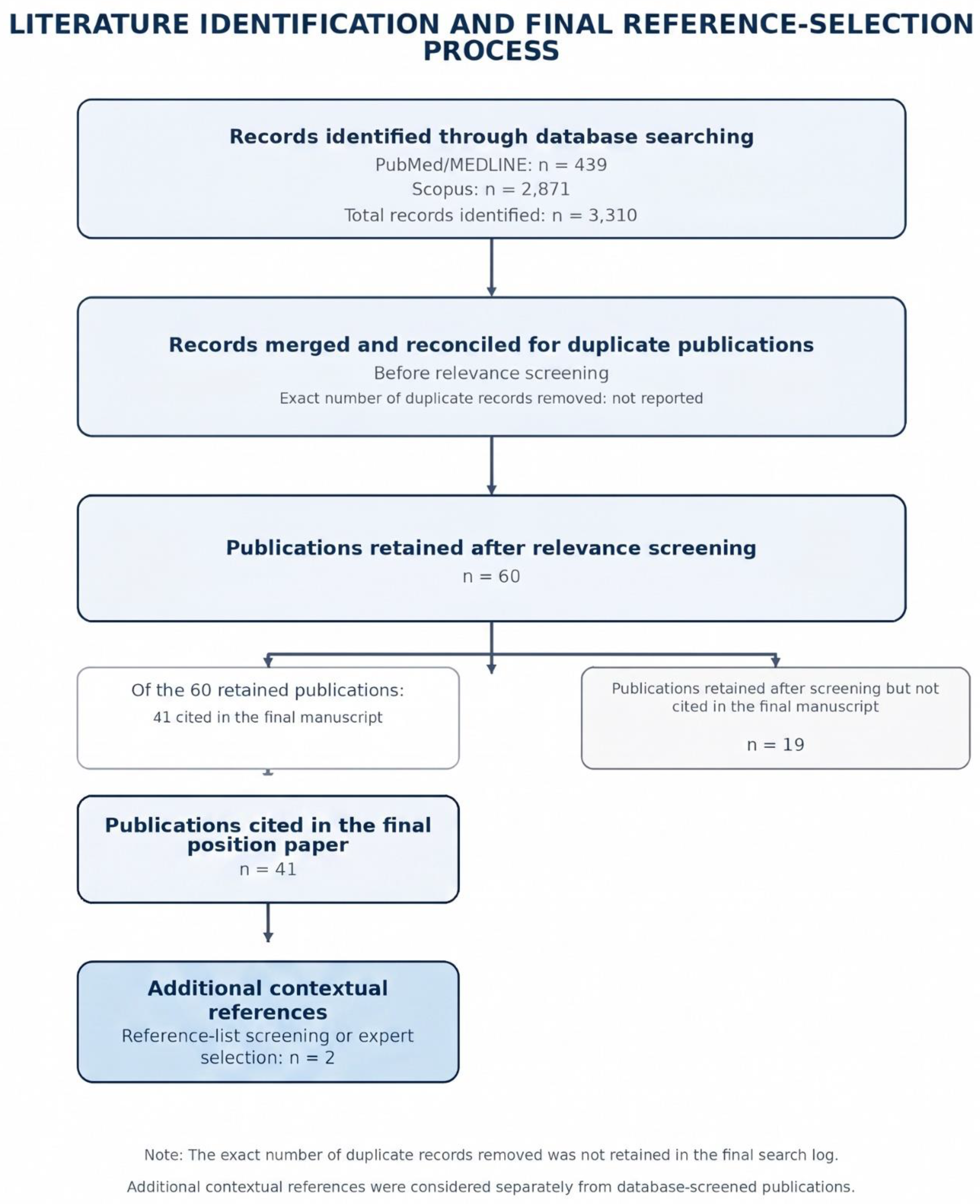
The database searches identified 439 records in PubMed/MEDLINE and 2871 records in Scopus, for a total of 3310 records. Records were merged and reconciled for duplicate publications by two of the Authors before relevance screening. After database merging, duplicate reconciliation, and relevance screening, 60 publications were retained as potentially relevant to the position paper. The final manuscript cited 41 of these publications, selected according to their direct relevance to the position statements, methodological contribution, and contextual value; 19 publications retained during screening were not cited in the final version. Two additional references identified through reference-list screening or expert selection were considered separately as contextual sources and were not included in this count. The literature identification and final reference-selection process is summarized in Figure 1.
Because the available literature was heterogeneous with respect to study design, patient population, injury classification, outcome definitions, and follow-up, quantitative meta-analysis was not performed. A qualitative synthesis was therefore undertaken, with findings grouped according to diagnostic approach, treatment strategy, lesion location, and patient age. This approach was also justified by the predominance of retrospective cohorts, case series, case reports, and other non-randomized evidence, together with the historical lack of large-scale prospective randomized trials in this surgical context.
2.4. Expert Panel and Consensus Process
This position paper was developed by a multidisciplinary panel of 20 experts appointed by SIET, comprising 12 thoracic surgeons, 7 interventional pulmonologists and 1 anesthesiologist. Panel members were selected based on their recognized expertise, clinical experience, relevant publications, procedural experience, and institutional or scientific roles within SIET. Seventeen recommendations were formulated through a structured expert-consensus process based on a critical appraisal of the literature and its application to the clinical management of post-intubation tracheal lacerations. The process included identification of key clinical issues, review of the available evidence, drafting of preliminary statements, and structured multidisciplinary discussion within the panel. All 17 statements were reviewed and refined during panel discussion until a shared position was reached. The consensus process consisted of two online meetings, each lasting approximately three hours. Disagreements were resolved through collective discussion and joint reformulation of the statements until unanimous agreement on the final version was reached among all 20 participants. The final statements reflect the collective judgment of the expert panel and are intended as consensus-based recommendations within a position paper.
2.5. Recommendations Framework
Given the position-paper nature of this document, the statements were developed as consensus-based recommendations intended to support clinical decision-making rather than as formal evidence-graded guidelines. The level of supporting evidence was interpreted descriptively, considering the type, consistency, and clinical relevance of the available literature. Given the rarity of PiTL and the limited quality of the evidence base, most recommendations rely on retrospective series, case reports, expert-based frameworks, and indirect evidence. Where direct evidence was lacking or inconsistent, recommendations were primarily based on multidisciplinary expert judgment and current clinical practice.
2.6. Epidemiology and Disease Burden
PiTL is an uncommon but potentially life-threatening complication of airway management. Although the reported incidence after orotracheal intubation ranges from approximately 0.05% to 0.37% [2,5,12], the true burden of disease remains uncertain because current estimates derive mainly from retrospective series and case reports, while prospective epidemiological studies are lacking. Several authors suggest that PiTL is substantially under-recognized and under-reported, leading to underestimation of its actual incidence [13,14,15]. The typical lesion consists of a longitudinal tear of the membranous posterior tracheal wall, most often located in the middle third and occasionally extending distally [12,13]. This characteristic morphology supports a pathophysiological mechanism related to endotracheal tube positioning, cuff pressure, and airway anatomy [16]. Several patient- and procedure-related factors have been associated with an increased risk of PiTL. Patient-related risk factors include advanced age, female sex, short stature, inflammatory lesions of the tracheobronchial tree and congenital tracheal abnormalities; while procedure-related risk factors include multiple attempts at forced intubation, operator inexperience and the use of rigid introducers or stylets [2,4,5,6,17]. However, the condition may also occur following apparently uncomplicated elective intubation, highlighting the importance of maintaining a high index of suspicion even in low-risk settings [18,19]. Notably, in case of emergency intubation, the incidence is reported in up to 15% cases (3) and those lesions have been associated with significantly worse outcomes and higher mortality rates, emphasizing its role as both an epidemiological and prognostic factor. Despite its rarity, PiTL carries a substantial clinical and economic burden. Management often requires a multidisciplinary approach involving thoracic surgeons, interventional pulmonologists, anesthesiologists, intensivists, and radiologists. These factors contribute to increased healthcare resource utilization, prolonged hospitalization, and higher costs. Moreover, delayed diagnosis is associated with poorer outcomes, more complex treatment pathways, additional procedures, and greater medico-legal implications. Collectively, these considerations suggest that PiTL is both more frequent and more impactful than reported incidence figures indicate, highlighting the need for greater awareness, standardized diagnostic pathways, and evidence-based management strategies.
2.7. Signs and Symptoms
The clinical presentation of PiTL is highly heterogeneous and often delayed. New-onset subcutaneous emphysema following intubation is the cardinal sign that mandates immediate workup [5,12,20], potentially extending to the pelvis, alongside pneumomediastinum and pneumothorax on chest radiographs or CT. Spontaneously breathing patients typically present with cough, dyspnea, or hemoptysis. Symptoms may be delayed by ≥3 days when the endotracheal cuff temporarily seals the defect [21]. In mechanically ventilated patients, PiTL should be suspected with high peak/plateau pressures, tension pneumothorax, persistent air leaks, or increasing cuff inflation requirements. Rare manifestations include pneumoperitoneum, pneumopericardium, and tension-induced hemodynamic collapse. A systematic review of 187 double-lumen tube ruptures highlighted pneumomediastinum, air leakage, hypoxemia, and subcutaneous emphysema as primary features, with most cases diagnosed intraoperatively (82.7%) [12]. Ultimately, the combination of recent airway instrumentation, subcutaneous emphysema, and unexplained respiratory deterioration strongly points to PiTL.
2.8. Diagnosis and Management: Available Evidence
Diagnosis is primarily driven by clinical suspicion following airway instrumentation. Chest radiography is often the initial step; it demonstrates indirect signs such as subcutaneous emphysema or pneumothorax, but its sensitivity is limited and a normal radiograph cannot exclude a laceration [12]. Chest CT with multiplanar reconstructions defines the exact presence of mediastinal air, injury level, and associated thoracic complications [2,12,15,22]. Flexible bronchoscopy remains the absolute diagnostic gold standard. It allows direct visualization, precise localization, and accurate assessment of length, depth, and circumferential involvement, which has major therapeutic implications for distinguishing superficial mucosal injuries from full-thickness ruptures [3,5].
2.9. Classification Systems
To guide therapeutic strategies, several morphological and functional classification systems have been published in the medical literature (Table 1).
The Cardillo morphological classification, proposed in 2010 and further modified in 2022 [3,23] was initially based on a prospective cohort of 30 patients and was subsequently adopted as the most widely referenced grading system for PiTL, operationalized in multiple institutional protocols and endorsed in authoritative reviews as a practical triage framework. This classification stratifies injuries according to the depth of tracheal wall involvement and the presence of mediastinal or esophageal extension, directly linking each grade to a therapeutic decision (Table 2).
The Cardillo morphological system does not stand alone in current practice. Boutros et al., in their 2022 multidisciplinary review [24], formalized a complementary functional (ventilatory) decision axis that intersects the morphological grade to determine management: the two critical questions are: does the patient require invasive mechanical ventilation? And, if so, can the injury be bypassed/bridged? This two-axis approach was operationalized in a prospective cohort of 30 consecutive patients at a single institution: non-ventilated patients were treated conservatively regardless of lesion length or grade, while ventilated patients were triaged based on the feasibility of endobronchial bridging (cuff repositioning distal to the tear, or bilateral selective endobronchial cannulation for pericarinal lesions), with surgery reserved for those in whom bridging was impossible or failed [5]. Schneider et al., in a 29-case retrospective institutional series [2], independently confirmed that "the decision for operative or nonoperative treatment is determined by the ventilating situation and the local extent of the injury", with nonoperative management safe in patients with uncomplicated ventilation, superficial or esophagus-covered tears, and moderate non-progressive emphysema. Beyond depth/grade, lesion location within the trachea modifies both the bridging strategy and the surgical approach, and is increasingly treated as a third axis:
- Upper-third lacerations: cervical/transcervical approach; cuff-bypass almost always feasible.
- Middle-third (most common phenotype): right posterolateral thoracotomy if surgical; standard cuff-repositioning for bridging.
- Lower-third / pericarinal: highest anatomical complexity; stenting (Y-stent) or bilateral selective endobronchial cannulation as bridging; right posterolateral thoracotomy for surgical repair.
Injuries caused by double-lumen tubes (DLTs) follow similar morphological principles; in a 187-case systematic review [12], the majority involved the trachea (52.4%) and left main bronchus (37.4%), suggesting that the Cardillo framework — designed for single-lumen tube injuries — can be applied to DLT injuries with the anatomical location serving as the primary additional modifier.
2.10. Therapeutic Modalities
There are several therapeutic approaches, ranging from active surveillance to endoscopic and surgical interventions. A brief overview of these modalities is presented below prior to addressing integrated treatment strategies.
Endoscopic management: includes several complementary techniques. Injury bridging by positioning the endotracheal tube cuff distal to the tear under bronchoscopic guidance remains the most adopted strategy. Fibrin glue can be used as a minimally invasive adjunct in selected cases [3,25]. Also, stromal vascular fraction, adipose-derived mesenchymal stromal cells, and autologous lipofilling have been reported as potential minimally invasive options for promoting fistula closure [26]. Airway stenting is reserved for specific scenarios, particularly ventilated patients with distal tracheal injuries [27]. Endoscopic suture repair via rigid bronchoscopy has recently emerged as a less invasive alternative to surgery [28].
Surgical management: generally, involves drainage and control of pleural infection, debridement of necrotic tissue, direct closure of the bronchial defect, and reinforcement with a vascularized muscle or omental flap. In chronic cases, the residual pleural cavity may require obliteration with tissue transposition, occasionally combined with thoracoplasty. The approach depends on injury location: cervicotomy for upper and mid-tracheal lesions [29,30], right posterolateral thoracotomy for distal tracheal, carinal, or main bronchial injuries [31,32], and sternotomy in selected cases requiring carinal exposure [33] or thoracoscopic alternatives [34]. Muscle-flap interposition is recommended for combined tracheoesophageal injuries [35], whereas ECMO may be necessary in complex cases [36]. Early repair, ideally within 24 hours, is generally advocated [37].
Conservative management: combines airway control, ventilatory optimization, and supportive care [38,39,40]. In spontaneously breathing patients, intubation should be avoided whenever possible, as spontaneous ventilation may facilitate healing. Management includes fasting until esophageal injury is excluded, broad-spectrum antibiotics, adequate analgesia, cough suppression and nutritional support.
2.11. Integrated Management Strategies
Management of PiTL should be guided by three interrelated factors: ventilatory status, lesion severity, and the presence of associated complications. Cardillo level I–II and selected IIIA injuries can often be managed conservatively with excellent outcomes, whereas surgery is generally reserved for more severe or complicated cases, including persistent air leaks, inability to maintain adequate ventilation, associated esophageal injury, progressive mediastinal emphysema causing respiratory compromise, or persistent pneumothorax despite effective chest drainage. An integrated management strategy relies on two principal decision axes: (1) the patient's ventilatory requirements and (2) the feasibility of bypassing or “bridging” the lesion (Table 3).
These factors are further influenced by lesion morphology and the presence of tracheoesophageal fistula or mediastinitis (Cardillo I–IV). Current evidence suggests that treatment decisions should be driven primarily by ventilatory status and secondarily by anatomical classification. The ability to maintain adequate gas exchange while minimizing pressure-related stress on the injured trachea is therefore the key determinant of treatment selection. In spontaneously breathing patients or those adequately supported with non-invasive ventilation, conservative management is generally the preferred first-line strategy [4,5,41], aiming to promote spontaneous healing while avoiding mechanical stress on the laceration. In selected cases, bronchoscopic instillation of fibrin glue (e.g., Tissucol) may complement conservative treatment by sealing the membranous tear, stabilizing its margins, and facilitating mucosal healing. In the Cardillo classification series, successful non-surgical treatment with fibrin glue was reported in all level I (n=3), level II (n=24), and level IIIA (n=2) patients, with complete healing by day 28 on scheduled bronchoscopy [3]. Conservative management includes close clinical monitoring, optimization of respiratory support, and scheduled bronchoscopic reassessment to document healing and detect deterioration. Most lesions in clinically stable patients without invasive ventilation heal within a few weeks without significant sequelae.
When invasive ventilation is required, the first consideration is whether the lesion can be bypassed. Under bronchoscopic guidance, the endotracheal tube cuff should be positioned distal to the laceration whenever possible to minimize pressure transmission. As the tube and inflated cuff may partially obscure the tear, controlled cuff deflation and/or slight tube withdrawal under direct bronchoscopic guidance should be considered when necessary to ensure complete visualization and accurate assessment of lesion extent and characteristics. Distal or pericarinal injuries may require selective mainstem bronchial or bilateral endobronchial intubation [5,24]. Ventilation should follow a lung-protective strategy, using the lowest feasible inspiratory pressures and minimum PEEP compatible with adequate oxygenation, while limiting air leakage and further tissue disruption [9,25]. Deep sedation and analgesia, with neuromuscular blockade, when necessary, may reduce coughing, straining, and patient–ventilator dyssynchrony. A peak airway pressure below 25 cmH₂O, however, should be considered a pragmatic institutional target rather than a universally validated evidence-based threshold. Serial bronchoscopies should document healing, confirm stable tube positioning, and guide ventilatory weaning or other de-escalation measures. Although no standardized surveillance schedule exists, reassessment at approximately 7 and 28 days, followed by examinations at 3 and 6 months when indicated, has been used in prospective PiTL series [3]. This approach has enabled successful non-operative management even in mechanically ventilated patients, particularly when surgical risk is substantial [5,24].
When bypass strategies are unfeasible or fail to ensure adequate ventilation, treatment escalation is required. Temporary airway stenting may offer a minimally invasive alternative in selected patients [27], particularly when operative risk is prohibitive or anatomy precludes conventional bridging. Covered stents, including silicone Y-stents for distal injuries, can isolate the defect while maintaining airway patency. However, stenting requires expertise, close follow-up, and a predefined removal strategy because of potential migration, granulation tissue formation, and infection. Surgical repair remains the definitive treatment for patients with tracheoesophageal fistula, associated esophageal injury, mediastinitis, or failure of optimal airway management to ensure adequate ventilation. Evidence supports early intervention when indicated, as delayed repair has been associated with increased morbidity and mortality. Contemporary series suggest that the need for surgery depends more on physiological and ventilatory factors than on the absolute length of the laceration [2,24]. Although morphology remains relevant, it should be interpreted within the overall clinical context: superficial Cardillo level I injuries are generally suitable for conservative treatment, whereas level II lesions require individualized assessment based on clinical stability and radiological evolution. More advanced injuries involving mediastinal tissues or esophageal damage (Cardillo IIIA–IV) warrant multidisciplinary evaluation, with surgery strongly favored in the presence of esophageal involvement or tracheoesophageal fistula. Nevertheless, even extensive tears may heal without surgery if adequate ventilation can be maintained without excessive airway stress [5,24]. Regardless of the initial strategy, continuous reassessment is essential. Worsening respiratory failure, inability to maintain ventilation without high airway pressures, progressive subcutaneous emphysema, enlarging pneumomediastinum or pneumothorax, persistent air leak, failure of lung re-expansion, or suspected esophageal injury should prompt immediate reconsideration and possible escalation to endoscopic or surgical intervention. In highly selected cases, extracorporeal membrane oxygenation (ECMO) may provide temporary support when conventional ventilation is ineffective or potentially harmful, serving as a bridge to healing or definitive treatment [36]. Optimal outcomes require multidisciplinary management involving intensivists, anesthesiologists, interventional pulmonologists, and thoracic surgeons [2]. For patients undergoing surgical repair, airway management should be planned jointly by thoracic surgeons, anesthesiologists, intensivists, and bronchoscopists. Intraoperative oxygenation may be maintained through distal endotracheal tube advancement, selective mainstem intubation, or cross-table/cross-field ventilation. In complex cases involving severe hypoxemia or inability to ventilate across the operative field, venovenous ECMO (VV-ECMO) may serve as a bridge to repair or recovery, although supporting evidence remains limited to case reports and small series [24,42,43]. When the trachea is open and electrocautery or laser is used, FiO₂ should be reduced to the lowest level compatible with safe oxygenation, according to institutional airway-fire protocols; no universal numeric threshold is established in the tracheal-injury literature. In selected PiTLs requiring definitive intraluminal closure when open repair is undesirable or high-risk, bronchoscopic endotracheal suturing through rigid bronchoscopy/tracheoscopy has been described as a minimally invasive option. This technique allows direct visualization and approximation of the tear margins, with continuous or interrupted sutures placed using endoscopic needle-holders through the rigid scope. It may represent an endoluminal alternative or bridge to surgery in highly selected patients and expert centers [3,24,28].
3. Future directions
SIET Position Statements
All statements were reviewed and approved by the SIET expert panel through structured discussion. No formal voting procedure was applied; agreement was reached by consensus judgment.
Diagnostic Statements
Statement D1
Early diagnosis of suspected PiTL should be based on a high index of clinical suspicion in any patient who develops unexplained ventilatory impairment, persistent air leak despite correct tube positioning, or rapidly progressive subcutaneous emphysema, pneumomediastinum, dyspnoea, dysphonia, or chest discomfort after intubation.
Statement D2
Chest X-ray should be promptly performed in patients with suspected PiTL; however, a normal examination does not exclude tracheal injury and should not delay further diagnostic evaluation when clinical suspicion remains high.
Statement D3
Chest CT with multiplanar reconstructions should be performed early in all hemodynamically stable patients, as it accurately identifies air leakage, localizes the injury, and detects associated thoracic complications.
Statement D4
Flexible bronchoscopy should be performed as early as possible to confirm the diagnosis, directly assess lesion morphology, extent, and depth, and guide therapeutic planning.
Statement D5
In intubated patients, flexible bronchoscopy should be performed through the endotracheal tube to simultaneously assess airway integrity and tube positioning; in extubated patients, bronchoscopy should follow CT evaluation to support definitive management.
Statement D6
Diagnosis should be established as early as possible, ideally within hours of clinical suspicion, to prevent progression of mediastinal contamination and allow timely treatment stratification.
Management Statements
Statement M1
Management of PiTL should be individualized according to ventilatory status, lesion characteristics, associated complications, and morphological stratification based on the Cardillo classification.
Statement M2
Conservative management should be considered the first-line treatment in clinically stable patients with Cardillo I–IIIA lesions, particularly when spontaneous breathing or noninvasive ventilation can be maintained.
Statement M3
In mechanically ventilated patients, conservative treatment may be feasible when the endotracheal tube cuff can be positioned distal to the laceration, effectively bridging the defect and ensuring adequate ventilation.
Statement M4
In ventilated patients selected for conservative management, a lung-protective ventilation strategy should be applied. Peak airway pressures should be kept <25 cm H₂O, PEEP optimized to limit air leakage, and deep sedation provided to eliminate coughing and patient-ventilator desynchrony.
Statement M5
Patients managed conservatively should receive appropriate antibiotic therapy, close clinical and radiological monitoring, and adequate nutritional support.
Statement M6
Adjunctive bronchoscopic therapies, including fibrin glue application or airway stenting, may be considered in selected patients to facilitate defect sealing or maintain airway integrity, particularly when surgery is undesirable or bridging cannot be achieved.
Statement M7
Surgical repair is indicated in patients with Cardillo IIIB–IV lesions, ventilatory instability, inability to maintain adequate ventilation or bridge the defect, esophageal injury, mediastinitis, persistent major air leak, progressive emphysema with respiratory compromise, or persistent pneumothorax despite drainage.
Statement M8
When surgery is indicated, early repair, preferably within 24 hours of diagnosis, is recommended to reduce morbidity, mediastinal infection, and long-term complications.
Statement M9
Intraoperative airway management during surgical repair should be multidisciplinary to balance adequate oxygenation with surgical exposure, utilizing distal tube advancement, cross-table ventilation, or VV-ECMO.
Statement M10
Prior to opening the airway, FiO2 should be reduced to the lowest safe level to minimize airway fire risk during electrocautery or laser use.
Statement M11
A structured bronchoscopic follow-up is recommended after treatment to assess healing and detect late complications such as tracheal stenosis.
4. Discussion
Available evidence confirms that PiTL require a highly structured multimodal diagnostic and therapeutic strategy. While historical management leaned heavily toward mandatory open surgical intervention, contemporary literature highlights that physiological stability and ventilatory status should dictate the care pathway rather than purely anatomical parameters. This SIET Position Paper establishes 17 consensus-based statements bridging diagnostic accuracy and therapeutic optimization [Statements D1–D6, M1–M11]. Flexible bronchoscopy remains the cornerstone for early diagnosis and treatment planning [Statements D4, D5], supported by early multiplanar CT [Statement D3]. Therapeutic decisions prioritize functional and ventilatory stability alongside Cardillo morphological grading [Statements M1, M2, M7]. Conservative management with protective ventilation (<25 cm H2O) and cuff bridging can be successful in selected candidates [Statements M3, M4], whereas early surgical intervention within 24 hours is recommended for severe, unstable, or tracheoesophageal lesions [Statements M7, M8].
4.1. Clinical, Organizational, and Medico-Legal Implications
The management of PiTL carries heavy organizational, financial, and medico-legal responsibilities due to the life-threatening potential of airway rupture complications. From an organizational standpoint, implementing a clear diagnostic pipeline (incorporating immediate flexible bronchoscopy and multiplanar CT reconstructions upon clinical suspicion) may reduce clinical deterioration and institutional operational variability.
From a medico-legal and clinical-governance perspective, the use of recognized morphological reference systems, such as the Cardillo classification, may support a structured and reproducible assessment of post-intubation tracheal lacerations. In conjunction with the recommendations issued by formally recognized guidelines, where applicable, and with consideration of the specific features of each clinical case, such an approach may contribute to sound clinical decision-making and documentation in accordance with the principles underlying Article 5 of the Italian Gelli–Bianco Law.
Given the rarity of this condition and the limited availability of high-quality comparative evidence, the recommendations presented in this position paper should be regarded as expert-consensus proposals rather than as legally binding standards of care. Detailed documentation of respiratory and ventilatory parameters, airway-device positioning and manipulation, diagnostic findings, treatment decisions, and the rationale for any deviation from the proposed pathway may improve continuity of care, clinical governance, and retrospective traceability.
This structured documentation may support quality improvement and risk management but should not be interpreted as guaranteeing a specific legal outcome or as replacing individualized clinical judgment.
4.1. Limitations
The primary limitation of this position paper stems from the lack of randomized controlled trials comparing conservative, endoscopic, and surgical modalities, as existing literature relies mainly on retrospective series and expert opinions subject to potential bias. Furthermore, no formal risk-of-bias assessment was performed, and selection or publication bias cannot be excluded. The true incidence of post-intubation injuries remains underreported, and available classification systems pose additional constraints: the Cardillo system lacks prospective multicenter validation and interobserver reproducibility data, while current ventilatory-based frameworks remain pragmatic algorithms rather than validated scores. Finally, lesion length alone—traditionally set at 4 cm—is increasingly recognized as insufficient for guiding surgery compared to lesion depth, esophageal involvement, and ventilatory feasibility. In addition, consensus was reached through structured discussion without a formal voting procedure or predefined agreement threshold, and the panel was drawn from a single national scientific society, which may limit the reproducibility of the process and the generalizability of the statements to other settings.
5. Conclusions
Post-intubation tracheal lacerations require early, highly structured diagnostic evaluation and individualized management. Adherence to the 17 SIET position statements is intended to support standardized care, prompt diagnostic confirmation via flexible bronchoscopy and CT, tailored non-operative or surgical intervention, and structured endoscopic surveillance to minimize long-term tracheal complications.
Supplementary Materials
The following supporting information can be downloaded at: Preprints.org, Figure S1: title; Table S1: title; Video S1: title.
Author Contributions
Conceptualization, F. Raveglia, G. Galluccio and G. Cardillo; Methodology, G. Brigiari; Software, F. Raveglia; Validation, G. Galluccio and G. Cardillo; Investigation, F. Raveglia; Resources, M. Ibrahim; Writing – Original Draft Preparation, F. Raveglia; Writing – Review & Editing, D. Brascia, P. Carlucci, A.C. Comel, G. Cusumano, A. De Palma, D. Divisi, T. Galasso, P. Lyberis, S. Lovadina, V. Luzzi, L. Michieletto, D. Nachira, S. Ricciardi, L. Tritapepe, C. Zirafa, G. Brigiari; Supervision, L. Corbetta; Project Administration, F. Raveglia.
Funding
This research received no external funding.
Institutional Review Board Statement
the study did not require ethical approval.
Informed Consent Statement
Not applicable.:
Data Availability Statement
no new data were created.
Conflicts of Interest
The authors declare no relevant financial or non-financial conflicts of interest related to the content of this position paper. Non-financial competing interest: G. Cardillo developed the classification system discussed and recommended in this manuscript.
Abbreviations
The following abbreviations are used in this manuscript:
| Fi02 | Fraction Inspired Oxigen |
| PEEP | Positive End Expiratory Pressure |
| PiTL | Post intubation Tracheal Laceration |
| SIET | Società Italiana Endoscopia Toracica |
| DLTs | double-lumen tubes |
| ECMO | Extra Corporeal Membrane Oxigenation |
Appendix A
Appendix A.1
Complete database-specific search strings, including the Boolean combinations and any database-adapted syntax
References
- Hofmann, H.S.; Rettig, G.; Radke, J.; Neef, H.; Silber, R.E. Iatrogenic ruptures of the tracheobronchial tree. Eur. J. Cardiothorac. Surg. 2002, 21, 649–652. [Google Scholar] [CrossRef] [PubMed]
- Schneider, T.; Storz, K.; Dienemann, H.; Hoffmann, H. Management of iatrogenic tracheobronchial injuries: a retrospective analysis of 29 cases. Ann. Thorac. Surg. 2007, 83, 1960–1964. [Google Scholar] [CrossRef] [PubMed]
- Cardillo, G.; Carbone, L.; Carleo, F.; Batzella, S.; Jacono, R.D.; Lucantoni, G.; Galluccio, G. Tracheal lacerations after endotracheal intubation: a proposed morphological classification to guide non-surgical treatment. Eur. J. Cardiothorac. Surg. 2010, 37(3), 581–7. [Google Scholar] [CrossRef] [PubMed]
- Gómez-Caro, A.; Ausin, P.; Boada, M.; et al. Role of conservative medical management of tracheobronchial injuries. J. Trauma. 2006, 61, 1426–1434. [Google Scholar] [CrossRef] [PubMed]
- Conti, M.; Pougeoise, M.; Wurtz, A.; et al. Management of postintubation tracheobronchial ruptures. Chest 2006, 130, 412–418. [Google Scholar] [CrossRef] [PubMed]
- Carbognani, P.; Bobbio, A.; Cattelani, L.; et al. Management of postintubation membranous tracheal rupture. Ann. Thorac. Surg. 2004, 77, 406–409. [Google Scholar] [CrossRef] [PubMed]
- Passera, E.; Orlandi, R.; Calderoni, M.; et al. Post-intubation iatrogenic tracheobronchial injuries: the state of the art. Front Surg. 2023, 10, 1125997. [Google Scholar] [CrossRef] [PubMed]
- Jones, T.S.; Sullivan, A.; Damle, S.; et al. Assessment and management of post-intubation airway injuries. Semin Cardiothorac. Vasc. Anesth. 2017, 21(1), 99–104. [Google Scholar] [CrossRef] [PubMed]
- Grewal, H.S.; Dangayach, N.S.; Ahmad, U.; et al. Treatment of tracheobronchial injuries: a contemporary review. Chest 2019, 155(3), 595–604. [Google Scholar] [PubMed]
- Donatelli, J.; Gupta, A.; Santhosh, R.; et al. To breathe or not to breathe: a review of artificial airway placement and related complications. Emerg. Radiol. 2015, 22(2), 171–179. [Google Scholar] [CrossRef] [PubMed]
- Marchioni, A.; Moretti, A.; Tabbi', L.; Livrieri, F.; Tartaglia, M.; Gentile, M.; Scorsone, A.; Mattioli, F.; Tonelli, R.; Clini, E.M. The forgotten airway: central airway pathology in the ICU. Front Med. 2026, 12, 1712110. [Google Scholar] [CrossRef] [PubMed] [PubMed Central]
- Miñambres, E.; Burón, J.; Ballesteros, M.A.; Llorca, J.; Muñoz, P.; González-Castro, A. Tracheal rupture after endotracheal intubation: a literature systematic review. Eur. J. Cardiothorac. Surg. 2009, 35(6), 1056–62. [Google Scholar] [CrossRef] [PubMed]
- Massard, G.; Rougé, C.; Dabbagh, A.; et al. Tracheobronchial lacerations after intubation and tracheostomy. Ann. Thorac. Surg. 1996, 61, 1483–1487. [Google Scholar] [CrossRef] [PubMed]
- Marty-Ané, C.H.; Picard, E.; Jonquet, O.; Mary, H. Membranous tracheal rupture after endotracheal intubation. Ann. Thorac. Surg. 1995, 60, 1367–1371. [Google Scholar] [CrossRef] [PubMed]
- Prokakis, C.; Koletsis, E.; Dedeilias, P.; et al. Airway trauma: a review on epidemiology, mechanisms of injury, diagnosis and treatment. J. Cardiothorac. Surg. 2014, 9, 117. [Google Scholar] [CrossRef] [PubMed]
- Delgado Moya, F.P.; Valiente Fernández, M.; Lesmes González de Aledo, A. Post-intubation tracheal laceration. Over-insufflation of the cuff as risk factor. Med Intensiva (Engl Ed) English, Spanish. 2020, 44(6), 394–395. [Google Scholar] [CrossRef] [PubMed]
- Sahin, M.; Anglade, D.; Buchberger, M.; et al. Iatrogenic bronchial rupture following the use of endotracheal tube introducers. Can. J. Anaesth. 2012, 59(10), 963–967. [Google Scholar] [CrossRef] [PubMed]
- Thiam, N.F.; Diom, E.S.; Ndiaye, C.; Sy, A. Post-intubation tracheal lacerations during cervical spine surgery: about 3 cases. Pan Afr. Med. J. 2017, 28, 168. [Google Scholar] [PubMed]
- Akeely, Y.; Vilke, G.M.; Alzahrani, H.; et al. Postintubation tracheal perforation while on long-term steroid therapy: a case report. J. Emerg. Med. 2021, 60(3), 380–383. [Google Scholar] [CrossRef] [PubMed]
- Borasio, P.; Ardissone, F.; Chiampo, G. Post-intubation tracheal rupture. A report on ten cases. Eur. J. Cardiothorac. Surg. 1997, 12(1), 98–100. [Google Scholar] [CrossRef] [PubMed]
- Nyi, T.; Chrastek, D.; Shah, S.; Kouritas, V. Tracheal laceration causing important post-intubation delayed subcutaneous emphysema and ventilatory deterioration in a COVID-19 patient with severe rheumatoid arthritis: a case report. Mediastinum 2021, 5, 30. [Google Scholar] [CrossRef] [PubMed]
- Jesani, S.; Elkattawy, S.; Ayad, S.; El-Feki, I.; Chapla, R.; Patel, H.; Garg, V. A Rare Yet Morbid Complication of Endotracheal Intubation: Tracheal Rupture. Eur. J. Case Rep. Intern Med. 2021, 8(7), 002722. [Google Scholar] [CrossRef] [PubMed] [PubMed Central]
- Cardillo, G.; Ricciardi, S.; Forcione, A.R.; et al. Post-intubation tracheal lacerations: risk stratification and treatment protocol according to morphological classification. Front Surg. 2022, 9, 1049126. [Google Scholar] [CrossRef] [PubMed]
- Boutros, J.; Marquette, C.H.; Ichai, C.; Leroy, S.; Benzaquen, J. Multidisciplinary management of tracheobronchial injury. Eur. Respir. Rev. 2022, 31(163), 210126. [Google Scholar] [CrossRef] [PubMed]
- Fiorelli, A.; Cascone, R.; Di Natale, D.; et al. Endoscopic treatment with fibrin glue of post-intubation tracheal laceration. J. Vis. Surg. 2017, 3, 102. [Google Scholar] [CrossRef] [PubMed]
- Van Dongen, J.A.; Pouw, R.E.; Bülbül, M.; Kemming, H.J.L.; Coert, J.H.; Hillegersberg, R.V. Multidisciplinary management of bronchoesophageal fistula using adipose-derived stromal vascular fraction and platelet-rich plasma. VideoGIE 2025, 11(2), 37–40. [Google Scholar] [CrossRef] [PubMed] [PubMed Central]
- Tazi-Mezalek, R.; Musani, A.I.; Laroumagne, S.; et al. Airway stenting in the management of iatrogenic tracheal injuries: 10-year experience. Respirology 2016, 21(8), 1452–1458. [Google Scholar] [CrossRef] [PubMed]
- Krassas, A.; Mallios, D.; Iliadis, I.; Agiannidou, A.; Boulia, S.; Iliadis, K. Bronchoscopical Repair of Tracheal Laceration Using Conventional Instrumentation: A Novel Technique. Ann. Thorac. Surg. 2020, 109(2), e141–e143. [Google Scholar] [CrossRef] [PubMed]
- Janni, A.; Menconi, G.; Mussi, A.; et al. Postintubation tracheal tear repair by cervicotomy and longitudinal tracheotomy. Ann. Thorac. Surg. 2000, 69(1), 243–244. [Google Scholar] [CrossRef] [PubMed]
- Evermann, M.; Roesner, I.; Denk-Linnert, D.M.; et al. Cervical repair of iatrogenic tracheobronchial injury by tracheal T-incision. Ann. Thorac. Surg. 2022, 114(5), 1863–1870. [Google Scholar] [CrossRef] [PubMed]
- Gabor, S.; Renner, H.; Pinter, H.; et al. Indications for surgery in tracheobronchial ruptures. Eur. J. Cardiothorac. Surg. 2001, 20(2), 399–404. [Google Scholar] [CrossRef] [PubMed]
- Koletsis, E.; Prokakis, C.; Baltayiannis, N.; et al. Surgical decision making in tracheobronchial injuries on the basis of clinical evidence and the injury's anatomical setting: a retrospective analysis. Injury 2012, 43(9), 1437–1441. [Google Scholar] [CrossRef] [PubMed]
- Carter, J.J.; Evans, D.; Shah, P.; Ura, M. Iatrogenic tracheal rupture: bovine pericardial patch repair without flap reinforcement. Interact. Cardiovasc Thorac. Surg. 2012, 14(4), 502–503. [Google Scholar] [CrossRef] [PubMed]
- Karpitski, A.; Shestiuk, A.; Panko, S.; et al. Thoracoscopic treatment of iatrogenic injuries of the tracheobronchial tree: a retrospective analysis of 5 cases and review of the literature. Wideochir Inne Tech Maloinwazyjne 2022, 17(1), 240–244. [Google Scholar] [CrossRef] [PubMed]
- Naamee, A.; Galvaing, G.; Chadeyras, J.B.; Farhat, M.; Page, J.P.; Bony-Collangettes, E.; Tardy, M.M.; Filaire, M. Tracheoplasty With Use of an Intercostal Muscle Flap for Caustic Necrosis. Ann. Thorac. Surg. 2015, 100(5), e103-5. [Google Scholar] [CrossRef] [PubMed]
- Antonacci, F.; De Tisi, C.; Donadoni, I.; et al. Veno-venous ECMO during surgical repair of tracheal perforation: a case report. Int. J. Surg. Case Rep. 2018, 42, 64–66. [Google Scholar] [CrossRef] [PubMed]
- Herrmann, D.; Volmerig, J.; Al-Turki, A.; et al. Does less surgical trauma result in better outcome in management of iatrogenic tracheobronchial laceration? J. Thorac. Dis. 2019, 11(11), 4772–4781. [Google Scholar] [CrossRef] [PubMed]
- Cunningham, L.C.; Jatana, K.R.; Grischkan, J.M. Conservative management of iatrogenic membranous tracheal wall injury: a discussion of 2 successful pediatric cases. JAMA Otolaryngol. Head Neck Surg. 2013, 139(4), 405–410. [Google Scholar] [PubMed]
- Ovári, A.; Just, T.; Dommerich, S.; Hingst, V.; Böttcher, A.; Schuldt, T.; Guder, E.; Mencke, T.; Pau, H.W. Conservative management of post-intubation tracheal tears-report of three cases. J. Thorac. Dis. 2014, 6(6), E85-91. [Google Scholar] [CrossRef] [PubMed] [PubMed Central]
- Vlahou, A.; Ampatzidou, F.; Bismpa, K.; Karaiskos, T. Conservative management of large post-intubation tracheal laceration. Ann. Card. Anaesth. 2024, 27(1), 85–88. [Google Scholar] [CrossRef] [PubMed]
- Nobre de Jesus, G.; Freitas, F.; Fernandes, S.M.; Alvarez, A. Post-intubation tracheal laceration. Intensive Care Med. 2019, 45(4), 521–522. [Google Scholar] [CrossRef] [PubMed]
- Parr, A.R.; Liles, J.T.; Malik, M.J.; Morgan, C.T. Narrative review of extracorporeal membrane oxygenation utilization in surgical management of tracheal intubation injuries. J. Thorac. Dis. 2026, 18(4), 423. [Google Scholar] [CrossRef] [PubMed]
- Sian, K.; McAllister, B.; Brady, P. The use of extracorporeal membrane oxygenation therapy in the delayed surgical repair of a tracheal injury. Ann. Thorac. Surg. 2014, 97(1), 338–40. [Google Scholar] [CrossRef] [PubMed]

Table 1.
This table shows the most relevant morphological and functional classification systems published in the medical literature to guide therapeutic strategies.
Table 1.
This table shows the most relevant morphological and functional classification systems published in the medical literature to guide therapeutic strategies.
| Classification System | Focus Type | Core Criteria and Clinical Impact |
|
Cardillo et al. (2010) Boutros et al. (2022) |
||
| Morphological | Reference system distinguishing four levels of increasing severity; serves as the primary standard for treatment stratification. | |
| Functional | Evaluates whether the patient is spontaneously breathing/stable on noninvasive ventilation versus requiring mechanical ventilation with inability to bridge the lesion. | |
| Schneider et al. (2007) Koletsis et al. (2012) |
Decision Framework | Prioritizes respiratory/ventilatory status and anatomical extent, emphasizing functional impact over pure morphology. |
| Anatomical / Zonal | Divides the tracheobronchial tree into four distinct zones, integrating precise anatomical location and lesion length (>2 cm) to guide surgery. | |
| Herrmann et al. (2019) |
Simplified System | Simplifies management by separating superficial lesions (managed conservatively) from transmural injuries requiring mechanical ventilation or surgical repair. |
Table 2.
Cardillo classification.
| Cardillo level | Morphological definition | Typical bronchoscopic finding | Treatment implications |
| I II IIIA |
Mucosal or submucosal injury without subcutaneous or mediastinal emphysema and without esophageal injury | Superficial mucosal or submucosal linear laceration; no deep wall disruption or tissue herniation | Usually non-surgical management, provided respiratory status is adequate; close clinical and endoscopic follow-up |
| Lesion extending to the muscular wall, with subcutaneous or mediastinal emphysema, but without esophageal injury, mediastinitis, or mediastinal/esophageal soft-tissue herniation | Deeper laceration involving the muscular layer; no herniation of mediastinal or esophageal tissue | Usually, non-surgical management in appropriately selected patients; individualized according to respiratory status, ventilation requirements, lesion length and location | |
| Complete laceration of the tracheal wall with herniation of esophageal or mediastinal soft tissue into the tracheal lumen, without esophageal injury or mediastinitis | Full-thickness defect with protrusion or herniation of mediastinal or esophageal soft tissue into the airway lumen | Conservative or endoscopic management may be considered in selected patients at experienced tertiary centers, particularly when respiratory status can be maintained and there is no carinal or main-bronchial involvement; surgery may be required if conservative management fails | |
| IIIB IV |
Any tracheal wall laceration associated with esophageal injury or mediastinitis | Tracheal defect with associated esophageal injury and/or endoscopic or clinical evidence of mediastinitis; esophageal protrusion is not required | Prompt surgical management is generally recommended, together with treatment of esophageal contamination and mediastinitis; the exact approach depends on anatomy and clinical condition |
| Extensive loss of tracheal wall substance and/or fracture of tracheal cartilage rings | Extensive defect, cartilage disruption or loss of structural airway support | Usually requires urgent surgical reconstruction or other advanced airway intervention; management depends on extent, location and physiological stability |
Table 3.
Integrated Two-Axis Decision Framework.
| Ventilatory status | Morphological grade | Management | Clinical Evidence |
| Spontaneous breathing / NIV-stable Mechanically ventilated — bridging feasible (cuff past tear; selective bronchial intubation) Mechanically ventilated — bridging NOT feasible (uncontrolled leak; distal/pericarinal lesion not bypassable) |
I–IIIa | Conservative (endoscopic surveillance ± NIV) | Boutros prospective cohort |
| I–IIIa | Conservative/bridging + protective ventilation + scheduled bronchoscopy | Boutros prospective cohort | |
| II–IIIa | Surgery (or stent in selected poor surgical candidates) | Schneider retrospective, 29 cases; Grewal review | |
| Any Ventilatory status |
IIIb-IV (esophageal involvement / mediastinitis) | Emergency surgery | Miñambres systematic review, 182 cases; Cardillo series |
Disclaimer/Publisher’s Note: The statements, opinions and data contained in all publications are solely those of the individual author(s) and contributor(s) and not of MDPI and/or the editor(s). MDPI and/or the editor(s) disclaim responsibility for any injury to people or property resulting from any ideas, methods, instructions or products referred to in the content. |
© 2026 by the authors. Licensee MDPI, Basel, Switzerland. This article is an open access article distributed under the terms and conditions of the Creative Commons Attribution (CC BY) license (http://creativecommons.org/licenses/by/4.0/).
Copyright: This open access article is published under a Creative Commons CC BY 4.0 license, which permit the free download, distribution, and reuse, provided that the author and preprint are cited in any reuse.